This is an Open Access article distributed under the terms of the Creative Commons Attribution License, which permits unrestricted use, distribution, and reproduction in any medium, provided the original work is properly cited.
Human development: from conception to maturity
Desenvolvimento humano: da concepção à maturidade
Abstract
The main objective of this review was to describe and emphasize the care that a woman must have in the period prior to pregnancy, as well as throughout pregnancy and after the birth of the baby, cares and duties that should continue to be followed by mother and child throughout the first years of the child’s life. Such cares are of nutritional, behavioral and lifestyle natures, and also involve the father and the whole family. Human development, from conception to maturity, consists of a critical and important period due to the multitude of intrinsic genetic and environmental factors that influence, positively or negatively, the person’s entire life. The human being, who originated and passed his/her first phase of development in the womb, receives influence from different factors: a) of parental origin (father and mother), including health and lifestyle of the father and mother, genetic inheritance, nutrition of the mother prior to and during pregnancy; b) events that affected the mother and hence the child under development in intrauterine life, at birth (delivery), during perinatal period, and throughout the early years of life. The fragility of development continues throughout the preschool, school and adolescent periods during which proper nutrition with a balanced lifestyle is essential and depends on guidance from the parents, caregivers and teachers.
Keywords: Human development; Nutrition; Health care; Genetic inheritance.
Resumo
O principal objetivo desta revisão é descrever - além de enfatizar – o cuidado que a mulher deve ter no período anterior à gravidez, bem como durante toda a gravidez e após o nascimento do bebê. Tais cuidados e deveres devem permanecer com a mãe e a criança nos primeiros anos de vida. Estes cuidados são de natureza nutricional, comportamental e de estilo de vida, e também envolvem o pai e toda a família. O desenvolvimento humano, desde a concepção até a maturidade, consiste em um período crítico e importante devido à multiplicidade de fatores genéticos e ambientais intrínsecos, que influenciam, positiva ou negativamente, a vida da pessoa em toda a sua longevidade. O ser humano, que é originário e tem sua primeira fase de desenvolvimento no útero, recebe influência de diferentes fatores: a) origem parental (pai e mãe), incluindo saúde e estilo de vida do pai e da mãe, herança genética, nutrição da mãe antes e durante a gravidez; b) eventos que podem ocorrer com a mãe e que afetam a criança em desenvolvimento na vida intrauterina, no parto (nascimento), no período perinatal e nos primeiros anos de vida. A fragilidade no desenvolvimento continua nos períodos pré-escolar, escolar e de adolescência, nos quais uma nutrição adequada, com um estilo de vida equilibrado, é essencial e dependente da orientação dos pais, cuidadores e professores.
Palavras-chave: Desenvolvimento humano; Nutrição; Cuidados de saúde; Herança genética. Valdemiro Carlos Sgarbieri1, Maria Teresa Bertoldo Pacheco2*
1 Universidade Estadual de Campinas (UNICAMP), Faculdade de Engenharia de Alimentos, Departamento de Alimentos e Nutrição, Campinas/SP - Brazil
2 Secretaria de Agricultura e Abastecimento do Estado de São Paulo (SAA), Agência Paulista de Tecnologia dos Agronegócios (APTA), Instituto de Tecnologia de Alimentos (ITAL), Centro de Ciência e Qualidade de Alimentos, Campinas/SP - Brazil
*Corresponding Author
Maria Teresa Bertoldo Pacheco, Secretaria de Agricultura e Abastecimento do Estado de São Paulo (SAA), Agência Paulista de Tecnologia dos Agronegócios (APTA), Instituto de Tecnologia de Alimentos (ITAL), Centro de Ciência e Qualidade de Alimentos, Avenida Brasil, 2880, Caixa Postal: 139, CEP: 13070-178, Campinas/SP - Brazil, e-mail: mtb@ital.sp.gov.br
Cite as: Human development: from conception to maturity. Braz. J. Food Technol., v. 20, e2016161, 2017.
1 Nutrition and human development
It is important to make the distinction between two terms that overlap in practice, namely, growth and development. Growth refers simply to the increase in body size, both weight and stature, while development represents changes in parameters that may or may not depend on growth, involving a very complex series of factors: genetic, epigenetic, nutritional, environmental and lifestyle, amongst others (HURLEY, 1980; STANNER et al., 2009). Development is the sequence of ordered alterations that occurs within the organism as from fertilization of the ovum formed in the ovaries, by sperm produced in the testicles, resulting in the oocyte (first diploid cell) of the new organism to be formed. The alterations are progressive and irreversible; they occur in a regular sequence with slight variations, and each change leads to an organism that is different from the previous one and unable to return to its original stage. These changes constitute the life cycle of the organism since, as part of the development process, theoretically a new adult will be formed, who will able to produce ova or sperm which may originate the next generation.
There are three aspects involved in development, as follows: 1) growth, which differs from development, despite being one of its aspects; it is a basic phenomenon that has been extensively studied for a long time in several different modes and regulatory mechanisms are involved in its initiation and termination, as well as in its maintenance; 2) differentiation, which refers to the transformation of the cell originally formed by fertilization, and subsequent cell division (mitosis) that transforms it into specialized cells. From a single cell (oocyte), cells of many different types are originated, varying both in structure and function. Some important questions are still made concerning this phenomenon, as follows: what are the biochemical processes that occur in cell differentiation and what are the mechanisms that control these processes? The interest is not only due to the type of cell that is formed, but also due to the control of the number of cells of each type. This is a basic problem in research on cancer. If the mechanisms controlling the numbers and types of cells were known, then in the case of the development of cancers, where there is excessive cell production, the control of this carcinogenesis might be more effectively carried out; 3) morphogenesis, this process includes the growth and development of the anatomical structures of the organism, involving the establishment of specific patterns of structural shapes and being the process whereby the adult reaches his/her final form. However, at the same time, development is both morphological and functional. Functional development represents an important aspect of the fetal and neonatal period (HURLEY, 1980).
Amongst the most important discoveries in cell biology are those relating to chromosomes, the structures
containing all the basic genetic information of an organism. In most eucaryote organisms, virtually all somatic cells contain duplicate chromosomes (2n: diploid) while the reproductive cells contain half, or only one chromosome (1n: haploid). In the human species, the total number of chromosomes is 46 (23 pairs) and 1 differentiated pair, known as XY in males or XX in females. Therefore, the Y chromosome, which appears only in the male (sperm), transmits the male characteristics, and if on fertilization the ovum receives that chromosome from the sperm, the future embryo will be male. If it receives the X chromosome from the sperm, the future embryo will be female. The definitive expression of sexuality of the individual depends on the hormonal balance and homeostasis during prenatal development and during the subsequent stages of postnatal development. Almost all the DNA in the chromosomes of eucaryote cells is associated with a set of different proteins called histones. The interaction between histones and DNA is fairly regular; that is, for each sequence of 150 to 180 DNA base pairs, one molecule of histone H1 and two molecules each of the histones H2A, H2B, H3 and H4, appear to be linked. The amino acid sequence of some of these histones has been remarkably well conserved during evolution (DARNELL et al., 1986).
2 Developmental periods
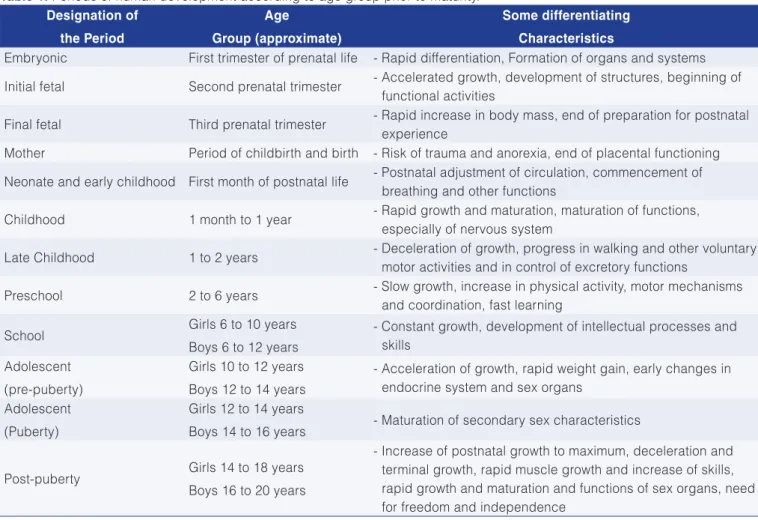
Table 1 briefly shows the periods of life in the human species prior to maturity. In the first trimester of prenatal life, the embryonic period, there is rapid differentiation and development of organs and systems. The initial fetal stage occurs during the second trimester. This stage is characterized by accelerated growth, the development of structures, and the start of functional activities, thus being a period of biochemical development; in the final stage of the fetal period considerable biochemical alterations occur. The period known as parturient consists exactly of the labor, and the neonatal period corresponds to the first month of postnatal life. The next stages correspond to what we call baby, toddler, preschool age, school age and adolescence. With sexual maturity the individuals acquire the ability to reproduce and can restart a new cycle.
2.1 Curve of human perinatal growth
way the parts do not stop growing at the same point in time. Once maturity is reached, the total size of the body remains relatively constant. Another characteristic of growth in a multicellular organism is that the growth of one part of the body is controlled by another part. For example, the growth of the skeleton can be controlled by the pituitary gland, which produces the necessary hormones. There is an integrative interaction between the parts to maintain the correct body proportions (HURLEY, 1980).
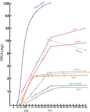
In an animal model (rat), Enesco and Leblond (1962) showed that cell multiplication and growth can also be divided into three distinct phases: the first is a period of rapid cell proliferation, during which there is virtually no increase in cell size. In the rat, this period extends for about 17 days after birth; in the second phase, cell proliferation decreases considerably, but cell size increases rapidly. In the rat, this period extends from 17 to 48 days after birth; in the third phase, there is almost no cell proliferation, but cell size increases rapidly. The authors demonstrated that in most rat organs the RNA content was proportional to that of the DNA during the first months of postnatal life, resulting in a constant value of RNA per nucleus; therefore the RNA/DNA ratio is immutable for each organ. Organs
Table 1. Periods of human development according to age group prior to maturity. Designation of
the Period
Age
Group (approximate)
Some differentiating
Characteristics
Embryonic First trimester of prenatal life - Rapid differentiation, Formation of organs and systems
Initial fetal Second prenatal trimester - Accelerated growth, development of structures, beginning of functional activities
Final fetal Third prenatal trimester - Rapid increase in body mass, end of preparation for postnatal experience
Mother Period of childbirth and birth - Risk of trauma and anorexia, end of placental functioning
Neonate and early childhood First month of postnatal life - Postnatal adjustment of circulation, commencement of breathing and other functions
Childhood 1 month to 1 year - Rapid growth and maturation, maturation of functions, especially of nervous system
Late Childhood 1 to 2 years - Deceleration of growth, progress in walking and other voluntary motor activities and in control of excretory functions
Preschool 2 to 6 years - Slow growth, increase in physical activity, motor mechanisms and coordination, fast learning
School Girls 6 to 10 years
Boys 6 to 12 years
- Constant growth, development of intellectual processes and skills
Adolescent (pre-puberty)
Girls 10 to 12 years Boys 12 to 14 years
- Acceleration of growth, rapid weight gain, early changes in endocrine system and sex organs
Adolescent (Puberty)
Girls 12 to 14 years
Boys 14 to 16 years - Maturation of secondary sex characteristics
Post-puberty Girls 14 to 18 years Boys 16 to 20 years
- Increase of postnatal growth to maximum, deceleration and terminal growth, rapid muscle growth and increase of skills, rapid growth and maturation and functions of sex organs, need for freedom and independence
Adapted from Timiras (1972).
with high protein synthesis, such as liver, heart and muscle, showed higher RNA/DNA ratios.
The organs in which DNA synthesis ended earlier were the lung and brain; and these were also the organs in which the cell proliferation stage ended earlier, which in the rat was observed about 9 days after birth. In the kidneys, the rapid cell proliferation phase ended 39 days after birth, and in the heart, 49 days after birth. Thus the various organs of the body presented different speeds of DNA synthesis and different times for the growth phases. This phenomenon is important in terms of the effects of nutrition on development. If the individual is submitted to malnutrition throughout the whole phase of rapid cell proliferation, the number of cells is diminished and cannot be reversed even with regular nutrition at a later stage. These discoveries concerning the stages of cell reproduction and growth were very important, because they demonstrated that precocious malnutrition, particularly in the stages of rapid cell proliferation, is detrimental to the future development of the individual, considering both physical and cognitive aspects, irreversibly in several species, including the human species (ENESCO; LEBLOND, 1962; WINICK; NOBLE, 1965; SUSSMAN, 1973; RICHTER, 1961; DOBBING, 1964, 1965; CRAVIOTO; ROBLES, 1962, 1965; CRAVIOTO et al., 1967; MONCKEBERG, 1968). These authors demonstrated that malnutrition in the intrauterine period and in the first phase of life (in humans, up to approximately 3 years of age) is detrimental not only to physical development (height, body weight) but also to learning and school performance, also impacting the physical and mental performance and sociability of these individuals in adulthood. Winick and Noble (1965) studied cell development in rats, in terms of (mgDNA) for different vital organs, showing the periods during which these organs were showing maximum cell reproduction activity, as well as the postnatal age around which some organs (heart, lung, kidneys, salivary gland and particularly the brain) stop producing new cells, and reach maturity. On the other hand, organs such as the liver and spleen continue to grow, while the thymus gland begins a process of partial atrophy around 39 days after birth (WINICK; NOBLE, 1965), Figure 2.
2.2 Evolution of clinical studies in humans
Under conditions of less severe malnutrition than those caused by a great lack of food, for example when the population is exposed to famine for prolonged periods, it is more difficult to assess the effects on prenatal development, and little information has been made available up to the present time, resulting from clinical studies in humans. Amongst the many issues involved, there is the difficulty in evaluating dietary intake, in establishing nutritional intake safely, and in controlling all the factors likely to affect development. However, thanks to the continuity of studies, currently the consensus indicates that the nature
of a pregnant woman’s diet may affect fetal development in many ways.
A clinical study investigating the relationship of maternal nutrition to neonate development, with a well-controlled double blind, was published in the early 1940s by Burke et al. (1943). Dietary surveys were carried out with women who attended a prenatal care clinic at Harvard University (USA) in which the nutritional profile was evaluated. When the babies were born, they were examined by pediatricians who did not know the mothers’ nutritional profiles. Mothers whose diets were classified as good or excellent gave birth to babies whose condition was classified as good or superior. A total of 42% of the mothers with diets considered good or excellent had babies classified as superior. For the rest of mothers with good or excellent diets, their babies were classified as good. In contrast, for mothers whose diets were considered regular, only 6% had babies classified as superior, 44.5% were considered good or regular, and 5% were considered inferior. Therefore, there was a well-defined correlation between the quality of the maternal diet during pregnancy and the condition of the babies at birth. It was concluded that the weight and length of the baby at birth correlated with the quantity of protein in the maternal diet. For a quantity of protein less than 45 g/day, the mean weight at birth was 2.63 kg; for diets ranging from 65 to 74 g
of protein per day, the mean weight of the babies was 3.63 kg; while in diets with 85 g or more of daily protein, the unborns presented a mean weight of 4.173 kg. Similar results were presented by Jeans et al. (1955). In this study, the dietary habits of 400 rural low-income pregnant women were assessed and correlated with the condition of their babies at birth. Again it was observed that lower weights, decreased vitality and a greater number of newborn deaths correlated with mothers showing poorer nutritional profiles. In addition, this study showed that the incidence of premature infants increased significantly as the nutritional status of the pregnant woman worsened. In the case of women whose dietary habits were classified as acceptable to excellent, 4% of the babies were born prematurely, while for the pregnant women whose diets were considered poor or very poor, the incidence of premature infants was 9.6%. Even more serious were the conditions of these mothers’ newborns (1 death in labor, 4 born with severe congenital abnormalities and 5 deaths in the neonatal period).
Primrose and Higgins (1971) carried out a study of nutritional intervention in Montreal (Canada). In this study, the dietary history of pregnant women was assessed, and poor women with deficient diets were provided with an extra supplement based on milk, eggs and oranges, as well as education on nutrition. Expectant mothers were also provided with a supplement containing mixtures of vitamins, iron and other essential minerals. In the group of pregnant women who received the nutritional supplements and education on nutrition, the incidence of deaths in labor, and perinatal and neonatal mortalities was significantly lower than those in the province of Quebec and in Canada as a whole, as observed in Table 2.
2.3 Fetal growth retardation in humans
Fetal malnutrition can occur in two different ways: 1) the nutritional status of the mother, before and during pregnancy, and the dietary intake can affect the supply of nutrients to the fetus; 2) abnormal or insufficient functioning of the placenta can deprive the fetus of essential nutrients due to inadequate transfer via the placenta. Furthermore, the functions of the placenta may be affected by factors such as diseases, drugs or abnormalities in the chromosomes. Poor fetal nutrition or malnutrition is usually studied in babies
with intrauterine growth retardation, which most specialists consider to be a sign of fetal malnutrition. Intrauterine growth retardation is defined as the child being born with size and weight below those considered normal for the gestational age. Metcoff (1973) published a study on fetal growth retardation as an index of malnutrition during pregnancy. In this study several biochemical parameters were analyzed in newborns, such as: the protein/DNA ratio (cell size) and the profiles of various enzymes in white blood cells isolated from umbilical cord blood. These results were compared with the same parameters obtained from white blood cells isolated from mothers who gave birth to children with normal sizes for their gestational age. The size of the leukocytes of infants who were submitted to fetal malnutrition and of their respective mothers was larger than those of any other group of newborns. Moreover, enzyme analyses showed alterations in the energy metabolism profiles of the leukocytes of the newborns. A reduced ATP content per cell was also observed, as well as for pyruvate and adenylate kinase. This profile was similar to that found in newborns with severe postnatal protein-calorie malnutrition, offering more evidence that fetal growth retardation may be taken as an index of prenatal malnutrition.
3 Metabolic programming
More recent research has introduced new concepts concerning the physiological, metabolic and genetic phenomena during pregnancy and after birth. The success of a pregnancy should not be defined only by the result obtained at birth, but also by the state of health throughout the life of the individual. Conditions in the intrauterine period and in the first months of life affect their general state of health, with increased risk of diseases in adulthood. Nutritional, hormonal and physiological alterations during pregnancy, in critical phases of development, may result in low birth weight, which is associated with metabolic diseases in adulthood, particularly type 2 diabetes (DM2) and cardiovascular disease (CVD) (BARKER 2007; BARKER, 1998). The hypothesis that came to be known as the “Barker hypothesis” assumes that fetuses that underwent intrauterine deprivation developed an energy saver phenotype (“thrifty phenotype”) (BARKER et al. 1989; HALES; BARKER, 1992). A selective economy is assumed that prioritizes oxygen and nutrients for more vital organs, such as the brain, to the detriment of other
Table 2. Influence of nutritional intervention on births in the study of dietary supplementation in Montreal. Events
(Nature)
No./1000 live births Study in Montreal (Dietary
supplementation)
Province of Quebec
Canada (entire country)
Deaths in labor 8.7 11.8 11.4
Neonatal mortality (1st week) 5.7 16.2 14.4
Perinatal mortality 14.4 28.0 25.8
“less vital” organs such as the pancreas. If, after birth, the food deprivation is replaced by food abundance, the phenotype starts to develop obesity and the metabolic syndrome.
Dutch youth who underwent intrauterine exposition to a period of starvation or famine from 1944 to 1945 (The Dutch Famine, 1944-1945 apud WATERLAND; GARZA, 1999) were examined at the age of 19. The children of mothers subjected to famine during the first two trimesters of pregnancy showed a prevalence for overweight that was 80% higher than those borne to mothers who were not subjected to the same condition (WATERLAND; GARZA, 1999). Nutritional deprivation during the critical period for the differentiation of the hypothalamus, resulted in alterations in the development of the hypothalamus centers that regulate the appetite. On the other hand, the youths whose mothers were exposed to conditions of food scarcity during the last trimester of pregnancy or during the first five months after birth showed 40% less prevalence for overweight as compared to those who were subjected to food deprivation early in the pregnancy. In the latter group, the nutritional deprivation occurred during the critical period for adipocyte replication. Thus depending on the time of dietary deprivation during pregnancy, the effects may impact as alterations in the organs and systems under development at that moment, for example: 1) maternal malnutrition during pregnancy may affect the health of adult individuals without influencing their size at birth; 2) the occurrence of metabolic adaptations to allow fetal development (ROSEBOOM et al., 2001). In the latter case, the mechanisms by which this phenomenon occurs came to be known as metabolic imprints and occur via variations in the structure of organs, alterations in the number of cells and metabolic differentiation, such as, for example: 1) changes in vascularization, innervation or the juxtaposition of different cell types within the organ; 2) alterations in the expression of certain genes, leading to variations in the production of enzymes, hormones, hormone receptors, transporters, etc. (WATERLAND; GARZA, 1999).
In another survey carried out in China, Li et al. (2010) reported a study with 800 adults born in the period from 1954 to 1964, who were exposed to malnutrition at the intrauterine stage (“The Chinese famine from 1959 to 1961), with a high risk of negatively affecting the homeostasis of the glucose metabolism in adult life. In these studies in humans it is difficult to differentiate between the effects of stress and of malnutrition, since interfering effects, such as infections and stress, can both lead the individual to develop health problems which are similar to those caused by famine. Prenatal stress is recognized as capable of affecting the functioning of the hypothalamic-pituitary-adrenal (HPA) axis and, particularly, the secretion of cortisol. This could be a plausible explanation for the mechanism of association
between prenatal programming and a higher prevalence of hyperglycemia in adult life.
4 From plasticity to epigenetics
The ability of the genotype to produce different phenotypes in response to different environments is known by the term of “plasticity”. The period of greatest plasticity seems to be during development. This ability of the organism to facilitate changes is called “adaptability” for the expression of a set of genes, particularly during developmental transitions, probably due to the fundamental plasticity of an organism. Developmental plasticity acts in order to adjust the expression of the genes to produce a more adequate phenotype for the predicted subsequent development (GLUCKMAN; HANSON, 2004). When the resulting phenotype is adjusted to its environment, the organism will remain healthy. When incompatibility occurs, the individual’s ability to respond to environmental changes may be inadequate and the risk of diseases increases. Therefore, the degree of genotypic/phenotypic incompatibility determines an individual’s susceptibility to chronic diseases (GODFREY et al., 2007).
4.1 The epigenome
Inheritance is not limited to the transmission of the genetic information contained in the DNA sequence. The DNA is packed in the form of chromatin, contained within the nucleus of each cell, and the DNA molecule is wrapped in a bundle in the three-dimensional structure of the chromosomes. In eukaryotes, the DNA molecules are wrapped in an octameric nucleus of histones consisting of two copies of each protein molecule: H2A, H2B, H3 and H4, as shown in Figure 3. These core particles or nucleosomes are the basic units of chromatin, and may form arrangements of higher structural levels due to the binding of histones
H1 and nonhistone proteins, as well as non-coding RNAs (PORTHA et al., 2014). The nucleosomes maintain the DNA-histonone complex compacted, and the degree of compaction creates an additional layer of genomic activity. The configuration of the histone-DNA complex is maintained by electrostatic bonds between histones with (+) charges and DNA with (-) charges. Alterations to the profile of these charges regulate the gene expression. The nucleosomes are also subject to covalent modifications in their histones and in the DNA. These epigenetic modifications (markers) can determine whether portions of the chromosomes are strongly or weakly associated, which influences whether the gene can be “turned on” or “turned off”. Other epigenetic parameters are also important to modulation of the structure and genomic activity including histone variants, noncoding RNAs, remodeling of the chromatinic complex and spatial organization of the chromatin within the nucleus (PORTELA; ESTELLER, 2010). Currently, it is recognized that the epigenetic information is crucial for the dynamic interpretation of gene information, so the correct genes are expressed at the correct time during critical decisions of the cellular phase.
4.2 Modifications of histones
Each core histone features a terminal amine tail that protrudes from the nucleosome and may be subject to a series of post-translational covalent modifications (SANTOS-ROSA; CALDAS, 2005; MARGUERON; REINBERG, 2010). Chromatin is usually compartmentalized in two main
types of domain: heterochromatin, which is condensed and poor in genes, and euchromatin, which is decondensed and rich in genes (MARGUERON; REINBERG, 2010). These domains feature different profiles of histone modifications and are associated in different arrangements of nucleosomes, with higher structural order and nuclear organization. In general, heterochromatin is associated with marker histones that are repressive to DNA methylation, while euchromatin is associated with active marker histones (MARGUERON; REINBERG, 2010). Acetylation of histones is restricted to lysines (k) preserved in the core histones. It is considered that an open marker, with active chromatin domains, corresponds to actively transcribed genes with high levels of acetylation in their promoting regions, initial transcription sites, which are CpG islands and regulatory functional elements. Acetylation levels as chromatin are determined by histone acetyltransferases (HATs) which catalyze the addition of acetyl radicals to lysine (k) residues, and histone deacetylases (HDACs) that remove acetyl groups from the lysine residues. The balance of the activities of these two types of enzyme determines the state of acetylation of the histones, which influences the levels of expression of the associated genes. The acetylation of the lysine 9 (K9) residue at the terminal amino of the histone tail H3 (H3K9ac), for example, neutralizes the positive charge of the histone tail, decreasing its affinity for the negative charge of the DNA, resulting in relaxation of the DNA which wraps around the histone octamer residues (Figure 4). Transcription factors and the transcription apparatus will
have access to the DNA, and the corresponding gene expression is facilitated (CEDAR; BERGMAN, 2012). On the other hand, deacetylation is associated with gene silencing. In fact, HDACs are generally considered as transcriptional corepressors.
Compared with acetylation, histone methylation is far more complex (ZHANG; REINBERG, 2001). Methylation can occur in preserved lysine (K) and arginine (R) residues and via the four types of histone. Up to 3 methyl groups can be added to the amino group of lysine (mono-, di-, and trimethylation), while arginine can only be mono- or dimethylated. Histone methyltransferases (HMTs) are enzymes responsible for the addition of methyl groups to lysine or arginine residues. Until recently it was believed that histone methylation represented the most stable or permanent modification, because the “turnover” of this marking was lower than the highly dynamic acetylation. However, the recent identification of enzymes capable of removing mytilo groups from histones has shown that this marking can be similarly dynamic (BLACK et al., 2012). Therefore, unlike acetylation, which affects the charge of the amino acid residue, directly impacting the histone-histone or histone-DNA bonds, the role of methylation can only be governed by the additional recruitment of regulatory factors (Figure 4).
5 Epigenetic deregulation: molecular mechanism of the origin of diseases
The phenomenon of epigenetic deregulation is an example of plasticity in the development, through which alternative phenotypes are generated from a specific genotype, by adjusting the developmental program in response to persistent environmental factors (GLUCKMAN et al., 2007). Therefore, epigenetics, and particularly epigenetic deregulation, go against the simplistic interpretation of the phenotype as a result set by the genotype. This interpretation dominated the thinking about developmental and evolutionary biology throughout the 20th century. Based on the hypothesis that body disorders may result from an imbalance between environmental factors in the womb and in infancy, it has been proposed that the memory of fetal history and the adaptive responses in aging cells and organs may be mediated through epigenetic gene regulation mechanisms (WATERLAND; GARZA, 1999; OZANNE; CONSTANCIA, 2007). The still unanswered question is whether the identity of the genes likely to be involved in this increased susceptibility to deregulation by environmental factors. Many investigations are being carried out to identify these genes by employing amplified genomic approaches in monozygotic (identical) and dizygotic (non identical) twins and rodent lineages (pure or “inbred”), in order to study the impact of the epigenome without interfering effects from genetic variability, such as the effects of chemical substances (food, toxins, drugs) or
non-chemical effects (e.g. behavior) or of environmental factors, on the epigenetic marking of specific genes.
In a recent review by Portha et al. (2014), they concluded that the idea is becoming increasingly accepted that environmental substances can produce alterations in genomic activity which, although not altering the DNA sequence, can produce important and stable translational alterations in the phenotype. Epigenetic alterations, in particular DNA methylation, produce a developmental “memory” of plastic responses in their primordial environment, which can be decisive in the generation and stability of phenotypes throughout life. Their effects may manifest only during prenatal life, for example, in terms of altered responses to environmental challenges. Evidence has accumulated that endocrine or nutritional interventions in early postnatal life may reverse epigenetic and phenotypic alterations induced, for example, by an unbalanced maternal diet during pregnancy. The elucidation of epigenetic processes may enable the identification of individuals at greater risk of future cardiovascular and metabolic diseases, and enable researchers and practitioners to develop intervention strategies to reduce the risk.
5.1 Significance of low birth weight
influenceable. Low birth weight is correlated with race and poverty, which correlate with malnutrition and subnutrition. Other complicating factors include, for example, a lack of education, poor integration of family units, inadequate hygienic conditions and diseases. All these factors acting in conjunction form, apparently, the syndrome of poverty, disease and human underdevelopment.
Considering all the organs affected by malnutrition or subnutrition in humans during the prenatal period and first year of postnatal life, the impact on the brain is that which has been the greatest concern of those responsible for the areas of health and education. Similar to animals, in humans, the most vulnerable period in the development of the brain is the last stage of prenatal growth and the first postnatal period (approximately 12 months), periods in which there is rapid proliferation and initial cell growth and differentiation. Myelination occurs rapidly, at the same time as the exponential growth of the brain. When malnutrition or subnutrition is imposed during this period of rapid myelination, there is a decrease in the quantity and concentration of cholesterol. Cholesterol is an essential component for the formation of myelin, along with cerebrosides, phosphatidylethanolamine and sphingomyelin. All these components suffer reductions in conditions of malnutrition in the most vulnerable period of development, which results in a decreased speed of myelination and reduced amount of myelin in the brain. A reduction in the number and composition of all types of brain cells will occur as a result of precocious malnutrition.
In general, studies in humans are considerably outnumbered by studies in animal models; however, similar results have been obtained in children exposed to malnutrition and/or subnutrition in early periods of their lives. Some studies were carried out in Chile, a country that has undergone serious problems with child malnutrition. They examined the brains of children who died of malnutrition, specifically of protein-calorie malnutrition (marasmus). The graph in Figure 5 compares the composition of well-nourished Chilean children who died in accidents or of other causes not related to malnutrition and which were within the range of values considered normal, even for North American children. The concentrations of RNA, protein and DNA were significantly lower in children who died of marasmus, in different periods after birth (WINICK, 1969).
5.2 Influence of malnutrition on brain function
Malnutrition in early life can produce permanent effects on the size, number of cells and composition of the brain. This fact has been demonstrated in animals and in humans (Figure 5). The question that arose at the time was what these changes could mean in terms of brain function. Currently, there is significant evidence that malnutrition or subnutrition, in critical periods of development, produces alterations of a behavioral nature in experimental animals.
Various techniques have been used such as: learning tests, behavior tests and emotionality tests. There are still many important issues to be studied on the applicability to humans of the results obtained with animals.
The important question in terms of human society is the following: does early malnutrition have a negative effect on learning ability? The difficulty in answering this question lies in the fact that the malnutrition factor is never isolated from a number of other factors that can also affect the learning ability and intelligence quotient (I.Q.) of an individual. Malnutrition is rarely found as an isolated factor but rather in combination with poverty, low socioeconomic level, unfavorable home environment, absence of intellectual stimulus and other factors that are related to the development and low learning ability. What we can conclude is that subnutrition and malnutrition at an early age (prenatal and in the first year of postnatal life) permanently alter some structures and the chemical composition of the brain, which leads to the hypothesis that the IQ and learning ability should also be affected. However, the direct cause-and-effect relationship with the permanent and irreversible detriment to intellectual capacity in humans, still demands future studies which seek to eliminate interfering factors of a non-nutritional nature as much as possible.
The period from 1950 to 1970 was of great importance since much relevant medical and nutritional research was carried out. These studies demonstrated the need for greater care in the food area in relation to pregnant women and their gestational products, so that they could generate healthy children that might develop into intelligent and productive citizens. Based on the literature published in the areas of pediatric medicine, nutrition and food science, particularly concerning pregnant women, newborns and children in preschool and school age, there was greater motivation by the government agencies responsible for public health to direct greater investment to these areas. The ultimate goal
Figure 5. Reduction of the number of brain cells in nine children who died of malnutrition in Santiago, Chile. The lines of the graph delimit the range of normality adopted in the United States (USA):
(•)DNAcontentofnormalChileanchildren;(Δ) Chilean children
was to better preserve the human capital through special programs that enabled greater and coordinated care for pregnant women, newborns and children in preschool and school age, the most critical periods for the development of future citizens who are healthy and productive in society. In the 1960s, institutions such as INAN (National Institute of Food and Nutrition), COBAL (Brazilian Food Company) and PRONAN (National Program of Food and Nutrition) were created in Brazil in order to increase food and nutritional assistance so as to implement an improvement in the nutrition and nutritional status of these segments of the population, particularly in the most destitute. In Chile, the Chilean Government created INTA (National Institute of Food Technology) with the goals of developing foods that were adequate for those segments of the population. Due to these initiatives, in a few years maternal and child malnutrition and subnutrition were virtually eradicated in the country. More recently, research has continued to be carried out in Brazil and in virtually all countries (developed and developing) with the objectives of better explaining the dynamics of human development in its various stages.
5.3 Later effects of precocious malnutrition
An excellent review on the effects of maternal and child malnutrition and its consequences on the health of the individual as an adult, was published by Victora et al. (2008). They addressed the associations between maternal and child malnutrition and the human capital and risks of diseases in low- and middle-income countries. The analysis was carried out using data from five long cohort studies including Brazil, Guatemala, India, the Philippines and South Africa, after maternal and infant malnutrition indices had been collected. The following indices were evaluated: maternal height, birth weight and intrauterine growth restriction, in addition to the weight, height and body mass index when the child reached 2 years of age, in accordance with the new growth standard of the World Health Organization (WHO). These indices were considered in relation to the results for adults with respect to: height, educational level and economic power of the family, as well as birth weight, body mass index, blood glucose concentration and blood pressure. Systematic reviews of studies in low and middle family income countries were carried out for these same parameters and for indicators related to blood lipids, cardiovascular diseases, pulmonary and immune functions, cancers, osteoporosis and mental illness. Subnutrition was shown to be strongly associated with short stature, low educational level, reduced economic productivity and, for women, lower birth weights. Associations with disease indicators in adults were not so clear in this research. Higher weight at birth and in childhood was positively associated with the adults’ body mass index, and, less clearly, with blood pressure values, but not with blood glucose concentrations.
Both in the analyses carried out by the aforementioned authors and in other published studies, lower birth weight and subnutrition appeared as risk factors for high blood glucose concentrations, blood pressure and undesirable lipid profiles. It was observed that adjustments were made to the body mass index and height, suggesting that rapid postnatal weight gain – especially after childhood – was connected to these unfavorable conditions.
life, and may result in irreversible detriment throughout the individual’s life, such as a reduction in adult stature, poor school performance, reduced competitiveness at work as an adult and lower birth weight of descendants (VICTORA et al., 2008).
A child’s brain volume doubles from birth to six months of life, triples from birth to 2 years of age, and reaches the adult volume by 5 years of age. The brain prototype is remodeled after birth, since unused neurons disappear and stimulated neurons increase in both number and quantity of connections, which results in a complex brain network. This fact illustrates the importance of stimuli during the early years, which are critical to brain development. Direct psychosocial stimuli by means of contact with parents are some of the most significant elements for brain development in early childhood. In the fetal period, the brain is well protected as long as the mother has correct and balanced nutrition. However, iodine deficiency can lead to mental retardation and a lack of folic acid in the first months of pregnancy can cause neural tube defects (GERMAN, 2007). When there is a low amount of iodine in the diet, as in some regions that are mountainous and far from the sea, the amount of iodine transferred to the fetus through the placenta and that which is contained in the breast milk may be insufficient. The addition of iodine to table salt (NaI) was the technological solution found to address this problem in several countries. A lack of iron is the main nutritional deficiency both in developed and developing countries. It is estimated that anemia caused by a lack of iron affects more than 25% of the children in the world and there is evidence that iron deficiency anemia is associated with the impairment of psychomotor and cognitive development. Children with iron deficiency anemia reach inferior levels of mental and motor development. Supplementation with ferrous sulfate improves both mental and motor performance in cases of established retardation; however, it is necessary to establish the needs of each child in treatment. Breast milk is adequate for the needs of babies because they are born with a reserve of iron. Infant formulas enriched with iron contain higher amounts of this mineral, but while the iron in breast milk is absorbed up to 70%, absorption of iron from formulas is only 11%. A child’s need for iron changes during the first year of life. The fuel used by the brain is practically only glucose. The neuron membranes comprise a double thin layer composed primarily of fatty acid molecules, and glucose, oxygen and micronutrients pass through the membrane to nourish the neurons and provide the fuel required for nerve impulses. Myelin, the protective layer that surrounds the axons, is composed of 30% protein and 70% lipid. The brain requires lipids for its protection and to provide speed to neural transmissions (GERMAN, 2007).
5.4 Congenital abnormalities: significance of birth defects
A second type of abnormal development consists of congenital abnormalities, which are synonymous with birth defects. The word congenital means that the condition already exists at birth, be it caused by genetic or environmental factors. Birth defects are defined as structural or metabolic disorders present at birth, caused by factors that act during the prenatal period. Often, congenital abnormalities are not apparent at birth and are discovered later due to producing functional problems. Congenital heart disease, for example, is commonly discovered after birth, to the extent that cardiac function presents as abnormal and insufficient. The study of congenital abnormalities and their etiologies is the object of Teratology. There are two ways in which the development of the embryo can be affected by environmental factors, including nutrition: 1) by direct effect on the embryo itself; 2) indirect effect through the mother.
In 1910, the percentage of children who died because of congenital malformation was low, around 5%, while 30% of deaths during the first year of life were caused by enteritis, diarrhea, pneumonia and other infectious diseases. The methods of care intended for children and the hygienic conditions of hospitals improved, and, especially, antibiotics and other drugs have emerged, leading specific infectious diseases to decrease significantly. Around 1950, child deaths by infectious diseases dropped to approximately 5% and, proportionately, deaths due to congenital malformations increased.
In 1964, 19% of child deaths occurred due to congenital malformation. It is likely that, in absolute terms, the incidence of deaths due to congenital malformation did not change in the period 1950-1964, but it became more important as other causes of death decreased because of improved care and medical knowledge. According to research published by Apgar and Stickle (1968), the vital statistics for the United States in 1964 indicated that birth defects were the leading cause of infant death (1st year of life). They also found that at least 21,000 cases of pregnancy indicated that at least 7% of all children born showed structural or functional defects of prenatal origin, detected in the first years of life. Less than half (50%) of these defects are detectable at birth. The authors, Apgar and Stickle (1968), also found that at least 62,000 deaths at any age, in each year, could be attributed to birth defects (Table 3 and 4).
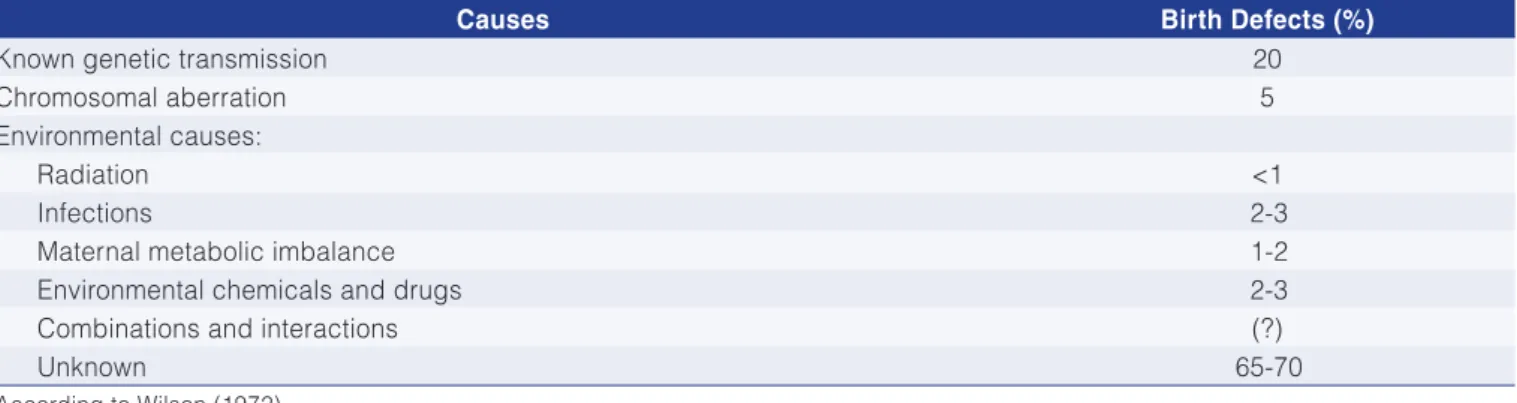
known causes of birth defects amount to only 30-35% and include: genetic transmission, chromosomal aberrations, radiation, German measles and other viral infections, diabetes, drugs and environmental chemicals. Wilson estimated that 65-70% of all birth defects in humans are of unknown causes. It is possible that nutrition is involved in some of the factors in this unknown group, whether due to nutrition by itself or in interaction with some other factors, already known to affect development.
The dangerous effects of some environmental chemical contaminants have recently been described. Rubin and Soto (2009) describe a possible association between the intake of the compound Bisphenol A in perinatal period and weight gain. Bisphenol A (BPA) is a component of polycarbonate plastics and of the resinous lining on the
inside of the packaging of drinks and food. BPA is known for transferring to the product with which it is in contact (food or drink) and being routinely ingested with food and drink. In a recent study of a sectional cohort, BPA was detected in urine samples of 92.6% of the population examined, in the United States. BPA’s potential influence on body weight was suggested by in vitro studies, showing effects of the substance on adipocyte differentiation, lipid accumulation, glucose transport and the secretion of adiponectin. Data from in vitro studies have demonstrated dose-dependent and sex-dependent effects on the weight of rodents exposed to BPA in the perinatal period. The mechanisms through which perinatal exposure to BPA produces persistent effect on body weight and increase of adiposity were not elucidated.
Table 3. Deaths caused by congenital conditions identified in the seventh revision of the international classification of diseases in the United States, 1965.
Cause of death No. of deaths
Congenital syphilis 34
Neoplasms, with less than 28 days 91
Myxedema and cretinism 342
Diabetes mellitus 33,174
Cystic Fibrosis 610
Lipidosis (lipidic metabolic disorder) 158
Amyloidosis 171
Other metabolic diseases 672
Family jaundice 130
Sickle cell disease 358
Hemophilia 59
Chromosomal abnormalities 267
Hernia of abdominal cavity 3,277
Muscular birth defect 689
Congenital malformations 19,512
Neonatal disorders originated from diseases of the mother during pregnancy 717
Hemolytic disease of the newborn (erythroblastosis) 1,485
Hemorrhagic disease of the newborn 477
Other congenital conditions 8
Total 62,231
According to Apgar and Stickle (1968).
Table 4. Causes of the development of birth defects in humans (in %).
Causes Birth Defects (%)
Known genetic transmission 20
Chromosomal aberration 5
Environmental causes:
Radiation <1
Infections 2-3
Maternal metabolic imbalance 1-2
Environmental chemicals and drugs 2-3
Combinations and interactions (?)
Unknown 65-70
Lu et al. (2006) determined the exposure of pre-school age children to organosulphur pesticides in 23 schools by way of urinary biomonitoring. The conventional diet of most children was replaced by organic farming food for 5 consecutive days, during which two samples of urine were collected daily in the morning and another before going to bed, during the 15 days of the study. It was found that the median urinary concentrations of specific metabolites for melation and chlorpyrophosphate decreased to undetectable levels immediately after introducing the organic diet, and remained undetectable until the conventional diet was reintroduced. The authors concluded that these children were exposed to organosulphur pesticides exclusively through their diets.
In recent years, there has been increasing concern about the potential role of chemicals that interfere with the endocrine system in the development of chronic diseases such as obesity and type-2 diabetes (CASALS-CASAS; DESVERGNE, 2011).
Recent experimental evidence suggests that prenatal exposure to chemicals that harm the endocrine system can increase the risk of postnatal obesity, and that these effects may be dependent on the diet and sex (VALVI et al., 2012). The authors explored the influence of the concentration of some organochlorine (OC) compounds such as polychlorinated biphenyl (PCB), dichlorodiphenyldichloroethylene (DDE) and dichlorodiphenyltrichloroethane (DDT), to determine if they associated with overweight in 6.5 year old children, and if the child’s sex and fat intake would alter the results. A total of 344 Spanish children were studied from a cohort
established in 1997-1998. Overweight at 6.5 years of age was defined as a body mass index (BMI) with a score-Z ≥ of 8.5%, according to the reference established by the World Health Organization (WHO). Concentrations of products in the umbilical cord blood were determined and treated as categorical variables. The childrens’ diets were estimated using the food intake frequency questionnaire and the relative risks (RRs) estimated using generalized linear models. After multivariate adjustments, the authors found an increase in overweight RR in the third tertile for exposure to PCB [RR=1.70; 95% (CI): 1.9; 2.64] and in the second tertile for the DDE compound [RR=1.67; 95% (CI): 1.0; 2.55], but no association was found for exposure to DDT in the population as a whole. Associations between overweight and the PCB and DDE concentrations were stronger for girls (p=0.01 and 0.28); DDT was associated with overweight only for boys. In the aforementioned study, the authors suggested that prenatal exposure to the chemicals PCB, DDE and DDT may be associated with overweight in children, and that sex and a high fat intake are factors that may influence the susceptibility to the phenomenon.
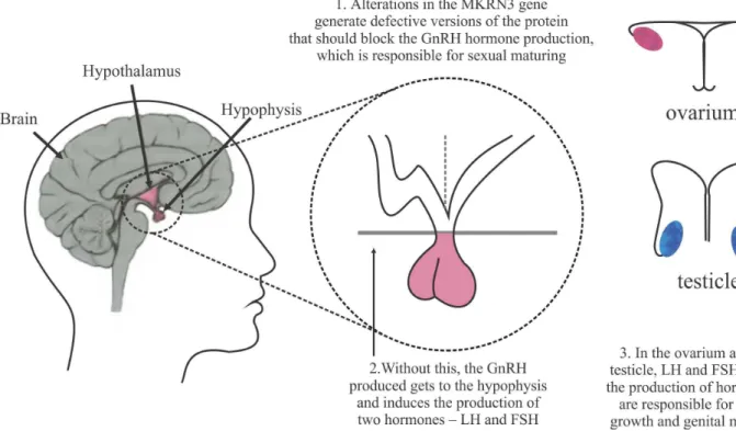
5.4.1 Central precocious puberty
An unusual heritage that affects the child in the first years of life is called central precocious puberty, caused by early activation of the hypothalamus-pituitary-gonads axis and occurring in both sexes, as illustrated in Figure 6. Several researchers from Brazil and from abroad have devoted themselves to this topic in the past decade,
according to Zorzetto (2014). In 12 Brazilian families and three foreign families, 32 individuals were shown to have entered puberty too early, generally at the age of 6 years, by way of clinical and hormonal tests, In all these cases, the accelerated development that marks the transition of the body to adulthood started ahead of time, due to a premature increase in the production of the hormone that releases the gonadotropins, the GnRHs, which are responsible for the sexual maturation of the organism. Produced in the brain by a small group of neurons in the hypothalamus, the GnRHs are released in faster pulses at puberty, inducing the pituitary gland to produce two other sex hormones: the luteinizing hormone (LH) and the follicle stimulating hormone (FSH). These hormones are released into the bloodstream and reach the ovaries and testicles, where they activate the release of other sex hormones that make the body grow and mature from the reproductive point of view (Figure 6). The reseachers then decided to sequence the genetic material of the 32 participants, investigating alterations that could explain the early onset of puberty. Eight of the 32 individuals showed defects in the same gene, the MKRN3 gene, this being the first gene identified as responsible for this inherited form of precocious puberty. The high incidence of genetic defects in a single gene (33%) surprised the researchers, as alterations in genes affect less than 10% of individuals with a particular genetic disorder. In addition to being very frequent in the cases where precocious puberty was present in more than one generation in the same family, the MKRN3 gene mutations were also shown to be common in people with central precocious puberty of nonhereditary origin. Puberty is usually associated with a series of long-term physical and psychological phenomena and hence a greater understanding of what defines its onset could create an opportunity to address health issues such as cancer, risky behavior and drug abuse.
A group of researchers in São Paulo, Ribeirão Preto and Campinas (all in Brazil) and in Macedonia (Eastern Europe), followed 215 children who presented isolated, nonhereditary precocious puberty. The proportion of individuals with altered MKRN3 genes was considered high, approximately 3% (ZORZETTO, 2014). In the first phase of the research, it was established that alterations in the MKRN3 gene generated defectuous versions of the kisspeptin protein, which should block the production of the hormone GnRH, which controls sexual maturation. With the mutation, this block was absent and the GnRH that was produced reached the pituitary gland, inducing the production of the hormones LH and FSH. These hormones will act making the body grow and the genitals develop precociously. The group of international researchers continued to seek for an explanation for this phenomenon, and recently published the results of their research (TELES et al., 2008; ABREU et al., 2013). According to Teles et al. (2008) the central or gonadotropin-dependent
precocious puberty is caused by an early maturation of the hypothalamus-pituitary-gonad axis, and in girls, this condition is mostly nonhereditary. Recently, a G protein-coupled receptor, GPR54, and its ligand to the protein kisspeptin, were described as an excitatory neuroregulator system for the secretion of the gonadotropin regulating hormone (GnRH). In this study, the authors identified an autosomal dominant mutation in the GPR54 receptor, consisting of the replacement of arginine (R) by proline (P) in the code 386 for this receptor (R386P), in the genetic profile of a girl with precocious puberty. The authors reported the case study of a girl aged 8 years which attempted to evaluate her precocious puberty. Premature breast development had been observed as from birth. By 7 years of age, breast growth was accelerated as well as pilosity in the pubic region. The mammalian animal model suggests an important role for kisspeptin, since intermittent infusion of this protein results in early sexual maturation in rats and an early release of the hormone GnRH in primates (NAVARRO et al., 2004a; PLANT et al., 2006). In their study, Teles et al. (2008) identified a heterozygotic mutation in GPR54 (R386P) in the aforementioned girl with nonhereditary precocious puberty.
Data in the literature suggests that an increased hypothalamic expression of kisspeptin in puberty contributes to maturation of the reproductive axis (NAVARRO et al., 2004b; HAN et al., 2005). Teles et al. (2008) speculated that a decrease in sensitization of the receptor GPR54 because of the mutation (R386P) could be expected to increase the stimulatory effects of kisspeptin on the secretion of GnRH, thus accelerating the maturation of the reproductive axis. In addition, in this study the development of the patient’s breasts in the neonatal period seems to be consistent with the kisspeptin-GPR54 system activity. The authors concluded they had identified a mutation in an autosomal dominant gene (R386P-GPR54), which prolonged the intracellular signaling of the GPR54 receptor in response to kisspeptin, and that all indications pointed to this being associated with the phenotype of central precocious puberty.
affected. A total of 4 mutations were identified, of which 3 involved encoding for truncated proteins and 1 was of the missense type that could block the production of the protein. The MKRN3 gene is located in a special region of chromosome 15 (15q11-q13) and all the affected individuals inherited the deficiency from their parents, a finding that indicates perfect segregation according to the expected inheritance from an imprint gene.
How puberty is initiated is still an enigma that defies an explanation from scientists. Much of the recent advance in the understanding of the mechanisms involved in reactivation of the central system, hypothalamus-pituitary-gonad, or reproductive axis in puberty, has been based on the characterization of genetic mutations associated with reproductive disorders in humans. Most mutations were identified in patients with hypogonadotropic hypogonadism, a disorder which is much less common than central precocious puberty (SEMINARA et al., 2003; ROUX et al., 2003). It is believed that the onset of puberty results from the reduction of factors that inhibit GnRH release combined with an increase in stimulatory factors. Studies on hypogonadotropic hypogonadism have resulted in the identification of genes that encode factors that have stimulatory action (BIANCO; KAISER, 2009; SEMPLE; TOPALOGLU, 2010). In contrast, MKRN3 seems to have an inhibitory role in humans, a role that disconnects as a result of the mutation that occurs in the GRP54 receptor, activating the central axis of precocious puberty.
5.4.2 Etiology of infantile leukemia
Another serious problem that affects human development and originates in the womb, are the infantile leukemias. Acute leukemias amount to approximately 30% of all malignant diseases that affect children in the western world. The peak incidence of B-cell precursors that transform into leukemic cells or acute lymphoblastic leukemia (ALL) emerged as a result of improved socioeconomic conditions in numerous countries throughout the world. In studies on twins and blood analyses in newborns, it has been possible to reproduce the first genetic events in hematopoietic cells that are critical for the “in utero” fetal development of the B-cell precursors to ALL, and, in some cases, of the precursors to AML or acute myelogenous leukemia (FORD et al., 1993; GALE et al., 1997; WIEMELS et al., 1999; WIEMELS et al., 2002; MORI et al., 2002). These events may occur as a normal part of fetal development, but it is not clear if other factors (constitutional or environmental) are involved and if they may increase the probability that the first events occur. For some leukemias (e.g.: ALL), the first event seems sufficient to create a clone of malignant cells; however, for most cases of ALL and AML, other genetic alterations are required, probably in the postnatal period.
Many environmental factors have been proposed as causes for leukemia, but only ionizing radiation and
certain chemicals (e.g., cytotoxic alkylating, benzene and topoisomerase II inhibitors) have been confirmed, especially for AML. It seems increasingly probable that delayed and unregulated responses to common infectious agents play a very important role in the conversion of preleukemic cell clones into B-cells that are precursors to ALL, the most common form of childhood leukemia. Polymorphic allelic variants, constitutional in genes of the immune response (especially HLA class II proteins) and cytokines, may play a role in determining the type of immune response. High-penetration mutations similar to germs are involved in only approximately 5% of childhood leukemia (more in AML and ALL). There is very little evidence supporting the participation of viruses as the cause of leukemia in humans. However, there is evidence for the role of other environmental factors such as nonradiating electromagnetic radiation and electric fields, although their mode of action on the genesis of leukemia remains unclear. Childhood leukemia has no single cause and for most individuals it is the result of a combination of factors; all involving gene-environmental interaction. To date, few preventive measures have emerged except completely avoiding x-ray exams in the first trimester of pregnancy; a healthy diet with an adequate amount of folic acid, both in the preconception period and in early pregnancy. It is important that the the child has contact with other children outside the home environment for stimulus and maturation of the immune system (EDEN, 2010). A summary of the main genetic and environmental factors involved in the development of the main childhood leukemias is presented in Table 5.
5.4.3 Normal birth versus surgical birth
caesarean section, with greater risk for mother and baby, in addition to unnecessary expense as a detriment to the national health system.
5.4.4 Viroses
Dengue, chikungunya, microcephaly (Zika virus). The virus of Zika, known in Brazil and in the media as the Zika virus, was first identified in 1947 in a “rhesus” monkey used by British scientists as sentry for the detection of yellow fever, while they researched in a laboratory located in the forest named Zika, in Uganda, Africa - hence the name “Zika virus” for the newly discovered virus. The first human infection was described in 1954 in Nigeria, Africa. Only in 2007 was there a massive infection of 75% of the population of a small island (Yap) between the Philippines and New Guinea, Western Pacific Ocean, followed by an even larger epidemic in 2013, in French Polynesia (CHANG et al., 2016).
There are two known strains of the Zika virus, one African strain and one Asian strain (DAFTY et al., 2009; HADDOW et al., 2012). Initially associated only with mild clinical symptoms, subclinical or even asymptomatic disease, the virus, at present, has come to be associated with multiple cases of neurological damage in the newborns of mothers infected with the Zika virus, even the Guillain Barré Syndrome, in epidemic areas of the virus. Initially, it was thought that the virus could only be spread by mosquitoes, but more recently it was found that individuals could also be infected by sexual contact, blood transfusion, saliva and other physiological fluids (MUSSO et al., 2015; VENTURI et al., 2016).
The pathogenesis of infection by Zika virus, in particular the more severe complications, are as yet unknown. Distinct cellular mechanisms may be at work, including autophagia, the balance between cytokines
and molecules as adhesion receptors that facilitate viral penetration. Interferon inhibitors have been shown to be effective against the Zika virus infection in vitro and may be an alternative (HAMEL et al., 2015). While the Zika virus infection spreads, the need to develop a vaccine is vital.
The main challenges of the research aimed at combating the Zika virus are:
1) Establishing clearly the connection between neurological sequelae and infection by the Zika virus and, in case of confirmation, determine the risk factors (genetic and/or environmental) associated with the complications;
2) Define tests that identify the Zika virus infection during pregnancy;
3) Determine the molecular signatures of the virus that are associated with the virulence and/or mechanisms of the neurological complications associated with microcephaly and the Guillain-Barré Syndrome;
4) Determine if the Zika virus isolated from its natural host in Africa differs from that isolated from natural vectors in the West.
Microcephaly is associated with multiple causes, including: genetic disorders (e.g., autosomal recessive microcephaly, Aicardi-Goutières Syndrome, chromosomal trisomia, Rett syndrome, and X-chromosomal microcephaly); intoxication of pregnant mother with drugs and chemicals (e.g., use of alcohol, cocaine, antiepileptic drugs, lead, mercury or radiation poisoning); malnutrition and any transplacental infection by virus or bacteria (VON DER HAGEN et al., 2014); maternal infections, including rubella, Cytomegalovirus, herpes simplex, varicella (Zoster Virus), HIV
Table 5. Major genetic and environmental factors involved in the development of infantile leukemia.
➢Genetic Factors
• Genetic mutations (∼5% TP53, NF1, AT, etc)
• Down’s Syndrome considerably increases risk (ALL and AML)
• Twins, no interference in prenatal period but postnatal influence may occur
• Polymorphic metabolic variants, immune response and gene repair may play important role (e.g.: NQ01, MTHFR, HLA class II)
• The profile of genes and protein expression may identify other susceptible genes
➢Environmental factors
• Ionizing radiation causes leukemia if exposure is high and/or involves vulnerable individuals (fetal DNA damage, defects in the repair system)
• Irradiation of the environment in most cases does not affect childhood leukemia
• Parents’ occupation/exposure to diagnostics (controversial, unclear mechanism)
and arbovirus such as chikungunya have been associated with microcephaly in newborns (GÉRARDIN et al., 2014).
Several species of mosquito have been identified as vectors of the Zika virus, including: Aedes africanus, Aedes luteocephalus, Aedes hensilli, Aedes polynesiensis, Aedes dalzielii, Aedes albopictus, Aedes apicoargenteus and Aedes aegypti, amongst others. Aedes aegypti is by far the predominant mosquito species in Brazil and is also associated with the transmission of other types of virus that cause dengue and chikungunya. The first case of infection by the Zika virus in Brazil was confirmed in May 2015 (ZANLUCA et al., 2015). The incidence of microcephaly in Brazil in 2015 was 20 times higher than in previous years. After a complete study of the genetic sequencing of the Zika virus by Calvert et al. (2016) and a comparison with genetic banks of the same virus found and studied in several continents, it was concluded that the Zika virus found in Brazil was probably closer to that found in French Polynesia of the Asian strain than that of the African strain. The determination of the Zika virus genome and the Zika virus IgM in the amniotic fluid of pregnant women and unborn children with microcephaly had not been reported in detail in the scientific literature (CALVERT et al., 2016).
Other severe neurological complications such as Guillain Barré Syndrome have been described in patients infected with the Zika virus (OEHLER et al., 2014). A perfect understanding of the range of neurological diseases which may be caused by the Zika virus is important not only for affected individuals but also to establish care plans for the health of the population. The Japanese experience with encephalitis in Asia has shown that the development of a vaccine is not sufficient. Public health planners must
understand the full extent of the disease to complement the implementation program with a vaccine (SOLOMON, 2006; CAROD-ARTAL et al., 2013), and such development will take a few years. For the moment, there is urgent priority in understanding the scale and complete range of the neurological problems associated with the Zica virus infection.
6 Nutrition in the perinatal period 6.1 Nutrition in pregnancy
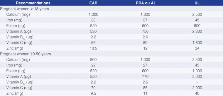
The subject of nutrition in pregnancy is complex due to the fact that it involves multiple factors that interfere with the health of mother and child, some of which were presented briefly in specific topics in this review. Special care should be exercised with some micronutrients as suggested in Table 6 according to the recommendations of Rauber and Vitolo (2012).
In the first trimester of pregnancy, the maternal energy intake should be similar to that of the pregestational period, that is, approximately 2,000 Kcal per day, considering a diet with a proper balance of nutrients. In the second and third trimesters, an additional intake of 300 Kcal is recommended, considering a diet such as that recommended by the RDA. Table 6 illustrates the recommended intakes of some nutrients for the different ages of pregnant women. Some of the nutrients mentioned in Table 6 are critical for the development of the fetus and newborn; therefore, they should be evaluated with criteria.
Calcium. Hormonal alterations that occur in pregnancy promote adjustments to the calcium metabolism, so as to promote increased biological use of that element during this period. Therefore, despite the calcium requirements being greater in pregnancy, dietary recommendations remain
Table 6. Dietary intake of the most important micronutrients in pregnancy for the different ages of pregnant woman.
Recommendations EAR RDA ou AI UL
Pregnant women < 18 years
Calcium (mg) 1,000 1,300 2,500
Iron (mg) 23 27 45
Folate (µg) 520 600 800
Vitamin A (µg) 530 750 2,800
Vitamin B12 (µg) 2.2 2.6
-Vitamin C (mg) 66 80 1,800
Zinc (mg) 10.5 12 34
Pregnant women 19-50 years
Calcium (mg) 800 1,000 2,500
Iron (mg) 22 27 45
Folate (µg) 520 600 1,000
Vitamin A (µg) 550 770 3,000
Vitamin B12 (µg) 2.2 2.6
-Vitamin C (mg) 70 85 2,000
Zinc (mg) 9.5 11 40