Patrícia Corrêa-Faria(a)
Paulo Antônio Martins-Júnior(a) Raquel Gonçalves Vieira- Andrade(a)
Leandro Silva Marques(b) Maria Letícia Ramos-Jorge(a)
(a)Department of Pediatric Dentistry, School of Dentistry, Univ Federal dos Vales do Jequitinhonha e Mucuri - UFVJM, Diamantina, MG, Brazil.
(b)Department of Orthodontics, School of Dentistry, Univ Federal dos Vales do Jequitinhonha e Mucuri - UFVJM, Diamantina, MG, Brazil.
Corresponding Author: Patrícia Corrêa-Faria
E-mail: [email protected]
Perinatal factors associated with
developmental defects of enamel in
primary teeth: a case-control study
Abstract: The present study was designed to evaluate associations be-tween developmental defects of enamel (DDE) in the primary dentition and aspects related to mothers and preschoolers in the city of Diaman-tina, Brazil. A case-control study was carried out involving children aged three to ive years. The case group was composed of 104 children with at least one dental surface affected by DDE. The control group comprised 105 children without DDE, matched for gender and age. The diagno-sis of enamel defects was performed using the Developmental Defects of Enamel Index. Information was collected through interviews investigat-ing socio-demographic aspects, gestation, birth weight, prematurity and breastfeeding. Simple and multiple regression analyses were performed, providing unadjusted and adjusted prevalence ratios (Poisson regression). DDE were more prevalent among children who had not been breastfed (PR = 1.57; 95% CI: 1.1–2.2) and those whose mothers were under 24 years of age at the birth of the child (PR = 1.41; 95% CI: 1.1–1.9). The prevalence of DDE in the primary dentition was higher among children who had not been breastfed and those whose mothers were under 24 years of age at the birth of the child.
Descriptors: Tooth, Deciduous; Dental Enamel Hypoplasia; Dental Enamel.
Introduction
Developmental defects of enamel (DDE) are deined as disturbances in hard tissue matrices and mineralization occurring during odontogen-esis. These defects are classiied as either enamel hypoplasia or enamel opacities, based on clinical appearance.1-3 Enamel hypoplasia is
associ-ated with a reduction in the thickness of the enamel in either a localized or a more widespread area, but the enamel matrix is mineralized nor-mally.1 It may manifest clinically in the form of pits and grooves, or else a
partial/total lack of surface enamel.1,2 In the case of enamel opacities, the
matrix is secreted to form a normal thickness, but parts fail to mature or mineralize properly, leaving regions with deicient mineral content, which appear as diffuse or demarcated opacities of yellowish or brown coloration.4
The prevalence of DDE in the primary dentition ranges from 24.4% to 81.3% in different countries.5,6 The etiological factors associated with
acquired DDE may act prenatally, perinatally or postnatally, and may
Declaration of Interests: The authors certify that they have no commercial or associative interest that represents a conflict of interest in connection with the manuscript.
Submitted: Oct 08, 2012
be either systemic or localized. Studies have demon-strated that premature birth, low birth weight and lack of breastfeeding are the main causes of DDE in primary teeth.7-8 Furthermore, enamel defects are
related to social aspects, as well as nutritional and systemic problems in childhood.9-11 The extent of
DDE depends on the intensity of the etiological fac-tor, its duration and the period in which the factor was present during the formation of the crown.12
Teeth with DDE are more susceptible to caries, insofar as these teeth have retentive areas that make them more susceptible to the accumulation of bac-terial plaque. Hypocalciication can lead to a more rapid progression of carious lesions. DDE can also affect the esthetics of the maxillary incisors, exert-ing a negative impact on social and psychological aspects.13 Thus, diagnosing and understanding the
factors associated with DDE may contribute toward the prevention or minimization of adverse outcomes stemming from such defects.6,14
The present study was designed to evaluate as-sociations between developmental defects of enamel (DDE) in the primary dentition and aspects related to mothers and preschoolers in the city of Diaman-tina, Brazil.
Methodology
A case-control study was conducted in the city of Diamantina, which is located in the northern portion of the state of Minas Gerais in southeast-ern Brazil. The study population included children from three to ive years of age treated at the ten ba-sic healthcare units in the city during immunization campaigns held in 2010. Diamantina has 90% vac-cine coverage.
This investigation was carried out in two steps. A cross-sectional study was initially conducted to estimate the prevalence of DDE in preschool chil-dren aged three to ive years. All teeth were evalu-ated by examiners submitted to a calibration pro-cedure. In the second phase, a case-control study was conducted to identify risk factors associated with DDE in the primary dentition. The case group comprised 104 children with DDE and the control group was made up of 105 children without DDE, matched for gender and age. Children who did not
cooperate during the exam and those with lip and/ or palate abnormalities15 or systemic problems, such
as asthma16 and celiac disease,17 which are
associ-ated with DDE, were excluded from the study. Data acquisition involved a clinical oral exam, an-thropometric measures and a questionnaire adminis-tered in the form of an interview. The selection of the children for the case and control groups and the data collection were carried out on two separate days dur-ing the 2010 National Child Vaccination Campaign. A team of three researchers (an examiner and two assistants) was installed in each healthcare unit.
Prior to the ieldwork, the examiners underwent a calibration and training exercise for DDE diagno-sis. Calibration was performed using images of dif-ferent clinical situations, on two separate occasions, with a one-week interval between sessions. Kappa values were calculated. Minimum intraexaminer agreement was 0.81 and minimum interexaminer agreement was 0.80. A training exercise was then carried out, involving the clinical oral exam of chil-dren in the same age group as those participating in the main study.
The exam was performed with a disposable tongue depressor under natural light and gauze to clean the teeth. The child remained seated in a chair in front of the examiner and facing a window to make maximum use of natural light. The three types of DDE were assessed (diffuse opacity, demar-cated opacity and enamel hypoplasia) and classiied based on the criteria of the Modiied Index of De-velopment Defects of Enamel.4 Teeth with carious
lesions that rendered the diagnosis of enamel defects impossible were excluded.
Anthropometric measures (weight and height) were used to assess nutritional status. The weight of the children was determined by placing them on a digital scale (Plenna, São Paulo, Brazil); food intake and excrement elimination were not taken into con-sideration. Height was determined by a stadiometer with a millimeter scale and two-meter limit (Welmy, Porto Alegre, Brazil), placed on a lat surface. Nutri-tional status was determined by comparing measure-ments with the reference standards stipulated by the American National Center for Health Statistics.18
ob-and hypoplasia (4.2%).
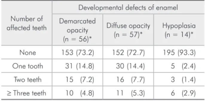
In the case-control study, the mean age of the children examined was 49.06 months (standard de-viation = 6.68). The most common type of DDE was diffuse opacity (27.3%; n = 57), followed by demar-cated opacity (26.8%; n = 56) and hypoplasia (6.7%; n = 14). Table 1 shows the distribution of DDE ac-cording to the number of affected teeth per child. Table 2 shows the results of the associations be-tween DDE and both socio-demographic and birth-related variables.
The following maternal aspects and variables related to the child were associated signiicantly with the occurrence of DDE (p < 0.05): mother’s age at the birth of the child (p = 0.012), breastfeed-ing (p = 0.016) and monthly household income (p = 0.030). In the unadjusted model, the mother’s age at the birth of the child (PR: 1.47; 95% CI: 1.1 to 1.9), absence of breastfeeding (PR: 1.67; 95% CI: 1.2 to 2.3) and monthly household income less than twice the minimum wage (PR: 1.36; 95% CI: 1.0 to 1.8) were signiicantly associated with DDE. In the adjusted multivariate regression model, the mother’s age at the birth of the child (PR: 1.41; 95% CI: 1.1 to 1.9) and absence of breastfeeding (PR: 1.57; 95% CI: 1.1 to 2.2) were signiicantly associated with a higher prevalence of DDE in the primary dentition, independently of monthly household income (Table 3).
Discussion
The present study examined associations between the occurrence of DDE in the primary dentition and tained from the vaccination card. The children were
classiied based on birth weight using the criteria of the World Health Organization:
• very low birth weight (less than 1500 g),
• low birth weight (less than 2500 g) and
• normal birth weight (equal to or greater than 2500 g).19
Interviews were conducted with parents/guard-ians to gather information on socio-demographic as-pects (monthly household income, mother’s school-ing, number of children in the family, and place of residence), mother’s age at the birth of the child, child’s age, gender and gestational age. Gestational age was classiied as
• “full term” (37 or more weeks of gestation) and
• “premature birth” (less than 37 weeks of gesta-tion).19
Data analysis was performed using the Statistical Package for the Social Sciences (SPSS for Windows, version 17.0, SPSS Inc., Chicago, USA) and included frequency distribution and association tests. Associ-ations between DDE and the independent variables were determined using the chi-squared test. Pois-son regression with robust variance was performed for the analysis of factors associated with DDE. The magnitude of each factor associated with the presence of DDE was determined using unadjusted prevalence ratios (PR), respective 95% conidence intervals (CI) and p-values (Wald test). Explanato-ry variables with a p-value ≤ 0.20 for the bivariate analysis were incorporated into the model.
This study received approval from the Human Research Ethics Committee of the Universidade Federal dos Vales do Jequitinhonha e Mucuri, Brazil. Parents/guardians signed a statement of in-formed consent.
Results
None of the mothers refused to participate in either phase of the investigation. The prevalence of DDE in preschool children aged three to ive years (29.9%) was determined in the irst phase of the study. The most common type was demarcated opacity (16.8%), followed by diffuse opacity (15.7%)
Table 1 - Distribution of types of developmental defects of enamel in primary teeth according to the number of affected teeth per child in the case group (n = 104).
Number of affected teeth
Developmental defects of enamel Demarcated
opacity (n = 56)*
Diffuse opacity
(n = 57)* Hypoplasia(n = 14)*
None 153 (73.2) 152 (72.7) 195 (93.3) One tooth 31 (14.8) 30 (14.4) 5 (2.4) Two teeth 15 (7.2) 16 (7.7) 3 (1.4)
aspects related to mothers and preschoolers, par-ticularly breastfeeding. Children who had not been breastfed had more enamel defects than those who had been breastfed. This result is in agreement with the indings of a previous case-control study car-ried out with Brazilian children, in which the lack of
breastfeeding was associated with a 3.2-fold greater chance of DDE in the primary dentition.20
Human milk is a child’s primary source of nutri-ents in the irst months of life, including calcium. Cal-cium ions regulate cell activities, such as cell commu-nication, signal transduction, enzyme activation and the polymerization of cytoskeletal proteins.21
Calci-um also participates in the stage of amelogenesis in which there is loss of proteins from the matrix and in-put of calcium and potassium from the blood vessels that contribute to forming tooth enamel.22 A
com-plete absence of calcium from one’s diet is reported to result in hypomineralization of the enamel.23 This
lends support to the hypothesis that the absence of breastfeeding deprives a child of calcium and causes DDE. However, this hypothesis does not appear to be valid for all teeth, insofar as the formation of the enamel matrix and calciication begin about the 15th
week of intrauterine life;24 however, this theory may
be true for teeth that complete their formation in the post-uterine period. Moreover, since the tooth forma-tion process may be altered by nutriforma-tional, vitamin and mineral deiciencies,2,9-11 the absence of
breast-feeding may reduce the amount of nutrients during tooth formation, thereby leading to DDE.
No statistically signiicant association was found between premature birth and DDE. This inding may be explained by the small number of study children who had been born prematurely. More-over, issues related to how this information associat-ing premature birth and DDE was acquired could be the result of an interview format conditioned by the memory bias of the respondent. Previous studies carried out in different countries report that enam-el defects are associated with premature birth.7-8,20
Children born prematurely may be affected by seri-ous disorders related to calcium metabolism during the neonatal period, caused not only by prematurity itself, but also by numerous other complications en-countered in these children.25
Maternal factors, such as age at the birth of the child, use of medications, infections and smoking during the prenatal period, have been associated with a higher prevalence of DDE.26 In the present
study, the mother’s age at the birth of the child was signiicantly associated with DDE. Children whose
Table 2 - Association between aspects related to child, mother and socio-demographic factors, and occurrence of developmental defects of enamel in the primary teeth of preschool children in Diamantina, Brazil (chi-squared test; p < 0.05).
Cases
(n = 104) (n = 105)Controls P Gestation, birth and breastfeeding
Age of mother at birth of child
• ≥ 24 69 (44.2) 87 (55.8)
• < 24 30 (65.2) 16 (34.8) 0.012 Gestation
• Full-term 89 (48.9) 93 (51.1)
• Premature 9 (45.0) 11 (55.0) 0.740
Birth weight
• < 2500 g 9 (39.1) 14 (60.9)
• ≥ 2500 g 94 (50.8) 91 (49.2) 0.291 Breastfeeding
• Yes 92 (47.7) 101 (52.3)
• No 12 (80.0) 3 (20.0) 0.016 Breastfeeding duration (months)
• ≥ 6 65 (51.2) 62 (48.8)
• < 6 27 (48.2) 29 (51.8) 0.711 Current child variables
Nutritional diagnosis
• Ideal weight range 84 (48.0) 91 (52.0)
• Overweight 6 (54.5) 5 (45.5)
• Malnourished 12 (60.0) 8 (40.0) 0.562
Socio-demographic factors Mother’s schooling
• ≥ 8 years 45 (48.4) 48 (51.6)
• < 8 years 59 (50.9) 57 (49.1) 0.722 Household income
• ≥ 2 times the
minimum wage 70 (45.5) 84 (54.5) • < 2 times the
mothers were under 24 years of age at the time of birth had a greater frequency of enamel defects. A mother’s age is a well-known determinant of com-plications during pregnancy and delivery.27
Preg-nancy in adolescence is associated with a higher risk of adverse outcomes, including low birth weight and premature birth.28 This hypothesis should be further
investigated in longitudinal studies aimed at deter-mining associations between complications during pregnancy and DDE in the primary dentition.
A mother’s age at the birth of the child was also signiicantly associated with monthly household income (p < 0.001) and schooling (p = 0.035), and may therefore be used as a complementary datum in the evaluation of socioeconomic status. It is suggest-ed that young mothers with a lower socioeconomic status are more susceptible to complications during pregnancy due to less access to healthcare services, lack of prenatal care or unsatisfactory assistance, resulting in premature birth and low birth weight, which are related to the etiology of DDE.
The association between breastfeeding and DDE found in the present study suggests the importance of breastfeeding in preventing these defects in the primary dentition. Breastfeeding contributes to re-duced illness rates and complications in early life,
and is related to the development of dental struc-tures.20 Therefore, breastfeeding and the follow-up
of growth and development in the initial years of life are strongly recommended in promoting both gener-al and orgener-al hegener-alth, as well as preventing undesirable conditions in children. In regard to enamel defects, these measures should be combined with the identi-ication of risk factors and conditions such as dental caries, to establish early control and minimization of problems resulting from DDE.
The common occurrence of premature birth, low birth weight, nutritional deiciencies and lack of breastfeeding in poor regions underscores the need for longitudinal studies addressing social and bio-logical factors associated with DDE.20 The
identii-cation of these factors is important for the adoption of preventive measures against not only these de-fects but also other oral and systemic problems with common risk factors.
Conclusion
The prevalence of developmental defects of enamel in the primary dentition was higher among children who had not been breastfed and those whose mothers were under 24 years of age at the birth of the child.
Variable Unadjusted PR (95% CI) p Adjusted PR (95% CI) p
Age of mother at birth of child (years)
• ≥ 24 1.0 1.0
• < 24 1.47 (1.1–1.9) 0.006 1.41 (1.1–1.9) 0.018 Breastfeeding
• Yes 1.0 1.0
• No 1.67 (1.2–2.3) 0.001 1.57 (1.1–2.2) 0.007
Household income
• ≥ 2 times the minimum wage 1.0 1.0
• < 2 times the minimum wage 1.36 (1.0–1.8) 0.034 1.26 (0.94–1.69) 0.123 Table 3 - Prevalence ratio
and confidence intervals for associations between developmental defects of enamel and variables analyzed (p < 0.05).
References
1. Suckling GW. Developmental defects of enamel – historical and present-day perspectives of their pathogenesis. Adv Dent Res. 1989 Sep;3(2):87-94.
3. Slayton RL, Warren JJ, Kanellis MJ, Levy SM, Islam M. Prevalence of enamel hypoplasia and isolated opacities in the primary dentition. Pediatr Dent. 2001 Jan-Feb;23(1):32-6. 4. A review of developmental defects of the enamel dental
in-dex (DDE Inin-dex). Commission on Oral Health, Research & Epidemiology. Report of an FDI Working Group. Int Dent J. 1992 Dec;42(6):411-26.
5. Lunardelli SE, Peres MA. Prevalence and distribution of devel-opmental enamel defects in the primary dentition of pre-school children. Braz Oral Res. 2005 Apr-Jun;19(2):144-9. 6. Targino AG, Rosenblatt A, Oliveira AF, Chaves AM, Santos
VE. The relationship of enamel defects and caries: a cohort study. Oral Dis. 2011 May;17(4):420-6.
7. Corrêa-Faria P, Martins-Júnior PA, Vieira-Andrade RG, Oliveira-Ferreira F, Marques LS, Ramos-Jorge ML. Devel-opmental defects of enamel in primary teeth: prevalence and associated factors. Int J Paediatr Dent. 2012 May 1. doi: 10.1111/j.1365-263X.2012.01241.x. Epud ahead of print. 8. Pinho JR, Lamy Filho F, Thomaz EB, Lamy ZC, Libério SA,
Ferreira EB. Are low birth weight, intrauterine growth restric-tion, and preterm birth associated with enamel developmental defects?. Pediatr Dent. 2012 May-Jun;34(3):244-48. 9. Massoni AC, Chaves AM, Rosenblatt A, Sampaio FC, Oliveira
AF. Prevalence of enamel defects related to pre-, peri and postnatal factors in a Brazilian population. Community Dent Health. 2009 Sep;26(3):143-9.
10. Takaoka LA, Goulart AL, Kopelman BI, Weiler RM. Enamel defects in the complete primary dentition of children born at term and preterm. Pediatr Dent. 2011 Mar-Apr;33(2):171-6. 11. Rugg-Gunn AJ, Al-Mohammadi SM, Butler TJ. Malnutrition and developmental defects of enamel in 2- to 6-year-old Saudi boys. Caries Res. 1998 May-Jun;32(3):181-92.
12. Regezi JA, Sciubba JJ. Oral Pathology. 3th ed. Rio de Janeiro: Guanabara Koogan; 2000. Chapter 16, Anomalies of the dentition; p. 405-25.
13. Vargas-Ferreira F, Ardenghi TM. Developmental enamel de-fects and their impact on child oral health-related quality of life. Braz Oral Res. 2011 Nov-Dec;25(6):531-7.
14. Carvalho JC, Silva EF, Gomes RR, Fonseca JA, Mestrinho HD. Impact of enamel defects on early caries development in preschool children. Caries Res. 2011 Sep;45(4):353-60. 15. Ruiz LA, Maya RR, D’Alpino PH, Atta MT, Syizero NR.
Prevalence of enamel defects in permanent teeth of patients
with complete cleft lip and palate. Cleft Palate Craniofac J. 2012 Jan 31. Epub ahead of print.
16. Widmer RP. Oral health of children with respiratory diseases. Paediatr Respir Rev. 2010 Dec;11(4):226-32.
17. Rashid M, Zarkadas M, Anca A, Limeback H. Oral manifes-tations of celiac disease: a clinical guide for dentists. J Mich Dent Assoc. 2011 Oct;93(10):42-6.
18. Hamill PVV, Drizd TA, Johnson CL, Reed RB, Roche AF, Moore WM. Physical growth: National Center for Healt sta-tistics percentiles. Am J Clin Nutr. 1979 Mar;32(3):607-29. 19. WHO: recommended definitions, terminology and format for
statistical tables related to the perinatal period and use of a new certificate for cause of perinatal deaths. Modifications recommended by FIGO as amended October 14, 1976. Acta Obstet Gynecol Scand. 1977 Jan;56(3):247-53.
20. Lunardelli SE, Peres MA. Breast-feeding and mother-child factors associated with developmental enamel defects in the primary teeth of Brazilian children. J Dent Child (Chic). 2006 May-Aug;73(2):70-8.
21. Posner AS, Perloff A, Diorio AF. Refinement of the hydroxy-apatite structure. Acta Crystallogr. 1958 Apr;11(4):308-9. 22. Warshawsky H, Nanci A. Stereo electron microscopy of
enamel crystallites. J Dent Res. 1982 Dec;Spec No:1504-14. 23. Bonucci E, Lozupone E, Silvestrini G, Favia A, Mocetti P. Morphological studies of hypomineralized enamel of rat pups on calcium-deficient, and of its changes after return to normal diet. Anat Rec. 1994 Aug;239(4):379-95.
24. Rythén M, Sabel N, Dietz W, Robertson A, Nóren JG. Chemi-cal aspects on dental hard tissues in primary teeth from pre-term infants. Eur J Oral Sci. 2010 Aug;118(4):389-95. 25. Seow WK. Effects of preterm birth on oral growth and
devel-opment. Aust Dent J. 1997 Apr;42(2):85-91.
26. Velló MA, Martínez-Costa C, Catalá M, Fons J, Brines J, Guijarro-Martínez R. Prenatal and neonatal risk factors for the development of enamel defects in low birth weight chil-dren. Oral Dis. 2010 Apr;16(3):257-62.