C
ritiCalreviewEndodontic Therapy
Manoel Damião de SOUSA-NETO(a)
Yara Correa SILVA-SOUSA(b)
Jardel Francisco MAZZI-CHAVES(c)
Kleber Kildare Teodoro CARVALHO(a)
Ana Flávia Simões BARBOSA(b)
Marco Aurélio VERSIANI(a)
Reinhilde JACOBS(c)
Graziela Bianchi LEONI(b)
(a) Universidade de São Paulo, School of
Dentistry of Ribeirão Preto, Department of Restorative Dentistry, Ribeirão Preto, SP, Brazil;
(b) Universidade de Ribeirão Preto, Faculty of
Dentistry, Ribeirão Preto, SP, Brazil.
(c) Katholieke Universiteit Leuven, Faculty of
Medicine, Department of Imaging and Pathology, Leuven, Belgium.
Root canal preparation using
micro-computed tomography analysis:
a literature review
Abstract: This literature review has critically analyzed the published
research related to the biomechanical preparation of root canals
with three-dimensional analysis using micro-computed tomography
(micro-CT). In December 2017, six databases (PubMed, Cochrane, Web
of Science, Embase, Scopus, and Science Direct) were accessed using
keywords to find articles including the use of the micro-CT analysis
in biomechanical root canal preparation. There were 60 full articles
that were selected, which were screened and read by two authors. The
research that was reviewed and analyzed included root canal anatomy
and sample selection, changes in canal shape and untouched canal areas,
canal transportation and centering ability, and kinematics (motion). Of
the studies selected, 49.18% discussed anatomical characteristics, with
54.1% of these studies describing mesial roots of mandibular molars
with moderate curvature. Only 35% used a stratified distribution based
on root canal system morphology and quantitative data obtained by
micro-CT. The analysis of canal transportation and centering ability
showed that transport values in the apical third exceeded the critical
limit of 0.3 mm in mesial roots of mandibular molars with moderate
curvature, especially in the groups in which a reciprocating system
was used. In relation to kinematics, 91.70% of the reviewed studies
evaluated continuous rotating instruments, followed by reciprocating
rotation (38.33%), vibratory (15%), and the adaptive kinematics, which
was in only 8.33%. The reciprocating kinematics was associated with
higher canal decentralization and transportation indexes, as well as
a greater capacity for dentin removal and debris accumulation. This
literature review showed that the anatomy, the type of design and
kinematics of instruments, and the experimental design are factors
that directly influence the quality of biomechanical preparation of root
canals analyzed in a qualitative and quantitative manner by micro-CT.
Keywords: Endodontics; Root Canal Preparation; X-Ray
Microtomography.
Introduction
Biomechanical root canal preparation is an important endodontic treatment
step. The goal is the complete removal of remaining pulp tissue, microorganisms,
and infected dentin; as well as shaping of the root canal system (RCS) through
Declaration of Interests: The authorscertify that they have no commercial or associative interest that represents a conflict of interest in connection with the manuscript.
Corresponding Author: Manoel Damião de Sousa-Neto E-mail: [email protected]
https://doi.org/10.1590/1807-3107bor-2018.vol32.0066
Submitted: May 03, 2018
Accepted for publication: May 29, 2018 Last revision: June 06, 2018
Sousa-Neto MD Silva-Sousa YC, Mazzi-Chaves JF, Carvalho KKT, Barbosa AFS, Versiani MA et al.
the mechanical action of endodontic instruments and
the chemical action of auxiliary solutions providing
adequate conditions for the sealing of the pulp cavity
and repair of the periapical tissues.
1,2,3,4,5Since 2012, new protocols for biomechanical root
canal preparation have been developed using
nickel-titanium (NiTi) instruments, whose flexibility and
resistance to torsion allow their use in a continuous
rotating movement; these in turn reduce the working
time, operator fatigue, and the risk of operative
accidents.
6,7,8,9,10,11,12,13,14,15NiTi instruments have been
developed with various geometric conformations,
8with the most significant design difference being the
taper with conicity ranging from 2% to 12%, compared
to the standard 2% taper of stainless steel instruments
established by ANSI/ADA in 1976 and updated in 1982.
8NiTi rotary instruments were first commercialized
in the 1990s, and since then about 700 studies have
been published in journals indexed in PubMed before
June 2010. They have evaluated the performance of
these instruments, as well as the newly developed
instrument designs. Studies have shown that despite the
flexibility, torsional strength, and elastic memory of NiTi
instruments, they still leave a significant percentage of
the canal surface untouched. This is mainly due to the
anatomical characteristics of root canals, such as flattening,
curvatures, isthmuses, recesses, and ramifications, which
hinder the performance of the instrument and may leave
tissue and bacterial remnants
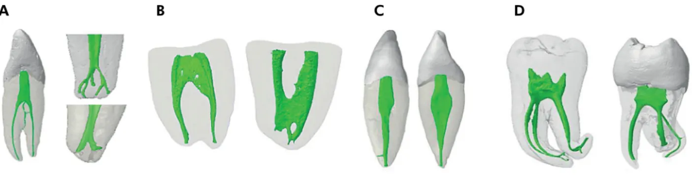
16,17,18,19,20,21,22,23(Figure 1).
Another concern noted when NiTi rotary
instruments first appeared was a screw effect, which
added to the difficulty of preparing challenging
anatomical areas. This led to the development of
more than 150 mechanized systems with differing
designs, including variable tapers as well as different
alloy treatments and movement types.
2,8,24,25,26,27,28,29,30In this review, various mechanical systems have
been reviewed and discussed according to the
innovations in the design, type of alloy treatment,
movements, and concept of recommended preparation,
without commercial bias (Table 1). It is noted that
some systems may incorporate more than one of the
mentioned characteristics.
An attempt to overcome the limitations imposed by
the anatomical complexity of the RCS resulted in the
development of the Self-Adjusting File (SAF) instrument
(Redent-Nova Inc., Ra’anana, Israel) with an innovative
manufacturing process. It is an instrument with a
distinctive design that features a hollow, compressible,
thin-walled body, composed of a delicate NiTi trellis
covered by an abrasive layer. The use of this single
instrument allows its adaptation inside the root canal,
and by its vibratory movement (3000 to 5000 vibrations
per minute) of low amplitude (0.4 mm), it promotes
uniform dentine wear, which results in a canal with a
cross section similar to the original but with slightly
larger dimensions.
2,29,30,31,32,33,34,35Several reciprocating systems have been released
26such as Waveone (Dentsply Maillefer, Baillagues,
Switzerland) and Reciproc (VDW GmbH, Munich,
Germany), which are based on the concept of root
canal preparation with a single instrument. These
instruments are manufactured with a M-Wire alloy
from a thermal treating process of the NiTi alloy,
A
B
C
D
Figure 1. Three-dimensional models of root canal system of human teeth obtained from microcomputed tomography scanning showing the importance of the diagnosis of anatomical challenges prior to the biomechanical preparation of the root canal system. (A) Presence of accessory and lateral canals, and apical deltas. (B) Isthmus. (C) Flattened canals. (D) Presence of moderate and severe curvatures.
Root canal preparation using micro-computed tomography analysis: a literature review
which provides greater flexibility and resistance to
cyclic fatigue than the conventional NiTi alloy.
4,9,11,13, 15,33,34,35,36,37,38,39,40,41,42,43,44,45The movement dynamics of
these instruments, known as reciprocating, is the
rotation in the counterclockwise direction (cutting
direction) followed by a less extensive clockwise
rotation (instrument releasing direction), facilitating
continuous and progressive movement toward
apical.
15,26According to some authors, the reciprocating
movement reduces the risk of torsion fracture because
the instrument is not subjected to the stress levels
caused by a continuous rotary motion.
15,26,46,47,48Another system for the preparation of the root
canal based on the concept of a single instrument,
but with continuous rotation, is the OneShape system
(MicroMega, Besançon, France).
11,49,50This system is
produced from conventional NiTi alloy using a tip
diameter of #25 and continuous 0.06 taper, with three
different cross-sections along the active part, variable
pitch, and idle spirals, with the goal of reducing the
tapping effect.
11,50,51,52This was also a goal for several
other systems, including Race (FKG Dentaire S.A., La
Chaux-de-Fonds, Switzerland), Rondo (FKG Dentaire
S.A., La Chaux-de-Fonds, Switzerland), EndoSequence
(Brasseler USA Dental, Savannah, USA), and EndoWave
(J. Morita Corporation, Osaka, Japan), which presented
a design of helical angles and alternating areas and,
more recently, systems including BioRace (FKG Dentaire
S.A., La Chaux-de-Fonds, Switzerland), Revo-S
(Micro-Mega, Besançon, France) and ProTaper Next (Dentsply
Maillefer, Ballaigues, Switzerland).
With a combination of continuous and reciprocating
motions, the Twisted File Adaptive system was
developed (SybronEndo, Orange, USA) with
instruments made from a phase-R thermal treatment
alloy that adapted the kinematics to the stress of the
Table 1. Mechanized systems cited in the reviewed studies according to the kinematics and alloys treatment used in their manufacture.Kinematics Alloy type
NiTi with no treatment NiTi with treatment
Continous
BioRace (FKG Dentaire) HyFlex CM (Coltene)
Hero (Micro-Mega) K3XF (SybronEndo)
Hyflex (Coltene) OneShape (Micro Mega)
K3 (SybronEndo) ProTaper Gold (Dentsply)
Mtwo (VDW) TRUShape (Dentsply)
ProTaper Next (Dentsply) Vortex Blue (Dentsply)
ProTaper Universal (Dentsply) XP-endo Shaper (FKG Dentaire)
Revo-S (Micro-Mega) XP-endo Finisher (FKG Dentaire)
EdgeFile (EdgeEndo) Twisted File (SybronEndo)
iRaCe (FKG Dentaire) RaCe (FKG Dentaire)
FlexMaster (VDW) ProGlider (Dentsply)
System GT (Dentsply) ScoutRace (FKG Dentaire)
Profile (Dentsply) Ease ProDesign Logic (Easy)
GT Rotary File (Dentsply) HyFlex EDM (Coltene)
EndoSequence (Brasseler) BT Race (FKG Dentaire)
EndoEZE AET (Ultradent)
-Vortex (Dentsply)
-Wizard Navigator (Medin)
-Reciprocating
Reciproc (VDW)
Reciproc Blue (VDW) WaveOne (Dentsply)
EZ-Fill Safesider system (EDS)
Adaptative - Twisted File Adaptive (SybronEndo)
Vibratory Self-Adjusting File (ReDent-Nova)
Sousa-Neto MD Silva-Sousa YC, Mazzi-Chaves JF, Carvalho KKT, Barbosa AFS, Versiani MA et al.
instrumentation. This system was designed to permit
switching from a continuous clockwise motion (when
the instrument is not subjected to stress within the
canal) to an interrupted reciprocation motion (when
undue tensions are generated by dentin) during
instrumentation.
37,44,45,47,53,54,55,56,57,58There are also instruments designed with center
of mass and/or center of rotation in offset that, in
rotation, produce a mechanical wave that runs through
the active part of the instrument. This results in
improvement of the flexibility along the active part of
the instrument and minimizes the instrument locking
in the dentin, in addition to reducing the formation
of debris.
8,30,34,41,59These features were incorporated
into additional systems including Protaper Next
(Dentsply Maillefer, Ballaigues, Switzerland),
TRUShape (Dentsply Tulsa Dental Specialties, Tulsa,
USA), Revo-S (Micro-Mega, Besançon, France), and
One Shape (Micro-Mega, Besançon, France).
Recently, the concept of mechanical finalization of
the biomechanical preparation was proposed. Following
the mechanical preparation of the root canal, the
instrument whips against the walls of the root canal
allowing its action in untouched areas such as isthmuses,
flattenings, and recesses. This action is possible with the
XP-endo Finisher instrument (FKG Dentaire S.A., La
Chaux-de-Fonds, Switzerland), which is produced with
highly flexible NiTi MaxWire alloy (25/.00)
(Martensite-Austenite Electropolish-FleX) that changes shape at
different temperatures. The instrument is straight in
the martensite phase (M-phase) of the alloy, which is
reached when it is cooled, and when it is exposed to
higher temperatures (such as body temperature) its shape
changes due to molecular memory of the alloy to the
austenite phase (A-phase). This makes the instrument
assume a semi-circular conformation that, in rotation,
allows it to reach an area of 6 mm in diameter that is
100 times greater than that of other instruments.
60,61In
2017, Leoni et al.
61evaluated the possibility of using the
XP-endo Finisher instrument as well as the SAF in the
mechanical finishing of root canal preparation with
isthmuses, demonstrating the reduction of accumulated
debris after biomechanical preparation.
Later, with the combination of this NiTi MaxWire
alloy and Booster Tip technology, the XP-endo Shaper
(FKG Dentaire S.A., La Chaux-de-Fonds, Switzerland)
offered greater flexibility, fatigue resistance, and
penetration of the canals with ease and speed, expanding
or contracting according to canal morphology and
preserving the three-dimensional structure of the
root canal.
62,63Similar to the preparation finalization
instrument mentioned above, this instrument can react
to temperature variations and acquires a predetermined
shape at body temperature, with the taper of the
instrument starting at .01 and reaching a minimum .04
taper when it expands into the root canal. The Booster
Tip has unique geometry that allows the operator to
start the preparation after an initial glide path of at
least ISO diameter 15, increasing its working range
gradually until reaching ISO diameter 30, following
the original canal path.
29,62In addition to the aforementioned innovations, the
thermomechanical treatment process of the alloys
used in the fabrication of these instruments alters the
molecular structure of the alloy, providing resistance
to cyclic fatigue and flexibility while reducing shape
memory, allowing pre-bending of the alloys.
28,62,64,65,66Use of this enhanced treatment process resulted in
the development of blue rotary files.
28The Vortex
Blue system (Dentsply Tulsa Dental Specialties, Tulsa,
USA) and, more recently, the Reciproc Blue system
(VDW GmbH, Munich, Germany) are features in a
reciprocating kinematics.
Parallel to the development of mechanized
instruments and systems for clinical use, experimental
models for the evaluation of the biomechanical
preparation of the RCS in human teeth were
perfected. In previously available experimental
models, biomechanical preparation was assessed by
radiographic images
67,68,69,70and root cutting series
using the muffle system or its variations;
71,72however,
these methods allowed only a two-dimensional
quantitative evaluation after preparation, and the
muffle system was destructive.
73,74,75A solution for the three-dimensional and
non-destructive evaluation of the RCS appears to be the
use of computed tomography and magnetic resonance
imaging in experimental procedures.
27,76,77,78The
development of micro-CT allows a more precise
RCS evaluation than a conventional CT scanner,
and more recent use of specific software has made
it possible to accurately assess the biomechanical
Root canal preparation using micro-computed tomography analysis: a literature review
preparation as well as the anatomy, root canal filling,
and retreatment.
2,3,4,5,7,9,11,12,13,14,15,25,27,28,29,30,31,34,35,37,38, 39,40,41,42, 43,44,45,47,56,57,58,59,61,62,63,68,79,80,81, 82,83,84,85,86,87,88,89,90,91, 92,93,94,95,96,97,98,99,100,101,102,103,104,105,106,107,108,109
The aim of this review is to present a discussion
of the studies that evaluated the preparation of root
canals by three-dimensional analysis based on the use
of micro-CT while considering the evolution of NiTi
instruments and biomechanical preparation evaluation
methods, as well as the limitations imposed by the
anatomical aspects of the RCS and the resulting need
for sample selection for biomechanical preparation
evaluation studies. The topics that will be addressed
include root canal anatomy and sample selection,
changes in canal shape and untouched canal areas,
canal transportation and centering ability, and
kinematics (motion).
Methodology
This literature review followed the PRISMA
(Preferred Reporting Items for Systematic Reviews
and Meta-Analyses) guidelines for literature review
to ensure the understanding, transparency, and
fidelity of the results.
110,111The search strategy was used in six databases
(PubMed, Cochrane, Web of Science, Embase,
Scopus, and Science Direct), with the same key
word combinations and MeSH Terms (Table 2),
including titles, abstracts, and full texts. The search
was performed in December 2017, and there was no
initial restriction regarding the year of publication,
article language, or journal publication.
After the initial search, all of the titles and abstracts
were screened by two authors to find eligible full
articles to be included in the literature review.
The evaluation of the mechanized (rotary,
reciprocating, oscillatory) biomechanical preparation
of the RCS by micro-CT was the inclusion criteria
used in the article selection. Furthermore, to reduce
the risk of bias, it included only studies in the English
language that evaluated mechanized endodontic
systems, used human permanent teeth (any type of
tooth groups and root canals), and analyzed
two-dimensional and three-two-dimensional parameters
(canal transportation, centering ability, changes
in the volume, surface area, perimeter and area,
and others) of the root canals before and after the
biomechanical preparation.
Studies written in a language other than English;
those which used endodontic hand files, deciduous
teeth, acrylic/resin blocks with simulated root canals;
and other types of analyses that did not include
biomechanical preparation assessment were all
excluded from the literature review. All studies
found in the search were individually evaluated by
two evaluators, and only after full agreement were
they included in our study.
Search results
Initially using only titles and abstracts, a total of
102 articles were selected in the database search, and
were saved in the Mendeley Reference Management
Software & Researcher Network to organize and
subsequently facilitate the search and reading of
articles. The articles did not meet inclusion criteria
for various reasons such as articles that duplicated
research or were not accessible in English or that
used different methodologies, samples, or analysis
are presented in the Table 3.
Only 60 papers met all inclusion criteria and were
included in this literature review for 2003 to 2017
(Figure 2; Table 4).
Antomical aspects that influence the
sample selection
and biomechanical
preparation in studies
in vitro
The study of the internal anatomy of human teeth
only aroused the interest of researchers at the end of
the 19th century. In 1901, Preiwerk
112conducted the
first studies with the injection of molten metal inside
human dental canals. In 1913, Prinz
113developed
the traditional diaphanization method, which was
then used by Okumura (1927),
114a pioneer in the
classification of root canals based on their anatomical
characteristics. Through diaphanization, Vertucci
(1984)
115also classified the RCS of permanent human
teeth into eight morphological types according to the
number of canals and the location of their divisions in
the same root, and this classification system was most
commonly cited in the studies of internal anatomy.
Sousa-Neto MD Silva-Sousa YC, Mazzi-Chaves JF, Carvalho KKT, Barbosa AFS, Versiani MA et al.
Subsequently, other studies added more than 30
morphological types to this classification
3,36,116,117,118,119,120,121evidencing that a root with a conical canal and a single
foramen was not commonplace.
1To increase the accuracy of the methods previously
proposed for the evaluation of dental anatomy,
the micro-CT allowed the non-destructive
three-dimensional analysis of additional canals, multiple
foraminas, apical deltas, isthmuses, C-shaped roots
and canals, and accessory canals
3,33,82,122,123(Figure 1).
In addition, it obtained three-dimensional quantitative
data of volume, surface area, and the structure model
index (SMI); and two-dimensional parameters of area,
perimeter, major and minor diameter, roundness and
form factor of the root canal.
3,32,33,108,122,123,124,125Obtaining these quantitative micro-CT anatomy
data may have contributed to
in vitro
studies producing
more reliable results from a more selective sample, with
the formation of homogeneous groups for the degree
of curvature, diameter, and internal morphology,
Table 2. The search strategy using during the literature review.Base Strategy
PubMed
(“x-ray microtomography”[MeSH Terms] OR (“x-ray”[All Fields] AND “microtomography”[All Fields]) OR “x-ray
microtomography”[All Fields] OR “microct”[All Fields]) AND (“dental pulp cavity”[MeSH Terms] OR (“dental”[All Fields] AND “pulp”[All Fields] AND “cavity”[All Fields]) OR “dental pulp cavity”[All Fields] OR (“root”[All Fields] AND “canal”[All Fields]) OR “root canal”[All Fields])
Cochrane Central
(“x-ray microtomography”[MeSH Terms] OR (“x-ray”[All Fields] AND “microtomography”[All Fields]) OR “x-ray
microtomography”[All Fields] OR “microct”[All Fields]) AND (“dental pulp cavity”[MeSH Terms] OR (“dental”[All Fields] AND “pulp”[All Fields] AND “cavity”[All Fields]) OR “dental pulp cavity”[All Fields] OR (“root”[All Fields] AND “canal”[All Fields]) OR “root canal”[All Fields]) AND biomechanical[All Fields] AND preparation[All Fields]
Web of Science
(“dental pulp cavity”[MeSH Terms] OR (“dental”[All Fields] AND “pulp”[All Fields] AND “cavity”[All Fields]) OR “dental pulp cavity”[All Fields] OR (“root”[All Fields] AND “canal”[All Fields]) OR “root canal”[All Fields]) AND (“x-ray microtomography”[MeSH Terms] OR (“x-ray”[All Fields] AND “microtomography”[All Fields]) OR “x-ray microtomography”[All Fields] OR
(“microcomputed”[All Fields] AND “tomography”[All Fields]) OR “microcomputed tomography”[All Fields])
Embase
(“root canal preparation”[MeSH Terms] OR (“root”[All Fields] AND “canal”[All Fields] AND “preparation”[All Fields]) OR “root canal preparation”[All Fields]) AND (“x-ray microtomography”[MeSH Terms] OR (“x-ray”[All Fields] AND “microtomography”[All Fields]) OR “x-ray microtomography”[All Fields] OR (“microcomputed”[All Fields] AND “tomography”[All Fields]) OR
“microcomputed tomography”[All Fields])
Scopus
(“root canal preparation”[MeSH Terms] OR (“root”[All Fields] AND “canal”[All Fields] AND “preparation”[All Fields]) OR “root canal preparation”[All Fields]) AND (“x-ray microtomography”[MeSH Terms] OR (“x-ray”[All Fields] AND “microtomography”[All Fields]) OR “x-ray microtomography”[All Fields] OR “microct”[All Fields])
Science Direct
(“root canal preparation”[MeSH Terms] OR (“root”[All Fields] AND “canal”[All Fields] AND “preparation”[All Fields]) OR “root canal preparation”[All Fields]) AND (“x-ray microtomography”[MeSH Terms] OR (“x-ray”[All Fields] AND “microtomography”[All Fields]) OR “x-ray microtomography”[All Fields] OR “microct”[All Fields]) AND (“evaluation studies”[Publication Type] OR “evaluation studies as topic”[MeSH Terms] OR “evaluation”[All Fields])
(“root canal preparation”[MeSH Terms] OR (“root”[All Fields] AND “canal”[All Fields] AND “preparation”[All Fields]) OR “root canal preparation”[All Fields]) AND (“x-ray microtomography”[MeSH Terms] OR (“x-ray”[All Fields] AND “microtomography”[All Fields]) OR “x-ray microtomography”[All Fields] OR (“micro”[All Fields] AND “ct”[All Fields]) OR “micro ct”[All Fields]) AND (“evaluation studies”[Publication Type] OR “evaluation studies as topic”[MeSH Terms] OR “evaluation”[All Fields])
(“root canal preparation”[MeSH Terms] OR (“root”[All Fields] AND “canal”[All Fields] AND “preparation”[All Fields]) OR “root canal preparation”[All Fields]) AND (“x-ray microtomography”[MeSH Terms] OR (“x-ray”[All Fields] AND “microtomography”[All Fields]) OR “x-ray microtomography”[All Fields] OR (“micro”[All Fields] AND “ct”[All Fields]) OR “micro ct”[All Fields])
(“root canal preparation”[MeSH Terms] OR (“root”[All Fields] AND “canal”[All Fields] AND “preparation”[All Fields]) OR “root canal preparation”[All Fields]) AND (“x-ray microtomography”[MeSH Terms] OR (“x-ray”[All Fields] AND “microtomography”[All Fields]) OR “x-ray microtomography”[All Fields] OR (“micro”[All Fields] AND “ct”[All Fields]) OR “micro ct”[All Fields]) AND (“evaluation studies”[Publication Type] OR “evaluation studies as topic”[MeSH Terms] OR “evaluation”[All Fields])
(“root canal preparation”[MeSH Terms] OR (“root”[All Fields] AND “canal”[All Fields] AND “preparation”[All Fields]) OR “root canal preparation”[All Fields]) AND micro-computed[All Fields] AND tomographic[All Fields] AND (“evaluation studies”[Publication Type] OR “evaluation studies as topic”[MeSH Terms] OR “evaluation”[All Fields])
(“root canal preparation”[MeSH Terms] OR (“root”[All Fields] AND “canal”[All Fields] AND “preparation”[All Fields]) OR “root canal preparation”[All Fields]) AND micro-computed[All Fields] AND tomographic[All Fields]
(“root canal preparation”[MeSH Terms] OR (“root”[All Fields] AND “canal”[All Fields] AND “preparation”[All Fields]) OR “root canal preparation”[All Fields]) AND micro-computed[All Fields] AND (“tomography, x-ray computed”[MeSH Terms] OR (“tomography”[All Fields] AND “x-ray”[All Fields] AND “computed”[All Fields]) OR “x-ray computed tomography”[All Fields] OR “tomography”[All Fields] OR “tomography”[MeSH Terms])
Root canal preparation using micro-computed tomography analysis: a literature review
which will result in a better understanding of the
action of each instrument according to the internal
anatomy of the RCS. In this way, we will next discuss
the anatomical aspects that were used in the 60 studies
included in our literature review.
In the studies we reviewed, teeth with immature
apices, resorptive defects, fractured roots, or root
canal fillings or obstructions were excluded. For
inclusion, the mandibular molar was generally the
dental group of choice in 60% (n = 36) of the studies
Table 3. Articles excluded during the strategy search.Article Exclusion reason
Rhodes et al., 1999 This study purposes the validation of the Micro-CT methodology.
Rhodes et al., 2000 This study used a manual/hand files.
Peters et al., 2000 This study evaluated only root canal anatomy.
Peters; Schonenberger; Laib., 2001 This study used a manual/hand files.
Peters et al., 2001 This study used a manual/hand files.
Bergmans et al., 2001 This study purposes the validation of the Micro-CT methodology to evaluate the root canal preparation.
Hubscher; Barbakow; Peters., 2003 This study did not evaluate biomechanical preparation by Micro-CT.
Chen et al., 2009 Article written in Chinese.
Kim et al., 2009 This study used simulated curved canals.
Paqué; Ganahl; Peters., 2009 This study used a manual/hand files.
Moore; Fitz-Walter; Parashos., 2009 This study used a manual/hand files.
Metzger et al., 2010 This study did not evaluate biomechanical preparation by Micro-CT.
Yin et al., 2010 This study used a manual/hand files.
Paqué; Zehnder; Marending., 2010 This study used a manual/hand files and did not evaluate biomechanical preparation by Micro-CT.
Li et al., 2011 This study used a manual/hand files.
Ounsi et al., 2011 This study used simulated curved canals.
ElAyouti et al., 2011 This study used a manual/hand files.
Narayan et al., 2012 This study did not evaluate biomechanical preparation by Micro-CT. Pasqualini et al., 2012 This study did not evaluate biomechanical preparation. Marending; Schicht; Paqué., 2012 This study did not evaluate biomechanical preparation.
Markvart et al., 2012 This study used a manual/hand files.
Ametrano et al., 2013 Article written in Italian.
Stavileci et al., 2013 This study used a manual/hand files.
Ordinola-Zapata et al., 2014 This study used prototyping teeth replicas.
Zeng et al., 2014 Article written in Chinese.
Muhaxheri et al., 2015 This study used a manual/hand files.
Kirchhoff et al., 2015 This study did not evaluate biomechanical preparation of the entire root canal. Liu & Bulling., 2016 This study used simulated canals in resin blocks.
De-Deus et al., 2016 This study did not evaluate biomechanical preparation only dentinal defects.
Chen; Chen; Liang., 2016 Article written in Chinese.
Keles et al., 2016 This study did not evaluate biomechanical preparation only the reduction of accumulated hard tissue debris after different irrigation protocols.
Bayram et al., 2017 This study did not evaluate biomechanical preparation only dentinal microcrack formation.
Kaya; Elbay; Yigit, 2017 This study used primary teeth.
Zanesco et al., 2017 This study used a manual/hand files.
Cassimiro et al., 2017 This study did not evaluate biomechanical preparation only dentinal defects.
Amoroso-Silva et al., 2017 This study used a manual/hand files.
Alovisi et al., 2017 This study used a manual/hand files.
S
ou
sa
-N
et
o M
D S
ilv
a-S
ou
sa Y
C
, M
az
zi
-C
h
av
es J
F
, C
ar
v
al
h
o K
K
T
, B
ar
bo
sa A
F
S
, V
er
si
an
i M
A e
t a
l.
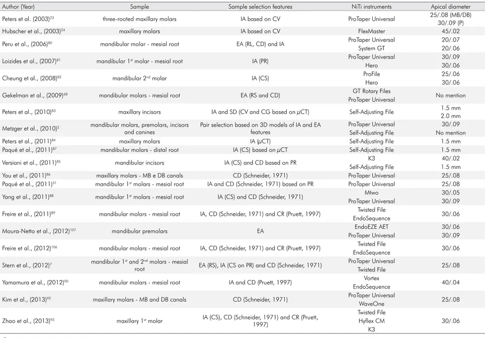
Table 4. Summary of studies included in this literature review according to author and year of publication, sample used, sample selection features, instrumentation system and final apical diameter evaluated.
Author (Year) Sample Sample selection features NiTi instruments Apical diameter
Peters et al. (2003)23 three-rooted maxillary molars IA based on CV ProTaper Universal 25/.08 (MB/DB)
30/.09 (P)
Hubscher et al., (2003)24 maxillary molars IA based on CV FlexMaster 45/.02
Peru et al., (2006)80 mandibular molar - mesial root EA (RL, CD) and IA ProTaper Universal 20/.07
System GT 20/.06
Loizides et al., (2007)81 mandibular 1st molar - mesial root IA (PR) ProTaper Universal 30/.09
Hero 30/.06
Cheung et al., (2008)82 mandibular 2nd molar IA (CS) ProFile 25/.06
Hero 30/.06
Gekelman et al., (2009)68 mandibular molars - mesial root EA (RS and CD) GT Rotary Files No mention
ProTaper Universal
Peters et al., (2010)83 maxillary incisors IA and SD (CV and CG based on µCT) Self-Adjusting File 1.5 mm
2.0 mm
Metzger et al., (2010)2 mandibular molars, premolars, incisors and canines
Pair selection based on 3D models of IA and EA features
ProTaper Universal 30/.09
Self-Adjusting File No mention
Peters et al., (2011)84 maxillary molars IA (µCT) Self-Adjusting File 1.5 mm
Paqué et al., (2011)87 mandibular molars - distal root IA (CS) based on µCT Self-Adjusting File 1.5 mm
Versiani et al., (2011)85 mandibular incisors IA (CS) and CD based on PR K3 40/.02
Self-Adjusting File 1.5 mm
You et al., (2011)86 maxillary molars - MB e DB canals CD (Schneider, 1971) ProTaper Universal 25/.08
Paqué et al., (2011)31 mandibular 1st molars - mesial root IA and CD (Schneider, 1971) based on PR ProTaper Universal 25/.08
Yang et al., (2011)88 mandibular 1st molars - mesial root IA (CS) and CD (Schneider, 1971) Mtwo 30/.05
ProTaper Universal 30/.09
Freire et al., (2011)89 mandibular molars - mesial root IA, CD (Schneider, 1971) and CR (Pruett, 1997) Twisted File 30/.06 EndoSequence
Moura-Netto et al., (2012)107 mandibular premolars EA EndoEZE AET 30/.06
ProTaper Universal 30/.09
Freire et al., (2012)106 mandibular molars - mesial root IA, CD (Schneider, 1971) and CR (Pruett, 1997) Twisted File 30/.06 EndoSequence
Stern et al., (2012)7 mandibular 1st and 2nd molars - mesial
root EA (RS), IA (CS on PR) and CD (Schneider, 1971)
ProTaper Universal
25/.08 Twisted File
Yamamura et al., (2012)90 mandibular molars - mesial root IA and CD (Pruett, 1997) Vortex 40/.04
EndoSequence
Kim et al., (2013)92 maxillary molars - MB and DB canals CD (Schneider, 1971) ProTaper Universal 25/.08
WaveOne
Zhao et al., (2013)93 maxillary 1st molar IA (CS), CD (Schneider, 1971) and CR (Pruett, 1997)
Twisted File
30/.06 Hyflex CM
K3
Continue
27
B
ra
z
. O
ra
l R
e
s. 20
18
;3
2
(s
u
p
p
l):
e
6
R
oo
t c
an
al p
re
p
ar
at
io
n u
si
n
g m
ic
ro
-c
om
p
u
te
d t
om
og
ra
p
h
y a
n
al
y
si
s: a l
it
er
at
u
re r
ev
ie
w
Continuation
Versiani et al., (2013)33 mandibular canine IA (CS on PR) and SD based on µCT
Self-Adjusting File 1.5 mm
WaveOne 40/.08
Reciproc 40/.06
ProTaper Universal 40/.06
McRay et al., (2014)39 mandibular molars - mesial root IA (CS on µCT) and CD (PR and µCT – Pruett, 1997)
WaveOne
25/.08 ProTaper Universal
Ceyhanli et al., (2014)94 mandibular molars - mesial root CD and CR (Pruett, 1997) and IA based on PR
RaCe 30/.04
ProTaper Universal 30/.09
Safesider 30/.04
Junaid et al., (2014)38 mandibular molars - mesial root IA and CD (Schneider, 1971) Twisted File 25/.08
WaveOne
Hwang et al., (2014)9 maxillary molars IA based on µCT and CD (Schneider, 1971) Reciproc 25/.08
Mtwo 25/.07
Al-Sudani et al., (2014)95 maxillary 1st premolars CD (Schneider, 1971) ProFile Vortex 30/.06
Revo-S
Gergi et al., (2014)37 mandibular molars - mesial root IA, SD and CD (Schneider, 1971)
Reciproc
25/.08 WaveOne
Twisted File Adaptive Sant’Anna Júnior et al.,
(2014)40 mandibular 1st molars - mesial root IA and CD (Schneider, 1971)
Mtwo 40/.04
Reciproc 40/.06
Zhao et al., (2014)41 mandibular 1st molars - all roots
IA and SD based on µCT WaveOne 25/.08 (M)/40/.08 (D)
CD (Schneider, 1971) based on PR ProTaper Universal 25/.08 (M)/40/.06 (D) ProTaper Next 25/.06 (M)/40/.06 (D)
Almeida et al., (2015)96 mandibular molars - mesial root CD K3 30/.04
K3XF
Gergi et al., (2015)44 mandibular molars - mesial root IA, SD and CD (Schneider, 1971)
Reciproc
25/.08 WaveOne
Twisted File Adaptive
Busquim et al., (2015)42 mandibular molars - distal root IA and CD (Schneider, 1971) based on PR Reciproc 40/.06
BioRace 40/.04
Marceliano-Alves et al.,
(2015)45 mandibular molars - mesial root IA, SD and CD (Schneider, 1971)
Reciproc
25/.08 WaveOne
Twisted File HyFlex CM
Ahmetoglu et al., (2015)34 maxillary 1st molars CD (Schneider, 1971)
Self-Adjusting File 1.5 mm (MB/DB)/2 mm (P)
Reciproc 25/.08 (MB/DB)/40/.06
(P)
Revo-S 25/.06 (MB/DB)/
40/.06 (P)
Continue
28
B
ra
z
. O
ra
l R
e
s. 20
18
;3
2
(s
u
p
p
l):
e
6
S
ou
sa
-N
et
o M
D S
ilv
a-S
ou
sa Y
C
, M
az
zi
-C
h
av
es J
F
, C
ar
v
al
h
o K
K
T
, B
ar
bo
sa A
F
S
, V
er
si
an
i M
A e
t a
l.
Continuation
Peters et al., (2015)97 mandibular molar - mesial root IA (CV), SD and CD based on µCT Vortex 30/.06
TRUShape
Pasqualini et al., (2015)98 maxillary 1st molar IA, CD (Schneider, 1971) and CR ProGlider/ProTaper Next 25/.06
ScoutRace/Bio-Race
Gagliardi et al., (2015)27 mandibular 1st molars - mesial root IA, SD and CD based on µCT and PR
ProTaper Universal 25/.08
ProTaper Next 25/.06
ProTaper Gold 25/.08
De-Deus et al., (2015)43 mandibular molar - mesial root IA (CV), SD and CD based on µCT and PR
Reciproc 40/.06
WaveOne 40/.08
BioRace 40/.04
Coelho et al., (2016)11 mandibular incisors IA (CS)
WaveOne 25/.08
Easy ProDesign Logic 25/.06
OneShape 25/.06
Pedullà et al., (2016)47 mandibular molars - mesial root IA, SD and CD (Schneider, 1971) based on PR Twisted File Adaptive 25/.06 Mtwo
Yang et al., (2016)12 mandibular molars - mesial root IA and CD (Schneider, 1971) based on µCT and
PR WaveOne 25/.08
Santa-Rosa et al., (2016)99 maxillary molars IA (CS and CV), CD and CR (Schneider, 1971) WaveOne 25/.08
OneShape 25/.06
Jardine et al., (2016)13 maxillary molars SD, CD and CR (Schneider, 1971)
Wizard Navigator 25/.08
WaveOne 25/.06
ProTaper Universal 25/.08
Vallaeys et al., (2016)100 mandibular 1st molar - mesial root IA (CS), CD (Schneider, 1971) and CR (Pruett, 1997)
Mtwo 30/.05
ProTaper Universal 30/.09
Revo-S 30/.06
Da Silva Limoeiro et al.,
(2016)101 mandibular molars - mesial root CD (Schneider, 1971)
ProTaper Next
25/.06 BioRace
Lopes et al., (2017)57 mandibular molars - mesial root IA (CS based on µCT) and CD (Schneider, 1971 based on PR)
ProTaper Next
25/.06 Twisted File Adaptive
Venino et al., (2017)103 all teeth IA and EA Hyflex EDM No mention
ProTaper Next
Continue
29
B
ra
z
. O
ra
l R
e
s. 20
18
;3
2
(s
u
p
p
l):
e
6
R
oo
t c
an
al p
re
p
ar
at
io
n u
si
n
g m
ic
ro
-c
om
p
u
te
d t
om
og
ra
p
h
y a
n
al
y
si
s: a l
it
er
at
u
re r
ev
ie
w
Continuation
Arias et al., (2017)102 mandibular molars - distal root IA and EA (CS, RS and RL) Vortex 30/.06
TRUShape
Guimarães et al., (2017)4 contralateral single-rooted mandibular
premolars IA (CS based on PR and µCT)
TRUShape
40/.06 Reciproc
Azim et al., (2017)62 mandibular incisors IA (AD, CS, CV, RL) and SD based on PR, CBCT and µCT
Vortex Blue 30/.04
XP-endo Shaper 30/.01
Serefoglu et al., (2017)105 mandibular 1st molar - mesial root IA (AD, CS and CV), SD, CD (Schneider, 1971) and CR (Pruett, 1997) based on PR and µCT
ProTaper Universal 40/.06
Self-Adjusting File 1.5 mm
Silva et al., (2017)58 mandibular 1st and 2nd molars- mesial root
IA (CS based on µCT), SD and CD (Schneider, 1971) based on PR
ProTaper Next
25/.06 Twisted File Adaptive
Elnaghy et al., (2017)59 mandibular 1st molars - mesial root IA (AD and CS), SD and CD TRUShape 25/.06
ProTaper Next
Brasil et al., (2017)14 mandibular 1st molars - mesial root IA (CS, CV and CG), SD and CD (Schneider, 1971) based on PR and µCT
ProTaper Next 30/.07
BT-Race 35/.04
Duque et al., (2017)104 mandibular molars - mesial root IA (CS) and CD based on µCT ProTaper Universal 25/.08
ProTaper Gold 30/.09
Lacerda et al., (2017)29 mandibular molars - distal root IA (CS) based on RP and CD (Schneider, 1971) and SD based on µCT
Self-Adjusting File 2 mm
TRUShape 30/.06
XP-endo Shaper 30/.01
Versiani et al., (2018)63 mandibular incisors IA (CS) and SD based on µCT
XP-endo Shaper 30/.01
iRace 30/.04
EdgeFile 30/.04
Espir et al., (2018)15 mandibular incisors IA (CS) based on RP and SD based on µCT Reciproc 40/.06
Mtwo
Zuolo et al., (2018)30 mandibular incisors IA (CV, CG, RL, CS and AD), SD and CD (Schneider, 1971) based on PR and µCT
BioRace 25/.06
Reciproc 25/.08
Self-Adjusting File 1.5 mm
TRUShape 25/.06
(1st) first; (2nd) second; (MB) mesiobuccal canals; (ML) mesiolingual canals; (DB) distobuccal canals; (IA) internal anatomy; (EA) external anatomy; (CV) canal volume; (CD) curvature degree; (RL) root length; (RS) root shape; (CG) canal geometry; (CS) canal shape; (PR) periapical radiographs; (CR) curvature radius; (SD) sample distribution; (CBCT) cone beam computed tomography; (µCT) microcomputed tomography; (AD) apical diameter.
30
B
ra
z
. O
ra
l R
e
s. 20
18
;3
2
(s
u
p
p
l):
e
6
Sousa-Neto MD Silva-Sousa YC, Mazzi-Chaves JF, Carvalho KKT, Barbosa AFS, Versiani MA et al.
reviewed, and in 83.33% of those (n = 30) in which the
mesial root was selected for the root canal preparation
evaluation. The curvature of the selected roots was
evaluated in 71.66% (n = 43) of the studies with a higher
prevalence of roots with moderate curvature and a
bend angle between 20° and 40° in 58.13% (n = 25).
It is worth noting that the degree of curvature can
influence the maintenance of the centering ability
of the root canals.
44,47It was observed in the description of the
methodologies that 50% (n = 30) of the studies
reported criteria of sample selection based on the
internal anatomy of the root canals, qualitatively by
Vertucci’s classification;
12,14,40,43,44,45,47,58,62,63,93,97,101,104,115isthmus classification;
57,58root canal shape;
4,11,15, 30,31,33,42,62,63,85,102anatomical variation;
82and/or by
quantitative composition of similar or homogeneous
groups in relation to bi- and tri-dimensional
parameters of the root canal.
13,14,15,29,30,33,37,41,43,44,45,47,5 7,58,59,62,63,83,105,126,127In view of this, a variation of the
root canal anatomy selected in these studies was
observed, which made it difficult to statistically
compare the data obtained.
Regarding selection of the t ype of canal,
Vertucci’s classification for the evaluation of the
root canal preparation showed the use of the
Type I (single canal that extends from the pulp
chamber to the apex) for incisors,
62Type II (two
canals that leave the pulp chamber and join near
the apex to form a single canal),
43and Type IV
(two distinct and separate canals that extend
from the pulp chamber to the apex) in mesial
roots of mandibular molars.
12,14,40,44,45,47,97,101,104Also,
in mesial roots of mandibular molars, isthmus
classification Type I (narrow sheet and complete
connection existing between two canals)
128or Type
III (incomplete isthmus existing above or below a
complete isthmus)
57,58was seen.
Another aspect of the internal anatomy used
for the sample selection in the analyzed studies
was the canal flattening, which classified the root
canals as oval in 21.67% (n = 13) of the studies rev
iewed,
11,15,29,30,33,45,62,102long-oval
31,42,63and flat-oval
85in 53.84% (n = 7) of these studies, evaluating the
degree of flattening by means of the ratio between
the buccolingual and mesiodistal dimensions of the
root canal on radiographs
15,29,30,33,42,85and in CBCT
images.
11The studies did not use quantitative
data obtained by micro-CT (roundness, factor
form, major and minor diameter, and SMI), which
Figure 2. Flow diagram with the database search and articles include (based on PRISMA).Records identified through database searching (n = 102)
Additional records identified through other sources (n = 0)
Records after duplicates removed (n = 5)
Records screened (n = 97)
Records excluded (n = 23)
Articles included (n = 60) Full-text articles assessed
for eligibility (n = 74)
Ful-text articles excluded, with reasons (n = 14)
Root canal preparation using micro-computed tomography analysis: a literature review
would have provided greater acuity of this type
of anatomical variation.
Regarding the composition of the experimental
groups, only 35% (n = 21) of the studies used a stratified
distribution proposed by Versiani et al.
33based on
the similar internal anatomy and morphological
dimensions of the root canal (statistically similar
dimensions of the root canal) from the two-dimensional
and three-dimensional data obtained by the micro-CT
prior to the preparation.
27,29,33,34,45,57,63,97,98Because studies have shown that the result of
the biomechanical preparation depends more on
the original anatomy of the root canal than on the
instrument or technique used,
6,27,33,42,45,64,129,130,131,132it
becomes important to properly select samples with
two-dimensional and three-dimensional values in
order for the experimental groups to be balanced in
terms of anatomical characteristics, which can improve
the understanding of the results of each instrument
against the different morphological characteristics
of the root canals.
33,63,129Changes in the canal geometrics and
untouched areas after biomechanical
preparation
The action of mechanized instruments inside the
RCS promotes dentin wear that results in changes
in the geometric configuration of the root canal, and
these can be qualitatively and quantitatively observed
through micro-CT
30,33,45,63by images obtained at
different steps in endodontic treatment by aligning
the three-dimensional spatial coordinates of x, y, and
z on specific software.
The quantitative parameters used for the
evaluation of changes in the geometric configuration
resulting from the biomechanical preparation are the
three-dimensional parameters of volume, surface
area, and (SMI); and two-dimensional parameters
of area, perimeter, roundness, form factor, and
major and minor diameter.
32,33,43,45,63The reviewed
studies corroborate that after the biomechanical
preparation, there is an increase in these parameters,
regardless of the type of instrument or technique
used.
4,27,36,41,44,47,102,104In addition, it is also possible to quantify the
percentage of root canal walls touched and not
touched by instruments. The touch or action of
the instrument on the canal walls and the changes
in volume and SMI are the parameters most used
(68.85%) to evaluate the preparation by means of
micro-CT.
4,11,15,42,47,57,62,83,96,102,103,105The change in canal volume is related to the
effects of biomechanical preparation on dentin
removal,
7,25,44,83,132,133showing that the volume increase
after preparation is proportionally higher in the
cervical and middle third than in the apical and can
be attributed to the cervical preparation
7,32,33,84and to
the greater taper of the instruments in the cervical
region.
11,47Clinically, the increase in canal volume
in the cervical third can mean the improvement of
the reach of irrigating solutions in the apical third,
or that the apical mechanical debridement was not
as effective as cervical
7,12(Figure 3).
An important aspect observed in this review
was that 50% (n = 30) of studies evaluated the
average percentage of canal walls untouched after
biomechanical preparation and showed that no system
or technique was able to touch all the walls of root can
als,
4,27,29,30,42,43,44,45,57showing a range of 2.6% to 80% of
the walls being untouched.
27,57This variation may be
related to changes in dental morphology,
25,27,45,79,84,101,104the characteristics of the instruments used,
4,11,30,32,62,63,102or the evaluation methodology used.
25,31,84In studies in which there was concern about sample
selection, the percentage of untouched walls ranged
from 8.17% to 58.8% for the whole canal in groups
of teeth with flattened canals,
62,63and from 3.13% to
51.03% for the apical third.
15,102Another aspect that may explain the range in
the percentage of untouched walls is the design
variability of instruments such as taper, diameter,
and cross-section.
4,11,30,32,62,102The SAF, XP-endo
Shaper, and TRUShape instruments showed highest
percentages of touched walls when compared to other
systems.
4,29,30,31,32,33,63,102Regarding the final diameter, the reviewed studies
showed that the final diameter of the instruments used
for the biomechanical preparation ranged from diameter
25 to diameter 40 with a diameter 30 being most common
in these canals. The mesial canals of mandibular molars
were the most frequently used sample among the
reviewed studies, and the diameter of these canals at
Sousa-Neto MD Silva-Sousa YC, Mazzi-Chaves JF, Carvalho KKT, Barbosa AFS, Versiani MA et al.
1 mm of the apical foramen varied between 0.28–0.40
mm in the buccolingual direction and 0.21–0.28 mm in
the mesiodistal direction.
134. The surgical diameter was
similar to the anatomical diameter, which may also explain
the large percentage of untouched walls. Therefore, the
standardization of the anatomic diameter should be a
concern for sample selection during the experimental
design as this data can be determined by the parameters
of major and minor diameter obtained by micro-CT as
well as being clinically described by Pécora et al.
135In addition to the anatomical features of the dental
group and instruments used for the preparation,
another factor that can interfere in the results of
touched walls is related to the evaluation methodology.
The parameter of untouched walls is calculated by
the difference between the number of static voxels
and the total number of voxels on the surface of the
root canal (voxels present at the same position on the
canal surface before and after preparation).
25Both the
overlap of the images and the resolution used for the
acquisition of these images may be important factors in
the interpretation of the obtained results
31,84. The values
of the resolutions used in the studies in this literature
review ranged from 11.88 μm to 36 μm,
25,42,79,88with
the best resolutions in Yang et al.,
88with more values
closer to 20 μm. Therefore, the observed differences
in the percentage of untouched walls between studies
may also be related to methodological differences.
It is known that bacteria may penetrate dentinal
tubules in depths of 200 μm or more,
83,136and the
full extension and even the root canal by 200 μm
seems to be a goal not yet achieved by any preparation
technique.
2,4,7,9,11,12,13,14,15,25,27,28,29,30,31,34,35,37,38,39,40, 41,42,43,44,45,47,57,58,59, 62,63,68,79,80,81,82,83, 84,85,86,87,88,89,90,91,92,93,94,95,96,97,98,99,100,101, 102,103,104,105,106,107The untouched walls, especially in areas of anatomical
challenges such as isthmus, recesses, and flattened root
canals can retain bacteria and serve as a potential cause
of persistent infection.
137,138This demonstrates the need
for new strategies such as improving the performance
of the irrigating solution, physical action of ultrasonic
activation, and intracanal dressing to complement
suboptimal mechanical action.
30,61,79Controversially, one study showed that there was no
significant correlation between the elimination of active
Figure 3. Representative 2D and 3D reconstructions of the internal and external anatomy of the mandibular molar before and after root canal preparation with mechanized instruments. (A) Buccal view of superimposed 3D models before (green) and after (red) root canal preparation. (B) Lingual view of superimposed 3D models before (green) and after (red) root canal preparation. (D) Representative cross-sections of the superimposed root canals before (green) and after (red) preparation at the cervical (c), middle (m) and apical (a) thirds.
A
B
C
c
m
a
Root canal preparation using micro-computed tomography analysis: a literature review
bacteria and the average percentage increase in volume,
surface area, and untouched walls when analyzed by
micro-CT.
138In addition, a recent study of the correlation
between data obtained after biomechanical preparation
using micro-CT and histology
29showed that even with
the presence of untouched walls, SAF, TRUShape, and
XP-endo Shaper can remove pulp tissue in oval canals.
29To analyze the changes in the dimensional shape
of the root canal, the commonly used parameter is
the SMI, which evaluates the convexity of the surface.
Studies have shown that SMI values increased after
preparation, indicating that the small and flatter
irregularly tapered canal changed to a rounder and
smoother tapered canal.
27,31,33,36,45,68,86,88,102,103Lower values
for changes in SMI are related to the maintenance of
the original shape of the root canal.
27,45,102Considering the limitations between the studies
for analyzing the preparation related to changes in
the geometry of the canals due to the methodological
differences, the instruments showed that the most
promising results regarding uniform dentin wear,
smaller percentages of untouched walls, and the canal
shape maintenance (evaluated by the SMI) especially
on flattening canals, were the SAF, TRUShape, and
XP-endo Shaper systems.
4,29,30,31,33,63,85,102Expression of canal transportation and
centering ability during the biomechanical
preparation
In 2017, the American Association of Endodontists
139defined the canal transportation as a removal of canal wall
structure on the outside curve in the apical half of the canal
due to the tendency of files to restore themselves to their
original linear shape during canal preparations, which
may lead to ledge formation and possible perforations.
Gambill et al.
140proposed the term “centering ability”
as a measurement of the ability of the instrument to
stay centered in the canal. These measurements are
still being used in various endodontic studies to assess
the quality of the biomechanical preparation of root
canals with instruments and techniques using different
methodologies
141. This was observed in this literature
review in which 63.33% (n = 38) of the studies evaluated
the canal transportation and the centering ability.
Several factors can influence the canal transportation
and centering ability such as errors in treating
endodontic cavities and in glide path, use of non-flexible
instruments, instrument design (cross-section, taper,
tip) and the absence of specific treatments and alloys
in endodontic instrument manufacturing (stainless
steel, nickel-titanium, thermic treatment). In addition,
negligence during irrigation protocols and the operator’s
experience in determining the most appropriate
preparation technique for each situation should be
considered.
140,141,142The importance of anatomical
knowledge of the RCS and its variations such as radius
and curvature degree, canal configuration, and dentin
thickness which can be observed in two-dimensional
radiographic examinations is noteworthy.
3,140,141,142This literature review showed that approximately
52.63% (n = 20) of the studies reviewed evaluated
the canal transportation and centering ability
using the method proposed by Gambill et al.
140for
a CBCT analysis. This methodology consists of the
measurement of the shortest distance, in mesial
and distal directions, from the limit of the
non-instrumented root canal to the limit of the tooth
in comparison with the same measurements of the
instrumented areas, represented by the formula
(X¹ − X²) − (Y¹ − Y²). The X¹/X² and Y¹/Y² values
represent the shortest distances from the outside
and inside of the curved root to the periphery of the
non-instrumented and instrumented areas of the
root canals, respectively. Values may be seen from 0
(indicating no canal transportation) to 1 (indicating
the perfect centering).
The high resolution and acuity of micro-CT allowed
the evolution of this analysis, as introduced by Peters
et al.,
131,143,144comparing each slice voxel by voxel from
all data sets obtained before and after root canal
preparation to obtain consistent results for centers
of gravity. In this case, each slice was defined by a
series of coordinated data for the x-, y-and z-axes,
and the centers of gravity were calculated for each
slice, connected along the z-axis by a fitted line which
can be analyzed to determine canal curvature as the
second derivative. Thus, the comparison of the centers
of gravity before and after preparation showed the
results of canal transportation,
45,97,131,143,144as shown in
47.36% (n = 18) of the studies in this review.
In general, besides the metallurgical properties
and design of instruments, other factors including
Sousa-Neto MD Silva-Sousa YC, Mazzi-Chaves JF, Carvalho KKT, Barbosa AFS, Versiani MA et al.