UNIVERSIDADE DE LISBOA Faculdade de Medicina Veterinária
CHANGES IN THE FELINE GUT MICROBIOTA ASSOCIATED TO TOXOCARA CATI INFECTIONS
ANA LÚCIA MIRANDA DUARTE
ORIENTADOR Doutora Cinzia Cantacessi
CO-ORIENTADOR Doutor Luís Manuel Madeira de Carvalho
2018 LISBOA CONSTITUIÇÃO DO JÚRI:
Doutor José Augusto Farraia e Silva Meireles Doutor Luís Manuel Madeira de Carvalho Doutora Maria Manuela Castilho Monteiro Oliveira
UNIVERSIDADE DE LISBOA Faculdade de Medicina Veterinária
CHANGES IN THE FELINE GUT MICROBIOTA ASSOCIATED TO TOXOCARA CATI INFECTIONS
ANA LÚCIA MIRANDA DUARTE
DISSERTAÇÃO DE MESTRADO INTEGRADO EM MEDICINA VETERINÁRIA
ORIENTADOR Doutora Cinzia Cantacessi
CO-ORIENTADOR Doutor Luís Manuel Madeira de Carvalho
2018 LISBOA CONSTITUIÇÃO DO JÚRI:
Doutor José Augusto Farraia e Silva Meireles Doutor Luís Manuel Madeira de Carvalho Doutora Maria Manuela Castilho Monteiro Oliveira
i
ii Acknowledgments
Lucky is the best word to describe the journey that lead to this point. There are so many people to thank to. A big thank you to Professor Cinzia Cantacessi for making it possible to for me to have my project done in Cambridge. Thank you for introducing me to this amazing and interesting subject and for the guidance. I feel thank you is not enough. Another big thank you to Professor Madeira de Carvalho who made it possible for me to finish this last part of the journey in England, I will never forget.
A thank you to Dr Joy Archer for allowing me to use the Parasitology Laboratory at The Queen´s Veterinary School Hospital and to Vikki Haverson for the help and sympathy especially after I showed up after a night shift at work.
Thank you to all the staff at the cat shelter in Cambridge for the help collecting the samples. Thank you to Professor Domenico Otranto and Dr. Stefania Latrofa from University of Bari in Italy and also a big thank you to Dr. Giada Annoscia and Dr. Bron Campbell for all the help and support during my short stay in Italy.
To everyone in the Microbiology Laboratory at Cambridge University also a very big thank you. Dr. Andrew Grant, Dr. Stefan de Vries and Dr. Srishti Gupta, thank you for all your help. Also from Cambridge University a special thank you to Timothy Jenkins for the incredible help. I couldn’t’ve done it without you.
As I said I am lucky.
Lucky that I had such amazing parents, specially my mother who fixed me ever since I was born and always believed in me. Lucky for my sister who was always there for me, and who spend hours with me studying. Lucky for my friends, especially Cristina Pereira who encouraged me and gave me the last push I needed to finish the degree.
iii Abstract
Changes in the feline gut microbiota associated to Toxocara cati infections
Investigations of the relationships between the gut microbiota and gastrointestinal parasitic nematodes are attracting growing interest by the scientific community. These studies have however been carried out mainly in humans and experimental animals, while knowledge of the make-up of the gut commensal microbiota in presence or absence of infection by parasitic nematodes in domestic animals is limited. In this study, we investigate the qualitative and quantitative impact that infections by a widespread parasite of cats (i.e. Toxocara cati) exert on the gut microbiota of feline hosts.
The faecal microbiota of cats with patent infection by T. cati (= Tc+), as well as that of negative controls (= Tc-) was examined via high-throughput sequencing of the V3-V4 hypervariable region of the bacterial 16S rRNA gene, followed by bioinformatics and biostatistical analyses of sequence data.
A total of 2,325,366 useable high-quality sequences were generated from the faecal samples analysed in this study and subjected to further bioinformatics analyses, which led to the identification of 128 OTUs and nine bacterial phyla, respectively. The phylum Firmicutes was predominant in all samples analysed (mean of 53.0%), followed by the phyla Proteobacteria (13.8%), Actinobacteria (13.7%) and Bacteroidetes (10.1%). Among others, bacteria of the order Lactobacillales, the family Enterococcaceae and genera Enterococcus and Dorea showed a trend towards increased abundance in Tc+ compared with Tc- samples, while no significant differences in OTU richness and diversity were recorded between Tc+ and Tc- samples (P = 0.485 and P = 0.581, respectively). However, Canonical Correlation and Redundancy Analyses were able to separate samples by infection status (P = 0.030 and P = 0.015, respectively), which suggests a correlation between the latter and the composition of the feline faecal microbiota.
Keywords: Gut microbiota, Cat, Toxocara cati, Lactobacilli, Microbial richness and diversity, 16S rRNA
iv Resumo
Alterações na microbiota intestinal felina associadas a infecções por Toxocara cati
A investigação das interações entre a microbiota intestinal e os nematodes intestinais tem vindo a atrair o interesse da comunidade cientifica. No entanto, a maioria destes estudos tem sido desenvolvida em humanos e animais de laboratório, e deste modo o conhecimento da composição da microbiota comensal do intestino na presença de nematodes intestinais é reduzido. Neste estudo, foi investigado o impacto qualitativo e quantitativo da infeção pelo parasita comum dos gatos (i.e. Toxocara cati) na microbiota intestinal do hospedeiro felino. A microbiota fecal de gatos com infeção patente por T. cati (Tc+), bem como controlos negativos (Tc-) foi avaliada através de sequenciação de alto débito da região hiper-variável V3-V4 do gene 16S, seguido de análise bioinformática e bioestatística.
Das amostras fecais incluídas no estudo foram obtidas um total de 2 325 366 sequências de alta qualidade e sujeitas a analise bioinformática, o que levou à identificação de 128 OTUs e nove filos bacterianos. O filo Firmicutes foi encontrado em predominância em todas as amostras (média de 53,0%), seguido do filo Proteobacteria (13,8%), Actinobacteria (13,7%) e por fim Bacteroidetes (10,1%). A abundância de determinados grupos de bactérias tendeu a aumentar nas amostras Tc+ quando comparadas com as amostras Tc-, tais como a ordem Lactobacillales, a família Enterococaceae e o género Enterococcus e Dorea. No entanto, a riqueza e diversidade das OTUs não apresentou diferenças significativas entre as amostras Tc+ e Tc- (P=0,485 e P=0,581, respetivamente). Todavia, a análise canónica de redundância demonstrou uma separação das amostras de acordo com o estado de infeção (P=0,030 e P=0,015, respetivamente), o que sugere uma correlação entre este e a composição da microbiota fecal felina.
Palavras chave: Microbiota intestinal, gato, Toxocara cati, Lactobacilli, Riqueza e diversidade microbiana, 16S rRNA
v Table of contents Acknowledgments ……… ii Abstract ……… iii Resumo ……… iv Table of contents ……….. v List of Figures ……….. vi List of Tables ………... vi
List of Abbreviations and Symbols ……….. vii
Introductory Note ………. ix
Chapter 1 – Literature Review ………. 1
1. Introduction ………... 1
2. Intestinal parasites of cats and dogs ……….. 2
2.1 Toxocara spp. ………... 2
2.1.1 Epidemiology ………. 2
2.1.2 Morphology ……… 3
2.1.3 Life cycle ………... 5
2.1.4 Pathogenesis and clinical signs ……….. 7
2.1.5 Immunological responses and evasion of the immune system ……….. 8
3. Gut microbiota in dogs and cats: composition and functions ……… 10
4. Helminth-microbiota interaction (HMI) ……… 18
5. Summary and research aims ……….. 21
Chapter 2 – Material and methods ……….. 22
1. Sample collection and parasitological testing ……….. 22
2. DNA extraction ……… 24
3. Library construction and 16S rRNA gene sequencing ……… 24
4. Bioinformatics and data analysis ………. 25
Chapter 3 – Results ……….. 27
Chapter 4 – Discussion/Conclusion ………. 31
Chapter 5 – Final Considerations and Future Directions ………. 38
References ……… 39
Annexes ……… 52
Annex A ……… 52
vi List of Figures:
Figure 1. Anterior end of Toxocara cati adult worm (Alho, 2010, p.36) …………...………4 Figure 2. Toxocara cati eggs showing the thick external layer, internal wall and
unicellularembryo (original) ………..………...4
Figure 3. Toxocara cati life cycle (original) ………...7 Figure 4. Composition and spatial distribution of microbiota along the mammalian intestinal
tract (Brown et al., 2013) ………11
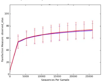
Figure 5. Agarose gel electrophoresis showing amplicons produced with universal primers for the V3-V4 region of the bacterial 16S rRNA gene ………...…..24 Figure 6. Rarefaction curves (number of observed OTUs on Y axis) for Tc+ (blue curve)
versus Tc- (red curve) ………..27
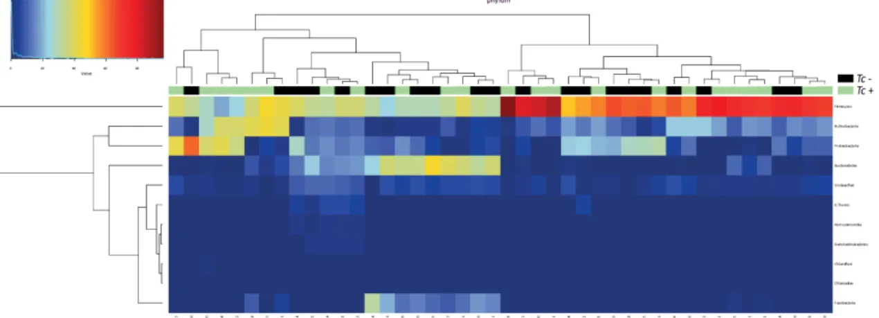
Figure 7. Hierarchical clustering heatmap of the composition of the faecal microbiota phyla of Toxocara cati-positive (Tc+) and T. cati-negative (Tc-) cats. Dendrograms at the top of the heatmap indicate relationships between samples. Colour intensity represents the relative abundance of sequences representing the corresponding bacterial family in each sample ….28 Figure 8. Rank plot of differentially abundant faecal bacteria (at the phylum - I, class - II, order - III, family - IV, genus - V and species - VI level) between Toxocara cati-positive (Tc+) and -negative (Tc-) cats, based on Linear Discriminant Analysis Effect Size (LEfSe) analysis. Taxa highlighted in green/black indicate overrepresentation in Tc+ and Tc- samples, respectively. Specific groups show trends towards increased abundance in Tc+ subjects
compared with Tc- samples………..………29
Figure 9. Differences in overall taxonomic species richness (a) and diversity (b) between the faecal microbiota of Toxocara cati-positive (Tc+) and T. cati-negative (Tc-) cats ……...….29 Figure 10. Redundancy analysis (RDA) and canonical correlation analysis (CCA) show that microbial samples from Tc+ and Tc- cats form separate clusters …………...………30
List of Tables:
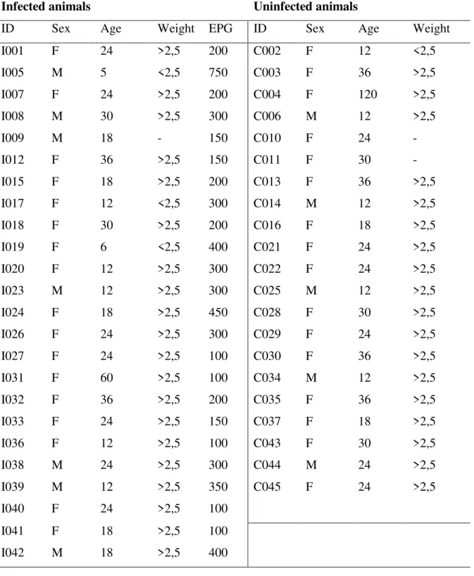
Table 1 – GI microbiota of healthy dogs based on 16S rRNA gene sequencing methods ….12 Table 2 – GI microbiota of healthy cats based on sequencing methods ………...……..13 Table 3 – Sex, age (months), and weight (kg) in evaluated cats. For those infected by T. cati (left column), the infection burden has been expressed in eggs per gram of faeces (EPG) …23
vii List of Abbreviations and Symbols:
~ - Approximately % - Percentage = - Equal to ± – Plus or minus
ALT - Alanine Transaminase AMPs - Antimicrobial Proteins APRIL - proliferation-inducing ligand Akt – protein kinase B
bp – base pairs
CCA – Canonical Correlation Analysis CLRs – C-type lectin receptors
CTL – C-type Lectin CP – Polysaccharides DC – Dendritic Cell
DNA – Deoxyribonucleic Acid dsDNA – double strain DNA EPG – eggs per gram
ES – Excretory-secretory FEC – faecal egg counts g – grams
GALT – Gut Associated Lymphoid Tissues GF - Germ Free
GI – Gastrointestinal
HMI – Helminth-microbiota interactions ICD – Idiopathic Chronic Diarrhoea IFN-g – Interferon-g
Ig - Immunoglobulins IL – Interleukins kg – Kilogram L1 – First stage larva L3 – Third stage larva L4 – Fourth stage larva L5 – Immature adult stage LDH - glutamate dehydrogenase
LEfSe – Linear discriminant analysis effect size LPS – Lipopolysaccharides
m – minutes
MAMPS – Microbe-Associated Molecular Pattern ml – Millilitre
MLN – Mesenteric Lymph Nodes MUC – Mucin
NCBI: National Centre for Biotechnology Information ng/µl – nanograms per microlitre
NGS - next-generation sequencing NLRs – NOD-like Receptors NOD - Oligomerisation Domains ºC – Centigrade degrees
OTU – Operational Taxonomic Units P – p-value
PCR – Polymerase Chain Reaction PGN – Peptidoglycan
PRR – Pattern Recognition Receptors
viii ® – Registered trademark
RDA – Redundancy Analysis RNA – Ribonucleic Acid rRNA – recombinant RNA s – seconds
sIgA - secretory IgA
SPF - specific pathogen free
sprr2A - small proline-rich protein 2A Tc+ - Toxocara cati-positive
Tc- - Toxocara cati-negative
Tc-PEB-1 – Toxocara phosphatidylethanolamine binding protein 1 TES - Toxocara ES
Th2 - T-helper type 2 cells TLR - toll-like receptor TM
- Trade mark
Treg - T regulatory cells µl – microlitre
ix Introductory Note
The present work was carried out in the Department of Veterinary Medicine of Cambridge University in England as part of the author’s training period during the 6th year of the Integrated Master in Veterinary Medicine. The training period went from October 2015 to June 2016 and was guided by Dr. Cinzia Cantacessi. This research has resulted in a paper published on Parasites and Vectors on December 2016 (Annex A).
During this time the student had also the opportunity to participate in a basic epidemiological study to verify the occurrence of cat lungworm infections in the Cambridge area. This study was sponsored by Merial. Faecal samples from 54 cats were collected from November of 2015 to March of 2016 from a cat shelter in Cambridge. All samples were also tested for other intestinal parasites using the floatation method. To test for the presence of the lungworm the Baermann technique was used. All samples were found negative for Aelurostrongylus abstrusus but some were positive for other intestinal parasites (Annex B).
1
Chapter 1 – Literature Review
1. Introduction
The human gastrointestinal (GI) tract harbours ~100 trillion (1013–1014) bacterial cells belonging to over 500 species, which together are referred to as the ‘gut microbiota’ (Brown, Sadarangani & Finlay, 2013; Giacomin, Croese, Krause, Loukas & Cantacessi, 2015). Particularly in veterinary species, the gut microbiota often shares its environment with large multicellular organisms, i.e. parasitic helminths (Peachey, Jenkins & Cantacessi, 2017). Moreover, both helminths and commensal bacteria have developed a range of strategies to modulate host immunity in order to establish themselves in the host gut (Reynolds, Finlay & Maizels, 2015). It is therefore plausible that the successful establishment of parasitic nematodes in the vertebrate gut is achieved, at least in part and directly and/or indirectly, via physical, molecular, and/or immunological interactions with the resident commensal microbiota (Glendinning, Nausch, Free, Taylor & Mutapi, 2014; Reynolds, Finlay & Maizels, 2015) and recent research on relations between the microbiome and parasites has shown a variety of interactions with implications to the host (Reynolds et al., 2014; Kreisinger, Bastien, Hauffe, Marchesi & Perkins, 2015; Cattadori et al., 2016).
However, most studies studying these interactions have been conducted on humans and rodents (Walk, Blum, Ewing, Weinstock & Young, 2010; Cooper et al., 2013; Rausch et al., 2013; Cantacessi et al., 2014; Lee et al., 2014; Reynolds et al., 2014; Giacomin et al., 2015a; Holm et al., 2015; Houlden et al., 2015; McKenney et al., 2015; Cattadori et al., 2016; Giacomin et al., 2016) and only a handful of studies have explored the relationships between GI parasitic nematodes and the commensal gut microbiota in non-experimental animals (Li, Wu, Li, Huang & Gasbarre, 2011; Li et al., 2012; Wu et al., 2012; Slapeta, Dowd, Alanazi, Westman & Brown, 2015; Li et al., 2016). Furthermore, to the best of our knowledge, so far the helminths involved in the studies investigating this interaction do not include ascarids. Ascarids are among the largest and commonest nematode parasites infecting the intestinal tract of domestic animals (Overgaauw, 1997). Toxocara canis and T. cati are large ascarids that infect dogs and cats, respectively (Overgaauw, 1997). Due to the zoonotic potential of T. canis, much is known of the pathogenicity and immunological features of this parasite, as well as of mechanisms of interaction with its hosts (Schneider, Laabs & Welz, 2011; Maizels, 2011; Resende et al., 2015). On the other hand, little is known of the complement of interactions occurring at the gut interface of cats infected by T. cati.
2
In this chapter in order to provide background information on the three way interaction between parasite, microbiota and host, a review of the relevant literature on the genus Toxocara, the microbiota and the helminth-microbiota interaction in veterinary species was undertaken. At the end of the chapter, conclusions drawn from the literature review to set the research aims of the present study will be presented.
2. Intestinal parasites of cats and dogs
Domestic cats and dogs can be infected with a wide range of intestinal parasites. Briefly, the most common parasites of cats are the hookworm Ancylostoma tubaeforme, the roundworms Toxascaris leonina and Toxocara cati, the tapeworms Dipylidium caninum and Taenia taeniaformis and protozoan parasites Giardia spp. and Cytoisospora spp. (Foreyt, 2001). Conversely, in dogs, the commonest intestinal parasites are the hookworms Ancylostoma caninum, A. braziliense and Uncinaria stenocephala, the roundworms Toxocara canis and Toxascaris leonina, the tapeworm Dipylidium caninum, the whipworm Trichuris vulpis and protozoans Cytoisospora spp. and Giardia spp. (Foreyt, 2001).
Amongst these, ascarids are one of the most prevalent intestinal parasites in Europe (Näreaho et al., 2012; Beugnet et al., 2014; Zanzani, Gazzonis, Scarpa, Berrilli & Manfredi, 2014; Paoletti et al., 2015) and other parts of the world (Savilla, Joy, May & Somerville, 2011; Hoopes et al., 2015; Tun et al., 2015; Yang & Liang, 2015).
Both larval and adult stages of ascarids can potentially be harmful to the vertebrate hosts: adult worms often cause poor growth and diarrhoea and, occasionally, intestinal obstruction in young animals, while larvae damage host tissues during the somatic migration phase of their life cycle (Taylor, Coop & Wall, 2007; Elsheika and Khan, 2011; Bowman, 2014).
2.1. Toxocara spp.
2.1.1. Epidemiology
In Europe, a study of privately owned cats from Austria, Belgium, France, Hungary, Italy, Romania and Spain, reported a prevalence of T. cati infection of 19.7% (Beugnet et al., 2014), while other studies reported a prevalence ranging from 5.4% in Finland (Näreaho et al., 2012) to 15.7% in the UK (Gow et al., 2009).
3
In dogs, reported prevalence of infection by T. canis range from 1.3% in Portugal (Ferreira et al., 2011), to 4.48%-22.2% in Italy (Zanzani et al., 2014) to 8% in Albania (Shukullari et al., 2015).
In Canada, ascarid infections were present in 16.5% and 12.7% in cat and dog populations, respectively (Villeneuve et al., 2015), although the prevalence in Canadian free-roaming cats was reported to vary from 1% to 15% in the same year (Hoopes et al., 2015). Another study in North America, observed a prevalence of 6.9% in dogs in south central West Virginia (Savilla et al., 2010). In Mexico, the prevalence of ascarid infections in owned dogs was 2.3% (Torres-Chablé et al., 2015).
In Australia, a study involving cats and dogs, reported a prevalence of ascarid infections of 3.2% and 1.2% respectively (Palmer, Thompson, Traub, Rees & Robertson, 2008). In Malaysia, infection prevalence was 11.9% and 9.9% in stray dogs and cats, respectively (Tun et al., 2015). A higher prevalence of 17.78% was detected in stray and shelter cats from central China (Yang & Liang, 2015).
Several factors have been associated with higher rates of Toxocara infections. For instance, infection rates are higher for dogs and cats with outdoor access, due to the likelihood of being exposed to contaminated soil and infected paratenic hosts (Näreaho et al., 2012; Beugnet et al., 2014; Shukullari et al., 2015). Studies have also shown that younger cats and dogs have a higher risk of being infected (Palmer et al., 2008; Savilla et al., 2010; Näreaho et al., 2012; Zanzani et al., 2014; Villeneuve et al., 2015), due to activation of latent larvae in the tissues of the bitch and queen and migration across the placenta and to mammary gland and milk infecting the puppies and kittens (Overgaauw, 1997).
The extensive distribution and strong concentration of Toxocara infections are mainly associated to: (i) female fecundity (~700 eggs per gram of faeces per day per worm); (ii) egg resilience to environmental challenge and (iii) the life cycle of the parasite, that involves encystment of larvae in the somatic tissue of the female hosts that thus act as reservoirs of infection for the offspring (Taylor et al., 2007; Bowman, 2014).
2.1.2. Morphology
Adult ascarids are large white opaque worms with no buccal capsule, and a mouth surrounded by three lips, one dorsal and two subventral. Some genera, like Toxocara, are characterised by two lateral cervical alae that make the anterior end of the worm resemble an arrowhead (Figure 1) (Taylor et al., 2007).
4
Ascarids have a few characteristic features: a mouth surrounded by three lips, one dorsal and two subventral, a large cervical alae and a glandular oesophageal bulb and the ventriculus, located at the junction between oesophagus and the intestine (Bowman, 2014). Male T. canis is around 10 cm long, while the female can be up to 18 cm long (Taylor et al., 2007). Males also feature a terminal narrow appendage and caudal alae. The female genital organs prolong from the anterior end to the vulvar region (Taylor et al., 2007). Adult Toxocara cati are smaller than T. canis, i.e. males 3-6 cm and females 4-10 cm long (Taylor et al., 2007).
Figure 1. Anterior end of Toxocara cati adult worm (x200) (Alho, 2010, p.36)
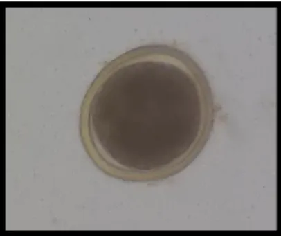
Toxocara eggs are characterised by a thick external layer, an evolutionary adaptation that provides a protective feature against harsh environmental conditions (Bowman, 2014). Eggs of T. canis and T. cati are morphologically undistinguishable, in spite of slight size differences. Indeed, T. canis eggs are 90 x 75 µm in average, whilst T. cati eggs are ~65 x 75 µm (Fahrion, Schnyder, Wichert & Deplazes, 2011). Eggs in faeces appear dark brown and subglobular, with a central unicellular embryo (Figure 2) (Araújo, 1972).
Figure 2. Toxocara cati egg showing the thick external layer, internal wall and unicellular embryo (aprox. x230) (original)
5 2.1.3. Life Cycle
The adult worm resides in the intestine of the definitive host, that can shed up to 200 000 eggs per day (Bowman, 2014). When shed, the eggs containing the first stage larvae (L1), are not infective, and undergo development for 3 – 6 weeks before becoming infectious (Overgaauw, 1997). The time needed for the unembryonated egg to moult into the infective stage larvae varies depending on environmental conditions. Azam, Ukpai, Said, Abd-Allah and Morgan (2011) reported that T. canis larvae can resist at a wide range of temperatures, as low as -2ºC for 6 weeks. Relative humidity and oxygen tension also play a role in the maturation into the infective stage (Araujo, 1972; Zajac & Conboy, 2012). While, for decades, the second larval stage was traditionally considered the ascarid infective stage, recent studies have shown that the latter is indeed a third stage larva (L3) (Schnieder, Laabs & Welz, 2011). L3s encapsulated in the eggshell can remain viable in the environment for at least 1 year (Overgaauw, 1997).
Following the ingestion by the definitive host, the eggs reach the duodenum and hatch within 2 to 4h. The infective larvae are then released and infiltrate the mucosa of the intestine. How the penetration occurs is still unclear, but a parasite-secreted elastase-like protease was hypothesized to play a role in digestion of host tissues (Schnieder et al., 2011). Direct mechanical disruption has also been hypothesized (Schnieder et al., 2011). After penetrating the intestinal wall, the larvae enter the circulatory system and reach the liver, via portal circulation, 24h post-infection (Webster, 1958). Within the subsequent 12h, the larvae continue their migration and reach the heart through the vena cava and the lungs via the pulmonary artery (Webster, 1958). Here, according to the age and immune status of the host, as well as the infecting burden, the larvae may infiltrate the alveoli and continue their migration through the bronchioles and trachea to the pharynx, where they are swallowed, thus reaching the intestine and developing into adults in this site (“tracheal route”) (Schnieder et al., 2011). Moult to a fourth stage larva (L4) is thought to occur in the bronchioles (Schnieder et al., 2011), while moult to the immature adult stage (L5) occurs after the larvae reach the small intestine (Schneider et al., 2011). The chance of the “tracheal route” happening was found to decrease after the puppy reaches 3 months old (Greve, 1971; Oshima, 1976) representing the phenomenon known as ‘age resistance’ (Schnieder et al., 2011), which is attributed to the development of immune competence and acquired immunity (Barriga, 1988).
6
In animals >6 months of age, larvae also infiltrate the alveoli and return to the circulatory system, to reach various somatic tissues (“somatic route”) (Schnieder et al., 2011), including skeletal muscles, kidneys, but also liver and central nervous system. In these tissues, the L3s encyst, remaining infective (Schnieder et al., 2011).
In female dogs, encysted larvae ‘reactivate’ during pregnancy and reach the foetus via the circulation and through the placenta (Schnieder et al., 2011). The larvae may also be transmitted via the lactogenic route, although it has been suggested that the latter way of transmission plays a minor role in the epidemiology of canine toxocarosis (Strube, Heuer & Janecek, 2013). The mechanism that determine reactivation of the larvae is yet unclear, but hormonal changes during pregnancy were hypothesized to play a role (Schnieder et al., 2011). After passing through the placenta, the larvae move to the liver of the foetus, where they remain until birth. At birth, larvae migrate to the lungs and follow the tracheal route (Schnieder et al., 2011).
In cats, reactivation of T. cati larvae encysted in somatic tissues of pregnant queen is unlikely and has only been shown to be lactogenically transmitted after acute infection during late pregnancy (Coati, Schnieder & Epe, 2004). Figure 3 illustrates the main stages of the Toxocara cati development in cats.
Besides ingestion of eggs, the definitive host may also become infected via ingestion of paratenic hosts that harbour Toxocara spp. larvae in their somatic tissues (Strube et al., 2013). Paratenic hosts include birds, mice, rabbits, foxes and humans (Schnieder et al., 2011). The migration pattern of Toxocara spp. larvae, especially T. canis, has been characterised in a variety of animal models (Strube et al., 2013), including rodents (mice, rats and gerbils), pigs, birds and primates (Strube et al., 2013). Tissues affected by encysted larvae, and distribution and survival of parasites are species dependent (Strube et al., 2013). However, for all of these species, somatic migration always affects the liver and the lungs (Strube et al., 2013). After ingestion of embryonated eggs, larvae penetrate the intestine wall and follow the same route as in the definitive host. After reaching the liver, some continue migration to the heart and lungs, whereas the others remain in the liver (Strube et al., 2013). After reaching the lungs, distribution to somatic tissues varies according to species of paratenic host (Strube et al., 2013). Larvae of T. cati migrate to the muscles of the latter, while T. canis larvae are more frequently detected in the central nervous system of these hosts (Overgaauw & van Knapen 2013).
7
No conclusive data is available regarding transmission of infective larvae to the offspring of paratenic hosts (Strube et al., 2013). However, lactogenic transmission is possible in mice infected by T. canis or T. cati (Strube et al., 2013). Interestingly, infection of mice with Toxocara resulted in behavioural changes, including a decreased activity and aversion to open areas, which makes them more likely to be caught by predators (Schneider et al., 2011).
Figure 3. Toxocara cati life cycle (original)
2.1.4. Pathogenesis and clinical signs
Clinical signs of toxocarosis vary with the age of the animal and the number, location and parasite developmental stage (Overgaauw, 1997). The infection with Toxocara spp. is highest in puppies and kittens until they are 6 months old (Overgaauw, 1997).
Cat ingests infectious eggs and after hatching the larvae begins migration through liver, lungs and trachea and swallowed to finish development in the
small intestine
Toxocara eggs are
passed in cat faeces Adult worms lay eggs
in the small intestine
Toxocara eggs embryonate in
the environment where they can survive for long periods of time Embryonated eggs
can be ingested by paratenic hosts and migrate to its tissue where it arrests Cat eats the tissue of
paratenic hosts and the larvae develops into adults in the
intestine
Nursing kittens ingest second stage larvae present in the milk and the larvae develops into adults in the intestine
8
At birth, puppies can develop pneumonia and oedema as a result of larval migration during the pulmonary phase (Urquhart, 1996).
The migration of larvae through the lungs can be accompanied by coughing and dyspnoea and a bubbly nasal discharge (Urquhart, 1996; Overgaauw, 1997). Presence of adult worms in the intestine is associated to a limited to moderate mucoid enteritis that may cause diarrhoea and vomiting. Distension of the abdomen (pot-belly) may occur, likely as a result of gas formation caused by dysbacteriosis (Overgaauw, 1997). In cases of heavy burdens of infection, the adult worms may cause obstruction and rupture of the intestine, as well as invasion of the bile ducts, perforation of the liver parenchyma and invasions of the abdominal cavity causing peritonitis (Overgaauw, 1997; Schnieder et al., 2011). However, it is during the pulmonary phase and pups which are heavily infected that most deaths happen, sometimes just a few days from birth (Urquhart, 1996).
As a result of age resistance, adult dogs are rarely heavily infected, and therefore clinical signs are rare, except in the case of immunocompromised subjects (Overgaauw, 1997). In puppies infected transplacentally, haematological findings include anaemia and eosinophilia (Schnieder et al., 2011). For both puppies and adult dogs, an increase of liver enzymes such as glutamate dehydrogenase (LDH) and alanine transaminase (ALT) can occur as a result of larval migration through the liver (Schnieder et al., 2011).
Histopathological changes observed in the tissues affected by the parasite include eosinophil infiltration and formation of granulomas, sometimes surrounding a larva (Schneider et al., 2011).
Kittens usually do not show clinical signs because infection is thought to be mainly acquired via the lactogenic route (Coati et al., 2004). Adult worms can only be found in the intestine ~28 days from birth, with clinical signs similar to those mentioned for dogs, albeit usually less severe (Overgaauw, 1997). If present, clinical signs in cats are usually limited to the intestine, and include pot-belly, diarrhoea, poor coat and failure to thrive (Urquhart, 1996). However, lung disease can still occur, even in the absence of symptoms (Dillon et al., 2013).
2.1.5. Immunological responses and evasion of the immune system
Data collected in paratenic hosts (i.e. murine models of infection) indicate that larval migration is rapidly followed by the onset of the host adaptive immune response. In particular, Toxocara-specific antibodies are produced, accompanied by an expansion of CD4+ T-helper type 2 cells (Th2) (Maizels, 2013). The Th2 response results in the production of the interleukins (IL)-4,-5, -10 and -13 (Maizels, 2013). IL-4 drives B cell differentiation and
9
switch from IgM into IgG, IgA and IgE (Maizels, 2013), while IL-5 drives the differentiation of eosinophils (Maizels, 2013).
Increased levels of IgE and eosinophils are an important feature of Toxocara infection (Schneider et al., 2011; Maizels, 2013). Host responses are partially stimulated by exposure to Toxocara excretory-secretory (TES) antigens (Maizels, Schabussova, Callister & Nicoll, 2006).
Prolonged larvae migration and long-term survival of arrested larvae in the host tissue occur as a result of an impressive ability of Toxocara to repeatedly slough off antibodies and cells from the outer cuticle (Maizels, 2013). This mechanism is preserved by constant expression and turnover of TES antigens, which results in the formation of an antigenic coat (Schnieder et al., 2011; Maizels, 2013). T. canis is also able to survive the activity of eosinophils (Maizels, 2013); while these rapidly adhere to the parasite surface, in the presence of specific antibodies, within 24 hours the larvae shed the outer cuticle to which immune cells are attached (Maizels, 2013). Indeed, electronic microscopy analysis of Toxocara larvae allowed to observe thickening and condensation of the surface coat upon incubation with immune serum for at least 30 minutes (Badley, Grieve, Rockey & Glickman, 1987).
Using different tools, such as peptide sequencing, monoclonal antibody binding and recombinant DNA techniques, three components of the TES-molecules were characterized, and two were completely identified (Maizels, 2013). TES-26 is a functional phosphatidyl-ethanolamine (PE)-binding protein with homology to a similar mammalian protein, and it was thus renamed as Tc-PEB-1 (Maizels et al., 2006; Maizels, 2013). Its function in the biology of the parasite remains thus far unclear (Maizels et al., 2006; Maizels, 2013). 32 and TES-70 are both members of the C-type lectin (CTL) family (Maizels et al., 2006; Maizels, 2013). Host lectins are known to play central roles in pathogen recognition and activation of the mammalian innate immune system and are therefore involved in host-host communications leading to the induction of inflammation (Maizels et al., 2006). Parasites may use soluble CTLs to compete for carbohydrates binding sites, thus blocking the inflammatory process (Maizels et al., 2006). TES-32 (CTL-1) is one of the major larval surface proteins; its 219-amino acid sequence is characterised by a carbohydrate-recognition domain with similarity to mammalian CTLs (Maizels et al., 2006). Recombinant TES-32 has been shown to bind monosaccharides in the presence of calcium, similarly to its mammalian homologue (Maizels et al., 2006). TES-70 (CTL-4), a larger protein than TES-32, is produced in smaller amounts; this protein binds to mammalian cells although its precise function is unclear (Maizels et al., 2006).
10
Several studies have shown that T. canis larvae express a range of mucin-like glycoproteins containing Serine-Threonine rich domains which act as a site for glycosylation, and that form the antigenic coat (Maizels, 2013).
Five different mucins (MUCs) have been identified using a combination of monoclonal antibody analysis and proteomics approaches (Maizels et al., 2006; Maizels, 2013); MUC-1 and MUC-3, together with MUC-2, form the TES-120 complex (Maizels et al., 2006).
In older definitive hosts, e.g. dogs >6 months of age, as previously mentioned, a phenomenon occurs known as ‘age resistance’ (Schnieder et al., 2011), which follows the development of immune competency and acquired immunity to the parasite. The onset of the latter occurs in the lung as a delayed hypersensitivity response (Schnieder et al., 2011). Also in immune dogs, L3s are, in some extent, prevented from penetrating the intestinal mucosa as a result of an inflammatory allergic reaction in which vasoactive amines are released by sensibilised mastocytes (Schnieder et al., 2011). Nevertheless, in immune dogs, somatic tissues still represent important reservoirs of hypobiotic larvae, which remain undetected by the host immune system (Schnieder et al., 2011).
3. Gut microbiota in dogs and cats: composition and functions
The intestinal microbiota is a compound and shifting ecosystem, made of 1010 – 1014 microbes residing in the gastrointestinal tract (Handl, Dowd, Garcia-Mazcorro, Steiner & Suchodolski, 2011; Swanson et al., 2011). Mammals are colonized by different species of bacteria starting from birth (Round & Mazmanian, 2009). The feline gut microbiota undergoes significant changes occurring in parallel with growth and dietary modifications (Jia et al., 2011). While the intestinal microbiota of adult cats and dogs is believed to stay more or less constant over time, some studies have suggested that older animals harbour a slightly modified commensal microbiota (Benno, Nakao, Uchida & Mitsuoka, 1992; Jia et al., 2011). In addition, that number of species (richness) and diversity of the gut microbiota increase substantially from duodenum to colon (Minamoto, Hooda, Swanson & Suchodolski, 2012). Species diversity can be defined as the number of different species that are found in a population (Tuomiso, 2010a); and it is dependent of the species richness and species enveness. Whereas the species richness is the sum of species present, the species evenness quantifies how similar the abundance of the species are (Tuomiso, 2010a; Tuomiso, 2010b).
11
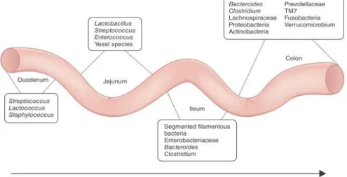
Each bacterial species colonizes a specific niche; for example, species belonging to Lactobacillus and Enterococcus mainly occur in duodenum, while those belonging to Bacteroides and Prevotellaceae are predominantly found in the colon (Figure 4) (Brown et al., 2013).
Figure 4. Composition and spatial distribution of microbiota along the mammalian intestinal tract (Brown et al., 2013)
Traditionally, culture-based methods have been used to identify and describe the bacterial groups that comprehend the gut microbiota of mammals (Minamoto et al., 2012). However these methods are extremely time-consuming and do not permit the identification of most bacterial groups present in the GI tract, which are currently uncultivable (i.e. 99%) (Nocker, Burr & Camper, 2007). High-throughput sequencing technologies, combined with bioinformatics tools for analyses of large-scale sequence data developed in the past decades have allowed to characterise several hundred bacterial genera present in the vertebrate GI tract (Minamoto et al., 2012).
The most predominant phyla in the canine and feline gut are the Firmicutes, Proteobacteria, Bacteroidetes, Fusobacteria and Actinobacteria (Deng & Swanson, 2015). The relative proportions of each of these phyla vary between studies, likely as a result of methodological differences (Deng & Swanson, 2015) alongside differences in animal breed, diet, age and living conditions (Deng and Swanson, 2015).
Details concerning the study design, animal characteristics and method of analyses are described in Table 1 and 2.
12
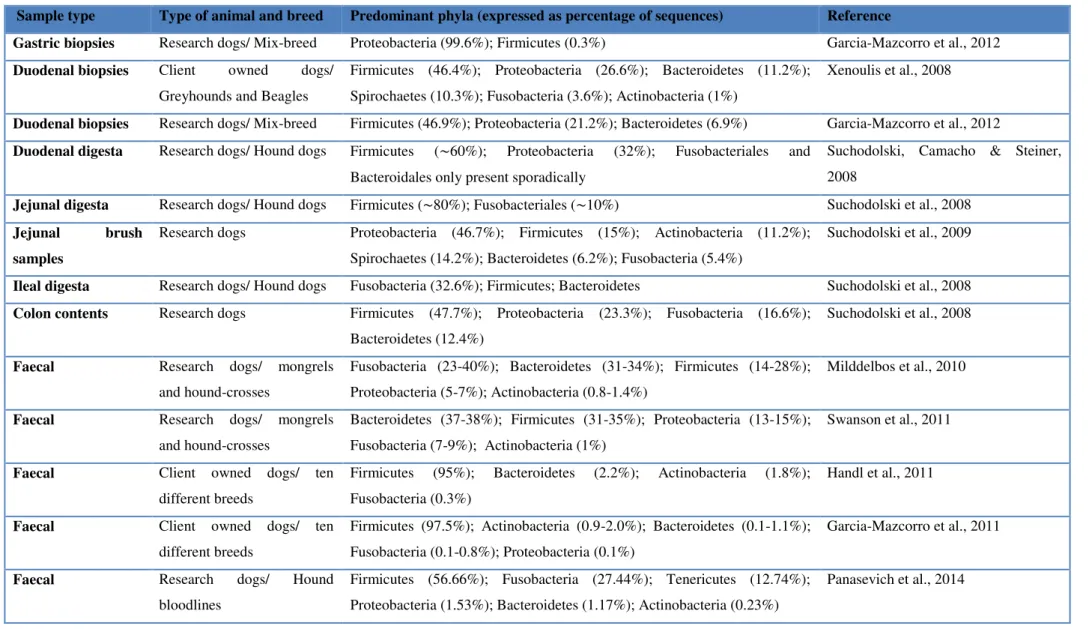
Table 1 – GI microbiota of healthy dogs based on 16S rRNA gene sequencing methods
Sample type Type of animal and breed Predominant phyla (expressed as percentage of sequences) Reference
Gastric biopsies Research dogs/ Mix-breed Proteobacteria (99.6%); Firmicutes (0.3%) Garcia-Mazcorro et al., 2012 Duodenal biopsies Client owned dogs/
Greyhounds and Beagles
Firmicutes (46.4%); Proteobacteria (26.6%); Bacteroidetes (11.2%); Spirochaetes (10.3%); Fusobacteria (3.6%); Actinobacteria (1%)
Xenoulis et al., 2008
Duodenal biopsies Research dogs/ Mix-breed Firmicutes (46.9%); Proteobacteria (21.2%); Bacteroidetes (6.9%) Garcia-Mazcorro et al., 2012 Duodenal digesta Research dogs/ Hound dogs Firmicutes (∼60%); Proteobacteria (32%); Fusobacteriales and
Bacteroidales only present sporadically
Suchodolski, Camacho & Steiner, 2008
Jejunal digesta Research dogs/ Hound dogs Firmicutes (∼80%); Fusobacteriales (∼10%) Suchodolski et al., 2008 Jejunal brush
samples
Research dogs Proteobacteria (46.7%); Firmicutes (15%); Actinobacteria (11.2%);
Spirochaetes (14.2%); Bacteroidetes (6.2%); Fusobacteria (5.4%)
Suchodolski et al., 2009
Ileal digesta Research dogs/ Hound dogs Fusobacteria (32.6%); Firmicutes; Bacteroidetes Suchodolski et al., 2008 Colon contents Research dogs Firmicutes (47.7%); Proteobacteria (23.3%); Fusobacteria (16.6%);
Bacteroidetes (12.4%)
Suchodolski et al., 2008
Faecal Research dogs/ mongrels
and hound-crosses
Fusobacteria (23-40%); Bacteroidetes (31-34%); Firmicutes (14-28%); Proteobacteria (5-7%); Actinobacteria (0.8-1.4%)
Milddelbos et al., 2010
Faecal Research dogs/ mongrels
and hound-crosses
Bacteroidetes (37-38%); Firmicutes (31-35%); Proteobacteria (13-15%); Fusobacteria (7-9%); Actinobacteria (1%)
Swanson et al., 2011
Faecal Client owned dogs/ ten
different breeds
Firmicutes (95%); Bacteroidetes (2.2%); Actinobacteria (1.8%); Fusobacteria (0.3%)
Handl et al., 2011
Faecal Client owned dogs/ ten
different breeds
Firmicutes (97.5%); Actinobacteria (0.9-2.0%); Bacteroidetes (0.1-1.1%); Fusobacteria (0.1-0.8%); Proteobacteria (0.1%)
Garcia-Mazcorro et al., 2011
Faecal Research dogs/ Hound
bloodlines
Firmicutes (56.66%); Fusobacteria (27.44%); Tenericutes (12.74%); Proteobacteria (1.53%); Bacteroidetes (1.17%); Actinobacteria (0.23%)
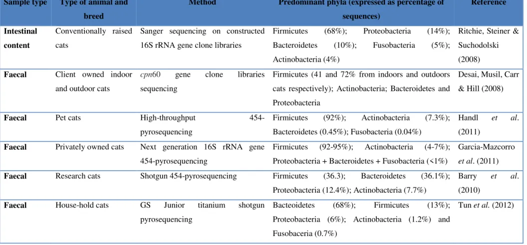
13 Table 2 – GI microbiota of healthy cats based on sequencing methods Sample type Type of animal and
breed
Method Predominant phyla (expressed as percentage of sequences) Reference Intestinal content Conventionally raised cats
Sanger sequencing on constructed 16S rRNA gene clone libraries
Firmicutes (68%); Proteobacteria (14%); Bacteroidetes (10%); Fusobacteria (5%); Actinobacteria (4%)
Ritchie, Steiner & Suchodolski (2008) Faecal Client owned indoor
and outdoor cats
cpn60 gene clone libraries sequencing
Firmicutes (41 and 72% from indoors and outdoors cats respectively); Actinobacteria; Bacteroidetes and Proteobacteria
Desai, Musil, Carr & Hill (2008)
Faecal Pet cats High-throughput
454-pyrosequencing
Firmicutes (92%); Actinobacteria (7.3%); Bacteroidetes (0.45%); Fusobacteria (0.04%)
Handl et al. (2011)
Faecal Privately owned cats Next generation 16S rRNA gene 454-pyrosequencing
Firmicutes (92-95%); Actinobacteria (4-7%); Proteobacteria + Bacteroidetes + Fusobacteria (<1%)
Garcia-Mazcorro et al. (2011) Faecal Research cats Shotgun 454-pyrosequencing Firmicutes (36.3); Bacteroidetes (36.1%);
Proteobacteria (12.4%); Actinobacteria (7.7%)
Barry et al. (2010)
Faecal House-hold cats GS Junior titanium shotgun pyrosequencing
Bacteoidetes (68%); Firmicutes (13%); Proteobacteria (6%); Actinobacteria (1.2%) and Fusobaceria (0.7%)
14
Albeit variations according the method, type of animal/breed and sample type, in dogs, within the phylum Firmicutes, the most abundant orders are Bacillales, Lactobacillales and Clostridiales (Suchodolski et al., 2008; Xenoulis et al., 2008; Suchodolski et al., 2009; Handl et al., 2011; Swanson et al., 2011; Panasevich et al., 2014). Within the order Clostridiales, the family Clostridiaceae is always reported as one of the most abundant (Suchodolski et al., 2008; Xenoulis et al., 2008; Suchodolski et al., 2009; Handl et al., 2011; Swanson et al., 2011; Panasevich et al., 2014); and Handl et al. (2011) was able to demonstrate the genus Clostridium as the most abundant within the family, followed by the genus Ruminococcus from the Ruminococcaceae family (Handl et al., 2011; Panasevich et al., 2014). The family Carnobacteriaceae is reported to be one of the most abundant within the order Lactobacillales (Xenoulis et al., 2008). Still within the Firmicutes, one further family within the Clostridiales and one further order were reported by Handl et al. (2011): the family Lachnospiraceae, represented mainly by the genus Dorea and Roseburia and the order Erysipelotrichia, represented mainly by the family Erisipelotrichaceae, respectively (Handl et al., 2011). According to Suchodolski et al. (2009) the phylum Bacteroidetes is found to be mainly represented by the order Bacteroidales; the phylum Fusobacteria by the family Fusobacteriaceae and the phylum Actinobacteria by the family Corynebacteriaceae. At last, within the phylum Proteobacteria the most abundant class is Gamma-proteobacteria, which is mainly represented by the families Pasteurellaceae, Moraxellaceae and Enterobacteriaceae (Suchodolski et al., 2008; Suchodolski et al., 2009 and Xenoulis et al., 2012).
Again, despite variations according to the method, type of animal/breed and sample type, in cats, within the phylum Firmicutes, the most abundant reported orders are Clostridiales, Lactobacillales and Erysipelotrichales (Desai et al., 2008; Ritchie et al., 2008; Handl et al., 2011). In the study performed by Handl et al. (2011) the order Lactobacillales consisted mostly of the genera Enterococcus and Lactobacillus, the order Erysipelotrichales, included primarily the genera Turicibacter, Catenibacterium, and Coprobacillus. Still in this study the phylum Actinobacteria, the second most abundant phylum, was mainly represented by the order Coriobacteriales (Handl et al., 2011). Within this order, all sequences belonged to the family Coriobacteriaceae, which consisted mainly of the genera Eggerthella and Olsenella (Handl et al., 2011).
The development of next-generation sequencing (NGS) and hence high-throughput comparative metagenomics have been responsible for the fast increase of research in this area that have added valuable information of the composition and function of bacterial populations in various environments (Jovel et al., 2016).
15
Tun et al. (2012) analysed pooled faecal samples from five healthy privately owned cats; the most predominant phylum was Bacteoidetes (68%), followed by Firmicutes (13%); Proteobacteria (6%); Actinobacteria (1.2%) and Fusobaceria (0.7%) (Tun et al., 2012). Within Bacteroidetes, the order Bacteroidetes was predominant whereas, within Firmicutes, the most abundant class was Clostridia (65%), followed by Bacilli and Mollicutes (Tun et al., 2012). The metagenomic approach used by these authors also allowed the characterization of individual microbial genes; microbial carbohydrate and protein metabolism represented 13 and 9% of the feline metagenome, respectively, while other key functional metabolic groups included DNA metabolism (8%), virulence factors (7%), and amino acid metabolism (6%) (Tun et al., 2012).
Barry et al. (2010) undertook a metagenomcis analysis of 12 individual faecal samples from four healthy cats; the predominant phyla were Firmicutes (36.1%) and Bacteroidetes (24-36%), followed by Proteobacteria (12.4%) and Actinobacteria (7.7%). The major functional metabolic categories identified were carbohydrates (15%); clustering-based subsystems (14%); protein metabolism (8%); amino acids and derivatives (8%); cell wall and capsule (7%); DNA metabolism (7%); virulence (6%); and cofactors, vitamins, prosthetic groups and pigments (6%) (Barry et al., 2010).
The symbiotic relationship between the intestinal microbiota and host is pivotal for host metabolism, organ development, immune system maturation, and defence against pathogens (Sommer & Bäckhed, 2013). In return, the microbiota inhabits a safe and nutrient-rich environment (Lee & Mazmanian, 2010).
A number of studies have demonstrated that the development of the gastrointestinal tract is dependent upon the gut microbiota (Sommer & Bäckhed, 2013). Comparative studies between wild-type and germ free (GF) mice reported that the intestine of the latter is characterised by an underdeveloped brush border and thinner intestinal villi (Abrams, Bauer & Sprinz, 1963; Reinhardt et al., 2012).
Other studies performed in GF mice support the part played by gut microbiota in preserving the structure and function of the intestinal villi (Crawford & Gordon, 2005). Furthermore, microbial wall peptidoglycan stimulates toll-like receptor 2 (TLR2) signalling, which is pivotal for the maintenance of tight junctions (Cario, Gerken & Podolsky, 2007). Also involved in the maintenance of the tight junctions is Bacteroides thetaiotaomicron, reported to prompt the expression of the small proline-rich protein 2A (sprr2A), that is required for the maintenance of the epithelial villus desmosomes (Hooper et al., 2001).
16
Prevention of cytokine apoptosis of the intestinal epithelial cells is another mechanism involved in the maintenance of structure and function of the intestinal villi by the gut microbiota (Rackoff-Nahoum, Paglino, Eslami-Varzaneh, Edberg & Medzhitov, 2004). For instance, Lactobacillus rhamnosus GG strain produces p40 and p75, this two soluble proteins are able to activate the Akt pathway, inactivate proapoptotic pathways (Hanada, Feng & Hemmings, 2004), reduce the apoptosis of the epithelial cell induced by cytokines and stimulate cell growth of the epithelial cells, both in humans and mice (Yan et al., 2007). The gut microbiota can also control gut barrier functions (Sommer & Bäckhed, 2013); for instance, Akkermansia muciniphilia leads to an increase in endocannabinoids levels which, in turn, results in a decrease of metabolic endotoxemia (Everard et al., 2013).
The gut microbiota is also important for the homeostasis of other tissues (Sommer & Bäckhed, 2013). Indeed, bone density is increased in GF mice when compared with wild-type mice, likely as a result of a decrease in number of serotonin receptors in the bone cells of the former (Sjögren et al., 2012). Bacterial colonization of GF mice resulted in the normalization of the bone density (Sjögren et al., 2012; Sommer & Bäckhed, 2013).
The presence of a large number of bacteria in the intestine of the animal, together with their proximity to epithelial tissues, represents a challenge to the mucosal immune system, which in one hand needs to tolerate the valuable commensals and on the other hand prevent the excessive expansion of the resident bacteria (Jandhyala et al., 2015). Despite this, the gut microbiota plays an important role in shaping both innate and adaptive immune systems and regulating intestinal immunity (Sommer & Bäckhed, 2013), via communications with the gut associated lymphoid tissues (GALT), effector and regulatory T cells, IgA producing B cells, and, resident macrophages and dentritic cells (DCs) in the lamina propria (Jandhyala et al., 2015). A role of the gut microbiota in promoting development of a ‘normal’ GALT has been demonstrated in studies conducted in GF mice (Round & Mazmanian, 2009). In these studies, the GALT of GF mice was characterised by reduced number of B, T and DCs and mesenteric lymph nodes (MLN), as well as by undeveloped Peyer´s patches (Round & Mazmanian, 2009). Colonization with commensal microorganisms resulted in the development of the GALT (Sommer e Bäckhed, 2013).
The surface of microorganisms presents molecules associated with different groups of microbes, such as peptidoglycans, lipopolysaccharides (LPS), lipid A, flagellin and bacterial RNA/DNA, fungal cell wall β-glucans, among others (Takeuchi & Akira, 2010). These components are part of microbe-associate molecular patterns (MAMPs), which activate immune responses (Takeuchi & Akira, 2010).
17
TLRs, C-type lectin receptors (CLRs) such as Dectin-1, and the cytosolic nucleotide-binding and oligomerisation domains (NOD) like receptors (NLRs) [all together, the pattern recognition receptors (PPR)] are present in the surface of macrophages, neutrophils and DCs (Takeuchi & Akira, 2010). The cross-talk between the PRRs and MAMPs leads to the activation of numerous signalling cascades, promoting the mucosal barrier functions and production of antimicrobial proteins (AMPs) such as cathelicidins, C-type lectins, and (pro)defensins by Paneth cells, and mucin glycoproteins and IgA (Takeuchi & Akira, 2010; Carvalho, Aitken, Vijay-Kumar & Gewirtz, 2012). Bacteroides thetaiotaomicron and Lactobacillus innocua are among the species responsible for increased production of AMPs, in the presence of a healthy microbiota (Hooper, Stappenbeck, Hong & Gordon, 2003; Cash, Whitman, Behrendt & Hooper, 2006).
By inducing production of IgA, the gut microbiota also prevents the overgrowth of pathogenic bacteria (Jandhyala et al., 2015). Gram negative bacteria like Bacteroides have been implicated in the activation of DCs leading, in turn, to the release of secretory IgA (sIgA) by the intestinal mucosa (He et al., 2007). sIgA binds to the gut microbiota, thus preventing the microbiota from entering the circulation (Jandhyala et al., 2015). Additionally, the cross-talk between the TLRs and MAMPs can induce the production of a proliferation-inducing ligand (APRIL) by the intestinal epithelial cells, which has the ability to switch a systemic sIgA1 phenotype to the intestinal mucosal sIgA2, more resistant to bacterial proteases (Macpherson & Uhr, 2004).
T cells differentiate into different subclasses implicated in different pro-inflammatory and anti-inflammatory pathways (Zhu, Yamane & Paul, 2010). Th1, Th2 and Th17, T helper cells, promote inflammation while Foxp3+ T regulatory cells (Treg) are contrast inflammatory responses (Sommer and Bäckhed, 2013). The Gram negative bacteria Bacteroides fragillis induces the differentiation of T cells CD4+ into Tregs, which leads to the production of anti-inflammatory cytokines like interleukin-10 (IL10) contrasting the pro-anti-inflammatory response mediated by Th17 cells (Maynard et al., 2007; Round et al., 2011). Induction of Tregs seems to be mediated by TLR2 signalling via the polysaccharide A on the surface of the bacterial capsule (Round et al., 2011).
18 4. Helminth-microbiota interactions (HMI)
The interactions between parasitic helminths and the gut microbiota have been investigated in a range of animal species, from mice (Walk et al., 2010; Reynolds et al., 2014; Holm et al., 2015; Houlden et al., 2015; Zaiss et al., 2015), pigs (Li et al., 2012), goats (Li et al., 2016) macaques (Broadhurst et al., 2012), to cats and dogs (Šlapeta et al., 2015).
Despite variations attributed to different range of animals and parasites, experimental designs and techniques, a few consistent alterations in the composition of the gut microbiota of the animals infected with helminths have been reported (Peachey et al., 2017).
For example, the abundance of Lactobacillaceae populations with an important role in carbohydrate metabolism (Felis & Pot, 2014) is frequently increased in the presence of helminths in the GI tract of animals.
In 2010, Walk et al. reported that infection of mice with the parasitic worm Heligmosomoides polygyrus, alters the relative abundance and distribution of gastrointestinal bacteria which, in turn, modulates host immunity. The authors also reported an increase in bacterial load/abundance in the ileum – but not in the cecum – of infected mice (Walk et al., 2010) and, in particular, an expansion of Lactobacilliaceae in the ileum (Walk et al., 2010).
Similarly, in 2015, Holm et al. also reported an increased abundance in Lactobacilli in response to Trichuris muris infection, also accompanied by a decrease in the diversity of intestinal microbiota (Holm et al., 2015). Other downstream effects of chronic infection included a skewed regulatory/inflammatory T cell balance in the intestine (Holm et al., 2015). Following on from this study, Houlden et al. (2015) demonstrated that parasite-associated changes in the metabolome and microbiota are reversed by anthelmintics.
In a study by Fricke et al. (2015) mice infected with Nippostrongylus brasiliensis presented a significantly increase of the members of the family Lactobacilliaceae in the ileum. These authors were also able to demonstrate the role of Th2 responses in parasite-associated modifications of the commensal microbiota (Fricke et al., 2015).
Lactobacilli by binding to PRRs expressed on immune cells prompt innate and adaptive immune responses in the host and several other tissues including the intestinal epithelium resulting in the expansion of Treg cells (Wells, 2011). Studies on mice indicate that specific strains of probiotics can be used to prevent or treat inflammatory diseases (Wells, 2011). Interestingly, Reynolds et al. (2014) reported that administration of the bacteria, commensal of rodents, Lactobacillus taiwanensis to BALB/c mice was associated with increased establishment of the parasitic nematode, H. polygyrus.
19
In addition, infections by the latter were associated with increased abundance of Lactobacillus species in the duodenum of C57BL/6 mice whereas SPF (specific pathogen free) BALB/c mice showed high levels of worm expulsion after the same days post infection (Reynolds et al., 2014). These data led the authors to hypothesize that the microbiota composition in the duodenum increases the persistence of H. polygyrus within the host, and that the parasite may actively change the microbiota in order to guarantee its own survival (Reynolds et al., 2014). In this cooperation, both promote the activation of T regulatory mechanisms, decreasing the chances of the host immune acting on the counterpart (Reynolds et al., 2014).
Other findings not as consistent as those found for Lactobacillaceae have been described. Broadhurst et al. (2012) reported that clinical improvements of macaques affected by idiopathic chronic diarrhoea (ICD) and experimentally infected with the whipworm, Trichuris trichiura, were associated to significant alterations in the composition of the microbiota of the colon, i.e. a decrease in bacteria belonging to the genus Streptophyta of phylum Cyanobacteria – which were increased in ICD macaques before helminth infection – and an increase in members of phylum Tenericutes (Broadhurst et al., 2012). Change in inflammatory signatures were profiled by flow cytometry analysis of leukocytes from pinch biopsies, as well as global analyses of gene expression, that showed reduced immune activation and increased mucosal TH2 responses (Broadhurst et al., 2012).
In small animals, a study by Šlapeta et al. (2015) showed infections of healthy cats and dogs by protozoa and helminths (Cystoisospora sp. in cats, and Ancylostoma caninum in dogs) and only protozoa (Giardia sp.) are associated with shifts in the composition of the host gut microbiota and its functions. In dogs, a significant difference between the Giardia-positive and -negative groups when assessing bacterial genera, was found. Unlike the faecal microbiomes of dogs infected with the canine hookworm (A. caninum) where no such difference was found. The exclusion of dogs infected by hookworms led to the separation of Giardia-positive and -negative groups. In Giardia-positive cats, no significant difference based on bacterial genera was found but the opposite was found in Cystoisospora-positive cats, which showed significantly different microbial structure when compared with Cystoisospora-negative cats. The genera Catenibacterium, Pseudobutyrivibrio and Subdoligranulum, significant producers of short-chain fatty acids, like butyric acid, which are associated with protection against pathogens and overall welfare of the gut (Kageyama & Benno, 2000; Wong, de Souza, Kendall, Emam & Jenkins, 2006; Fernandez-Rubio et al., 2009), presented significantly different abundances in parasitized animals.
20
Pseudobutyrivibrio and Subdoligranulum abundances were significantly lower in asymptomatic dogs and cats with Giardia, respectively, and Catenibacterium was significantly more abundant in dogs with Giardia. This led the authors to suggest an indirect effect of the Giardia infection in the upper gastrointestinal tract on the colon metabolism even in non-diarrhoeic animals and that Giardia may be stimulating growth of Catenibacterium. In 2012, Li et al. reported that experimental infection of pigs with the intestinal parasitic nematode Trichuris suis was associated with changes in abundances of 15 phyla and 13% of the bacterial genera. Approximately 26% of all identified metabolic pathways identified from the luminal content from proximal colon revealed were affected, including carbohydrate metabolism, lysine biosynthesis, and fatty acid absorption (Li et al., 2012).
Other studies have also reported changes in specific groups of bacteria and metabolic indicators linked to the fibrolytic potential and carbohydrate and protein transport and metabolism, following parasite infections (Li et al., 2012; Wu et al., 2012; Houlden et al., 2015; Li et al., 2016). For instance, studies in pigs infected with T. suis and mice infected with T. muris have implied a depression of metabolic pathways aforementioned in the colon (Li et al., 2012; Wu et al., 2012; Houlden et al., 2015). However the opposite, i.e. an increase in carbohydrate, protein and lipid metabolism, has been suggested to happened as a result of H. polygirus and Haemonchus contortus infections in mice colons and caprine abomasa, respectively (Kreisinger et al., 2015; Li et al., 2016).
Alpha diversity usually associated with a healthy gut homeostasis has been found to be reduced in many inflammatory bowel and/or systemic diseases in humans (Ott et al., 2004; Manichanh et al., 2006; Sepehri, Kottowski, Bernstein & Krause, 2007; Giloteaux et al., 2016) and thus is subject to investigation in many researches. Studies in rabbits and mice reported a clear reduction of alpha diversity during the acute phase of infection with Trichostrongylus retortaeformis and T. muris, respectively (Holm et al., 2015; Houlden et al., 2015; Cattadori et al., 2016). On the other hand, the alpha diversity was found to increase in humans and primates naturally or experimentally infected with nematodes like Trichuris trichiura and Necator americanus (Broadhurst et al., 2012; Lee et al., 2014; Giacomin et al., 2015; Giacomin et al., 2016), and remained unaffected in most studies conducted in a variety of animals/parasites (Li et al., 2011; Fricke et al., 2015; Kreisinger et al., 2015; McKenney et al., 2015; Slapeta et al., 2015; Li et al., 2016). The different findings on these studies might not only be explained by variations in the implicated animal and parasite species, and experimental circumstances but also by the phase of parasite infection in which samples are collected (Peachey et al., 2017).
21
It is reasonable to expect the decrease of microbial alpha diversity in the acute inflammatory phase of the infection, which can be restored or augmented during the development of chronic infections (Peachey et al., 2017).
Explanations on the causality of these interactions have arisen. For instance, helminth- associated changes in gut microbiota could be explained as a host immune response to the infection (Reynolds et al., 2014; Fricke et al., 2015; Holm et al., 2015; Cattadori et al., 2016). Indeed several studies describe an association between upregulation of cytokines after parasite invasion with changes in microbial composition (Rausch et al., 2013; Reynolds et al., 2014; Holm et al., 2015; Cattadori et al., 2016). For example, in a study where rabbits were infected with T. retortaeformis the increase in the abundance of Pasteurellaceae, Clostridiaceae, Ruminococcaceae, Peptos-treptococcaceae, and Flammenovirgaceae was related with the gene expression of interferon-g (IFN-g), whereas the reduction of Enterobacteriaceae was in negatively related with the expression of IL13 and IL14 (Cattadori et al., 2016).
Another hypothesis, that the changes in the microbiota are induced directly by the host in an attempt to generate an unfriendly environment for the parasite, has been supported by the increased production of AMPs in response to helminth infection in cattle and mice (D’Elia et al., 2009; Li et al., 2015), although it has been suggested that these responses could be the result of the Th2-mediated immunity (Fricke et al., 2015).
Finally, although the direct interaction between the TES products and the microbiota has not been reported yet, it would not be surprising if this products, some of them known to contain lysozymes (Hewitson et al., 2011), had an effect on the gut microbiota (Peachey et al., 2017).
5. Summary and research aims
Despite these efforts, knowledge HMI remains fragmentary. Capitalising on the sampling opportunities provided by a large European multicentric clinical study, with the aim to assess the efficacy of a combination of fipronil, (S)-methoprene, eprinomectin and praziquantel (i.e. BroadlineTM) against feline lungworm infections (Giannelli et al., 2016) the goal of investigate the qualitative and quantitative impact that infections by GI nematodes (i.e. Toxocara cati) exerts on the gut microbiota of cats was set. More accurately, the ultimate aim was to collect information on the effects of parasite infections on the composition of the commensal microbiota of domestic animals.
22
Chapter 2 - Materials and methods
1. Sample collection and parasitological testing
Individual faecal samples from household cats with acess to outdoor areas and able to hunt, living in Tessaloniki, Greece, were collected as part of a broader study on feline lungworm infection (Giannelli et al., 2016). All the cats were fed with identical dry commercial cat food (i.e. Purina Friskies®) for at least 6 months prior to sampling, were clinically healthy (e.g. absence of signs of GI disease or any other concomitant disease) and received no antibiotic and/or antihelminths treatment in the 12 and 3 months preceding the sample collection., respectively. None of the female cats included in this study was pregnant or lactating at the time of sample collection.
After collection the samples were immediately transported to the Laboratory of Parasitology and Parasitic Diseases, School of Veterinary Medicine of the Aristotle University of Thessaloniki (Greece) and kept at 4°C.
Here, individual samples were aliquoted for use in standard parasitological procedures (i.e. faecal egg counts [FEC] using a standard McMaster technique), as well as DNA extraction and high-throughput sequencing of the bacterial 16S rRNA gene (see below). Briefly, aliquots of 2 g of faeces were suspended in 28 ml zinc sulphate solution (ZnSo4, specific gravity = 1.180); the suspension was homogenised, filtered using a double-layer gauze, and pipetted into McMaster chambers for microscopical examination and let to stand, allowing the eggs to float to the surface and the residues to reach the bottom of the chamber (Euzeby, 1981). Only cats with or without patent T. cati infection and negative for other helminths and protozoan parasites at the fecal examination were included. Thus of the examined samples, 24 were positive for T. cati only and 21 were negative for any intestinal helminth parasite. The negative samples were used from this point onward as a negative control. Of the 24 cats that tested positive for T. cati, seven were males and 17 were females. The egg count showed an arithmetic mean of 254.2 eggs/g of faeces (EPG). Five cats that tested negative for T. cati were males and 16 were females (Table 3). The age of the cats ranged from 5 months to 10 years, with an arithmetic mean and median of 2 years.