Instituto de Ciências Biomédicas Abel Salazar, Universidade do Porto
Mestrado Integrado em Medicina
Dissertação – Artigo de Revisão Bibliográfica
Accuracy of transthoracic ultrasonography in diagnosis of
post procedure pneumothorax
Ana Soares Jorge1, António Pedro Esteves da Gama Gonçalves2, Evren Üstüner3
1[email protected], Instituto de Ciências Biomédicas Abel Salazar,
Universidade do Porto
2 [email protected], Médico Assistente no Centro Hospitalar do Porto,
Instituto de Ciências Biomédicas Abel Salazar
3 [email protected], Professor Associado na unidade de Radiologia do
Hospital İbni Sina Tıp Fakültesi - Ankara Üniversitesi
Orientador – António Pedro Esteves da Gama Gonçalves
Co-Orientador – Professora Doutora Evren Üstüner
Ana Soares Jorge
António Pedro Esteves da Gama Gonçalves
Evren Üstüner
Dedicatória
Para ti Mãe. “ Se alguém ama uma flor da qual só existe um exemplar em milhões e milhões de estrelas, isso basta para fazê-lo feliz quando as contempla. Ele pensa: Minha flor está lá, em algum lugar... Mas se o carneiro come a flor, é, para
ele, como se todas as estrelas se repentinamente se apagassem! E isto não tem importância?” Antoine de Saint-Exupéry, O pequeno Príncipe
Agradecimentos
O que inicialmente parecia ser fácil, ganhou novo significado agora, à conclusão deste percurso. À minha casa, o Instituto de Ciências Biomédicas Abel Salazar, que fez mais do que o seu papel enquanto instituição académica, mas me fez crescer e aprender a ser humana e médica.
Agradeço ao meu orientador, o Dr. António Gonçalves, pela orientação e pelo compromisso demonstrado ao longo da preparação desta dissertação. Pela autonomia e responsabilidade que me conferiu, ensinando-me a seguir o meu próprio caminho, e claro por ter reforçado, que não se coloca vírgulas entre sujeito e predicado. Ao Professor Doutor José Barros pela oportunidade de preparação para a futura vida profissional que esta dissertação implica.
Ao Hospital Ibni Sina em Ankara, por me acolher, e me fazer sentir mais do que uma estudante de Erasmus, mas mais um dos seus colaboradores. Ao Professor Doutor Serdar Akyar, por me ter permitido uma breve estadia no seu território, com a sua sabedoria e cafés turcos. Obrigada pelos poemas que declamamos, pelas conversas longas que culminaram com Edith Piaf e Je ne regrette rien. À minha co-orientadora a Professora Doutora Evren Üstüner, obrigada pelos seus ensinamentos, pela possibildade de trabalhar consigo, e me apresentar esta nova realidade da ultrassonografia. A toda a equipa técnica do departamento de Radiologia, pela sua colaboração. Aos professores Erdem e Atila, que enfrentaram os meus dados estatisticos, digerindo-os e fazendo-me quase acreditar que nasci matemática. Ao meu amigo de sempre e para a vida, que sente sempre, mesmo à distância os meus momentos difíceis, Pedro estamos juntos. À família do Arda, que se tornou a minha família em Ankara, que mesmo sem falarmos a mesma lingua, mostrou sempre o seu apoio. Ao Yavuz especificamente, pelos jogos de FIFA na playstation nos momentos mais críticos. Ao Arda é claro, que leu vezes sem conta esta tese, nunca concordando comigo em nada, crescemos juntos. À minha familia, que é grande o suficiente para não especificar, mas ciumenta o bastante para me fazer arrepender disso depois, um obrigada por toda a minha vida. Ao meu irmão, que é muito melhor do que eu, e um símbolo de sanidade mental para mim. À minha mãe, que foi e que é, mesmo sem querer e eu sem me aperceber o centro do meu mundo.
Resumo
Introdução: O pneumotórax é um problema frequente nos serviços de emergência
e cuidados intensivos, podendo ocorrer espontaneamente, após trauma, ou como complicação de procedimentos como toracocentese e biópsia pulmonar. O método de diagnóstico gold standard é a imagem radiológica, incluindo a tomografia computorizada e a radiografia do tórax. Para além dos efeitos da radiação, estas modalidades não podem ser realizadas à cabeceira do paciente. A ultrassonografia point-of-care (POCUS) está a tornar-se uma parte integral da prática clínica, podendo ser realizada rapidamente e facilmente.
Objetivo: O nosso objetivo foi analisar os dados obtidos a partir da literatura
disponível, avaliando a acuidade da ultrassonografia torácica no diagnóstico do pneumotórax após procedimentos torácicos invasivos. O nosso segundo objetivo foi propor uma metodologia para o diagnóstico de pneumotórax iatrogénico, a ser aplicada neste contexto e no futuro, incluída em um estudo que poderá fortalecer os resultados obtidos por outros autores.
Métodos: Em maio de 2018, realizámos uma revisão da literatura de artigos de
pesquisa publicados em inglês, entre janeiro de 1995 e março de 2018 no Medline e EMBASE, avaliando as características de teste da ultrassonografia em comparação com a radiografia torácica após intervenção. Artigos elegíveis foram definidos como estudos em pacientes submetidos a procedimentos torácicos e submetidos a investigação diagnóstica de pneumotórax com ultrassonografia e radiografia torácica. Os dados extraídos foram analisados, sendo calculados a sensibilidade, especificidade e o diagnostic odds-ratio global da ultrassonografia, utilizando o software freeware Meta-DiSc, versão 1.4.
Resultados: Oito artigos foram considerados adequados para a análise estatística,
compreendendo um total de 1.123 pacientes. A ultrassonografia mostrou uma sensibilidade de 89,7% (95% CI, 82,8 - 94,6) e especificidade de 97,9% (95% CI, 96,8-98,7) para a detecção do pneumotórax pós-procedimento. A radiografia de tórax apresentou uma sensibilidade de 48,6% (95% CI, 38.8-58.5) e uma especificidade de 99,3% (95% CI, 98.2-99.8). O diagnostic odds-ratio obtido para ultrassonografia e radiografia torácica foi 397.80 (95% CI,79.204-1998.0), e 113.52 (95% CI, 15.378 to 837.96) respetivamente.
a radiografia do tórax. Possivelmente, pode substituí-la no diagnóstico e exclusão do pneumotórax iatrogénico. A radiografia pode ser apenas necessária quando a qualidade do estudo ultrassonográfico não é considerada adequada.
Palavras-chave: Pneumotórax, pós-procedimento, ultrassonografia.
Abstract
Background: Pneumothorax (PNX) is a frequent problem in the emergency and
critically care settings, and can occur spontaneously, due to trauma, or as complication of interventional procedures as thoracentesis, and lung biopsy. The gold standard in the diagnosis of PNX is radiographic imaging including computerized tomography (CT) and chest radiography (CR). Beside the effects of the irradiation, CT and CR cannot be performed at the bedside of the patient. Thus, Point-of-care ultrasonography (POCUS) is becoming an integral part of the clinical practice, being quickly and easily performed.
Purpose: We aimed to analyse the data gained from the available literature
evaluating the accuracy of thoracic ultrasound in the diagnosis of post-procedure PNX. Our second goal was to propose a methodology for the diagnosis of pneumothorax, that could be applied in a post-interventional setting, and in the future included in a study that could support the findings reported by previous studies.
Methods: In May 2018, we performed a literature review of published research
articles in English, between January 1995 and March 2018 in Medline and EMBASE, evaluating the test characteristics of US in comparison with CR at a post-interventional setting. Eligible articles were defined as studies on patients submitted to thoracic procedures who underwent PNX screening with chest US and CR. The data extracted were analysed and pooled sensitivity, specificity and diagnostic odd ratio of US calculated with freeware Meta-DiSc, version 1.4 software.
Results: Eight articles were found suitable for the statistical analysis, comprising a
total of 1,123 patients. Overall, ultrasonography was 89.7% sensitive (95% CI, 82.8 -94.6) and 97.9% specific (95% CI, 96.8-98.7) for the detection of post-procedure PNX. Chest radiography had a pooled sensitivity of 48.6% (95% CI, 38.8-58.5) and a specificity of 99.3% (95% CI, 98.2-99.8). The pooled DOR for US was 397.80
(95% CI, 79.204 to 1998.0), whereas for CR, the pooled DOR was 113.52 (95% CI, 15.378 to 837.96).
Conclusion: Although the gold standard diagnosis modality of pneumothorax is still
computerized tomography, ultrasonography is more sensitive and at least as specific as CR, and possibly can substitute CR in diagnosis and exclusion of iatrogenic pneumothorax. CR may only be needed, when the quality of sonography findings is poor.
Abreviations
BLUE Bedside lung ultrasound in emergency
CR Chest radiography
CT Chest tomography
e-FAST extended FAST
FAST Focused assessment with sonography for trauma
PNX Pneumothorax
POCUS Point-of-care ultrasonography TBLB Transbronchial lung biopsy
TNAB Transthoracic needle aspiration and biopsy
Index
Resumo ... ii
Abstract ... iii
Tables Index ... vii
Figures Index ... viii
Introduction ... 1
Material and Methods ... 2
Studies inclusion criteria ... 3
Data extraction and management ... 3
Methodology for a future prospective study... 3
Patient Eligibility ... 3
Study design ... 4
Lung Ultrasound Examination ... 4
Results ... 5 Discussion ... 5 Conclusion ... 9 APPENDIX I... 10 APPENDIX II... 11 APPENDIX III... 12 APPENDIX IV ... 13 APPENDIX V ... 14 References ... 15
Tables Index
Figures Index
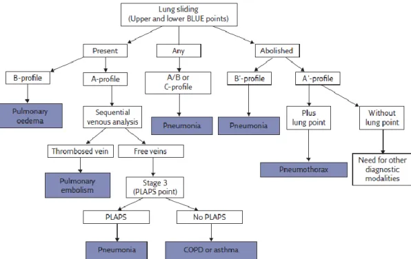
Figure 1 Decision tree of the BLUE protocol 10
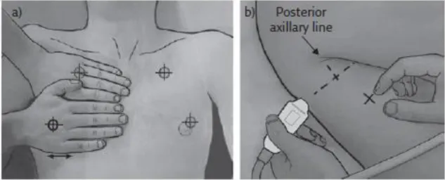
Figure 2 Areas of US investigation and the BLUE-points 12
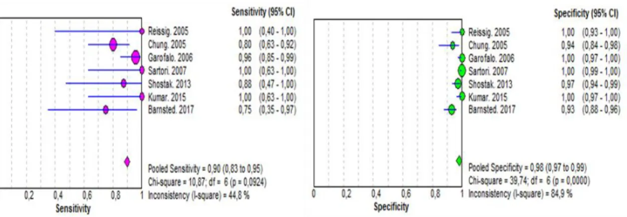
Figure 3 Forest plot for sensitivity, specificity of US 14
Figure 4 Forest plot for sensitivity, specificity of CR 14
Introduction
Pneumothorax (PNX) is frequently seen in emergency and critical care settings with significant morbidity and mortality rates. It can occur spontaneously, as a result of trauma or as a complication of various procedures, such as thoracentesis and lung biopsy.1
Defered diagnosis or treatment can lead to hemodynamic or respiratory compromise.1-3
Thus, early diagnosis is important for prognosis. The diagnosis of PNX starts with suspicion according to the clinical findings3 and is confirmed by imaging. The gold
standard diagnosis modality of PNX is computerized tomography (CT) of the chest but it has major drawbacks such as the necessity of patient transfer, delay in the diagnosis during transfer, irradiation, and cost.4 Chest radiography (CR) is commonly the first
imaging method in daily practice5 although it has been demonstrated to be an insensitive
and unreliable test in the diagnosis of limited PNX2 with misdiagnosis rates between 30%
and 40%4. Therefore there is increasing interest in alternative techniques, that can be
immediately implemented at bedside and accurate exclude pneumothorax.4
Point-of-care ultrasonography (POCUS) is now an integral part of the clinical practice, mainly in emergency and critical care but also has other diagnostic and procedural applications.6,7 It is a focused examination defined as bedside ultrasonography that
allows immediate and repeated assessments of the patient using real-time dynamic images, directly correlated with the patient’s signs and symptoms.7 Through the
identification of major signs that yield specific and standard profiles8,9 POCUS allows to
safely and efficiently diagnose or rule out conditions presented as hypotension, chest pain, or dyspnoea, reconsider priorities by the use of life-saving protocols7,9
(BLUE-protocol, Figure 1 - Appendix I), reduce the need for CT or other diagnostic procedures (as diagnostic laparotomy or arterial blood gas) and reduce the time to appropriate intervention, resulting in a shorter hospital stay, lower costs and overall mortality.7
Ultrasonography (US) of the chest, was previously limited to the study of pleural effusion and superficial thoracic masses.10 Recently, it has been highlighted that the lung is highly
sensitive to variations of the pulmonary content and balance between air and fluids.11 As
aeration of lung tissue is reduced, interstitial and alveolar fluids increase or when air and fluids are collected in the pleural space, visual assessment of several pathologic entities becomes possible by the identification of characteristic artefacts, that differ from the healthy lung.11-13
The dynamic and static analysis of a combination of this artefacts has a high diagnostic accuracy for common thoracic conditions, especially for pleural and parenchymal abnormalities. 6
Considering the impact of lung US in the assessment of respiratory and hemodynamic status of the patient, the integration of the technique in different settings is essential and indeed can be done easily without complex adaptation being cost-effective.8
Several early trials showed a strong superiority in favor of US over CR,14 and US may
be a better diagnostic strategy as an initial diagnostic study in critically ill patients with suspected pneumothorax.15 Despite those and other accumulating original research
evidence favoring ultrasonography, US remained underused due to the ready availability of CR.14
Experience with ultrasonography in the identification of iatrogenic pneumothorax is limited.16 In the few reports published, a small number of patients were enrolled and
conflicting results were obtained.17 So, there is the need of further studies that can
validate the superiority of lung ultrasound, not only in ruling out post interventional complications as pneumothorax but also confirm enhanced health outcomes.6
We aimed to conduct a review of the literature evaluating the accuracy of thoracic ultrasonography in the detection of post procedure pneumothorax, and calculate the pooled test characteristics, with the data extracted. Based on the literature available and considering the need of larger prospective studies to validate the utility of US in this field, we planned and present a methodology of a future study that aims to support the findings in previous studies.
Material and Methods
We performed a literature review of published research articles evaluating the diagnostic accuracy of US in comparison with CR at a post-interventional setting. No institutional review board approval or consents were needed, as it evaluated published studies without individually identifiable human subjects information.
We searched original articles published in English language from 1995 up to march 2018 in Medline and EMBASE. Our initial search was broad and included subject headings, truncated terms, and text words: “ultrasound” or “sonography” and “ultrasonography” or “radiography” or “chest film” or “chest radiograph” and “CT” or “chest tomography”, “pneumothorax”, and “iatrogenic” or “post procedure” or “post interventional”.
References of the initially chosen articles were also subjected to a secondary hand search, and relevant articles were extracted. No attempt was made to include unpublished data.
Studies inclusion criteria
The inclusion criteria used to select the articles are as follows:
(a) Prospective studies that evaluate the diagnostic performance of ultrasonography, CR, or both for the detection of procedure related PNX;
(b) Comparison of US imaging results with CR or gold standard (CT scan); (c) Description of diagnostic criteria for pneumothorax on US in clear details; Exclusion criteria were:
(a) Differential verification methods (two different reference standards);
(b) Long waiting time (defined as more than 6 hours) between index test and reference standard or vice versa;
(c) Insufficient details of the execution of the index test or the reference standard; (d) Lack of either CT-scan or CXR verification or US arm.
(e) Studies that included populations with known pneumothorax.
Data extraction and management
We completed our initial search strategy in march 2018. One of the authors independently reviewed the articles based on manuscript titles and abstracts, and extracted data. The data were analyzed with freeware Meta-DiSc, version 1.4 software. To explain the observed heterogeneity within studies, a random-effect model was used to calculate pooled sensitivity and specificity and Diagnostic odds ratio (DOR) with corresponding 95% confidence intervals (CI). Statistical heterogeneity was measured using the Chi 2 tests (p < 0.10 was representative of significant statistical heterogeneity)
18, and inconsistency index (I2). Possible causes of the variation observed were not
explored.
Methodology for a future prospective study
Patient Eligibility
We propose to include patients with 18 years or older undergoing CT or US guided transthoracic biopsy. Patients will be excluded if they (a) develop an intraoperative pneumothorax ,(b) declined to provide informed consent, (c) decline to undergo an ultrasound examination or CR, (d) have a preexisting pneumothorax, (e) have a chest drain in situ or other objects that would make ultrasound examination difficult, (f) if they do not undergo biopsy. Written informed consent will be obtained from enrolled participants. The local Ethics Comittee must grant a previous approval to the study.
Study design
Patients will undergo a preprocedure lung ultrasound, to identify any preexisting anatomic variants, pneumothorax, lung pathology, and for comparison with postprocedure images. Intraoperatively and immediately postbiopsy, patients will be examined for pneumothorax – with chest CT or US, according to the biopsy method. Thoracic ultrasonography will be performed within 60 minutes after transthoracic biopsy. As per institutional protocol, a CR will be done to all patients until 2 hour after biopsy to assess for pneumothorax. The CR findings will be reported by a consultant radiologist blinded to the study protocol and the patient’s clinical status. Because of the aditional radiation exposure, CT examination of the chest is not planned to be performed routinely, but only in case of disagreement between US and CR findings. Concordance between the results of transthoracic sonography and chest radiography will be considered sufficient for diagnosis or exclusion of pneumothorax. When diagnosed, complications will be treated according to international recommendations and sonographically monitored.
Lung Ultrasound Examination
All sonographic examinations will be performed by a 6th year medical student that
previously attended to an 80 hours ultrasound training workshop and underwent an initial training period of 1 week. An experienced radiologist in chest sonography will confirm the findings. Both operators will be blind to CR/CT scan results to eliminate any bias in the study.
The scanning technique planned is adapted from the emergency setting – BLUE points (Figure 2 - Appendix III; Lichtenstein, 2017).9 The images will be obtained in a
longitudinal scanning plane with the transducer indicator in a sagittal position along standardized thoracic points, starting from the biopsy site. Posterior thoracic wall will be also examined, considering the biopsys realized in that localization. The pleural surface will be identified as a hyperechoic line between 2 ribs shadows.
Pneumothorax will be diagnosed by a sequential approach, that envolved the identification of 4 signs, according to recent recomendations by an expert panel15 and
the BLUE prototocol (Figure 1- Appendix I; Lichtenstein, 2017)9: (1) recognise the
A′-profile; (2) find the lung point extending from the area with the A′-profile, laterally until the appearance of lung sliding and B-lines. If the lung point sign is absent, (3) identify the lung pulse (the lung pulse excludes a pneumothorax)19. If pneumothorax is suspected,
ultrasonography based diagnosis of PNX can be found in Appendix IV.
Results
We found 8 articles that met all the inclusion criteria and were considered eligible for a statistics analysis (Table I - Appendix II). The studies involved a total of 1,123 patients that underwent to invasive thoracic procedures as CT-guided lung biopsy (289 patients), transbronchial lung biopsy (379 patients), transthoracic needle aspiration and biopsy of the lung (TNAB) (97 patients), transthoracic sonographically guided lung biopsy (285 patients), thoracentesis (78 patients) and CT-guided cryoablation of a lung mass (1 patient).
Three of the studies4,16,20 did not report chest radiograph data. Therefore, chest
radiograph data were available for 660 of the 1,123 patients. One study17 didn´t report
clearly the number of patients in whom pneumothorax was diagnosed or excluded by US, and so was not included in US test characteristics analysis.
Chest radiographs were performed in the semierect position in 34 patients, in supine position in 247 patients, and in the erect position in 876 patients.
Overall, ultrasonography was 89.7% sensitive (95% CI, 82.8 – 94-6) and 97.9% specific (95% CI, 96.8-98.7) (Figure 3 – Appendix V). In comparison, chest radiography had a pooled sensitivity of 48.6% (95% CI, 38.8-58.5), a specificity of 99.3% (95% CI, 98.2-99.8) (Figure 4 – Appendix V). The pooled DOR for US was 397.80 (95% CI, 79.204 to 1998.0), whereas for CR, the pooled DOR was 113.52 (95% CI, 15.378 to 837.96) (Figure 5 – Appendix V).
The significant Chi2 values, shown in the forest plots for each test, implied that there
were causes of heterogeneity other than a cutoff effect. The possible source of heterogeneity in US and CR findings were not studied in our analysis.
Discussion
The stastistical analysis of the reviewed articles demonstrates significantly superior pooled sensitivity, odds of accurate diagnosis and similar pooled specificity in the use of ultrasonography compared with CR for the diagnosis of PNX.
Previous meta-analyses, studied and compared the test characteristics of US in the diagnosis of PNX within studies, in diferrent settings (trauma, and non-trauma), but few articles related to diagnosis of post-procedure PNX by US, were included. 1,2,14,21
The pooled specificity in our analysis (97.9%) is similar to the results reported by Ding et al.2, Alrajhi et al.1, Ebrahimi et al.21 and Alrajab et al.14 ( 99%, 98.2%, 99% and 98.4%
respectively). With the exception of one study, that reported a pooled sensitivity for US of 78.6%14, our result (89,7%) is also comparable with the results obtained by other
authors (88%2, 90.9.%1, 87%21). The value of DOR reported by the previous analysis,
is also in the same line of our results (397.80 versus 279.314 ,465.5221 with the exception
of the results obtain by Ding et al.2 (993.52).
Our results, revealed a high degree of heterogeneity within studies. Possible sources were considered but not analysed as in previous studies: operator, ultrasound probe used, ultrasonographic signs used as criteria to diagnose PNX.1,2,14,21 The operator,
differences between their skill, experience, knowledge of chest ultrasonography, and the type of subject (trauma/non trauma settings) were considered to be the main causes of this heterogeneity within studies.2,21 Recent meta-analysis revealed the pooled
sensitivity and specificity of US performed by nonradiologist physicians to be 89% and 99%, respectively.2 In Alrajab et al.14 emergency physicians performed better US than
nonemergency physicians, with a sensitivity of 82.3% and 72.8% respectively, possibly because their experience with chest US, now included in the eFAST (Extended Focused Assessment with Sonography for Trauma)21. However, this fact does not underestimate
the value of US, or limit its use in other settings. Studies show the learning curve for sonographic pneumothorax detection is short and steep16 and can be accuratly
performed by operators with no prior ultrasound experience after 2 hours of training.22,16
Although still performing better than chest radiography, ultrasonography sensitivity was low in 3 3,16,20 of the included studies for comparison.
Kumar et al (4) investigated the utility of bedside ultrasound for diagnosis of post-TBLB
pneumothorax. In their study the sensitivity, specificity, PPV, and NPV of US was 100%. Similarly Bensted et al.20 reviewed the use of ultrasound in screening for pneumothorax
following TBLB in post–lung transplant patients. The sensitivity and specificity of ultrasound was 75% and 93%, respectively, with a PPV of 35% and a NPV of 99%. In this study, the operator was not specifically trained in lung ultrasound, and an ultraportable handheld ultrasound device was used which could have affected the interpretation of the ultrasonography findings.20 Other important consideration was the
conservative approach of the operator, in the screening of patients with already compromised lung function and limited quality US findings, thereby over diagnosing PNX and lowering PPV.20
In 2013, a meta-analysis 14 assessed the efficacy of sonography in detecting PNX, only
including studies that compared ultrasound and CR with CT. In Chung et al. 3 , CT scans
were also performed in all the patients. Their finding of a lower sensitivity on ultrasonography examination may be explained by the fact that CT was detecting small, subclinical pneumothoraces not seen by ultrasound or CR.14 In the majority of studies
discussed, CT was not routinely performed postprocedure. Therefore, the true prevalence of occult pneumothoraces is unknown and the diagnostic accuracy of transthoracic sonography may have been overestimated.
Previous studies underlined the fact that lung sliding may be absent or difficult to recognise in cases of bullous emphysema, paralysis of the phrenic nerve and pachypleuritis, thereby leading to false positive diagnoses when absent lung sliding is considered sufficient for the diagnosis.23,16 Moreover this sign can be difficult to
appreciate in the upper chest quadrants in healthy patients as well.23 Volpicelli 19,
reported high values of sensitivity (100%), PPV (100%) and specificity (96.5%) with use of the simultaneous absence of lung sliding and B-lines as a criterion to exclude pneumothorax. In the articles reviewed, the M-mode4,13 and power Doppler24 were used
as adjuncts on the diagnosis of PNX, but the signs acessed by these (Seashore sign and power slide sign) are different graphical displays of lung sliding, being unlikely to affect significantly the test characteristics.1 The data available about the use of this
aditional methods, are also not enough to analyse the individual performance of the US signs detected by them.1
Some of the studies analyzed included patients with underlying lung disease.16,20
Shostak et al.16 could not obtain an adequate image the pleural surface in 43 of 185
patients (23%). In Bansted et al.20, 3 of the 11 patients (27.3%) who had a false positive
diagnosis of pneumothorax demonstrated reduced lung sliding on their preprocedure ultrasound. This could explain the lower specificity and PPV reported in comparison with the other studies.
Slater et al.25 determinded the diagnostic accuracy of US in the diagnosis of
pneumothorax in patients with COPD. The sensitivity of ultrasonography was 58.9% and specificity, 99.1%. This study concluded that in some patients ultrasonography only can exclude but not confidently diagnose PNX without the use of other imaging modalities.25
So, some authors20,16recommend a pre-procedure ultrasound scan, in order to identify
patients in whom a post-procedure ultrasound would not be diagnostic.
Goodman et al.17 studied the use of ultrasonography on detection of pneumothorax after
CT-guided biopsy in 29 patients.In this study the ultrasound examination was limited to the needle entry site, failing to detect six of 13 pneumothoraces.17 These data suggest
that it is insufficient to restrict sonographic examination solely to the needle entry site. 24
Although the details of the technique used to perform the ultrasound examination varied within studies, and we did not find any studies comparing them, the majority of the authors agreed that the examination should include the needle entrance point and more than one intercostal space in both hemithoraxes, from the midclavicular line to the midaxillary line,1 and even if not routinely the posterior axillary line if necessary. It is
possible that this methodology may improve diagnostic accuracy and the exclusion of occult pneumothoraces.16
The determination of the size of a PNX is important in making procedural and therapeutic decisions.4 Even with a limited chest wall examination, US was still found more sensitive
at detecting small post-biopsy pneumothoraces than erect chest radiographs.17 Small
pneumothoraces might not require invasive treatment, but are important to recognize, avoiding unnecessary therapeutic procedures.14 With the dynamic technique of
assessing lung points, pneumothorax extension can be derived from the site of lung-point projection with an accuracy almost as high as the reference standard (CT scanning).4 Garofalo et al.23 also reported complete concordance between US and CT
findings in quantification of severity of the pneumothorax and confirm the superiority of US over CR, in identification of very small pneumothoraces with a sensitivity of 95.65% and 42% respectively.
Considering some authors3 the supine incidence of CR is less sensitive (37%) than the
erect view (59%) in detecting small pneumothorax. Most of the patients included on the analyzed studies had an erect chest radiograph, possibly not underestimating the accuracy of CR. Even considering the possible confounder effect of the patient´s position on CR, the sensitivity reported in some studies was only 52%.14
The lung point is an inconstant, not very sensitive sign (75%),8,19 but it is highly specific
(100%) even in the identification of radio-occult pneumothorax.8,22 An anterior lung point
indicates mild or moderate pneumothorax whereas an absent lung point is suspicious for complete pneumothorax, requiring CT to determine the extension.4,24 US is still not
recognized as the method of choice to differentiate between small and large PNX,15 with
serial CR being recommend16.
This study has several limitations. We did not attempt to identify unpublished studies, or include articles in other languages, so our analysis included a small number of articles with small sample size and low frequency of post-interventional complications. This may have an effect on the pooled test characteristics obtained and limit the ability to generalize the data.
We noted several advantages of ultrasound over chest radiography. Ultrasonography allows a significantly quicker diagnosis of PNX compared with CR, considering the portability and almost immediate availability of the equipment, after the procedure.3,4,13,17,20 US is a relatively easy to learn, dynamic, non-ionizing, less
technically demanding tool, independent from specific acoustic windows and therefore suitable for use in different settings.26
If further prospective studies confirm the findings reported, transthoracic sonography should be considered for diagnosing pneumothorax after interventional procedures.13
Chest radiography may be reserved for assessment of the extension of pulmonary collapse, if there is discrepancy between sonographic findings and patient’s clinical manifestations13, limited studies20, or as control after pneumothorax drainage. This
approach will reduce ionizing radiation exposure, time delays and costs associated with radiography.20
Conclusion
Fast, reliable pneumothorax detection is vital at the bedside following interventional procedures. Ultrasound does have potential in fulfilling this role. US is a safe and noninvasive method, with high specificity, and so could be use as a screening tool to exclude post-interventional pneumothorax quicker and as accurately as CR.
US is reported to have good interobserver reproducibility when the operators are skilled, however is still an operator dependent test, that requires training and experience.
Larger prospective studies are needed to validate the utility of US not only in diagnosis but also to guide clinicians regarding management of PNX.
APPENDIX I
APPENDIX II
Table I Characteristics of included studies; NR not reported, CT comet tail (B-lines), LP lung point, LS lung sliding
Study Year Country Modality No. US operator US signs US probe
Goodman et al. 17 1999 United Kingdom US, CR , CT 41 Radiologist LS, CT Linear Reiβig and Kroegel24
2005 Germany US, CR 53 Pneumologist LS, CT,LP Linear
Chung et al.3 2005 Korea US, CR 97 Radiologist LS, CT Linear
Garofalo et al.23
2006 Italy US, CR 184 Emergency Phisician
LS, CT,LP Convex
Sartori et al.13 2007 Italy US, CR 285 NR LS, CT,LP Convex
Shostak et al.16 2013 United States US,CR 185 Radiologist/Clinical Investigator LS, CT Linear
Kumar et al.4 2015 India US,CR 113 Clinical
Investigator
LS, CT, LP Linear Bensted et
al.20
2017 Australia US, CR 165 Clinical Investigator
APPENDIX III
Figure 2 Areas of US investigation and the BLUE-points. Adapted from Lichtenstein, 2017.9
The upper BLUE hand is applied parallel and below the clavicle; The lower BLUE hand is applied just below; The upper BLUE point is defined at the middle of the upper BLUE hand. The lower BLUE point is defined at the middle of the palm of the lower BLUE hand.
APPENDIX IV
Lung Ultrasound Examination
The normal lung surface, defined in the BLUE protocol as the A profile (Figure 1-
Appendix I), is recognized on US by the presence of lung sliding and horizontal
repetitions of the pleural line, called A-lines.9 The A′-profile is defined anteriorly in supine
patients. It includes abolished lung sliding and the A-line sign (no B-line, should be observed).9 This profile is suggestive of pneumothorax, and definite if a lung point is also
present.9
Lung sliding is the representation of the movement that occurs between the parietal and visceral pleura synchronized with respiration.15 The B lines appear as vertical lines that
originate from the pleural surface, and result from the difference in acoustic impedance between two structures, such as visceral pleura and aerated lung.16 These artefacts are
sporadic in healthy lung and more numerous in diffuse parenchymal disease.13 The
presence of one or both signs refered previously, imply contact of the visceral pleura with the parietal pleura and can rule out PNX,2,15,23 but absence of lung sliding sign or B-lines
cannot confirm the existence of PNX.2,27 In this setting, a highly specific sign is the lung
point, which represents the transition point between the typical sonographic pattern of PNX (absence of lung sliding and B-lines) into the normal pattern of lung sliding, and represent the physical limit of PNX.15 The lung point is a pathognomonic sign of
pneumothorax however must never be sought if no A′-profile has been identified.9 The
lung pulse refers to the rhythmic movement of the visceral upon the parietal pleura with cardiac oscillations15 being useful for prompt diagnosis of an atelectasis.9 M-mode
reveals the seashore sign. Above the pleural line, the motionless chest wall displays a stratified pattern, below the pleural line, the movement of lung sliding show a sandy pattern.8 When pneumothorax occurs, the presence of air within the pleural spaces
generates reverberation artefacts that form parallel horizontal echoic lines motionless during breathing movements.13
In extreme emergency, absence of movement of the pleural line, horizontal (sliding) or vertical (pulse), combined with absence of B-lines, immediate and safely diagnosis pneumothorax without the need for searching the lung point.15
APPENDIX V
Figure 3 Forest plot for sensitivity, specificity of US for detection of pneumothorax.
Figure 4 Forest plot for sensitivity, specificity of CR for the detection of pneumothorax.
References
1. Alrajhi K, Woo M, Vaillancourt C et al. Test Characteristics of Ultrasonography for the Detection of Pneumothorax. Chest. 2012;141(3):703-708.
2. Ding W, Shen Y, Yang J, et al. Diagnosis of Pneumothorax by Radiography and Ultrasonography: a Meta-analysis. Chest. 2011;140(4):859-866.
3. Chung M, Goo J, Im J, et al. Value of high-resolution ultrasound in detecting a pneumothorax. Eur Radiol. 2005;15(5):930-935.
4. Kumar S, Agarwal R, Gupta D. Role of Ultrasonography in the Diagnosis and Management of Pneumothorax Following Transbronchial Lung Biopsy. J Bronchol
Intervent Pulmonol. 2015;22(1):14-19.
5. Kosiak W . Sonography of iatrogenic pneumothorax in pediatric patients. Journal of
Ultrasonography. 2013;13(55):379-393.
6. Hew M, Tay TR. The efficacy of bedside chest ultrasound: from accuracy to outcomes.
Eur Respir Rev. 2016; 25(141):230-246.
7. Moore C, Copel J. Point-of-Care Ultrasonography. The New England Journal of
Medicine. 2011;364(8):749-57.
8. Lichtenstein D. Lung ultrasound in the critically ill. Ann Intensive Care. 2014;4(1):1. 9. Lichtenstein D. Novel approaches to ultrasonography of the lung and pleural space: where are we now?. Breathe. 2017;13(7):100-111.
10. Gargani L, Volpicelli G. How I do it: Lung ultrasound. Cardiovascular Ultrasound. 2014;2:25-35.
11. Volpicelli G. Lung Sonography. J Ultrasound Med. 2013;32:65-171.
12. Rudas M, Orde S, Nalos M. Bedside lung ultrasound in the care of the critically ill.
Crit Care Resusc. 2017;19(4):327-336.
13. Sartori S, Tombesi P, Trevisani L, et al. Accuracy of Transthoracic Sonography in Detection of Pneumothorax After Sonographically Guided Lung Biopsy: Prospective Comparison with Chest Radiography. Am J Roentgenol. 2007;188(1):37-41.
14. Alrajab S, Youssef A, Akkus N, et al. Pleural ultrasonography versus chest radiography for the diagnosis of pneumothorax: review of the literature and meta-analysis. Crit Care. 2013;17(5):R208.
15. Volpicelli G, Elbarbary M, Blaivas M, et al. International evidence-based recommendations for point-of-care lung ultrasound. Intensive Care Med.
2012;38(4):577-591.
16. Shostak E, Brylka D, Krepp J, et al. Bedside Ultrasonography In Detection Of Post Procedure Pneumothorax. J Ultrasound Med. 2013;32(6):1003–1009.
17. Goodman TR, Traill ZC, Phillips AJ, et al. Ultrasound detection of pneumothorax.
Clin Radiol.1999;54(11):736-739.
18. Higgins JP, Thompson SG, Deeks JJ, et al. Measuring inconsistency in meta-analyses. BMJ. 2003;327(7414):557- 60.
19. Volpicelli G. Sonographic diagnosis of pneumothorax. Intensive Care Med. 2011;37(2):224-232.
20. Bensted K, McKenzie J, Havryk A, et al. Lung Ultrasound After Transbronchial Biopsy for Pneumothorax Screening in Post-Lung Transplant Patients. J Bronchology Interv
Pulmonol. 2018;25(1):42-47.
21. Ebrahimi A, Yousefifard M, Kazemi H, et al. Diagnostic Accuracy of Chest Ultrasonography versus Chest Radiography for Identification of Pneumothorax: A Systematic Review and Meta-Analysis. National Research Institute of Tuberculosis and
Lung Disease,Iran. 2014;13(4):29-40.
22. Galbois A, Ait-Oufella H, Baudel J, et al. Pleural Ultrasound Compared With Chest Radiographic Detection of Pneumothorax Resolution After Drainage. Chest. 2010;38(3):648-655.
med. 2006;111(4):516-525.
24. Reißig A, Kroegel C. Accuracy of transthoracic sonography in excluding post-interventional pneumothorax and hydropneumothorax. Eur J Radiol. 2005;53(3):463-470.
25. Slater A, Goodwin M, Anderson KE, et al. COPD can mimic the appearance of pneumothorax on thoracic ultrasound. Chest. 2006;129(3):545-550.
26. Volpicelli G. Point-of-Care Lung Ultrasound. Praxis (Bern 1994). 2014;103 (12):711-716.
27. Lichtenstein D. Ultrasound in the management of thoracic disease. Crit Care Med. 2007;35 (5):S250-S261.