Research Signpost
37/661 (2), Fort P.O., Trivandrum-695 023, Kerala, India
Vanadium Biochemistry
Chapter 14
VANADATE EFFECTS ON BONE METABOLISM: FISH
CELL LINES AS AN ALTERNATIVE TO MAMMALIAN
IN VITRO SYSTEMS
Daniel M. Tiago, Vincent Laizé, Manuel Aureliano and M. Leonor Cancela
Centre of Marine Sciences (CCMAR), University of Algarve, Faro, Portugal
Abstract
Vanadate, one of the most relevant forms of vanadium in solution, has been associated with the regulation of
various enzyme activities (e.g. phosphatases,
ribonucleases, ATPases, etc.) and shown to exhibit important biological effects. Several in vivo and in vitro studies have clearly demonstrated that any deficiency or excess of vanadium can seriously affect bone formation and its metabolism. Bone-related effects result largely from vanadium insulino-mimetic capabilities mediated by specific inhibition of protein tyrosine phosphatases (PTPases) and consequent activation of tyrosine kinase receptors (e.g. insulin receptor). Although mammals have been repetitively shown to be appropriate models to study vanadate mechanisms of action, fish have recently emerged as alternative models. Fish has been recognized as suitable model to study vertebrate bone formation and the natural presence of high quantities of vanadium in water makes it even more suitable to investigate vanadium effect on bone formation. Recent data obtained using fish
bone-derived cells revealed that micromolar
concentrations (5
µ
M) of monomeric and decamericvanadate slightly stimulate growth performances while strongly inhibiting extracellular matrix mineralization through mechanisms involving both alkaline phosphatase and MAPK pathways. Recent data obtained in fish cells will be discussed here and further compared to results obtained in mammalian systems.
.
*Correspondence/Reprint request: M. Leonor Cancela, CCMAR, Universidade of Algarve, Campus de Gambelas, 8005-139 Faro, Portugal. E-mail: lcancela@ualg.pt
1. INTRODUCTION
Vanadium is a trace element that has been shown to be essential for biological activity in various organisms [1-3]. In plants and algae, vanadium is directly involved in enzyme activity, acting as a cofactor in vanadium nitrogenases and vanadate-dependent haloperoxidases. In mammals, reduced doses of vanadium have been associated with poorly developed animals and higher rates of spontaneous abortion or mortality [4].
Biological effects of vanadium in vertebrates, in particular in mammals, essentially come from its capacity to interfere with several key enzymes, such as ATPases, phosphorylases, phosphatases (alkaline, acid and protein phosphatases) and ribonucleases [2]. This interference has been attributed to vanadium oxidation states VV and VIV (vanadate and vanadyl, respectively), which exhibit some analogy with phosphate, although to different extents (strong for vanadate and weak for vanadyl).
Inhibition of phosphatases by vanadate was attributed to the formation of good transition state analogues during enzymatic catalysis [2,5] and a similar mechanism was proposed for vanadyl although its geometry of coordination is somewhat different to that of phosphate [2].
An important feature of vanadate resides in its capacity to promote distinct effects depending on its oligomerization (monomeric to decameric species according to pH and concentration [6]) as shown using in vitro and in vivo models [7-9]. For example, decameric species were shown, in fish, to induce higher oxidative stress, haemoglobin oxidation, stimulate antioxidant enzyme activities and generate more tissue damages than other oligomeric species [10-12], while monomeric species apparently accumulated at higher rates and doses in cardiac muscle, red blood cells and plasma [13]. Aspects related with decavanadate in vivo effects are been reviewed in Chapter 9 of this book. Decameric species also revealed stronger inhibition and affinity for ATPases, in particular myosin ATPase and sarcoplasmic reticulum (SR) Ca2+- ATPase [9,14-16]. Decavanadate effects in the contractile system and in SR calcium pump are being reviewed in Chapters 5 and 7 of this book, respectively.
The most important biological effect of vanadium is probably insulin mimicking. It was first demonstrated in the early 80’s, when vanadate and vanadyl treatments applied to isolated rat adipocytes [17,18] were shown to stimulate hexose transport, glucose oxidation and lipogenesis, as insulin [19]. Additional in vivo studies showed that oral administration of vanadate to streptozocin-treated diabetic rats (STZ rats) resulted in decreased blood glucose to normal levels [20,21], confirming its action as a substitute of insulin. Vanadate was consequently proposed as a possible therapeutic agent for diabetes mellitus (insulino-dependent). However, the therapeutic use of vanadate has been seriously hampered by its relatively high toxicity [22,23]. Recent efforts have therefore focused on the discovery of chelators, which would reduce vanadyl and vanadate toxicity while preserving its insulino-mimetic properties [24].
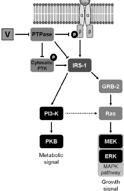
Insulino-mimetic properties of vanadium have been associated to specific inhibition of protein tyrosine phosphatases (PTPases) and consequent activation of receptor-associated protein tyrosine kinase (PTK), including that of insulin receptor [23,25,26]. Alternatively, non-receptor PTK, e.g. insulin receptor substrate 1 (IRS-1), or cytosolic PTK can be activated [27]. Two pathways involved in vanadate intracellular signalling have been identified, both resulting in distinct effects (Figure 1). A common feature of both pathways is the activation of PTK domain in IRS-1 through phosphorylation of tyrosines. While metabolic effects are mediated by phosphatidylinositol-3 kinase (PI-3K) activation, effects on cell proliferation and differentiation result from the activation of mitogen-responsive protein kinases (MAPK) [28]. Alternatively, specific insulin-like effects, e.g. glycogen synthase activation, have been associated to PI-3K/Ras/ERK activation [27].
While most studies investigating vanadium effects have been done in adipose tissue, bone has recently emerged as a system of interest after its regulation by insulin and IGF1 was demonstrated [1,29-31] and after vanadium was shown to accumulate in mammalian bones [32] and affect bone formation when absent from diet [4,33]. Accumulation of vanadium within bone, was first proposed to be related to a detoxification process [34,35]. Severe malformations occurring in bone of animals lacking vanadium however suggested that it could interact with specific growth factor-dependent signalling, in particular those known to control bone development: platelet-derived growth factor (PDGF), fibroblast growth factors (FGF1 and 2), insulin, insulin-like growth factors (IGF1 and 2), transforming growth factors beta (TGF-β1, 2 and 3), and bone morphogenetic proteins (BMP) [30,36]. Because insulin, IGFs and TGF-β
bind to specific tyrosine kinase receptors and are involved in osteoblast differentiation, they have been therefore considered as putative targets for vanadium. Effects due to insulin and IGF mimicking action of vanadium, were first demonstrated in vitro by Canalis and colleagues, who showed that concentrations below 15 µM of sodium vanadate increased DNA synthesis and mitotic index of rat calvaria primary cultures [31]. Since then, two mammalian cell lines, MC3T3-E1 (developed from newborn mouse calvaria, composed of pre-osteoblasts and able to mineralize extracellular matrix (ECM) [37]) and UMR106 (developed from rat osteosarcoma, composed of differentiated osteoblasts and unable to mineralize the ECM [38]) were shown to be particularly useful in understanding vanadium effects in bone insulin and IGF mimicking effects in bone and important features like tyrosine kinase receptors phosphorylation (with consequent activation) and activation of signalling mechanisms have been characterized [39].
Figure 1. Insulin signalling pathways and putative regulation by vanadium. Ins indicates insulin peptide; V indicates vanadium; α and β represent the subunits of insulin tyrosine kinase receptor; circled P represents phosphorylation of tyrosines in PTK domain within the β subunit of insulin receptor; PKB indicates protein kinase B, the activated intermediate in PI-3K pathway. GRB-2, Ras, MEK and ERK indicate intermediate adaptor proteins in MAPK pathways.
Until recently, effects of vanadate on bone metabolism have been exclusively characterized in vitro using these two mammalian bone-derived cell lines [39]. The recent contribution of a fish-bone derived cell line to this characterization is hereby described and results obtained compared to those derived from studies on mammalian cell lines and its suitability as an in vitro model system to study these effects is further discussed.
2. MAMMALIAN CELL LINES AS IN VITRO MODELS TO STUDY VANADIUM MECHANISMS OF ACTION IN BONE
Primary cultured cells from rat and chicken calvaria were the first in vitro systems used to investigate vanadium insulin-like effects in bone [31,40] and have contributed to the characterization of osteogenic action of vanadium in mammals including the inhibition of alkaline phosphatase (ALP) activity and the stimulation of type-I collagen synthesis [39], two mechanisms essential to the correct mineralization of the extracellular matrix (ECM) of bone cells [41]. Osteogenic actions of vanadate are being reviewed in Chapter 13 of this book. The key role of growth factors, e.g. IGF1, and tyrosine protein phosphorylation in the mediation of vanadium action was later demonstrated using mouse MC3T3-E1 and rat UMR106 cell lines [39]. Osteoblastic differentiation of these cells, monitored through the measurement of ALP activity, was show to be inhibited by vanadate, vanadyl, hydroperoxo- and peroxo-vanadium, in agreement with previous observations in primary bone-derived cultures [42]. Interestingly, enzymatic activity of PTPases was show to be inhibited in treated cells, suggesting a probable signalling interference with insulin and other growth factors that bind to tyrosine kinase receptors. In more recent experiments, vanadate, vanadyl and pervanadate were shown to stimulate cell proliferation at low doses, while higher doses severely arrested growth [43]. Primitive vanadium complexes (vanadium oxalate, citrate, tartrate and nitrilotriacetate) tested in mammalian MC3T3-E1 and UMR106 bone-derived cell lines revealed effects similar to those of vanadate: stimulation of cell proliferation, glucose consumption and protein content. It is worth to note that both vanadate and vanadium nitriloacetate exhibited a stronger inhibition of UMR106 cells differentiation [44]. However, complex toxicity was still high.
The new generation of vanadium complexes (e.g.
bis(maltolato)oxovanadium(IV) - BMOV - and bis(maltolato)dioxovanadium(V) - BMV) also exhibited effects similar to those of vanadium but revealed lower toxicity rates (BMV). All treatments with these complexes promoted cell growth in a biphasic manner, as well as increased phosphorylation of tyrosine residues, consistent with previous observation of strong inhibition of PTPases. In addition, BMOV, like vanadate, strongly inhibited ALP activity [45,46]. Similar results have been observed when using vanadyl/aspirin complex [47,48]. Relevant information concerning vanadium regulatory mechanisms associated to insulin signalling pathways were obtained only recently. Involvement of MAPK and PI-3K pathways in vanadium osteogenic effects was investigated using two complexes, trehalose vanadyl (TreVO) and vanadyl ascorbate (VOAsc) [49,50] and specific inhibitors of these pathways. While stimulation of cell proliferation and type-I collagen synthesis were shown to be mediated by an insulin-dependent mechanism, stimulation of glucose consumption and inhibition of ALP activity were apparently mediated by an insulin-independent mechanism. Another important observation was the increased phosphorylation of key intermediates of these cascades upon vanadium treatments. One of the major subfamilies of the MAP kinases, the extracellular signal-regulated kinase (ERK), has
been shown to be phosphorylated / activated in UMR106 and MC3T3-E1 cells after vanadium treatments. This effect is apparently mediate by a PI-3K/ras/ERK (extracellular signal-regulated kinase) pathway [39]. This mechanism is probably essential to explain vanadium effect in bone and will be further discussed.
Although vanadium insulin-dependent and insulin-independent actions have been already described in mammalian bone-derived in vitro systems, there is a lack of information concerning other vertebrate bone-derived systems, in particular those derived from marine vertebrate species.
3. TELEOST FISH BONE-DERIVED CELL LINES AS AN ALTERNATIVE TO MAMMALIAN SYSTEMS TO STUDY VANADIUM EFFECTS IN BONE
VSa13 bone-derived cell line has been recently developed from the vertebra of
Sparus aurata, a marine teleost fish, and fully characterized [51]. This cell line is
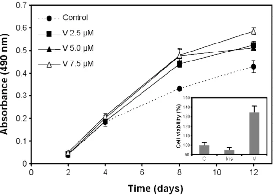
capable of mineralizing its extracellular matrix under appropriate culture conditions and has been associated to the chondrocyte lineage as suggested by a relatively high expression of matrix gla protein (a calcification inhibitor normally produced by chondrocytes in cartilage), no expression of osteocalcin (an osteoblastic marker) and strong alcian blue staining (normally associated with cartilage extracellular matrix). The suitability of VSa13 cells to study in vitro vanadium insulin-like action on bone was confirmed by its responsiveness to insulin treatments (10 nM insulin was shown to decrease by 25% the degree of extracellular matrix (ECM) mineralization in VSa13 cell [52]). To date, only vanadate oligomeric solutions have been tested in VSa13 cells. Metavanadate (containing n-meric species with n = 1-5) and decavanadate (n = 1 or 10) effects on VSa13 cells viability, growth, accumulation and ECM mineralization have been investigated [52]. Vanadate oligomerization did not originate differences in toxicity levels after prolonged exposures (metavanadate and decavanadate solutions were shown to be non-toxic at concentrations below 10 µM during 15 days). However, decameric species exhibited higher toxicity after 2 h of treatment using 1 mM concentration (acute toxicity). Similarly, accumulation of vanadium in VSa13 cells was oligomerization-independent from 4 to 24 h but much slower for decavanadate within the first 2 h of treatment. These results were shown to be coherent with decameric species decomposition rate into monomeric species (half-life time of approximately 150 min), suggesting that decavanadate long-term effects resulted mainly from monomeric species. However, in short-term treatments in which decameric species were still present in solution, vanadate was shown to accumulate much slower in cells, and to induce higher toxicities. These results reinforced the hypothesis of the higher toxicity of species highly oligomerized, which are probably formed in specific cellular environments (e.g. in which the pH is low). Merged results of long-term treatments by monomeric and decameric vanadate indicate that cell proliferation was stimulated (Figure 2) in a dose-dependent manner. The observation that insulin did not stimulate VSa13 cell proliferation (inset of Figure 2) suggested that mitogenic pathways may not be controlled by this hormone in these cells. It also suggested that vanadate effects on cell proliferation were not mediated through insulin signalling pathways, and possibly involving other PTK.
However, mineralization of VSa13 ECM was later show to be negatively regulated by insulin [52] (Figure 3), demonstrating the presence of insulin receptors and signalling pathways in these cells. ECM mineralization was also inhibited by vanadate in VSa13 cells, and combined treatments with insulin and vanadate resulted in stronger inhibition (31%, 76% and 87% inhibition in cells treated with insulin, vanadate and
insulin + vanadate, respectively; Figure 3). Effect of vanadium on extracellular matrix (ECM) mineralization in mammals has been reported in a recent study [50]. Surprisingly, vanadyl(IV)-ascorbate complex strongly increased mineral deposition within ECM of MC3T3-E1 (up to 20-fold using concentrations from 5 to 25 µM). This result, at the opposite of that observed in fish bone-derived cells, was even more surprising considering the mild effects of vanadium on bone markers: ALP activity was maintained or slightly decreased and type-I collagen synthesis was only slightly increased (approximately 10%). The opposite effect of vanadium on extracellular matrix (ECM) mineralization observed in VSa13 and MC3T3-E1 cells may be related to the species studied (fish versus mammal) or the cell types used (osteoblast versus chondrocyte) and will require additional studies in the future.
Figure 2. Vanadate and insulin effects on VSa13 cell proliferation. VSa13 cells were seeded in 96-well plates at 1.5×10³ cell/well then treated with metavanadate and decavanadate using concentrations ranging from 2.5 to 7.5 µM. Cell viability was evaluated at appropriate times using MTS assay. Inset indicates vanadate (5 µM) effect on VSa13 cell proliferation compared with that of insulin (10 nM) after 12 days of treatment. Vanadate values are the mean of metavanadate and decavanadate merged data resulting from at least six independent experiments. Insulin values are the mean of at least 3 independent experiments.
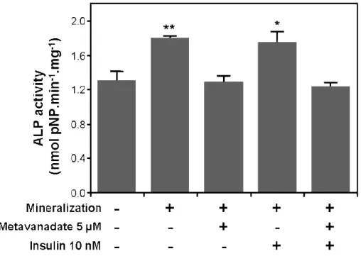
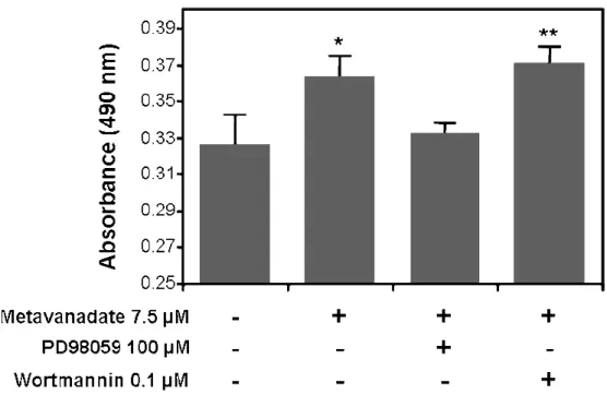
Data obtained from VSa13 cells suggested that vanadate effect on ECM mineralization probably involve insulin-signalling pathways, but the contribution of other mechanisms must also be considered. In that aspect, preliminary data obtained in VSa13 cells indicated a strong decrease in ALP activity in vanadate-treated cells during ECM mineralization (Figure 4; our unpublished results). ALP activity in VSa13 cells was demonstrated to be up regulated during ECM mineralization [51], which is consistent with a differentiation process, but treatments with 5 µM monomeric vanadate have shown to massively decrease this activity. Specific inhibitors should be tested, in order to evaluate to what extent, and which insulin signalling pathways are mediating vanadate inhibition of ECM mineralization. Preliminary data from experiments using MAPK and PI-3K inhibitors (PD98059 and wortmannin, respectively) have involved
mitogenic pathways (possibly through PI-3K/ras/MAPK signalling pathways). This issue will be further discussed in the next section of this chapter.
Figure 3. Effect of vanadate and insulin on VSa13 extracellular matrix (ECM) mineralization. Mineral deposition was revealed by von Kossa staining and evaluated by densitometric analysis. Values are the mean of metavanadate and decavanadate merged data resulting from at least six independent experiments. Asterisks indicate statistically significant differences compared to control (or between conditions) according to Student’s test (* p < 0.05, *** p < 0.001).
Figure 4. Effect of vanadate and insulin on alkaline phosphatase (ALP) activity of VSa13 cells during extracellular matrix (ECM) mineralization. Extracts from ECM mineralized cells alternatively treated with either insulin alone or metavanadate alone, or both, were collected in 0,1% (v/v) Triton X-100; samples ALP activity was evaluated in 50 mM glycine plus 0.5 mM MgCl2 reaction buffer using 0.5 mM
pNP-P (p-nitrophenyl-phosphate) as substrate. Values are the mean of at least three independent experiments. Asterisks indicate statistically significant differences compared to control according to Student’s test (* p < 0.05, ** p < 0.01).
4. MECHANISMS OF ACTION OF VANADIUM IN BONE-DERIVED CELL LINES
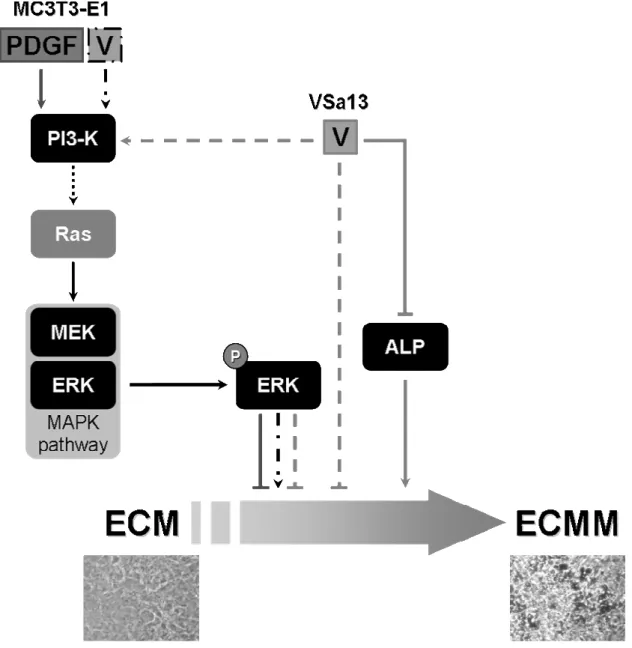
Different in vitro systems (different species and cell type) have been used to investigate proliferative and mineralogenic effects of vanadium. Available data (summarized in Table 1) suggest that vanadium may promote distinct effects depending on the system used. Although similar toxic and proliferative effects have been recognized in both systems (i.e. mammalian and fish), effects on ECM mineralization were contradictory (i.e. stimulation in mammalian cells and inhibition in fish cells). Interestingly, vanadium-treated MC3T3-E1 cells exhibited increased ERK phosphorylation. ERK activation by vanadium salts was previously demonstrated in experiments using Chinese hamster ovary cells overexpressing insulin receptor [27]. Stimulation of ras/ERK pathway mediated by PI-3K activation (resulting in the PI-3K/ras/ERK pathway) promoted important insulino-mimetic effects, e.g. glycogen synthesis and glucose homeostasis. Nevertheless, this pathway is commonly associated to growth promotion effects [53]. Recently, the ECM mineralization of MC3T3-E1 cells was show to be significantly inhibited by PDGF, also through the activation of ERK pathway. This result is, on one hand, in contradiction with the ERK-dependent mineralogenic effect of vanadium in MC3T3-E1, but, on the other hand, in total agreement with the anti-mineralogenic effect of vanadium in VSa13. It also suggests that vanadium effect could be mediated in VSa13 through ERK/MAPK pathway, an hypothesis which is strengthened by preliminary data showing the reversion of vanadium effect on cell proliferation by PD98059, a specific inhibitor of MAPK pathway early events (Figure 5; our unpublished results). However, increased ERK phosphorylation in VSa13 cells is still to be confirmed.
Figure 5. Effect of MAPK and PI-3K inhibitors on vanadate stimulation of VSa13 cells proliferation. VSa13 cells were seeded in 96-well plates at 1.5×10³ cell/well then treated with 7.5 µM metavanadate Cell viability was evaluated at 8 days using MTS assay. Values are the mean of at least three independent experiments. Asterisks indicate statistically significant differences compared to control according to Student’s test (* p < 0.05, ** p < 0.01).
Alkaline phosphatase (ALP) activity (and cell differentiation) was show to be similarly affected by vanadium in all systems. Preliminary data suggest that vanadate may not directly inhibit ALP activity in VSa13 cells (our unpublished result). We hypothesize that vanadate may affect indirectly the levels of ALP activity during ECM mineralization by down-regulating gene expression. A putative model for vanadium action during ECM mineralization is presented in Figure 6.
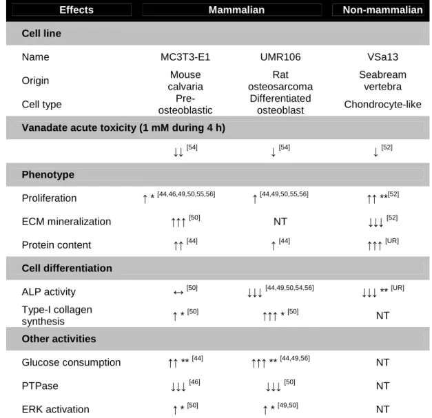
Table 1. Effects of vanadium on vertebrate bone-derived cell systems.
Effects Mammalian Non-mammalian
Cell line
Name MC3T3-E1 UMR106 VSa13
Origin Mouse calvaria Rat osteosarcoma Seabream vertebra
Cell type
Pre-osteoblastic
Differentiated
osteoblast Chondrocyte-like
Vanadate acute toxicity (1 mM during 4 h)
↓↓[54] ↓[54] ↓[52]
Phenotype
Proliferation ↑ * [44,46,49,50,55,56] ↑ [44,49,50,55,56] ↑↑ **[52]
ECM mineralization ↑↑↑[50] NT ↓↓↓[52]
Protein content ↑↑[44] ↑[44] ↑↑↑[UR]
Cell differentiation
ALP activity ↔[50] ↓↓↓[44,49,50,54,56] ↓↓↓ ** [UR]
Type-I collagen synthesis ↑ * [50] ↑↑↑ * [50] NT Other activities Glucose consumption ↑↑ ** [44] ↑↑↑ ** [44,49,56] NT PTPase ↓↓↓[46] ↓↓↓[50] NT ERK activation ↑ * [50] ↑ * [49,50] NT
* indicates insulin-like effect; ** indicates non-insulin-like effect; NT indicates not tested; UR indicates unpublished result
Figure 6. Vanadium putative regulation mechanisms for extracellular matrix (ECM) mineralization in VSa13 and MC3T3-E1 cells. ECMM indicates mineralized ECM; P indicates phosphorylation of ERK; V indicates vanadium; light grey arrows and dashed lines indicate vanadium putative effects in VSa13 cells; dark grey arrows and lines indicate PDGF effect in MC3T3-E1 cells; black dashed/dotted arrows indicate vanadium putative effects in MC3T3-E1 cells; dotted arrow indicates PI-3K/ras/MAPK pathway; below are represented micrographs of non-mineralized and mineralized VSa13 cells.
5. CONCLUDING REMARKS
This chapter presents a comprehensive collection of in vitro data regarding the effects of vanadium on bone metabolism in mammals. It also describes the recent work performed in fish bone-derived cells shown to respond to insulin, but also to be finely regulated by vanadate. In these cells, like in mammalian bone-derived cell lines, vanadate behaves as a growth factor, stimulating VSa13 cell proliferation through an insulin-independent mechanism. On the contrary, ECM mineralization of VSa13 cells is negatively regulated by insulin and vanadium, with a stronger inhibition resulting from the combination of both treatments. Based on previous studies, it is suggested that this effect may be mediated through activation of ERK pathway and consequent down-regulation of mineralization. However, in order to confirm specific involvement of this
pathway further investigation will be necessary. Preliminary results in VSa13 cells have so far indicated (1) the involvement of MAPK pathways in the vanadate effects on cell proliferation through a non-insulin related mechanism, and (2) the vanadate-dependent inhibition of cell differentiation through an effect on ALP activity. In general, it is important to notice that full characterization of vanadium effects in bone-derived systems will require further investigation. Additional suggested studies include analysis of: a) combined action of vanadium and other growth factors (e.g. IGF1, TGF-β); b) general gene expression during vanadium treatments (using microarray technology); c) specific bone-related gene expression; d) specific bone-related protein synthesis. The development of non-mammalian in vitro systems is likely to bring new insights on vanadium mechanisms of action in bone.
ACKNOWLEDGEMENTS
Daniel M. Tiago thanks to Ph.D. fellowship (BD/12773/2003) from the Portuguese Science and Technology Foundation.
REFERENCES
[1] Nielsen, F.H., and Uthus, E.O. 1990, Vanadium in biological systems, N.D. Chasteen (Ed.), Kluwer Academic Publishers, Boston, 51.
[2] Crans, D.C., Smee, J.J., Gaidamauskas, E., and Yang, L. 2004, Chem Rev, 104, 849. [3] Rehder, D. 2003, Inorg. Chem. Commun., 6, 604.
[4] Anke, M. 2004, Anal. Real Acad. Nac. Farm., 70, 961. [5] Chasteen, N.D. 1983, Structure and Bonding, 53, 105.
[6] Amado, A.M., Aureliano, M., Ribeiro-Claro, P.J.A., and Teixeira-Dias, J.J. 1993, J Raman Spectrosc, 24, 699.
[7] Crans, D.C., Mahroof-Tahir, M., and Keramidas, A.D. 1995, Mol Cell Biochem, 153, 17.
[8] Aureliano, M., and Madeira, V.M.C. 1998, Vanadium in the environment: Chemistry and Biochemistry, J.O. Nriagu (Ed.), J. Wiley, New York, 333. [9] Aureliano, M., and Gandara, R.M. 2005, J Inorg Biochem, 99, 979.
[10] Borges, G., Mendonca, P., Joaquim, N., Coucelo, J., and Aureliano, M. 2003, Arch Environ Contam Toxicol, 45, 415.
[11] Aureliano, M., Joaquim, N., Sousa, A., Martins, H., and Coucelo, J.M. 2002, J Inorg Biochem, 90, 159.
[12] Soares, S.S., Aureliano, M., Joaquim, N., and Coucelo, J.M. 2003, J Inorg Biochem, 94, 285.
[13] Soares, S.S., Martins, H., and Aureliano, M. 2006, Arch Environ Contam Toxicol, 50, 60.
[14] Tiago, T., Aureliano, M., and Gutierrez-Merino, C. 2004, Biochemistry, 43, 5551. [15] Aureliano, M., and Madeira, V.M. 1994, Biochim Biophys Acta, 1221, 259. [16] Tiago, T., Aureliano, M., and Moura, J.J. 2004, J Inorg Biochem, 98, 1902. [17] Shechter, Y., and Karlish, S.J. 1980, Nature, 284, 556.
[18] Dubyak, G.R., and Kleinzeller, A. 1980, J Biol Chem, 255, 5306. [19] Shechter, Y., and Ron, A. 1986, J Biol Chem, 261, 14945.
[20] Heyliger, C.E., Tahiliani, A.G., and McNeill, J.H. 1985, Science, 227, 1474. [21] Meyerovitch, J., Farfel, Z., Sack, J., and Shechter, Y. 1987, J Biol Chem, 262,
6658.
[23] Goldwaser, I., Gefel, D., Gershonov, E., Fridkin, M., and Shechter, Y. 2000, J Inorg Biochem, 80, 21.
[24] Thompson, K.H., and Orvig, C. 2000, J Chem Soc Dalton, 2885.
[25] Shechter, Y., Li, J., Meyerovitch, J., Gefel, D., Bruck, R., Elberg, G., Miller, D.S., and Shisheva, A. 1995, Mol Cell Biochem, 153, 39.
[26] Shisheva, A., and Shechter, Y. 1993, J Biol Chem, 268, 6463.
[27] Pandey, S.K., Anand-Srivastava, M.B., and Srivastava, A.K. 1998, Biochemistry, 37, 7006.
[28] Cheatham, B., and Kahn, C.R. 1995, Endocr Rev, 16, 117. [29] Canalis, E. 1993, Bone, 14, 273.
[30] Canalis, E., McCarthy, T.L., and Centrella, M. 1989, J Endocrinol Invest, 12, 577. [31] Canalis, E. 1985, Endocrinology, 116, 855.
[32] Etcheverry, S.B., Apella, M.C., and Baran, E.J. 1984, J Inorg Biochem, 20, 269. [33] Anke, M., Groppel, B., Gruhn, K., Langer, M., and Arnhold, W. 1989, S.I.T.E.
Symposium (Ed.), Fredrich- Jena, Schiller Universität, Jena, 17.
[34] Fukui, K., Fujisawa, Y., OhyaNishiguchi, H., Kamada, H., and Sakurai, H. 1999, J Inorg Biochem, 77, 215.
[35] Setyawati, I.A., Thompson, K.H., Yuen, V.G., Sun, Y., Battell, M., Lyster, D.M., Vo, C., Ruth, T.J., Zeisler, S., McNeill, J.H., and Orvig, C. 1998, J Appl Physiol, 84, 569.
[36] Canalis, E., Pash, J., and Varghese, S. 1993, Crit Rev Eukaryot Gene Expr, 3, 155. [37] Sudo, H., Kodama, H.A., Amagai, Y., Yamamoto, S., and Kasai, S. 1983, J. Cell
Biol., 96, 191.
[38] Ituarte, E.A., Halstead, L.R., Iida-Klein, A., Ituarte, H.G., and Hahn, T.J. 1989, Calcif Tissue Int, 45, 27.
[39] Barrio, D.A., and Etcheverry, S.B. 2006, Can J Physiol Pharmacol, 84, 677. [40] Lau, K.H., Tanimoto, H., and Baylink, D.J. 1988, Endocrinology, 123, 2858. [41] Murshed, M., Harmey, D., Millan, J.L., McKee, M.D., and Karsenty, G. 2005,
Genes Dev, 19, 1093.
[42] Cortizo, A.M., Salice, V.C., and Etcheverry, S.B. 1994, Biol Trace Elem Res, 41, 331.
[43] Cortizo, A.M., and Etcheverry, S.B. 1995, Mol Cell Biochem, 145, 97.
[44] Etcheverry, S.B., Crans, D.C., Keramidas, A.D., and Cortizo, A.M. 1997, Arch Biochem Biophys, 338, 7.
[45] Barrio, D.A., Braziunas, M.D., Etcheverry, S.B., and Cortizo, A.M. 1997, J Trace Elem Med Biol, 11, 110.
[46] Salice, V.C., Cortizo, A.M., Gomez Dumm, C.L., and Etcheverry, S.B. 1999, Mol Cell Biochem, 198, 119.
[47] Etcheverry, S.B., Williams, P.A., Barrio, D.A., Salice, V.C., Ferrer, E.G., and Cortizo, A.M. 2000, J Inorg Biochem, 80, 169.
[48] Etcheverry, S.B., Williams, P.A., Salice, V.C., Barrio, D.A., Ferrer, E.G., and Cortizo, A.M. 2002, Biometals, 15, 37.
[49] Barrio, D.A., Williams, P.A., Cortizo, A.M., and Etcheverry, S.B. 2003, J Biol Inorg Chem, 8, 459.
[50] Cortizo, A.M., Molinuevo, M.S., Barrio, D.A., and Bruzzone, L. 2006, Int J Biochem Cell Biol, 38, 1171.
[51] Pombinho, A.R., Laizé, V., Molha, D.M., Marques, S.M., and Cancela, M.L. 2004, Cell Tissue Res, 315, 393.
[52] Tiago, D.M., Laizé, V., Cancela, M.L., and Aureliano, M. 2007, Submitted to J Inorg Biochem,
[53] Kitagawa, D., Tanemura, S., Ohata, S., Shimizu, N., Seo, J., Nishitai, G.,
Watanabe, T., Nakagawa, K., Kishimoto, H., Wada, T., Tezuka, T., Yamamoto, T., Nishina, H., and Katada, T. 2002, J Biol Chem, 277, 366.
[54] Cortizo, A.M., Bruzzone, L., Molinuevo, S., and Etcheverry, S.B. 2000, Toxicology, 147, 89.
[55] Cortizo, A.M., Caporossi, M., Lettieri, G., and Etcheverry, S.B. 2000, Eur J Pharmacol, 400, 279.
[56] Williams, P.A., Barrio, D.A., Etcheverry, S.B., and Baran, E.J. 2004, J Inorg Biochem, 98, 333.