UNIVERSIDADE DA BEIRA INTERIOR
Ciências
The role of phytochemicals in Arcobacter butzleri
resistance
Vanessa Cristina Gouveia Sousa
Dissertação para obtenção do Grau de Mestre em
Bioquímica
(2º ciclo de estudos)
Orientadora: Doutora Susana Margarida Paraíso Ferreira
Coorientador: Doutor Ângelo Filipe Santos Luís
iii
Acknowledgements
First, I would like to transmit my gratefulness to the University of Beira Interior and to the Health Sciences Research Centre for providing the facilities and all the necessary conditions for the realization of this work.
I would like to express my profound gratitude to my supervisors, Professor Susana Ferreira and Professor Ângelo Luís, not only for the opportunity to let me develop this work but also for their endless patience, availability and immense knowledge.
Besides my advisors, I would also like to give my sincere thanks to Professor Ana Paula Duarte for providing most of the composites here studied, and to Professor Fernanda Domingues that not only also provide some of the compounds, but above all for her encouragement.
I thank my fellow lab mates for the companionship, the fun and the support throughout all the highs and lows of this last year.
To my friends both those who are close and those who are far away, a humble thank you for the affection, the strength and for always being there for me when I need you.
I am so grateful to my family, especially my mother, for all the moral and emotional support in my life and for always pushing me to do my best. I could not have done it without you. And lastly, to my grandfather, who I miss dearly, a thank you for all the teachings and love.
v
Abstract
Arcobacter butzleri is an emergent pathogen found in a wide range of habitats and hosts, which
has developed resistance to several antibiotics. Efflux pumps are an important mechanism of antimicrobial resistance, therefore, the use of efflux pump inhibitors (EPIs) may have the potential to restore A. butzleri susceptibility to old antibiotics. Plants have shown the ability to fight off infections despite the moderate antimicrobial action of some phytochemicals, so we aimed to test several bioactive compounds as putative EPIs, evaluating their role in the improvement of antibiotics’ performance against A. butzleri. To achieve this goal, the tolerance or resistance profile of A. butzleri strains regarding phytochemicals and antibiotics was traced through the determination of the minimum inhibitory concentration (MIC); assays of ethidium bromide accumulation were performed to assess the inhibition of the efflux pumps; the MIC of the phytochemicals in the presence of known EPIs was determined to examine the potential role of efflux pumps as resistance mechanism to the phytochemicals; checkerboard assays were made to investigate if the phytochemicals had a synergic interaction with the antibiotics; and finally, quorum sensing inhibition tests were carried out, since this mechanism is a promisor target to fight off bacterial infection.
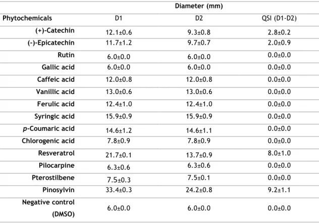
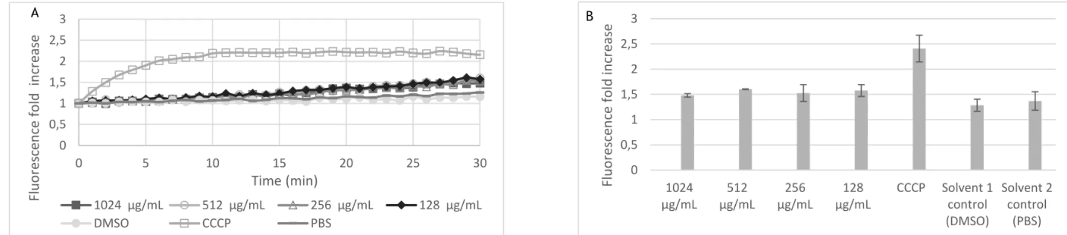
The determination of the MIC of the phytochemicals demonstrated that none of the compounds had antimicrobial activity at the concentrations tested, except for stilbenes, which MIC ranged from 64 to 512 μg/mL. Ethidium bromide accumulation assays showed that some of the tested phytochemicals presented a fluorescence folding increase higher than the controls, indicating that they may inhibit efflux pumps; however only the stilbenes presented a typical EPI profile. The assessment of the MIC of the phytochemicals in the presence of a sub-inhibitory concentration of EPIs, revealed that the importance of efflux pumps in the bacteria resistance to phytochemicals is dependent on the strain. Several phytochemicals were selected for checkerboard titration assays revealing no synergism with antibiotics, however, several cases of additivity were detected. Quorum sensing assays revealed that resveratrol and pinosylvin were able to inhibit this mechanism.
In conclusion, some of the phytochemicals tested presented potential to reduce A. butzleri resistance to antibiotics as demonstrated by the results obtained to resveratrol, pinosylvin and gallic acid, which have shown an additive effect when combined with the antibiotics. According to the ethidium bromide accumulation assay, the additive action of resveratrol and pinosylvin may be associated with efflux pump inhibition. Furthermore, these two stilbenes also possess the capacity to inhibit quorums sensing, suggesting that they may be able to inhibit A. butzleri virulence traits.
Keywords
vii
Resumo alargado
Arcobacter butzleri é um patogéneo emergente normalmente associado a doenças
gastrointestinais em humanos e animais, e a problemas reprodutores, nomeadamente abortos, em animais. Como muitos agentes patogénicos, A. butzleri tem vindo a desenvolver resistência e multirresistências a vários antibióticos. Considerando que as bombas de efluxo são um importante mecanismo de resistência antimicrobiana, sendo essenciais para o desenvolvimento de multirresistências, a estratégia de usar inibidores de bombas de efluxo para restaurar a suscetibilidade desta bactéria a antibióticos comuns é deveras promissora. Tendo em conta que as plantas estão constantemente expostas a stresses bióticos e abióticos e, apesar de alguns fitoquímicos apresentarem fraca atividade antimicrobiana contra bactérias Gram-negativas, as plantas conseguem combater infeções bacterianas com sucesso através do sinergismo entre compostos, surgindo assim como uma potencial fonte de compostos a explorar. O objetivo deste trabalho foi avaliar a capacidade de 14 fitoquímicos em inibir as bombas de efluxo de A.
butzleri, e avaliar o seu potencial na melhoria da atividade de vários antibióticos contra esta
bactéria.
Para alcançar este objetivo, o perfil antimicrobiano dos fitoquímicos e de vários antibióticos foi avaliado através da determinação da concentração mínima inibitória. Ensaios de acumulação de brometo de etídio foram realizados para determinar a possível inibição das bombas de efluxo pelos compostos em estudo. A concentração mínima inibitória dos fitoquímicos na presença de inibidores de bombas de efluxo conhecidos foi definida, a fim de investigar se as bombas de efluxo são o principal mecanismo de resistência da bactéria aos fitoquímicos. Também foram realizados ensaios de checkerboard para avaliar o potencial sinergismo entre os fitoquímicos e antibióticos e por fim também foram realizados ensaios de inibição do quorum sensing. A determinação da concentração mínima inibitória dos fitoquímicos e dos antibióticos revelou que todos os fitoquímicos têm uma concentração mínima inibitória superior a 1024 μg/mL, exceto o resveratrol, o pterostilbeno e o pinosilvino, cujos valores variam entre 64 e 512 μg/mL, para as estirpes em estudo. Os resultados obtidos relativos aos ensaios de acumulação de brometo de etídio mostraram que alguns fitoquímicos, nomeadamente (+)-catequina, (-)-epicatequina, rutina, ácidos cafeico e clorogénico, resveratrol, pterostilbeno e pinosilvino levam a um aumento de fluorescência superior ao aumento de fluorescência verificado para os controlos dos solventes. Isto é, eles levam a uma acumulação de brometo de etídio dentro das células superior aos controlos, o que sugere que estes compostos podem estar a inibir as bombas de efluxo. Porém, somente os estilbenos registaram um aumento de fluorescência superior ao verificado para o inibidor de bombas de efluxo usado como controlo. Estes compostos são também os únicos que apresentam um perfil típico de um inibidor de bombas de efluxo. A fim
de determinar se as bombas de efluxo são um mecanismo relevante de resistência aos fitoquímicos, a concentração mínima inibitória dos fitoquímicos foi determinada na presença de concentrações sub-inibitórias de inibidores de bombas de efluxo para as estirpes de A.
butzleri mais suscetível (DQ46M1) e mais resistente (CR50-2), de entre as estudadas.
Verificou-se que a importância das bombas de efluxo na resistência da bactéria aos fitoquímicos é dependente da estirpe, sendo a mais resistente mais dependente das bombas de efluxo do que a mais suscetível. Com base nos resultados do ensaio da acumulação de brometo de etídio, vários fitoquímicos foram selecionados para testes de checkerboard. Os resultados mostraram que várias combinações fitoquímico/antibiótico apresentaram um efeito aditivo, não se observando interação antagonista para nenhuma das combinações avaliadas. Os estilbenos, mais uma vez, foi a classe de fitoquímicos que apresentou os melhores resultados. Por fim, ensaios de inibição do quorum sensing foram realizados a fim de determinar se os fitoquímicos têm a capacidade de inibir estes mecanismos de comunicação celular. Os ensaios mostraram que o resveratrol e o pinosilvino conseguem inibir estes sistemas. Assim, uma vez que o quorum
sensing é fundamental para a regulação de diversos fatores de virulência como é o caso da
formação de biofilmes, estes compostos bioativos podem ter o potencial de contribuir para o controlo de A. butzleri ao atuar sobre a formação de biofilmes, inibindo-os.
Concluindo, apesar do reduzido potencial antimicrobiano da maioria dos fitoquímicos testados, alguns destes compostos apresentaram potencial no aumento de atividade de antibióticos, como foi o caso do resveratrol, pinosilvino e ácido gálico, os quais mostraram ter uma interação aditiva com os antibióticos. De acordo com o ensaio da acumulação de brometo de etídio, o efeito aditivo apresentado pelo resveratrol e pelo pinosilvino pode estar associado à inibição das bombas de efluxo. Estes dois estilbenos também demostraram a capacidade de inibir o
quorum sensing, o que sugere que podem ter a capacidade de inibir fatores de virulência
associados a A. butzleri.
Palavras-chave
Arcobacter butzleri, resistência a antibióticos, fitoquímicos, inibidores de bombas de efluxo,
ix
Index
Chapter 1- Introduction ... 1
1.1. Genus Arcobacter ... 1
1.2. Clinical relevance of Arcobacter ... 3
1.2.1. Arcobacter in humans ... 3
1.2.2. Arcobacter in animals ... 5
1.3. Distribution and transmission of Arcobacter ... 6
1.3.1. Transmission person-to-person ... 6
1.3.2. Distribution and transmission through contact with pets ... 6
1.3.3. Distribution and transmission in farm animals ... 7
1.3.4. Arcobacter distribution in water and its transmission ... 8
1.3.5. Arcobacter distribution in food and its transmission ... 9
1.3.5.1. Control of Arcobacter in food ... 10
1.4. Antibiotics resistance ... 11
1.4.1. Classes of antibiotics ... 12
1.4.2. Arcobacter resistance to antibiotics ... 14
1.5. Mechanisms of bacterial resistance ... 16
1.5.1. Target modification ... 16
1.5.2. Antibiotic inactivation ... 16
1.5.3. Outer membrane permeability ... 17
1.5.4. Efflux pumps ... 17
1.6. Phytochemicals ... 18
1.6.1. Plants as medicine ... 18
1.6.2. Classes of phytochemicals ... 19
1.6.3. Phytochemicals as inhibitors of efflux pumps ... 25
Chapter 2 – Aims ... 27
Chapter 3 - Materials and Methods ... 29
3.1. Microorganisms ... 29
3.2.1. Phytochemicals ... 29
3.2.2. Antibiotics ... 30
3.2.3. Efflux pump inhibitors ... 31
3.3. Growth curves determination ... 31
3.4. Determination of Minimum Inhibitory Concentration ... 31
3.5. Determination of Minimum Inhibitory Concentration of phytochemicals in the presence of EPIs ... 33
3.6. Ethidium bromide accumulation assays ... 33
3.7. Checkerboard assays ... 34
3.8. Quorum sensing inhibition by phytochemicals ... 35
Chapter 4 - Results and discussion... 37
4.1. A. butzleri’ susceptibility to antimicrobial agents ... 37
4.2. Phytochemicals as efflux pump inhibitors for A. butzleri strains ... 41
4.3. Efflux pumps as a resistance mechanism against the phytochemicals ... 47
4.4. Evaluation of synergistic interaction between phytochemicals and antibiotics ... 50
4.5. Quorum sensing inhibition ... 53
Chapter 5 - Conclusions and future perspectives ... 55
Chapter 6 – Bibliography ... 57
Appendix 1 ... 87
xi
List of figures
Figure 1. Chemical structure of some phenolic acids. ... 20
Figure 2. Chemical structure of some flavonoids. ... 22
Figure 3.Chemical structure of some stilbenes. ... 23
Figure 4. Chemical structure of the alkaloid pilocarpine... 25
Figure 5.Growth curves of all Arcobacter butzleri strains. ... 42
Figure 6.Fluorescence fold increase measured at 30 minutes for Arcobacter butzleri DQ46M1 strain in the presence of sub-inhibitory concentrations of the phytochemicals ... 43
Figure 7. Fluorescence fold increase measured at 30 minutes for Arcobacter butzleri CR50-2 strain in the presence of sub-inhibitory concentrations of the phytochemicals.. ... 45
Figure 8. Fluorescence folding increase measured at 30 minutes for Campylobacter jejuni 71/09 in the presence of sub-inhibitory concentrations of gallic acid... ... 46
Figure 9. Ethidium bromide accumulation assay for DQ46M1 strain in the presence of sub-inhibitory concentrations of (-)-epicatecin over 30 minutes (A) and at 30 minutes (B). .... 87
Figure 10. Ethidium bromide accumulation assay for DQ46M1 strain in the presence of sub-inhibitory concentrations of (+)-catechin over 30 minutes (A) and at 30 minutes (B). ... 87
Figure 11. Ethidium bromide accumulation assay for DQ46M1 strain in the presence of sub-inhibitory concentrations of gallic acid over 30 minutes (A) and at 30 minutes (B). ... 88
Figure 12. Ethidium bromide accumulation assay for DQ46M1 strain in the presence of sub-inhibitory concentrations of rutin over 30 minutes (A) and at 30 minutes (B). ... 88
Figure 13. Ethidium bromide accumulation assay for DQ46M1 strain in the presence of sub-inhibitory concentrations of vanillic acid over 30 minutes (A) and at 30 minutes (B)... 89
Figure 14.Ethidium bromide accumulation assay for DQ46M1 strain in the presence of sub-inhibitory concentrations of caffeic acid over 30 minutes (A) and at 30 minutes (B). ... 89
Figure 15.Ethidium bromide accumulation assay for DQ46M1 strain in the presence of sub-inhibitory concentrations of ferulic acid over 30 minutes (A) and at 30 minutes (B). ... 90
Figure 16.Ethidium bromide accumulation assay for DQ46M1 strain in the presence of sub-inhibitory concentrations of syringic acid over 30 minutes (A) and at 30 minutes (B). ... 90
Figure 17.Ethidium bromide accumulation assay for DQ46M1 strain in the presence of sub-inhibitory concentrations of chlorogenic acid over 30 minutes (A) and at 30 minutes (B). . 91
Figure 18.Ethidium bromide accumulation assay for DQ46M1 strain in the presence of sub-inhibitory concentrations of p-Coumaric acid over 30 minutes (A) and at 30 minutes (B). . 91
Figure 19.Ethidium bromide accumulation assay for DQ46M1 strain in the presence of sub-inhibitory concentrations of pilocarpine over 30 minutes(A) and at 30 minutes (B). ... 92
Figure 20.Ethidium bromide accumulation assay for DQ46M1 strain in the presence of sub-inhibitory concentrations of resveratrol over 30 minutes (A) and at 30 minutes (B). ... 92 Figure 21.Ethidium bromide accumulation assay for DQ46M1 strain in the presence of sub-inhibitory concentrations of pterostilbene over 30 minutes (A) and at 30 minutes (B). ... 93 Figure 22.Ethidium bromide accumulation assay for DQ46M1 strain in the presence of sub-inhibitory concentrations of pinosylvin over 30 minutes(A) and at 30 minutes (B). ... 93 Figure 23.Ethidium bromide accumulation assay for CR50-2 strain in the presence of sub-inhibitory concentrations of (+)-catechin over 30 minutes (A) and at 30 minutes (B). ... 94 Figure 24.Ethidium bromide accumulation assay for CR50-2 strain in the presence of sub-inhibitory concentrations of (-)-epicatechin over 30 minutes(A) and at 30 minutes (B). .... 94 Figure 25.Ethidium bromide accumulation assay for CR50-2 strain in the presence of sub-inhibitory concentrations of gallic acid over 30 minutes (A) and at 30 minutes (B). ... 95 Figure 26.Ethidium bromide accumulation assay for CR50-2 strain in the presence of sub-inhibitory concentrations of rutin over 30 minutes(A) and at 30 minutes (B). ... 95 Figure 27.Ethidium bromide accumulation assay for CR50-2 strain in the presence of sub-inhibitory concentrations of vanillic acid over 30 minutes(A) and at 30 minutes (B). ... 96 Figure 28.Ethidium bromide accumulation assay for CR50-2 strain in the presence of sub-inhibitory concentrations of caffeic acid over 30 minutes (A) and at 30 minutes (B). ... 96 Figure 29.Ethidium bromide accumulation assay for CR50-2 strain in the presence of sub-inhibitory concentrations of ferulic acid over 30 minutes (A) and at 30 minutes (B) ... 97 Figure 30.Ethidium bromide accumulation assay for CR50-2 strain in the presence of sub-inhibitory concentrations of syringic acid over 30 minutes (A) and at 30 minutes (B) ... 97 Figure 31. Ethidium bromide accumulation assay for CR50-2 strain in the presence of sub-inhibitory concentrations of p-Coumaric acid over 30 minutes (A) and at 30 minutes (B) .. 98 Figure 32.Ethidium bromide accumulation assay for CR50-2 strain in the presence of sub-inhibitory concentrations of chlorogenic acid over 30 minutes(A) and at 30 minutes (B). .. 98 Figure 33.Ethidium bromide accumulation assay for CR50-2 strain in the presence of sub-inhibitory concentrations of pilocarpine over 30 minutes (A) and at 30 minutes (B). ... 99 Figure 34.Ethidium bromide accumulation assay for CR50-2 strain in the presence of sub-inhibitory concentrations of resveratrol over 30 minutes (A) and at 30 minutes (B). ... 99 Figure 35.Ethidium bromide accumulation assay for CR50-2 strain in the presence of sub-inhibitory concentrations of pterostilbene over half an hour (A) and at 30 minutes (B). .. 100 Figure 36.Ethidium bromide accumulation assay for CR50-2 strain in the presence of sub-inhibitory concentrations of pinosylvin over 30 minutes (A) and at 30 minutes (B). ... 100
xiii
List of tables
Table 1. Arcobacter species identified so far and their original sources. ... 2 Table 2. Principal antibiotic classes, their mechanisms of action and year at which they were clinically introduced. ... 13 Table 3. Arcobacter butzleri strains used in this study. ... 29 Table 4. Phytochemicals used in this study. ... 30 Table 5. Lowest and highest limits of the range of concentrations tested for each compound. ... 32 Table 6. Minimum inhibitory concentration of the antibiotics for the four Arcobacter butzleri strains in study. ... 37 Table 7. Minimum inhibitory concentration of the antibiotics being studied for the
Campylobacter jejuni 71/09 strain. ... 38
Table 8. Minimum inhibitory concentration of the fourteen phytochemicals under evaluation for the four Arcobacter butzleri strains in study. ... 39 Table 9. Minimum inhibitory concentration of gallic acid for the control strain Campylobacter
jejuni 71/09. ... 40
Table 10. Minimum inhibitory concentration of ethidium bromide for the Arcobacter butzleri strains being studied. ... 41 Table 11. Minimum inhibitory concentration of four known efflux pumps inhibitors for the four Arcobacter butzleri strains in study. ... 47 Table 12. Minimum inhibitory concentration of several phytochemicals in the presence of sub-inhibitory concentrations of efflux pump inhibitors for the Arcobacter butzleri DQ46M1. ... 48 Table 13. Minimum inhibitory concentration of several phytochemicals in the presence of sub inhibitory concentration of efflux pump inhibitors for the Arcobacter butzleri strain CR50-2. ... 48 Table 14. Fractional Inhibitory Concentration Index and correspondent classification of the effect of the combination phytochemical-antibiotic in Arcobacter butzleri DQ46M1 strain. ... 50 Table 15. Fractional inhibitory concentration index and correspondent classification of the combination phytochemical-antibiotic in Arcobacter butzleri CR50-2 strain... 51 Table 16. Fractional inhibitory concentration index and correspondent classification of the combination gallic acid-antibiotic in Campylobacter jejuni 71/09i strain. ... 52
Table 17. Screening of phytochemicals for quorum sensing inhibition using Chromobacterium
xv
List of acronyms
ABC ATP Binding Cassette ATP Adenosine Triphosphate BHI Brain-Heart Infusion medium BRU Brucella Blood Agar
CCCP Carbonyl-Cyanide m-Chlorophenylhydrazone CFU Colony-Forming Units
DMSO Dimethyl Sulfoxide DNA Deoxyribonucleic Acid EPI Efflux Pump Inhibitor EtBr Ethidium Bromide
FDA Food and Drug Administration FIC Fractional Inhibitory Concentration FICI Fractional Inhibitory Concentration Index HIV Human Immunodeficiency Virus
LB Luria-Bertani LPS Lipopolysaccharide
MATE Multidrug And Toxic compound Extrusion MDR Multidrug Resistance
MFS Major Facilitator Superfamily MHB Müeller-Hinton Broth
MIC Minimum Inhibitory Concentration
MRSA Methicillin-Resistant Staphylococcus aureus NMP 1-(1-naphthylmethyl)-piperazine
PAβN Phenylalanine– Arginine β-Naphthylamide PBS Phosphate-Buffered Saline solution QRDR Quinolone Resistance Determining Region QSI Quorum Sensing inhibition
RNA Ribonucleic Acid
RND Resistance Nodulation Division SMR Small Multidrug Resistance TSA Tryptic Soy Agar
TSB Tryptic Soy Broth UHT Ultra-High Temperature UV Ultraviolet
1
Chapter 1- Introduction
1.1. Genus Arcobacter
The genus Arcobacter is a diverse group of Gram-negative bacteria that, together with the
Campylobacter and Sulfurospirillum genera, constitute the Campylobacteraceae family
(Collado and Figueras, 2011). Recently, this member of the Epsilonproteobacteria class, has been gaining increasing attention since some species are considered emergent pathogens and potential zoonotic agents (Collado et al., 2011; Mansfield et al., 2000).
Currently, this genus is composed of 27 species, the majority isolated in the last decade from several environments and hosts (Table 1).
The first Arcobacter was isolated by Ellis et al. in 1977 from bovine foetuses (Fera et al., 2009). However, this genus was only proposed in 1991 to reclassify Campylobacter cryaerophila and
Campylobacter nitrofigilis, two aerotolerant Campylobacter species, as Arcobacter cryaerophilus and Arcobacter nitrofigilis, respectively (Vandamme et al., 1991). One year
later, the genus was enlarged with the reclassification of Campylobacter butzleri as Arcobacter
butzleri and the description of the new species Arcobacter skirrowii (Vandamme et al., 1992a). A. butzleri had originally been isolated in the previous year from humans and animals with
diarrhoea (Kiehlbauch et al., 1991).
The name of this genus has Latin roots and means “bow-shaped rod” (Mansfield and Forsythe, 2000). True to its name, Arcobacter spp. are small, non-spore forming, curved rods, often helical or S shaped (0.2-0.9 μm wide and 0.5-3 μm long) (Ferreira et al., 2015; Vandamme et al., 1992a), although sometimes cells as long as 20 μm can be found (Mansfield and Forsythe, 2000).
With the exception of A. anaerophilus, which is an obligate anaerobe without flagella (Sasi Jyothsna et al., 2013), the members of this group move in darting or corkscrew-like movements due to a polar unsheathed flagellum at one or both ends of the cell (Vandamme et al., 1992a). This microorganism can grow in aerobic or microaerobic (3-10% oxygen with no hydrogen required) conditions, having an optimal growth temperature of 37°C in microaerophilic conditions and of 30°C in aerobic conditions. Though, Arcobacter spp. can grow at higher or lower temperatures, depending on the strain and conditions (Collado and Figueras, 2011; Ferreira et al., 2015; Mansfield et al., 2000; Vandamme et al., 1992a).
Table 1. Arcobacter species identified so far and their original sources.
Specie Source Reference
Arcobacter nitrofigilis
Roots of Spartina alterniflora Loisel (a salt march plant) and in root‐associated
sediments
(McClung et al., 1983)
Arcobacter cryaerophilus
Faeces, reproductive tracts, aborted foetuses of different farm animals and
from milk of cows with mastitis (Neill et al., 1985)
Arcobacter butzleri Humans and animals with diarrhoeal disease (Kiehlbauch et al., 1991)
Arcobacter skirrowii
Preputial fluids of bulls Bovine, porcine, and ovine isolates obtained from aborted foetuses and diarrhoeic faeces.
(Vandamme et al., 1992a)
Arcobacter cibarius Broiler carcasses in Belgium (Houf et al., 2005)
Arcobacter
halophilus Hypersaline lagoon in Hawaii (Donachie et al., 2005) Arcobacter mytili and brackish water in Spain Mussels (Mytilus sp.) (Collado et al., 2009a)
Arcobacter thereius
Kidney and liver of Danish pigs’ abortions and
cloacal content of ducks (Houf et al., 2009)
Arcobacter marinus Seawater with seaweeds and Starfish in Korea (Kim et al., 2010)
Arcobacter
trophiarum from fattening pigs in Belgium Faecal samples taken rectally (De Smet et al., 2011a)
Arcobacter defluvii Sewage samples (Collado et al., 2011)
Arcobacter molluscorum
Mussels (Mytilus sp.) and
oysters (Figueras et al., 2011a)
Arcobacter ellisii Mussels (Mytilus sp.) (Figueras et al., 2011b)
Arcobacter
bivalviorum Mussels (Mytilus sp) (Levican et al., 2012) Arcobacter
venerupis
Clam (Venerupis pullastra) (Levican et al., 2012)
Arcobacter cloacae
Mussels (Mytilus sp.) and sewage from the Waste Water
Treatment Plant (Levican et al., 2013)
Arcobacter suis Pork meat (Levican et al.,2013)
Arcobacter
anaerophilus Estuarine sediment (Sasi Jyothsna et al., 2013) Arcobacter
ebronensis Mussels (Levican et al., 2015) Arcobacter
aquimarinus Seawater (Levican et al., 2015) Arcobacter
lanthieri Pig and dairy cattle manure (Whiteduck-Léveillée et al., 2015)
Arcobacter
pacificus Seawater (Zhang et al., 2016) Arcobacter faecis Human waste septic tank (Whiteduck-Léveillée et al., 2016)
Arcobacter acticola Seawater on the East Sea in South Korea (Park et al., 2016)
Arcobacter porcinus Aborted piglet foetus (Figueras et al., 2017)
Arcobacter
lekithochrous Molluscan hatchery in Norway (Diéguez et al., 2017) Arcobacter haliotis Molluscan collected in Japan (Tanaka et al., 2017)
In a broad sense, the exception being A. pacificus, all species are oxidase positive, but catalase is only present in some species (Ferreira et al., 2017). Organic acids and amino acids are utilized as carbon sources (Vandamme et al., 1992a).
3
Campylobacter and Arcobacter are morphologically very similar, the key feature to distinguish
them is that Arcobacter can grow in aerobic conditions and at lower temperatures than the former (Collado and Figueras, 2011). However, with the classification and recognition of new species in recent years, this is not an absolute principle anymore, with, for example, A.
anaerophilus being an obligate anaerobe (Sasi Jyothsna et al., 2013).
1.2. Clinical relevance of Arcobacter
Arcobacter spp. are classified as emergent food and water-borne pathogens, with A. butzleri, A. cryaerophilus, and A. skirrowii being associated with human and animal disease (Vandenberg
et al., 2004; Kayman et al., 2012a). In fact, A. butzleri and A. cryaerophilus have been classified as severe hazards to human health by the International Commission on Microbiological Specifications for Foods (ICMSF, 2002).
Among Arcobacter species, A. butzleri stands out as the more prevalent in clinical and environmental samples, as well as in food of animal origin (Van den Abeele et al., 2016; Collado and Figueras, 2011; Fernandez et al., 2015).
1.2.1. Arcobacter in humans
A. butzleri has been associated with gastrointestinal diseases such as enteritis and colitis,
bacteraemia and septicaemia (Van den Abeele et al., 2016; Fera et al., 2010; Fernandez et al., 2015). Furthermore, it has been repeatedly classified as the fourth most common pathogen associated with diarrhoeal illness (Van den Abeele et al., 2014; Collado et al., 2013; Ferreira et al., 2014a; Prouzet-Maulon et al., 2006; Vandenberg et al., 2004).
Although Arcobacter spp. have been isolated in asymptomatic hosts (Houf and Stephan, 2007),
A. butzleri is typically associated with watery diarrhoea, abdominal pain, nausea, vomiting and
fever (Arguello et al., 2015; Jiang et al., 2010; Kayman et al., 2012a; Kiehlbauch et al., 1991; Teague et al., 2010; Vandamme et al., 1992b; Vandenberg et al., 2004). Though, there are cases where these symptoms are not all present, as illustrated by an A. butzleri outbreak in a school in Italy, where the infected children only reported abdominal pain (Vandamme et al., 1992b). Once again, it is easy to confuse an Arcobacter spp. infection with a Campylobacter spp. infection as they share many symptoms; however, Campylobacter jejuni is usually associated with bloody diarrhoea versus the watery one of A. butzleri (Vandenberg et al., 2004).
A study made with infected human colonic epithelial cells (HT-29/B6) concluded that the process by which A. butzleri induces diarrhoea is mediated by the reduced expression of tight-junction proteins claudin-1, -5 and -8, which causes an epithelial barrier dysfunction and,
consequently, epithelial apoptosis. This leads to diarrhoea through a leak flux mechanism (Bücker et al., 2009).
Arcobacter spp. has also been associated with a few cases of bacteraemia. A. butzleri
bacteraemia cases include an 85 year old man with chronic lymphocytic leukaemia (Arguello et al., 2015), a 69 years old woman with acute gangrenous appendicitis (Lau et al., 2002) and a neonate in the United Kingdom (On et al., 1995). On the other hand, A. cryaerophilus bacteraemia was diagnosed in an uremic patient with hematogenous pneumonia (Hsueh et al., 1997) and a 7 year old boy that had developed acute respiratory distress and renal failure (Woo et al., 2001). Furthermore, Arcobacter spp. was also linked with enteritis (Van den Abeele et al., 2014) and peritonitis (Monzon and Coronel, 2013).
Host characteristics, such as the state of the immune system, may play a role in the development of A. butzleri infection and pathogenicity, as studies made in India with human immunodeficiency virus type 1 (HIV-1) infected patients (Kownhar et al., 2007) and in Italy with type 2 diabetic individuals (Fera et al., 2010) showed. Both studies found a higher prevalence of A. butzleri in the ill patients versus the control group of healthy subjects. Moreover, a study in Canada found the prevalence of A. butzleri in diarrhoeic (56.7%) and non-diarrhoeic (45.5%) individuals very similar (Webb et al., 2016), suggesting that infection only occurs when certain circumstances are met.
In general, A. butzleri infections are not very severe, with cases of bacteraemia typically occurring in immunocompromised hosts. However, these infections can persist from a couple of days to a couple of months leading to a loss of life quality and leaving the immune system debilitated (Prouzet-Maulon et al., 2006; Tee et al., 1988; Vandamme et al., 1992b; Vandenberg et al., 2004).
Most laboratories do not use the appropriate conditions for the identification of Arcobacter spp., so they tend to be wrongfully classified as campylobacters. As such, the prevalence of
Arcobacter infections is not truly known (Taylor et al., 1991; Prouzet-Maulon et al., 2006).
However, globally, reports from Europe show an A. butzleri percentage of 0.07% in healthy patients in Denmark (Engberg et al., 2000), 0.4% in patients suspected of infectious gastroenteritis in the Netherlands (De Boer et al., 2013), 1% in patients suspected of having a
Campylobacter infection in France (Prouzet-Maulon et al., 2006), 3.5% in hospitalized patients
(Vandenberg et al., 2004), 0.7% in stools of patients with enteritis in Belgium (Van den Abeele et al., 2014) and 1.3% in diarrhoeal stools collected from 22 hospitals of Portugal (Ferreira et et al., 2014a). In South Africa two studies were made, one studied a heterogeneous population and had an Arcobacter spp. prevalence of 6.2% (Samie et al., 2007), the other analysed diarrhoeic stools obtained from a hospital and had a prevalence of only 0.33%. Additionally, it was reported an A. butzleri prevalence in patients with diarrhoea of 1.4% in Chile, (Collado et al., 2013), 0.51% in New Zealand (Mandisodza et al., 2012) and 2.38% in Thailand (Taylor et al., 1991). Lastly, India reported a prevalence of Arcobacter spp. of 1.25% (Kownhar et al., 2007).
5 The discrepancy in the results may be a reflex not only of the diverse prevalence of Arcobacter spp. in the different countries, but also due to the different methods of detection used and populations studied (Collado and Figueras, 2011).
There are some reports of travellers that developed A. butzleri infections while aboard. For example, a diabetic German man was admitted in the hospital with A. butzleri infection three months after visiting Thailand, Singapore and Hong Kong (Lerner et al., 1994), and a man that was returning from an European cruise was hospitalized with A. butzleri bacteraemia (Arguello et al., 2015). A larger study also analysed European and US travellers that acquired acute diarrhoea in Mexico, Guatemala and India and reported that 8% of them were hosts to A.
butzleri. Yet, as other microorganisms were also identified in some of the tourists, the role of A. butzleri as the causative agent was not certain (Jiang et al., 2010).
Currently, the precise mechanisms of pathogenicity of Arcobacter spp. remains relatively unexplored. Human and animal cell culture in vitro assays have shown that several Arcobacter species can adhere and invade eukaryotic cells (Fallas-Padilla et al., 2014), and produce toxins that damage host cells (Carbone et al., 2003). Arcobacter spp. also seems to be involved in inflammatory processes, as it is possible to find leukocytes (Kayman et al., 2012b; Vandenberg et al., 2004) and lactoferrin (Samie et al., 2007) in stools of patients with A. butzleri infection. Also, it was been demonstrated that A. butzleri is highly susceptible to human blood serum, being possibly able to activate the complement by an alternative pathway (Wilson et al., 2010).
1.2.2. Arcobacter in animals
A. butzleri, A. cryaerophilus, and A. skirrowii are the species most commonly recovered from
animals (Kabeya et al., 2003; On et al., 2002; De Smet et al., 2011a).
Arcobacter spp. has been found in healthy animal hosts (De Smet et al., 2011b; Stirling et al.,
2008; Van Driessche et al., 2004), however, they are also associated with diarrhoea (Kiehlbauch et al., 1991; Anderson et al., 1993), mastitis (Logan, 1982), reproductive problems, namely aborts (Oliveira et al., 1997; On et al., 2002; Vandamme et al., 1992a), and a few cases of active colitis (Anderson et al., 1993). One study also reported the development of lesions in the gastric mucosa in piglets infected with Arcobacter spp. but it was not possible to definitively link the lesions with the presence of the bacterium (Suarez et al., 1997).
It was been suggested that Arcobacter strains associated with infertility could be opportunistic pathogens that infect the foetus after the placenta being compromised as a study found that the strains isolated from reproductively impaired and in normal sows were similar (de Oliveria et al., 1999).
1.3. Distribution and transmission of Arcobacter
Arcobacter spp. has been isolated worldwide from healthy and diseased animals and humans,
water, food and food processing facilities (Ferreira et al., 2017). The vast distribution of this microorganism is supported by genomic studies, as the analysis of the human strain A. butzleri RM4018 shows that a substantial portion of the bacteria’s genome is associated with its adaptation to different environmental conditions (Miller et al., 2007).
The most likely route of human contamination is the consumption of contaminated water and food (Miller et al., 2009), though transmission by contact with a human or animal host is also a possibility (Fera et al., 2009; Vandamme et al., 1992b).
1.3.1. Transmission person-to-person
In 1983, in a period of two months, ten children that frequented the same nursey school in Italy, started to suffer from abdominal pain, vomiting and fever. When the children’ stools were analysed, it was discovered that not only was A. butzleri present in all the samples, but that all the strains shared phenotypic and genotypic characteristics. That, combined with the fact that the other children and staff that used the school dining room did not get sick, plus the conspicuous timing of the infections, all very close together, raised the hypothesis that person-to-person transmission had occur (Vandamme et al., 1992b).
A few years later, it was reported a case of a neonate with A. butzleri bacteraemia. This report is important because it was suggested that the infection has been contracted in utero, likely due to a prenatal bleeding experienced by the mother. This was the first study that indicated the possibility of vertical transmission in humans (On et al., 1995).
Venereal transmission of this bacterium has been suggested for animals (Ho et al., 2006a), but no information is available regarding humans.
1.3.2. Distribution and transmission through contact with pets
Arcobacter spp. has also been isolated from the oral cavities and faeces of pets, namely cats
and dogs. As such the contact with them and the faecal contamination of the environment has been suggested as a possible route of human infection.
A study performed in Denmark detected A. butzleri in the saliva of one cat (12.5%) and seven dogs (58%) (Petersen et al., 2007). In the same year, a study in Chile reported a 3.3% prevalence of A. butzleri in the faeces of dogs (Fernández et al., 2007), while, other study in Turkey did not found any isolates in dog’s stools (Aydin et al., 2007). In the next year, a study in Belgium found no arcobacters in cats, while only two dogs (0.75%) and five dogs (1.87%) carried arcobacters in the mouth and faeces, respectively (Houf et al., 2008). By contrast, in Italy, it
7 was detected a high prevalence (78.8%) of Arcobacter spp. in cats, of which 77.6% were A.
butzleri positive. The detection of this microorganism was higher in oral samples than in blood
and lymph nodes (76.5% vs 2.3%) (Fera et al., 2009). Recently, a study in Czech Republic tested oral samples from cats and dogs and confirmed the presence of A. butzleri in one cat (1.4%) and four dogs (3.7%) (Pejchalova et al., 2017).
1.3.3. Distribution and transmission in farm animals
Several studies have reported the occurrence of Arcobacter spp. in healthy farm animal’s faeces, namely, cattle (3.6%-39.2) (Van Driessche et al., 2003; Van Driessche et al., 2005; Giacometti et al., 2015; Kabeya et al., 2004; Öngör et al., 2004; Shirzad Aski et al., 2016; Wesley et al., 2000), pigs (7.1-85%) (Van Driessche et al., 2003; Hume et al., 2001; Kabeya et al., 2004; Van Driessche et al., 2004), chicken (14.5-64.3%) (Collado et al., 2009b; Kabeya et al., 2004), goats (10.7%) (De Smet et al., 2011b), sheep (16.1-43.1%) (Van Driessche et al., 2003; Shirzad et al., 2016; De Smet et al., 2011) and horses (15.4%) (Van Driessche et al., 2003). Being A. butzleri the overall most prevalent species (Van Driessche et al., 2004; González et al., 2010; Kabeya et al., 2004; Öngör et al., 2004; Shah et al., 2013), and co-infection with multiple species of Arcobacter an usual observation (Van Driessche et al., 2004; Shah et al., 2013). The prevalence of Arcobacter can be largely influenced by factors such as the farm where the study was made (reflecting the farm practices), the period of collection of the samples and the methodology used for sampling and isolation (Nieva-Echevarria et al., 2013). Regarding the transmission of Arcobacter spp. among farm animals, it is thought that the main factors for this are the consume of contaminated water (Wesley et al., 2000; Giacometti et al., 2015) and living in a contaminated environment (Van Driessche et al., 2004; 2005; Eifert et al., 2003). Additionally, vertical transmission was also suggested by a study that isolated Arcobacter spp. from the amniotic fluid of sows and from the rectal samples of new-born piglets. The similarity between the isolates led the authors to propose that intra-uterine transmission occurred. The same study also detected horizontal transmission from the mother or the environment to the piglets, showing that post-natal contamination occurred (Ho et al., 2006a). The fact that healthy livestock animals may be a reservoir for Arcobacter spp. is a public concern as it was hypothesised that Arcobacter spp. are introduced in slaughterhouses by the gut contents of asymptomatic animals leading to co-contaminations that are reflected in the high prevalence of genetic diverse Arcobacter spp. isolates in carcasses (Amare et al., 2011; Andersen et al., 2007; Van Driessche and Houf, 2007; Ho et al., 2008; Kabeya et al., 2004). Besides pets and farm animals, Arcobacter spp. has also been found in more exotic or unsuspected animals such as pigeons (Giacometti et al., 2015), ducks (Fernández et al.,2010), pelicans and sparrows (Fernández et al., 2007), raccoons (Hamir et al., 2004), rainbow trout, (Yildiz and Adyn, 2006) white and black rhinoceros, gorillas, alpacas, gazelles, rhea (Wesley and Schroeder-tucker, 2011), lizards, serpents and chelonians (Gilbert et al., 2014).
1.3.4. Arcobacter distribution in water and its transmission
Water is suggested as playing a major role in the transmission of Arcobacter spp. to animals and humans. In fact, it is estimated that 63% of human A. butzleri infections are due to the consumption of contaminated water (Shah and Saleha, 2011).
Arcobacter spp. has been isolated from several water sources such as rivers (Collado et al.,
2008, 2010; Fernández et al., 2010; Šilha et al., 2015; Laishram et al., 2016), lakes (Collado et al., 2008), seawater (Fera et al., 2004; Maugeri et al., 2004; Collado et al., 2008), wells (Fong et al., 2007; Rice et al., 1999), sewages and sludge (Collado et al., 2008; McLellan et al., 2011; Merga et al., 2014; Rodriguez-Manzano et al., 2012; Šilha et al., 2015;), drinking water, water that has received tertiary treatments (Jacob et al., 1993; Rodriguez-Manzano et al., 2012) and water used in aquafarming (Xiong et al., 2015). Additionally, it was suggested that the seasons influence the prevalence of Arcobacter spp. in water, as it is detected more frequently in the warmer months (Andersen et al., 2007; Collado et al., 2010).
Several reports have established an association between the isolation of Arcobacter spp. from water samples and its level of faecal contamination (Collado et al., 2008; Collado et al., 2010; Fong et al., 2007; Merga et al., 2014; Newton et al., 2013), with A. butzleri being the dominant species in most studies (Collado et al., 2010; Collado et al., 2008; Merga et al., 2014). It is understood that the inflow of faeces from human (Collado et al., 2008) and animal carriers (Newton et al., 2013; Stampi et al., 1993), transports the bacteria into the sewages and serves as a source of nutrients. That, allied with Arcobacter’s ability to survive in harsher environmental conditions than other faecal bacteria (Merga et al., 2014), has led to the high prevalence of this microorganism in the sewage system. Moreover, A. butzleri has shown the capacity to adhere and to form biofilms in various materials used in pipes (stainless steel, cooper and plastic) which indicates that it may be able to spread through the water distribution system (Assanta et al., 2002), a point that highlights the importance of the disinfection processes and supports the dissemination of the bacteria through the water system.
A. butzleri is susceptible to chlorination (Rice et al., 1999; Moreno et al., 2004), but the
membrane integrity and nucleic acids remained intact for more than five hours, and so, continuous chlorination is recommended to control its spread (Moreno et al., 2004).
Studies made in Spain and South Africa have not found Arcobacter spp. in chlorinated drinking water (Diergaardt et al., 2004; Collado et al., 2010). However, other studies have detected this microorganism in non-chlorinated drinking water (Jacob et al., 1993; Rodriguez-Manzano et al., 2012; Shah et al., 2013). Thus, the depuration treatments applied in some water treatment plants are not able to completely remove this pathogen. It is interesting to note that the number of Arcobacter spp. isolated from drinking water was much higher than the number of
Campylobacter spp. found. However, it is not possible to establish if this fact is due to Arcobacter resistance to the treatments applied or if it is a reflection of the different optimal
9 Despite the chlorine susceptibility of Arcobacter spp., there have been some reported cases of outbreaks related with this bacterium, and so supporting water as a route of contamination. One occurred in 1996 at a Girl Scout camp in Idaho, where the outbreak was associated with the consumption of water from an A. butzleri contaminated well when the chlorination system was broken. It was estimated that 81% of the people there became ill with nausea, vomiting, diarrhoea and cramps (Rice et al., 1999). Other case, happened in South Bass Island, Ohio in 2004, affecting many residents and tourists that developed diarrhoea. Arcobacter spp. was found, once again, in contaminated wells around the area (Fong et al., 2007). More recently, in 2008, an outbreak of acute gastroenteritis affected residents in Slovenia, where 2.3% of the faecal samples analysed were positive for A. cryaerophilus. Assumedly, the water system distribution was contaminated due to the constructions made to build of a new connection (Kopilović et al., 2008).
1.3.5. Arcobacter distribution in food and its transmission
The use of sludge and animal manure to fertilize the soil is an old practice. However, it has the side effect of potentially introduce pathogens into the food chain (Udeigwe et al., 2015).The consumption of raw or undercooked contaminated food is another of major route of transmissions suggested to Arcobacter spp.(Lappi et al., 2013).
Arcobacter spp. has been found in carcasses and offal of farm animals (beef, pork, poultry,
rabbit and lamb) (Rivas et al., 2004; Ho et al., 2006; Šilha et al., 2015), fish (Palareti et al., 2016), mussels (Fernández et al., 2010), raw milk (Giacometti et al., 2015), cheese and fresh (González and Ferrús, 2011) and ready-to-eat (Mottola et al., 2016) vegetables.
Moreover, Arcobacter spp. as also been detected, at a higher prevalence than Salmonella and
Campylobacter, in several restaurants popular among tourists in Bangkok. It was determined
that, independently of the restaurant, the risk of exposure per meal was 13%, rising to 75% once 10 or more meals are eaten (Teague et al., 2010). A. butzleri, particularly, has been identified as the likely etiologic agent of an outbreak of foodborne illness associated with the consumption of roasted chicken served during a wedding reception (Lappi et al., 2013). As mentioned above, Arcobacter spp. is frequently found in asymptomatic farm animals, contributing to faecal contamination of the carcasses during evisceration, either directly or using the equipment as an intermediate, and so being an unaccounted contamination risk during slaughter (Ho et al., 2008; Houf et al., 2002; Shah and Saleha, 2011; De Smet et al., 2010). In food, Arcobacter spp. is found more frequently in meat, namely poultry (13.1%-100%) (Atabay et al., 2006; Atabay et al., 1998; Ho et al., 2008; Kabeya et al., 2004; Nieva-Echevarria et al., 2013; Rahimi, 2014; Rivas et al., 2004; De Smet et al., 2010; Villarruel-López et al., 2003), followed by pork (7%-96.4%)(Van Driessche and Houf, 2007; Kabeya et al., 2004; Nieva-Echevarria et al., 2013; Rivas et al., 2004; Villarruel-López et al., 2003), beef (2.2%-37%) (Ho et al., 2006b; Kabeya et al., 2004; Nieva-Echevarria et al., 2013; Rivas et al., 2004; De Smet
et al., 2010; Villarruel-López et al., 2003) and lamb (15%) (Rivas et al., 2004). Being the most prevalent species A. butzleri, though A. cryaerophilus and A. skirrowii are also not uncommon. Relatively to other animal products, so far, studies indicated that, although breeding hens can be infected with Arcobacter spp., there is no contamination of the eggs (Lipman et al., 2008). Regarding dairy products, there are reports of a high prevalence (3.2%-80%) of Arcobacter spp. in raw milk (Scullion et al., 2006; Pianta et al., 2007; Ertas et al., 2010; Nieva-Echevarria et al., 2013; Giacometti et al., 2014) and cheese (Serraino et al., 2013; Yesilmen et al., 2014).
Arcobacter spp. has also been isolated from seafood, which consume presents a relevant hazard
as this is a food product often eaten undercooked or raw. The bacterium has been found in fish (19%-25%) (Laishram et al., 2016; Rathlavath et al., 2016), clams (100%) (Collado et al., 2009b), shellfish (14.7%-73.3%) (Nieva-Echevarria et al., 2013; Laishram et al., 2016) and mussels (22.7%-41.1%) (Collado et al., 2009a; Fernandez et al., 2001; Maugeri et al., 2000). No arcobacters were found in oysters or frozen shrimps (Collado et al., 2009b). The most prevalent specie isolated was A. butzleri (Fernandez, 2001; Collado et al., 2009b; Rathlavath et al., 2016).
Additionally, Arcobacter spp. was also found in carrot (Hausdorf et al., 2011) and spinach wash water (Hausdorf et al., 2013), in fresh lettuces (20%) (González and Ferrús, 2011) and ready-to-eat (Mottola et al., 2016) vegetables. These foods are especially dangerous as they are often eaten raw and, especially in the case of the ready-to-eat, not properly washed.
Furthermore, A. butzleri is not able to survive in beer (Šilha et al., 2013) or apple and pear purees (Lee and Choi, 2013). High sugar content, acidic pH and the presence of polyphenols and alcohol are some of the factors probably responsible for this (Lee and Choi, 2013; Šilha et al., 2013).
1.3.5.1. Control of Arcobacter in food
The treatments that meat is subjected to, in order to be commercialized seems to affect the survival of Arcobacter spp., as several studies have showed a decrease of its prevalence. Namely, in the case of chickens, a study isolated A. butzleri in 95% of the fresh carcasses, but only in 23% of the frozen carcasses (Atabay et al., 2003). Another study found a prevalence of
Arcobacter spp. of 96.8% in broiler carcasses pre-scalding, 61.3% in the carcasses pre-chill and
only 9.6% in the carcasses post-chill (Son et al., 2007). Concerning pork, a study isolated
Arcobacter spp. in 96.4% carcasses, but only in 21% of the pork at retail (Van Driessche and
Houf, 2007). For beef, a study found that Arcobacter spp. has present in 37.4% of the carcasses collected from two slaughterhouses, but after 24 hours of cooling at 7ᵒC, the percentage of
Arcobacter spp. isolated lowered significantly (7%) (De Smet et al., 2010). Arcobacter spp. has
11 packaged chill stored beef (Balamurugan et al., 2013). Moreover, A. butzleri is more tolerant to radiation under vacuum in ground pork than C. jejuni (Collins et al., 1996).
Relatively to scalding, survival tests also indicate that some Arcobacter species are able to survive for several minutes at 52ᵒC (Ho et al., 2008). It seems that the application of mild heat (50ᵒC) followed by cold shock (4ᵒC-8ᵒC) is more effective than these treatments applied separately (D’Sa and Harrison, 2005).
Regarding milk, it has been shown that, although A. butzleri and A. cryaerophilus cannot grow, they remain viable in Ultra-High Temperature (UHT), pasteurized and raw milk for six days
when stored between 4ᵒC and 10ᵒC. In raw milk A. butzleri increases when stored at 20ᵒC. These findings show that, although it is unlikely that Arcobacter spp. survives the pasteurization or UHT processes, it is possible that bad hygiene and storage leads to contamination (Giacometti et al., 2014).
Several plant extracts have also shown the capacity to inhibit Arcobacter spp. growth, namely the ones from cinnamon, bearberry, chamomile, sage and rosemary (Cervenka et al., 2006). Compounds like cinnamaldehyde, thymol, carvacrol, caffeic and tannic acids, eugenol and resveratrol presented activity against Arcobacter spp. (Cervenka et al., 2008; Duarte et al., 2015) Thus, phytochemicals are presented as a viable alternative to the traditional preservatives.
1.4. Antibiotics resistance
Until the commercialization of antibiotics, infections were a major detriment to human health. However, selective pressure exerted by the excessive and inappropriate use of a narrow repertoire of antimicrobials has contributed to the development of bacterial resistance (Okeke et al., 2005).
As many of the antibiotics used in humans are also applied in sub-therapeutic doses to food animals and plant agriculture to promote growth and prevent disease, there is the possibility that human pathogens that have reservoirs in animals, such as Arcobacter, will develop resistance to drugs employed in human medicine (Angulo et al., 2004; de Souza and Hidalgo, 1997; Wegener, 2003). Furthermore, the natural human microflora may exchange antibiotic resistance determinants, by horizontal gene transfer with ingested bacteria, as they pass through the colon, enhancing the resistance of these food-borne pathogens and of the bacterial flora (Salyers et al., 2004). The newly acquired resistance phenotypes tends to stabilize and stays ingrained in the bacteria, which means that reducing the use of antibiotics is not enough to reverse the resistance (Barbosa and Levy, 2000).
The increase of antibiotic resistant bacteria coincides with a reduction in the production of new antibiotic molecules. In fact, of the 48 drugs approved by the Food and Drug Administration (FDA) between 1998 and 2003, only 6 (14%) were considered new molecular entities, the other 86% were drugs structurally similar to one or more compounds that are already in the market (Brunton et al., 2011).
Nowadays, the scientific community faces two major challenges in this field: conserving the effectiveness of the existing antibacterial and developing new ones.
1.4.1. Classes of antibiotics
Antibiotics may be produced biosynthetically, by bacteria or fungi in order to kill competing microorganisms or, as is the case of many second and third generation antibiotics, be the result of semisynthetic modifications (Walsh, 2000; Hansen et al., 2003). Antibiotics act by killing the bacteria (bactericidal) or by stopping its growth (bacteriostatic) by inhibiting DNA replication/repair, or protein or cell wall synthesis (Fair and Tor, 2014; Walsh, 2000) (Table 2). For example, chloramphenicol, a member of the amphenicol class, binds reversibly to the peptidyl transferase centre of the 50S ribosomal subunit preventing its binding to the amino acid–end of tRNA, inhibiting peptide bond formation and, consequently, the elongation step of translation (Brunton et al., 2011). This antibiotic has a broad-spectrum activity and it is fairly used as it is inexpensive (Fair and Tor, 2014). However, there are safety concerns, namely haematological disorders such as aplastic anaemia, bone marrow suppression and leukaemia, as well as neurotoxicity and Grey syndrome (Aminov, 2017).
Erythromycin is a macrolide; this class of antibiotics inhibit protein synthesis by binding reversibly to the 50S ribosomal subunit and causing premature dissociation of peptidyl tRNA from the ribosome. Macrolides are the second most prescribed antibiotic class after the β-lactams, targeting the same range of pathogens but with lesser efficiency against Gram-negative bacteria (Aminov, 2017; Katz and Ashley, 2005).
The tetracycline family is constituted by natural and semisynthetic broad-spectrum agents that have activity against either Gram-positive and Gram-negative bacteria as well as protozoan parasites. They inhibit bacterial protein synthesis by preventing the attachment of aminoacyl-tRNA to the ribosomal acceptor (A) site (Following and Therapy, 2001). Tetracyclines do not exhibit any major adverse effect and are one of the more cheap antibiotics on the market, as such they have been extensively used in human and animal therapy (Following and Therapy, 2001; Roberts, 2005), inclusively in prolonged treatments of non-infectious conditions at sub-therapeutic levels (e.g. acne) (Roberts, 2003).
13
Table 2. Principal antibiotic classes, their mechanisms of action and year at which they were clinically introduced.
Class Examples Start of clinical use
Mechanism of
action Reference
Sulfonamides Prontosil 1935 Inhibit synthesis of
folic acid (Aminov, 2017) β-lactams Penicillin G 1938 Inhibit cell wall
biosynthesis
(Fair and Tor, 2014) (Page, 1984) Aminoglycosides Streptomycin 1946 Mistranslation of
protein (Fair and Tor, 2014) Amphenicols Chloramphenicol 1948 Inhibit protein
synthesis (Brunton et al.,2011) (Aminov, 2017) Polymyxins Colistin 1950 Increased cell membrane permeability (Falagas and Kasiakou, 2005) Macrolides Erythromycin 1952 Inhibit protein
synthesis
(Katz and Ashley, 2005) Tetracyclines Clortetracycline 1952 Inhibit protein
synthesis
(Fair and Tor, 2014) (Following and Therapy, 2001) Rifamycins Rifampicin 1958 Inhibit protein
synthesis (Fair and Tor, 2014) Glycopeptides Vancomycin 1958 Inhibit cell wall
biosynthesis (Reynolds, 1989) Quinolones Ciprofloxacin 1968 Inhibit DNA
synthesis
(Oliphant and Green, 2002) Streptogramins Pristinamycin 1999 Inhibit protein
synthesis
(Cocito et al., 1997) (Fair and Tor, 2014) Oxazolidinones Linezolid 2000 Inhibit protein
synthesis (Bozdogan and Appelbaum, 2004) Lipopeptides Daptomycin 2003 Disruption of the membrane structural integrity (Pirri et al., 2009) Pleuromutilins Retapmulin 2007 Inhibit protein
synthesis
(Brown and Dawson, 2015) Macrolactones Fidaxomicin 2011 Inhibit RNA
synthesis
(Venugopal and Johnson, 2012) Diarylquinolines Bedaquiline 2012 Inhibit ATP
Ciprofloxacin is a second-generation quinolone. Quinolones inhibit topoisomerases II (DNA gyrase) and IV promoting cleavage of bacterial DNA, quickly killing the cell. Most quinolones favour action upon either DNA gyrase or topoisomerase IV, though some later generation drugs target both. Ciprofloxacin is still one of the better antibiotics against Pseudomonas aeruginosa and has also garnered attention for its activity against extremely virulent bacteria such as
Bacillus anthracis and Yersinia pestis (Fair and Tor, 2014; Oliphant and Green, 2002).
1.4.2. Arcobacter resistance to antibiotics
Information about the susceptibility of Arcobacter spp. is scarce. The most prescribed drugs to treat Arcobacter spp. infections are erythromycin or fluoroquinolones, such as ciprofloxacin, though tetracycline, doxycycline, and gentamicin are also considered good alternatives (Shirzad Aski et al., 2016). However, numerous studies report that Arcobacter spp. is becoming increasingly resistant to several antibiotic classes.
Regarding human isolates, a ten years long study from Belgium found that 100% of the A.
butzleri isolates were susceptible to tetracycline and gentamicin, and 96.7% to ciprofloxacin,
while 21.3% were resistant to erythromycin and ampicillin (Vandenberg et al., 2006). In New Zealand, it was reported that 100% of the A. butzleri species isolated from diarrhoeal faeces were susceptible to ciprofloxacin, 85.7% to erythromycin, 57% to tetracycline and 42.8% to ampicillin (Mandisodza et al., 2012). Moreover, a few years later, a study, also from Belgium, performed in isolates from patients with gastroenteritis illnesses shown that 100% of the A.
butzleri strains were susceptible to gentamicin, 87% to ciprofloxacin, 86% to tetracycline and
76% to erythromycin, while 90% were resistant to ampicillin and 63% to doxycycline (Van den Abeele et al., 2016).
Studies involving farm animals suggest that tetracycline and gentamicin are effective antibiotics, as resistance to them are relatively low, varying from 0% to 7.4% for tetracycline (Shah et al., 2013; Shirzad Aski et al., 2016) and 0% to 3.7% for gentamicin (Shah et al., 2013; Shirzad Aski et al., 2016); A. butzleri is also susceptible to erythromycin (66.7%-100%) (Ünver et al., 2013; Shirzad Aski et al., 2016). Regarding ciprofloxacin one study reported that 100% of the A. butzleri strains isolated were susceptible to it (Shirzad Aski et al., 2016), while another point to a resistance of 33.4% (Shah et al., 2013). On the other hand, chloramphenicol, ampicillin and vancomycin are associated with high levels of resistance: 7.4% to 66.7% for chloramphenicol (Shah et al., 2013; Ünver et al., 2013; Shirzad Aski et al., 2016), 55.6% to 84.1% for ampicillin (Shah et al., 2013; Shirzad Aski et al., 2016; Ünver et al., 2013) and 100% for vancomycin (Shirzad Aski et al., 2016; Ünver et al., 2013;).
Considering food samples, several studies made with different kinds of retail meats have reported a high susceptibility of A. butzleri to tetracycline (96.6%-100%) (Atabay and Aydin, 2001; Harrass et al., 1998; Kabeya et al., 2004; Rahimi, 2014; Son et al., 2007; Villalobos et al., 2013), erythromycin (87.1%-100%) (Atabay and Aydin, 2001; Kabeya et al., 2004; Villalobos
15 et al., 2013) ciprofloxacin (100%) (Son et al., 2007), ampicillin (97.7%-100%) (Kabeya et al., 2004; Ferreira et al., 2013) and gentamicin (97%-100%) (Atabay and Aydin, 2001; Son et al., 2007; Abay et al., 2012; Ferreira et al., 2013; Rahimi, 2014). However, numerous cases of resistance to these same antibiotics have also been described. The percentage of isolates resistant to tetracycline is among the lowest (21%) (Zacharow et al., 2015); for erythromycin, the results obtained are vastly different: 4.2% (Son et al., 2007) in the USA and 62% in Poland (Zacharow et al., 2015); regarding ciprofloxacin Portugal reported the highest resistance (55.8%), while other countries reported resistance between 1.6% and 28% of the strains (Villalobos et al., 2013; Rahimi, 2014; Zacharow et al., 2015). The results for chloramphenicol are more controversial varying from 0% to 75%, undoubtedly a reflection of the veterinary practices of each country (Atabay and Aydin, 2001; Ferreira et al., 2013; Harrass et al., 1998; Kabeya et al., 2004; Rahimi, 2014; Villalobos et al., 2013). The resistance reported to ampicillin is particularly high ranging from 57.8% to 87% (Harrass et al., 1998; Atabay and Aydin, 2001; Villalobos et al., 2013; Rahimi, 2014; Zacharow et al., 2015); and lastly, vancomycin is associated with extremely high levels of resistance (95.8%-100%) (Ferreira et al., 2013; Kabeya et al., 2004; Rahimi, 2014). Regarding milk and cheese a study reported that 100% of the A.
butzleri strains found in these products were resistant to tetracycline and ampicillin. Moreover,
90% of the strains were resistant to vancomycin and 80% to erythromycin (Yesilmen et al., 2014). A work regarding edible bivalve molluscs reported a high percentage of susceptibility: 100% for erythromycin and gentamicin, 96.8% to ciprofloxacin and 54.8% for ampicillin (Collado et al., 2014).
Furthermore, when considering multidrug resistance in A. butzleri isolates, a study from Japan on retail meats shown that 56.3% of the strains were resistant to three or more antibiotics (Kabeya et al., 2003), while a study from Malaysia regarding healthy cattle and goats found 20% of the isolates resistant to four or more antibiotics (Shah et al., 2013). In the USA, a study in broiler carcasses reported that 71.8% of the Arcobacter spp. isolates were resistant to two or more antibiotics, while only 28.4% of the Campylobacter spp. isolates presented that level of resistance (Son et al., 2007).
The differences among studies may reflect the medical or livestock rearing practices of each country or result from the lack of a standardized method for antibiotic susceptibility determination and breakpoint recommendations for Arcobacter (Ferreira et al., 2013). Nonetheless, tetracycline and gentamicin have an overall effective action, with erythromycin being a possible alternative antibiotic to clinical and veterinary uses. Nonetheless, ciprofloxacin starts to show an increase in the number of resistant strains reported, especially in Portugal.
1.5. Mechanisms of bacterial resistance
Alexander Fleming, who discovered penicillin, was among the first to warn to the possibility of bacteria developing resistance to antibiotics (Aminov, 2017). In general, the development of resistances are quick, happening in months or years (Zhang et al., 2006).
Bacteria may be intrinsically resistant to certain antibiotics or may acquire resistance by de
novo mutation or through the acquisition of resistance genes from other microorganisms
(Livermore, 2003), this may happen through several genetic mechanisms such as transformation, conjugation or transduction (Tenover, 2006).
Resistance may be achieved by target modification (resulting in an alteration of the sensitivity to the antibiotic), by antibiotic inactivation, by outer membrane permeabilization or due to efflux pumps (reducing the concentration of the antibiotic inside the cell) (Livermore, 2003; Simões et al., 2009).
1.5.1. Target modification
Antibiotic’s targets tend to be involved in vital functions of the cell and, as such, cannot be eliminated. However, most antibiotics bind to their targets with high affinity, so a small mutation in the target is enough to hinder the binding between the two. Sometimes the modification needed in the target requires other changes in the cell to compensate the altered characteristics of the target (Spratt and Spratt, 2017).
In Arcobacter spp., the only resistance mechanism described regards the resistance to fluoroquinolones and has been associated with a point mutation on the gyrA gene, that results in a cytosine to thymine transition within the DNA gyrase subunit GyrA, in the quinolone resistance determining region (QRDR) (Abdelbaqi et al., 2007).
1.5.2. Antibiotic inactivation
This resistance mechanism relies on enzymes that destroy or modify the antibiotics before they can exert its effect (Tenover, 2006). There are three mechanisms that bacteria uses to achieve this: hydrolysis, group transfer and redox mechanisms (Dzidic et al., 2008). A classic example of hydrolysis is the inactivation of the β-lactam ring in penicillins and cephalosporins by the action of β-lactamases which bacteria releases into the periplasmic space to intercept the antibiotics before they reach their target in the cytoplasmic membrane (Walsh, 2000). Despite not experimentally validated, the described presence of β-lactamase genes in A. butzleri RM4018 genome indicates that this can be a resistance mechanism to β-lactam antibiotics (Miller et al., 2007). Also, the addition of certain chemical groups (adenylyl, phosphoryl, or