UNIVERSIDADE TÉCNICA DE LISBOA
Faculdade de Medicina Veterinária
CRYPTOSPORIDIOSIS IN PRE-WEANED CALVES – OBSERVATIONS IN
GERMANY AND PORTUGAL
Anna Katharina Rau
DISSERTAÇÃO DE MESTRADO EM MEDICINA VETERINÁRIA
CONSTITUIÇÃO DO JÚRI: ORIENTADOR
Presidente:
Doutor Miguel Luís Mendes Saraiva Lima Professor Dr. Axel Wehrend
Vogais:
Doutora Isabel Maria Soares Pereira da Fonseca CO-ORIENTADOR de Sampaio
Doutor George Thomas Stilwell Doutor George Thomas Stilwell
2012 LISBOA
iii
Acknowledgements:
At this final step and facing the end of an important period of my life I would like to thank some important people that made this possible.
First of all I want to thank my family for always standing by my side, supporting my decisions and helping me in every step. Thank you for all your love, understanding, encouragement and positivity.
I want to thank all my friends that I met throughout this wonderful period. To all the special people I met in Portugal, Hungary and Germany, thank you, obrigada, köszönöm, danke! for your support, help, friendship, fun, smiles, travels and fantastic moments.
For the realization of this dissertation, my thanks go to my supervisors Prof. Dr. Axel Wehrend and Dr. Marlene Sickinger for the introduction to the german reality in bovine medicine and the increase of my interest towards this field. Thank you for your support, knowledge, patience and rigor.
To my supervisor in Lisbon, Prof. Dr. George Stilwell, for the help, patience, encouragement, knowledge and disposal I owe gratitude.
I want to thank all the vets and employees from the Clinics for Ruminants – Internal Medicine and Surgery (Klinik für Wiederkäuer – Innere Medizin und Chirurgie) and from the Clinics for Obstetrics, Gynecology and Andrology with veterinary ambulance (Klinik für Geburtshilfe, Gynäkologie und Andrologie der Gross- und Kleintiere mit Tierärztlicher Ambulanz) from the Justus Liebig University Giessen, Germany. Thank you for your teaching, patience, sympathy and nice moments.
Sincere thanks also go to everybody from the veterinary clinic Dr. Karsten von Brehm in Niesgrau. Thank you for the introduction to real farm work, support, friendship and for the teaching.
For the gathering of information and academical help, I want to thank Prof. Dr. Miguel Saraiva Lima, Prof. Dr. Isabel Fonseca, Carla Mendonça, Joana Gonçalves, Inês Grenho Ajuda, Teresa Duarte, Lídia Nabais.
And last but not least, a very special thank you goes to the cows, sheep, goats, horses, pigs, dogs, cats, rabbits and all the other animals that give all this work a meaning.
v
Resumo: Criptosporidiose em vitelos pré-desmame
– Observações na
Alemanha e em Portugal
A criptosporidiose é uma das mais importantes causas de diarreia neonatal bovina, sendo uma potencial ameaça à produção e uma importante causa de prejuízos económicos.
Vitelos recém-nascidos e até ao desmame são especialmente susceptíveis à infecção e desenvolvimento de sinais clínicos pelo parasita protozoário Cryptosporidium parvum, já que nascem quase completamente desprovidos de mecanismos de imunidade naturais.
Este parasita é considerado existente mundialmente, ubiquitário, sem especificidade ao hospedeiro e oportunista. Os efeitos da infecção, nomeadamente a diarreia, desidratação e desequilibrios electrolíticos, juntamente com acidose metabólica, enfraquecem os animais e podem originar elevados níveis de morbilidade e mortalidade moderada, especialmente quando associado a outros agentes.
Actualmente não é conhecido nenhum tratamento específico completamente eficaz no combate à criptosporidiose bovina. O tratamento baseia-se na terapia de suporte por meio de fluidoterapia oral e endovenosa. Medidas preventivas assentes em melhorias de higiene e administração correcta de colostro são consideradas as mais importantes.
O potencial zoonótico do parasita, especialmente em indivíduos imuno-deprimidos, levanta preocupações de saúde pública.
No presente trabalho, foram elaborados 2 estudos clínicos, tentando confirmar a existência, prevalência e sinais clínicos de criptosporidiose em regiões na Alemanha e em Portugal. Na Universidade Justus-Liebig em Giessen, Alemanha, 35 vitelos foram examinados tendo em conta os seus sinais clínicos. Vinte e dois dos animais examinados demonstraram sinais de diarreia e, entre estes, 36% excretavam oocistos. Seguiu-se um exame mais detalhado dos animais infectados, incluindo o estudo dos sinais clínicos e dos valores laboratoriais. Os sinais clínicos e laboratoriais detectados confirmaram os esperados (característicos da criptosporidiose), incluindo diarreia fluida, desidratação, fraqueza e hipotermia; acidose metabólica, aumento dos níveis de lactato e hiponatremia. Todos os vitelos recuperaram após fluidoterapia, controlo da acidose e antibioterapia. Lactato de halofuginona foi administrado em alguns animais mas não demonstrou efeitos evidentes.
Em Portugal, 30 vitelos foram examinados numa exploração de engorda na área do Ribatejo, considerando os seus sinais clínicos e a presença de oocistos após análise pela técnica de Ziehl-Neelsen modificada. A prevalência foi elevada (43%). Os sinais clínicos variaram e a excreção de oocistos nem sempre estava acompanhada de diarreia (portadores assintomáticos). A evolução de quinze vitelos re-examinados variou, evidenciando por um lado a existência do carácter autolimitante (3 vitelos melhoraram) e por outro um possível aumento da mortalidade (2 vitelos morreram) associados à doença.
Palavras-chave: Cryptosporidium parvum, diarreia, vitelos, desidratação, fluidoterapia,
vii
Abstract: Cryptosporidiosis in pre-weaned calves – Observations in Germany
and Portugal
Cryptosporidiosis is one of the most important causes of bovine neonatal diarrhea, being considered an important threat to bovine production and a cause of economical losses. Pre-weaned, neonatal calves are particularly prone to the infection and development of clinical signs due to the protozoan parasite Cryptosporidium parvum, as they are born almost completely deprived of natural immunity.
This parasite is a worldwide existing, ubiquitous, non host specific and opportunistic agent of diarrhea. Dehydration and electrolyte imbalances, coupled with metabolic acidosis, weaken the animals and may cause high morbidity rates and moderate mortality, especially when occurring in combination with other infectious agents.
No completely effective specific treatment for calf cryptosporidiosis is known to this date. Treatment is mainly based on supportive care by oral and intravenous fluid therapy. Prevention by hygiene measures, correct colostral administration and management seem to be most important.
Public health concerns arise due to its zoonotic potential, especially in immune compromised individuals.
In the present dissertation, two case studies were developed, trying to confirm the existence, prevalence and clinical signs of cryptosporidiosis in Germany and Portugal.
In the Clinics for Obstetrics, Gynecology and Andrology with veterinary ambulance from the Justus Liebig University Giessen, Germany, 35 calves were examined for their clinical signs. 22 of the examined calves showed diarrhea and of these, 36% were positively excreting oocysts. A more detailed examination of the infected calves was performed and clinical signs and laboratory values studied. Detected clinical and laboratory signs could be confirmed by the expected (characteristic signs of cryptosporidiosis), including watery to thin mushy diarrhea, dehydration, weakness and hypothermia; metabolic acidosis, increased levels of lactate and hypernatremia. All calves recovered after alkalinizing fluid therapy and antibiotherapy. Halofuginone lactate was administered to some animals but it was not clear if it caused a major improvement in the recovery.
In Portugal, 30 calves were examined in a fattening unit in the Ribatejo area concerning their clinical signs and presence of oocysts after modified Ziehl-Neelsen technique.
Prevalence was high with 43% of animals showing oocysts in the faeces. Clinical signs varied and excretion was not always accompanied by diarrhea (assympthomatic carriers). Fifteen of the examined calves were reexamined one month later. Results varied greatly, evidencing on one hand the self-limiting character of the disease (3 calves improved) and, on the other hand, the possible connection of the disease to an increased mortality (2 calves died).
ix
Contents
INTRODUCTION:... 1
THE INTERNSHIP AND THE PERFORMED ACTIVITIES: ... 3
THE PARASITE: ... 8
MORPHOLOGY AND LIFE-CYCLE: ... 10
EPIDEMIOLOGY: ... 12
CLINICAL SIGNS: ... 13
PATHOGENESIS / PATHOPHYSIOLOGY: ... 15
Attachment and formation of the parasitophorous vacuole: ...16
Malabsorptive diarrhea due to cell loss: ...17
Secretory diarrhea due to prostaglandins: ...18
DIAGNOSIS OF CRYPTOSPORIDIOSIS: ... 19
THERAPY: ... 21
PHYSIOPATHOLOGY OF DIARRHEA: ... 23
METABOLIC ACIDOSIS: ... 25
FLUID THERAPY: ... 28
Oral electrolyte solutions: ...30
Administration of oral rehydration fluids: ...33
Intravenous Fluid Therapy: ...34
Types of intravenous rehydration solutions: ...34
Correct administration of IV fluid therapy: ...37
SYSTEMIC COMPLICATIONS: ... 39
Antimicrobials: ...40
Anti-infammatory drugs: ...41
Other ancillary treatment: ...42
x
Cleaning and disinfection:... 44
Vaccination: ... 45
The correct colostrum administration: ... 47
Preparing the cow for birth: ... 48
Housing: ... 48
CRYPTOSPORIDIOSIS AS A ZOONOSIS:... 48
OTHER AFFECTED SPECIES: ... 51
CASE STUDY IN HESSEN, GERMANY: ... 53
Introduction: ... 53
Purposes: ... 53
Materials and Methods: ... 53
Results: ... 56
Patient examination: ... 56
Discussion: ... 64
Conclusion: ... 77
CASE STUDY IN THE RIBATEJO AREA, PORTUGAL: ... 79
Introduction: ... 79
Purposes: ... 79
Materials and Methods: ... 79
Results: ... 82
Prevalence: ... 85
Correlation and regression: ... 85
Discussion: ... 86
Conclusion: ... 91
REFERENCES: ... 93
xi
Figures:
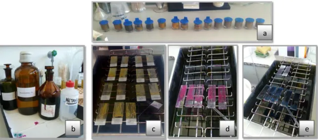
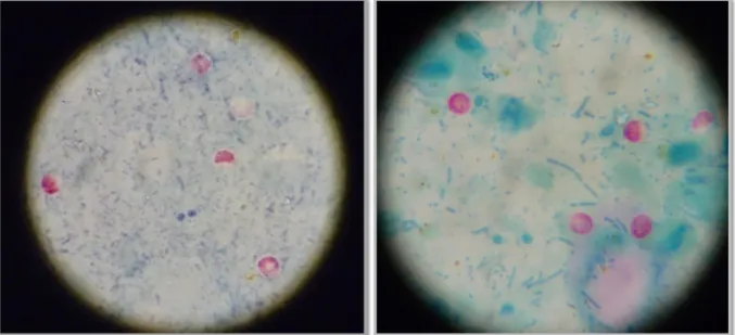
Fig. 1 and 2: Claw trimming and CMT test and udder examination……… ……….. Fig. 3, 4, 5 and 6: Blood sample collection, Claw amputation and recovery, Right abomasal displacement solution by the Dirksen method, Left abomasal displacement solution by the Jannowitz method ……….. Fig. 7 and 8: Lamb after assisted birth, critical care in a calf ……….,……. Fig. 9 and 10: Inhalation of saline solution in a calf suffering from respiratory disease (left), Arthromyodisplasia syndrome bandaging (right)………... Fig. 11: Picture of a pre-term piglet... Fig. 12: Housing of the calves suffering from diarrhea………. Fig. 13: Carbolfuchsin staining of the fecal samples………....……….………... Fig. 14 and 15: Appearance of Cryptosporidium oocysts under the microscope after carbolfuchsin staining. In the positive slides, round, colourless, bright structures representing the oocysts are seen……….. Fig. 16 - 20: Material and procedures for the modified Ziehl-Neelsen technique. Fecal containers. Solutions and reagents (Malachite green, hydrochloric alcohol, fuchsine, methanol). Fecal smears. Slides after hydrochloric alcohol application (bottom left). Malachite green application……….……….….………. Fig. 21 - 23: Microscopical observation of the colored slides. Prepared slides. Positive slides. Negative slides. ……….……….……… Fig. 24 and 25: Positive samples. ……….………...………. Fig. 26 and 27: Housing systems from the examined fattening unit in the Ribatejo area…….. Attached Fig. 1: Placement of an ear vein catheter……… Attached fig. 2: Intravenous fluid therapy in a dehydrated calf as a continuous drip infusion…
Graphs:
Graph. 1: Most frequently encountered infectious agents in neonatal calves displaying the most affected age-groups………..………..….…… Graph 2: Symptoms/ diseases shown by the examined calves……….……..……… Graph. 3: Prevalence of the excretion of oocysts in the first 15 examined calves……… Graph. 4: Prevalence of the excretion of oocysts in the second group of 15 calves (calf 16 to 30)………...……… Graph. 5: Overall prevalence of the excretion of oocysts in the 30 tested calves……….. Graph. 6: Linear regression between the incidence of diarrhea and a positive modified Ziehl-Neelsen result………. 3 4 5 6 6 54 55 55 80 81 81 86 108 109 1 56 85 85 85 85
xii
Graph 7: Linear regression between the presence of diarrhea and the presence of dehydration in the analyzed calves………...……… Attached Graph 1: Pathogenesis of dehydration………..
Tables:
Table 1: Overview of the examined calves, the calves that showed diarrhea and the ones positive to Cryptosporidium spp………..…………. Table 2: Normal laboratory values for new-born calves………..………..………. Table 3: Results of the first examination of 15 randomly selected calves from the ribatejo area (18. July 2011). ……….. Table 4: Results of the second examination of the previously examined calves from the ribatejo area (16th August 2011)………..………...…… Table no. 5: Results of the second group of randomly selected calves from the Ribatejo area
in Portugal (26th September 2011)……….……….……
Attached table no. 1: Clinical findings that identify a healthy calf……….…… …….…… Attached table no. 2: Scoring system to determine the severity of metabolic acidosis in calves ………..…….………... Attached table no. 3: Hydration status evaluation………..……... Attached table no. 4: Examples of oral rehydration solutions for calves as described in the Canadian Compendium of Veterinary Products……….………... Attached Table no. 5: General examination of the calf no. 1 that excreted Cryptosporidium spp. oocysts. ……….. Attached table no. 6: General examination of the calf no. 2 that excreted Cryptosporidium spp. oocysts………. Attached table no. 7: General examination of the calf no. 3 that excreted Cryptosporidium spp. oocysts………..……...
Attached table no. 8: General examination of the calf no. 4, that excreted Cryptosporidium spp. oocysts………... Attached table no. 9: General examination of the calf no. 4, that excreted Cryptosporidium spp. oocysts, during its second stay at the clinic. ………..…………
Attached table no. 10: General examination of the calf no. 5, that excreted Cryptosporidium spp. oocysts……… Attached table no. 11: General examination of the calf no. 6, that excreted Cryptosporidium spp. oocysts……… 86 104 56 63 82 83 84 103 105 106 107 110 111 112 113 114 115 115
xiii
Attached table no. 12: General examination of the calf no. 7, that excreted Cryptosporidium spp. oocysts. ………. Attached table no. 13: General examination of the calf no. 8, that excreted Cryptosporidium spp. oocysts………
List of abbreviations:
18S r DNA : sequence of recombinant DNA Acetyl-CoA: Acetyl Co enzyme A.
AIDS: Acquired immune deficiency syndome AMDS: Arthromyodisplasia syndrome
BD: Base deficit BE: Base excess
BID: bis in die ―twice daily‖
BW: Body weight
cAMP: cyclic adenosine monophosphate Cl-: Chloride ion
CMT: California mastitis test CNS: Central Nervous System CO2: Carbon dioxide
CSF: Cerebrospinal fluid ECF: Extracellular fluid
e. g.: exempli gratia. ―example given‖ H+: hydrogen ion
H2CO3: Carbonic acid
H2O: water
HACCP: Hazard analysis and critical control points
HCO3-: Bicarbonate ion
Hsp 70: 70 kilodalton heat shock protein i. e.: id est. ―that is‖
Ig: Immune globulin IM: Intramuscular
INF- γ: Interferon-gamma IPM: Integrated pest control IV: Intravenous
K+: Potassium ion Kg: kilogram Na+: Sodium ion
NaCl: Sodium chloride (salt)
NSAID: Non-steroidal anti-inflammatory drug OH: hydroxide
PGE2: Prostaglandin E2
PGI2: Prostaglandin I2
RT-PCR: Reverse transcriptase polymerase chain reaction
SIBO: Small intestine bacterial overgrowth SID: Strong ion difference
SID: ―once daily‖
TNF-1: Tumour necrosis factor 1
ZnCl2 – NaCl: Zinc chloride and sodium
chloride solution
116
1
Introduction:
―Infectious diarrhea remains one of the biggest health challenges in both the beef and dairy industries.‖ (page 13, Foster & Smith, 2009).
In dairy farms 50% of the calf mortality is assigned to diarrhea and in beef cattle it is thought to have an important impact in economic productivity (Foster & Smith, 2009).
Taking this fact into account, it is important to enlarge the knowledge of the causes, consequences, signs, therapy and prevention of this morbid condition.
There are innumerable causes that originate diarrhea in calves, some of them with worse consequences than others and hence some of them more urgent in matter of treatment and prevention.
Diarrhea can be infectious or non-infectious in its origin. Non-infectious diarrhea is normally associated to feeding mistakes (such as mixing or temperature errors). Infectious diarrhea is, on the other hand, mainly caused by four agents: enterotoxigenic Escherichia Coli (ETEC), Rotavirus, Coronavirus and Cryptosporidium parvum (Kaske & Kunz, 2003; Foster & Smith, 2009). Most of these agents affect calves in their first 4 weeks of life (Berchtold, 2009).
Graph. 1: Most frequently encountered infectious agents in neonatal calves displaying the most affected age-groups.
Addapted from: Kaske & Kunz (2003).
In this thesis, special attention will be given to a frequently encountered cause: Cryptosporidiosis.
―C. parvum is recognized as one of the most common infectious causes of intestinal disease in neonatal calves, lambs and kids…‖ (O’Handley & Olson 2006; de Graaf et al.1999b). As a very prevalent agent, it is thought to be present in almost every region worldwide (Fayer, Trout & Jenkins, 1998; Naciri, Lefay, Mancassola, Poirier & Chermette, 1999; Tzipori & Ward, 2002). 0 7 14 21 28 35 Age (days) E. Coli Rotavirus Coronavirus Cryptosporidium Eimeria Salmonella
2
Cryptosporidium is a protozoan, unicellular, opportunistic and worldwide existing parasite that affects a wide range of hosts. It was found that neonatal and pre-weaned calves are especially susceptible to the infection.
When a calve is born, its immune system is still very weak, given the fact that most of the immunity in new born calves will be attained after birth, through the ingestion of the maternal colostrum (Kaske & Kunz, 2003). In addition to the weak immunity, at birth a calve has to cope with the changes of environment by adapting its respiratory and cardiovascular systhems, thermoregulation and other organic functions to the extrauterine environment and by dealing with the increased infective factors upon leaving the sterile intrauterine surroundings (Kaske & Kunz, 2003).
Given these risk factors, it becomes more understandable that calves are highly susceptible to infectious agents, including Cryptosporidium spp.
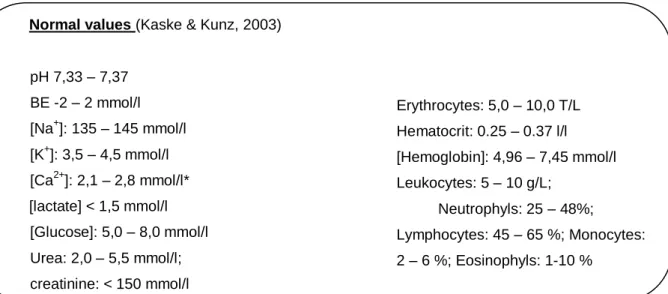
Special attention should be given to any alterations in their health status in their first days and weeks of life, detecting any signs that indicate a sick calf. The table no. 1 in the attachments shows the most important signs observed in a healthy calf.
The first description of Cryptosporidium was performed by Tyzzer, who identified it in a laboratory mouse in 1907. Only about 70 years later, the real significance of the parasite as a potential pathogenic agent became known, also when C. parvum was first characterized as a potential agent able to infect humans, in 1976 (Saini, Ransom & McNamara, 2000; Tzipori & Ward, 2002; O’Hara & Chen, 2011). In the 80’s its importance as a primary cause of diarrhea and as a potential threat for immune compromised individuals (AIDS) became evident (Tzipori & Ward, 2002; O’Hara & Chen, 2011). And in 1993, finally, it was characterized as ―the most serious, and difficult to control, cause of waterborne-related diarrhea‖ (page 1047, Tzipori & Ward, 2002; by McKanzie et al. 1994).
Infection by Cryptosporidium spp. in cattle was first completely reported in the beginning of the 70’s (Saini et al. 2000; Tzipori & Ward, 2002 - described by Morin, Lariviere & Lallier (1976) and Pohlenz, Moon, Cheville & Bemrick (1978)). Its role as a primary pathogen was first questionable as it usually coexisted with other enteropathogens. Only in the 80’s the role as a primary cause for neonatal diarrhea in calves was discovered by Tzipori et al. (de Graaf, Vanopdenbosch, Ortega-Mora, Abbassi & Peeters, 1999b; Naciri et al. 1999). Studies in the 1980’s revealed that the infection was much more prevalent in neonatal calves than thought before. Reaching epidemic to endemic level in some cases, it was by then recognized as an important cause of neonatal calf diarrhea (de Graaf et al.1999b; Tzipori & Ward, 2002; Divers & Peek, 2008).
Nowadays, even with the high number of studies performed, this parasite continues to be rather enigmatic. Studies continue to be of difficult performance because the propagating of
3
the parasite in vitro continues to be impossible on a continuous matter and because of the inability to cryopreserve this microorganism (Tzipori & Ward, 2002).
The present essay describes the most important facts about this parasite, focusing on biology, clinical signs, diagnosis, treatment, prevention and public health issues.
Two clinical case studies were performed, in the Ribatejo area of Portugal and in the region of Hessen in Germany, in order to understand the reality of the infection and its clinical course.
The internship and the performed activities:
The official curricular internship that gave rise to this dissertation had a total duration of 4 months, beginning on the 1st of October 2010 and lasting until the 31st of January 2011. It was spent in two different locations, the first part being at the Clinic for Ruminants – Internal Medicine and Surgery (Klinik für Wiederkäuer – Innere Medizin und Chirurgie) (around 340 Hours) and the second at the Clinic for Obstetrics, Gynecology and Andrology with veterinary ambulance (Klinik für Geburtshilfe, Gynäkologie und Andrologie der Gross- und Kleintiere mit Tierärztlicher Ambulanz) (around 380 Hours, including nightshifts) at the Justus Liebig University Giessen, Germany. The total duration of the internship was, thus, of around 720 hours.
In addition, an extracurricular internship with the duration of around 260 hours was spent from the 3rd of March to the 9th of April 2011 at the Veterinary Clinic Karsten von Brehm in Niesgrau, Germany.
In the Clinic for Ruminants, under the supervision of Dr. Marlene Sickinger, several clinical and diagnostic methods were applied.
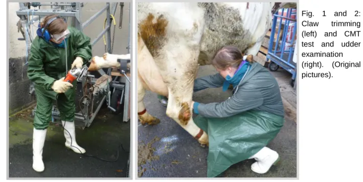
Fig. 1 and 2: Claw trimming (left) and CMT test and udder examination (right). (Original pictures).
4
Diagnostic methods included: general physical examinations and special examination of the respiratory tract, the gastro-intestinal tract, the circulatory system, the musculoskeletal system, the urinary tract and the nervous system: auscultation of the heart and lung, determination of the heart and respiratory rate, rumen activity auscultation, reticular pain probes, abomasal displacement test (steel band effect, lung percussion and ballottement), intestinal activity, fecal analysis (parasite, bacteria, virus), ruminal fluid quality testing (diagnosis of ruminal acidosis), rectal palpation with focus on the abdominal organs (rumen, intestines, kidneys, vessels and lymph nodes), diagnosis of udder disorders (CMT test, udder and milk evaluation), facial reflexes evaluation, urine testing (direct collection or via urinary catheter), ketosis diagnosis, blood analysis, cerebrospinal fluid collection, bone marrow collection, X-ray, amongst others.
Therapeutic methods included: vaccinations, injections of all type (subcutaneous, intramuscular, intravascular, intraperitoneal, epidural, intradermic), ear vein and jugular vein
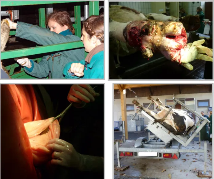
Fig. 3, 4, 5 and 6: Blood sample collection (up left), Claw amputation and recovery (up right), Right abomasal displacement solution by the Dirksen method (bottom left), Positioning for left abomasal displacement solution by the Jannowitz method (bottom right) (Original pictures).
5
catheterization, infusions, whole blood transfusions, oral administrations, drenching (ruminal fluid transfer, ruminal stimulants), bandaging and fracture treatment, claw trimming, euthanasia.
I also was able to assist in surgical procedures such as the surgical resolution of left abomasal displacement (by the Jannowitz method), surgical resolution of right abomasal displacement (Dirksen method), ruminotomy and production of a ruminal fistula, ceacal tympany resolution, claw amputation, deep digital flexor tendon tenotomy, rectal prolaps solution in an Alpaca.
The second part of the internship in the clinic of Reproduction, Obstetrics and Andrology was done under the supervision of Prof. Dr. Axel Wehrend. Different to the previous clinic, where only ruminants were treated, in this clinic in addition to ruminants, horses, pigs, dogs, cats and rabbits where present.
Diagnostic methods included similar ones to the learned before, but major focus was given to the examination of the reproductive tract, including vaginal specula positioning with mucosal and cervix evaluation, semen collection and evaluation, rectal palpation and determination of reproductive status, vaginal palpation, fetal position evaluation, transrectal ultrasound in cows, abdominal ultrasound to determine vitality of the lamb in sheep, ultrasound of the
6
teats, uterine cytology and biopsy in cows and mares, mastitis evaluation, evaluation of calves with diarrhea, respiratory conditions, septicemia, articular fluid collection, amongst others.
Implemented treatment methods included all type of injections, infusion therapy, teat injectors, estrus synchronization, teat amputation, theloresectoscopy, birth assistance in cows, sheep, horses and pigs, castration of pigs and rams, cesarean sections (in cows, sheep, sows and dogs), fetotomy in cow and mare, resuscitation and critical care of calves, Bühner band in sheep, umbilical surgery in calves, bandaging of calves suffering from AMDS (Arthromyodisplasia syndrome), tenotomy to solve AMDS, approach to calve diarrhea and respiratory diseases (antibiotherapy, pain management, supportive treatment), acupuncture and homeopathy.
Further performed activities included: feeding techniques of calves, farm visits and evaluation, euthanasia.
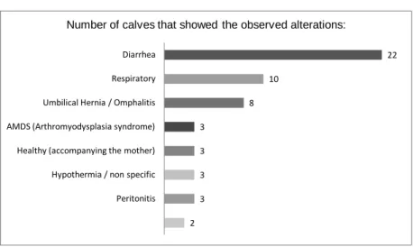
During the stay in the Clinic for obstetrics, ginecology and andrology, several calves suffering from diarrhea were identified, evaluated and tested for the presence of Cryptosporidium oocysts in their feces. The detailed case study and its results are outlined in greater detail elsewhere in this essay.
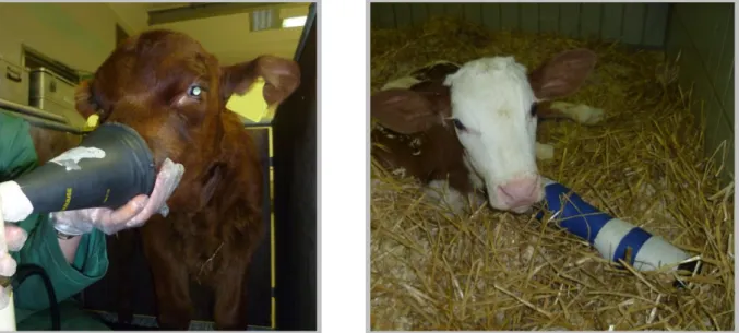
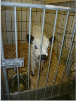
Fig. 9 and 10: Inhalation of saline solution in a calf suffering from respiratory disease (left), Arthromyodisplasia syndrome bandaging (right) (Original pictures).
7
In the extracurricular internship under the supervision of Dr. Karsten v. Brehm, in contrast to the 2 previous internships, the activities consisted mainly of direct veterinary work at the farms.
The activities included basic diagnostic methods, injections, infusions, antibiotherapy, artificial inseminations, estrus synchronization, pregnancy diagnosis, rectal examination of the reproductive tract of cows, birth assistance in cows and sheep, resuscitation and critical care of calves, fetotomy in cows, left sided abomasal displacement solution (Laparoscopic abomasopexy technique by Christiansen), fecal analysis, diarrhea treatment, euthanasia, ketosis treatment, hypocalcemia and hypophosphatemia treatment, hypomagnesiemia treatment, ruminal bloat in calves, vaccination programs, milk sampling and mastitis treatment.
In Lisbon another clinical case study was performed with examination and fecal sample collection of 30 calves and posterior analysis of the samples in the parasitology laboratory of the Faculty of Veterinary Medicine – UTL Lisbon.
8
Literature review
The Parasite:
Parasites of the Cryptosporidium genus are well-known agents of ruminant’s disease and have been deeply reviewed over the past years.
Cryptosporidium is classified as an ubiquitous, obligate, intracellular parasite, which affects all domestic animals and even humans (Naciri et al. 1999; Fayer, Morgan & Upton, 2000; Kaske & Kunz, 2003; Hamnes, Gierde & Robertson, 2006).
Studies conducted by Naciri, et al. (1999) showed that Cryptosporidium is one of the major etiological agents in neonatal calves from 4 to 10 days of age. Blume (2007) also evidenced Cryptosporidium as the most common cause of diarrhea in neonatal calves.
These parasites belong to the Kingdom Protozoa, phylum Apicomplexa, class Sporozoea, subclass Coccidia, Order Eucoccidiida, suborder Eimeriina and family Cryptosporidiidae (Rommel, Eckert, Kuntzer, Körting & Schnieder, 2000; Tzipori & Ward, 2002).
Comparing members of the cryptosporidiidae family with other coccidia, it becomes evident that they have many similarities in their life-cycle (presence of both sexual and asexual forms) (Divers & Peek, 2008). However, unlike other coccidian, Cryptosporidium oocysts are excreted in the feces in the already sporulated form and can cause auto-infection (de Graaf et al. 1999b; Tzipori & Ward, 2002; Hamnes et al. 2006; O’Handley & Olson, 2006). They differ in their host specificity, C. parvum being much less host specific, and in their size (Cryptosporidium spp. is much smaller in size and is much more difficult to detect by traditional fecal flotation methods) (Tzipori & Ward, 2002; Divers & Peek, 2008). Their high resistance as well as their characteristic location in the cell, are two additional features which differentiate Cryptosporidium from other coccidians (Tzipori & Ward, 2002; Hamnes et al. 2006).
In the last years, 13 valid species of Cryptosporidium were described (Olson, O’Handley, Ralston, McAllister & Thompson, 2004; Thomson, Palmer & O’Handley, 2007). Recently, further Cryptosporidium species have been identified, so nowadays the number of species rose to 23, and further non-identified species might exist (Muhid, Robertson, Ng & Ryan, 2011).
We can highlight 6 main species of cryptosporidia that affect animals. Those are: C. parvum and C. andersoni (mammalian), C. maleagridis and C. baileyi (avian), C. serpentis (reptiles) and C. nasorum (fish).The species from dogs (C. canis), cats (C. felis), pigs (C. suis) guinea pigs (C. wrairii) and marsupials are less well defined (Tzipori & Ward, 2002; Thompson et al. 2007).
9
Because most of the diagnosis were made by microscopy, to this date many unknown species may exist which were wrongly classified as C. parvum (because of the absence of morphological differences) (Thompson & Smith, 2011).
There are 3 main species of cryptosporidia that have an impact in the bovine species (Fayer, Santín, Trout & Greiner, 2006; Hamnes et al. 2006; O’Handley & Olson, 2006):
Cryptosporidium parvum is a pathogenic, non host specific, unicellular parasite that colonizes the small and large intestine of over 150 animal species, including all domestic animals (calves, lambs and young pigs especially) and also humans (Kaufmann, 1996). It is the most extensively studied because of its zoonotic potential as well as its clinical relevance as an important cause of neonatal diarrhea in ruminants, where it usually colonizes the ileum and the proximal large intestine (Laurent, McCole, Eckmann & Kagnoff, 1999; Naciri et al. 1999; Rommel et al. 2000; O’Handley & Olson, 2006).
Cryptosporidium andersoni (once called muris) can be found in the mammal’s stomach
(abomasum of ruminants). At this site, it causes dilation of the gastric glands, hypertrophy of the mucosa and atrophy of the epithelium. The protein digestion seems to be impaired (Olson et al. 2004). Even though it was declared to be non pathogenic, it can be held responsible for economic losses in both beef and dairy industry and is nowadays seen as a possible emerging pathogen. Its global prevalence is considered to be low (2 – 5%) (Santín et al. 2004) and the disease seems to be limited to cattle. Affecting primarily post-weaned beef and dairy cattle as well as mature animals, it is classified as a chronic infection (with a clinical course of months or even years) (Olson et al. 2004; Santin et al. 2004; Fayer et al. 2006; O’Handley & Olson, 2006). Pre-weaned calves can be already infected at an early age with this pathogen, but only show clinical signs when they are older (Kváč, Hromadová, Kvĕtoňová, Rost & Sak, 2011).
A new species of Cryptosporidium (C. bovis) was isolated in the USA (Divers & Peek, 2008). Encountered usually at calves aged from 2 to 7 months (Fayer et al. 2006, O’Handley & Olson, 2006), it is not certain yet if it is involved in neonatal diarrhea in calves (Divers & Peek, 2008).
A deer-like genotype (C. ryanae) was identified and can occur predominantly in weaned calves aged 2 to 11 months, without showing clinical signs (Fayer et al. 2006; Kváč et al. 2011).
Despite the multiplicity of Cryptosporidium species, one species remains the most important in mammals: Cryptosporidium parvum (Tzipori & Ward, 2002). Especially in pre-weaned calves, C. parvum is the most prevalent species and the only one with clinical relevance (Mendonça et al. 2007; Divers & Peek, 2008; Imre et al. 2011; Kváč et al. 2011; Muhid et al. 2011). To avoid the creation of a too extensive essay, major focus will be given to this species of Cryptosporidium and future reference to the disease will be concerning C. parvum infection.
10
Morphology and life-cycle:
The oocysts of Cryptosporidium parvum are circular shaped, with a size of approximately 5,0 x 4,5 µm. A double layered lipoprotein membrane, forming the oocyst membrane, encloses 4 sporozoites and a crystalline residual body (de Graaf et al. 1999b; Rommel et al. 2000). C. parvum develops in the small intestine, especially in the caudal part of the jejunum and in the ileum and sometimes also in the large intestine (de Graaf et al. 1999b; Rommel et al. 2000). All developmental stages are localized in the apical pole of the epithelial cells, but even though it multiplies in the cell, it is not included in the cell cytoplasm (O’Handley & Olson, 2006). Hence, they are localized intracellularly but extracytoplasmatically (Heine, Pohlenz, Moon & Woode, 1984; Kaske & Kunz, 2003).
The whole development of the parasite is located in one single host, classifying the life cycle as monoxenous (de Graaf et al. 1999b; O’Hara & Chen, 2011).
The infection is usually carried out through the fecal-oral route, by ingestion of oocysts in the feces (Rommel et al. 2000; Tzipori & Ward, 2002; Hamnes et al. 2006; Coklin et al. 2009). In addition, contaminated ground water and feedstuffs can be an important source of infection (Hamnes et al. 2006; Divers & Peek, 2008; Coklin et al. 2009); as are rubber and plastic fomites which, when insufficiently cleaned, can be a way of transport for the infective oocysts (Bopp, 2003). In general, it is believed that farm effluents are probably the most important sources of environmental contamination with oocysts (Tzipori & Ward, 2002).
Fonseca (2000) confirmed the importance of transmission of cryptosporidiosis by the drinking water, as 55% of the analyzed water samples in a study in Portugal contained Cryptosporidium oocysts.
Assymptomatic adult cattle are rather frequent and can easily contribute to the spreading of the infection to young calves by excreting the infectious oocysts (de Graaf et al. 1999b; Naciri et al. 1999; Tzipori & Ward, 2002). This is thought to be the most important source of C. parvum infections in dairy calves that suckle the mother after birth (Tzipori & Ward, 2002). The contact to wildlife species carrying the same parasite to which also bovine species are susceptible constitutes another possible way of infection (Bopp, 2003; Olson et al. 2004). After ingestion of the sporulated oocysts, the changes in temperature and pH (gastric acid), as well as the concentration of carbon dioxide, pancreatic enzymes and bile salts act on the oocysts and lead to their excystation (Heine et al. 1984; Laurent et al. 1999; de Graaf et al. 1999b; Tzipori & Ward, 2002; O’Handley & Olson, 2006; Foster & Smith, 2009). Parasite endogenous factors probably contribute to the successful excystation mechanism. These include: serine and cystine proteases associated to the sporozoites, secretory phospholipase A2, molecules associated with protein synthesis (proteins related to ribossomes or
11
This way the 4 motile and infective sporozoites of C. parvum are freed into the gut lumen and start colonizing areas between microvilli in the small intestine, mainly in the ileum (sporadically also in the colon) (Tzipori & Ward, 2002; Gookin, Nordone & Argenzio, 2002; O’Handley & Olson, 2006; Foster & Smith, 2009). The fusion of the parasite’s pellicula with the microvilli membrane and invagination of the luminal membrane follows (forming the parasitophorous vacuole which has an extracytoplasmic but intracellular location), with the simultaneous formation of feeder organelle between the parasite and the cellular cytoplasm (Laurent et al. 1999; Gookin et al. 2002; Tzipori & Ward, 2002; Foster & Chen, 2009; O’Hara & Chen, 2011). C. parvum is thought to be completely dependent of the nutrient support by the host through the feeder organelle, as the parasitic biochemical mechanisms are rather underdeveloped (O’Hara & Chen, 2011).
The trophozoite is formed and undergoes asexual reproduction to give rise to a type 1 schizont (or meront) (5 x 5,6 µm), enclosing 8 banana-shaped merozoites (by a process called endopolygeny) (Rommel et al. 2000; Tzipori & Ward, 2002; Gookin et al. 2002; O’Handley & Olson, 2006; Foster & Smith, 2009; O’Hara & Chen, 2011).
After rupturing, the merozoites are freed in the gut lumen and form either new type 1 schizonts on other sites or type 2 schizonts which enclose only 4 merozoites. From the latter, micro- and macrogametocytes are formed in which, respectively, 16 microgamettes or a 4,6 µm large macrogamette is formed (Rommel et al. 2000; Tzipori & Ward, 2002; Gookin et al. 2002; Foster & Smith, 2009; O’Hara & Chen, 2011).
A diploid zygote is formed when the non-flagelated microgamete fertilizes the macrogametocyte (Tzipori & Ward, 2002; Gookin et al. 2002; O’Handley & Olson, 2006; O’Hara & Chen, 2011). The sporogony process (a mechanism similar to meiosis) originates the sporulated oocyst (enclosing 4 haploid sporozoites). This structure is either excreted to the environment (thick walled oocysts) after a prepatent period of 3 to 6 days or originates an autoinfection if thin walled oocysts are formed. The thin walled oocysts, instead of being excreted to the environment, are freed still inside the host, restarting the whole endogenous cycle (Rommel et al. 2000; Gookin et al. 2002; Foster & Smith, 2009; O’Hara & Chen, 2011). The excreted oocysts are immediately infective to other animals when they are ingested and remain so for a long period of time in the environment (Gookin et al. 2002; Foster & Smith, 2009; O’Hara & Chen, 2011).
This autoinfection by thin walled oocysts is thought to be the cause of extended clinical conditions which, more likely, may lead to cachexia and death of the newborns (Tzipori & Ward, 2002; Divers & Peek, 2008).
The shedding begins often at 3 days of age and peaks at 14 days. The animal usually shows clinical signs at 3 to 5 days and during 4 to 17 days, being often able to excrete up to 107 oocysts per gram of feces (Fayer et al. 1998; Olson et al. 2004; Hamnes et al. 2006; Radostits, Gay, Hinchcliff & Constable, 2007).
12
Usually the infection remains restricted to the gastrointestinal tract, however there can be cases of extra-intestinal phases in other organs (Tzipori & Ward, 2002).
Epidemiology:
Cryptosporidiosis is a frequent disease in neonatal ruminants occurring world-wide, especially in dairy calves.
Longitudinal studies indicated that the incidence in dairy herds could easily reach 100%, whereas in beef herds the incidence was much lower (3 – 25%) (Jäger et al. 2005; Gow & Waldner, 2006; O’Handley & Olson, 2006).
The prevalence in dairy calves was determined in studies conducted by de la Fuente et al. (1999) (Spain), Almeida, Oliveira & Teixeira (2008) (Brasil), Kváč et al. (2011) (Czech Republik) and Imre et al. (2011) (Romania). Prevalences in calves ranged from 25 to 61%. In a study by Coklin et al. (2009) (Canada), however, prevalence in dairy calves reached only 6%.
A study of the prevalence of infection by Cryptosporidium spp. in beef calves was conducted by Ralston, Mc Allister & Olson (2003) in Canada and did not exceed 5%. Gow and Waldner (2006) detected a prevalence of 3,1% amongst beef-calves in Canada, and Jäger et al. (2005) a prevalence of 20 – 25% in beef-calves in Germany.
In the study in France, conducted by Naciri et al. (1999), a higher prevalence in suckling compared to dairy calves was detected, which somehow contradicts the previously established facts.
Dairy calves may have a higher incidence of cryptosporidiosis because the beef calves are born in a single period in spring while dairy calves are born all year round. So, considering their short calving season, the transmission is much lower in beef than in dairy calves. Dairy calves are housed in pens that are kept close together with other animals, whereas beef calves are released to a large open area (Olson et al. 2004).
Even though it was thought that the lower prevalence in beef calves is related to the lower stock density when comparing to dairy calves, recent studies showed that even when keeping beef calves in a similarly confined environment as dairy calves, the incidence in beef calves never was higher than 25%, which strongly suggests other factors that influence the different susceptibility in beef and dairy calves (Kaufmann et al. 1996; Jäger et al. 2005). The necessary dose for a successful infection is very variable and depends on the individual resistance of the host. Nevertheless, susceptible or immune-compromised animals need a very low dose (as low as 10 oocysts sometimes) to get infected (Divers & Peek, 2008; O’Hara & Chen, 2011). As an infected calve sheds several million oocysts in the diarrheic feces (up to 107), the concentration and risk of infection in an affected farm is very high
13
(infective pressure) and the problem can easily turn into a herd problem (de Graaf et al. 1999b; Hamnes et al. 2006; Divers & Peek, 2008).
In addition to the low infectious dose, another feature that aids the transmission is the extremely high resistance of the oocysts in the environment, mainly due to their highly protective carbohydrate wall (Rommel et al. 2000; Bopp, 2003). At 4ºC with a sufficient degree of humidity, they can survive for 6 months, only being destroyed immediately at temperatures under – 18º C or over 65º C (Naciri et al. 1999; Rommel et al. 2000). Standard levels of chlorination are also insufficient to destroy the oocysts, allowing an easy infection through the waterborne way in animals and humans (Betancourt & Rose, 2004; Olson et al. 2004). The presence of chloride can even increase the infectivity of the oocysts, as it can hasten the excystation (Fonseca, personal communication on the 26th January 2012).
A relation between the incidence of cryptosporidiosis and the season of the year has been suggested, but no significant correspondence has been identified. In winter, when the confinement of the calves is higher in some countries, it might occur more often (Hamnes et al. 2006, Radostits et al. 2007).
Clinical signs:
The clinical signs of a C. parvum infection are nonspecific and can not be easily differentiated from bacterial or viral infections that cause diarrhea. They are similar in calves, lambs and kids and include diarrhea, dehydration, abdominal pain, reduced appetite and depression (Naciri et al. 1999; de Graaf et al. 1999b; Olson et al. 2004; O’Handley & Olson, 2006; Divers & Peek, 2008; Kváč et al. 2011). Anorexia, combined with the diarrhea, usually gives rise to weight loss and retarded growth in affected calves (de Graaf et al. 1999b). Following the study from Vos, Constable & Kuhlenschmidt (2005) (USA), abomasal emptying in cases of a Cryptosporidium parvum infection was increased at the beginning of infection, resuming to a normal rate 1 week after the start of the diarrhea.
The infection is characterized by profuse and persistent watery, yellow-greenish colored diarrhea with putrid smell, containing mucus (Kaufmann et al. 1996; de Graaf et al. 1999b; Rommel et al. 2000; Olson et al. 2004; O’Handley & Olson, 2006; Radostits et al. 2007; Thompson et al. 2008). These clinical signs can appear in animals as young as 3 days of age, rarely affecting calves of 30 days and older (Santín et al. 2004; Fayer et al. 2006). Mostly calves with 5 to 14 days of age are affected and the infection lasts for approximately 4 to 18 days (Rommel et al. 2000; Kaske & Kunz, 2003; Olson et al. 2004; O’Handley & Olson, 2006; Thompson et al. 2008). Even though it occurs rarely, the signs are less severe if the infection occurs in older (over 4 weeks of age) calves (Rommel et al. 2000, Hamnes et al. 2006).
14
Several studies carried out by Naciri et al. (1999) in France, de la Fuente et al. (1999) in Spain, Santín et al. (2004) in the USA, Mendonça et al. (2007) in Portugal, Muhid et al. (2011) in Malaysia, Imre et al. (2011) in Romania and Tiranti et al. (2011) in Argentina proved the higher incidence of cryptosporidiosis in preweaned calves in comparison to post-weaned ones. Being the most affected group, animals aged between 8 and 14 days.
Generally, when comparing to other agents (E. Coli, Rotavirus and Coronavirus), findings suggest that calves affected by Cryptosporidium spp. get sick later and during a longer time, but die less often. The degree of dehydration seems, though, higher in Cryptosporidium spp. infections (Blume, 2007).
Even though the parasite infects preferably the distal part of the small intestine, it can spread throughout the gut. In this way, the diarrhea is most watery and intense when the parasite colonizes the proximal parts of the small intestine, if the infection affects mainly the distal parts of the intestine, the infection might even be assymptomatic (Tzipori & Ward, 2002). Some data suggests that older cattle can get immunized to future infections with this parasite because of previous exposure (Harp, Woodmansee & Moon, 1990; Fayer et al. 1998; Coklin et al. 2009). Studies conducted by Harp et al. (1990) showed that rechalenging calves that previously suffered from cryptosporidiosis, after 3 months, evidenced neither clinical signs related to cryptosporidiosis nor oocyst shedding.
Together with the signs of diarrhea, oocyst shedding takes place. The period of oocyst shedding, however, is determined by host factors (age and immune status). After the shedding begins, the number of excreted oocysts increases, reaching a maximum 5 to 6 days after the infection and decreasing suddenly 10 to 15 days after the day of infection (de Graaf et al. 1999b; Hamnes et al. 2006; Radostits et al. 2007). The excretion of oocysts can persist for several months even after disappearance of the clinical signs (de Graaf et al. 1999b; Naciri et al. 1999; Tzipori & Ward, 2002).
In a study conducted by Fayer et al. (1998) the prepatent period in infected calves ranged from 3 to 6 days and at the peak of infection (6-8 days post infection), calves were able to excrete up to 107 oocysts per gram of feces. Most of the calves excreted oocysts during 69 days.
Blume (2007) detected oocyst excretion already on the 3rd day of age in some animals and a study conducted by Vos, Constable & Kuhlenschmidt (2005) evidenced the oocyst shedding cyclic pattern (low in the beginning, increasing later on and decreasing again).
Morbidity of affected calves with less than 3 weeks of age can reach values greater than 50%, however mortality is usually low if supportive fluid therapy is sufficient and no mixed infections exist (Divers & Peek, 2008); as the main cause of death remains the high loss of fluids due to diarrhea and not the direct action of the infective agent (Kaske & Kunz, 2003).
15
In rare cases, severe cryptosporidiosis can lead to extreme dehydration, metabolic acidosis and death due to cardiovascular collapse (O’Handley & Olson, 2006; Radostits et al. 2007; Thompson et al. 2008).
Frequently the infection with Cryptosporidium parvum occurs together with other infective agents (mixed infection) (Naciri et al. 1999; de la Fuente et al. 1999; Kaske & Kunz, 2003; Radostits et al. 2007). In fact, in a study by Blume (2007), half of the collected fecal samples contained a mix of several diarrhea causing agents.
The reason for the mixed infections is that this parasite is mostly opportunistic and infects especially immune supressed organisms (Kaske & Kunz, 2003). In addition, lesions of the intestinal mucosa predispose the calf to the infection by other agents such as E. Coli, Salmonella sp. and viruses (especially Rotavirus and Coronavirus) (Divers & Peek, 2008). The fecal samples are likely to contain several agents in addition to the Cryptosporidia. However, as C. parvum oocysts are shed throughout and even after the presence of clinical signs and Rota or Coronavirus are shed only in the beginning, C. parvum is more often found. Hence, the early collection and analysis of fecal samples is crucial for the correct determination of the etiology of the calves’ diarrhea (Divers & Peek, 2008). i.e. C. parvum is a pathogenic entity by itself, whose symptoms can be worsened by the presence of other diarrhea causing agents (E. coli, Corona virus, Rota virus…) (Naciri et al. 1999; Rommel et al. 2000).
In case of a monoinfection with C. parvum, if the calf is not too weak and the density of infective forms in the environment is not too high, the infection is usually self-limiting and mild (Kaufmann et al. 1996; Divers & Peek, 2008). This is especially seen if the calves continue nursing (Divers & Peek, 2008).
In mixed infections, the clinical signs and prognosis tend to be worse (acidosis, electrolyte imbalances, dehydration and possible dysentery) and treatment is more complicated. Thus, there is usually a higher mortality rate (Divers & Peek, 2008).
As to the impact on production, to this date there are no data that support any long-term effects on production due to the infection with C. parvum in calves. No differences regarding weight gain have been noted between treated and untreated animal groups. Economic losses are, thus, only related to the cost of treatment and death of infected animals, which nevertheless can be substantial (Divers & Peek, 2008).
Pathogenesis / Pathophysiology:
The exact mechanism of pathogenesis of cryptosporidiosis is not well known to this date. Even though some structural evidences are understood, much research is still needed to know the molecular processes that are involved (Tzipori & Ward, 2002).
16
As mentioned before, Cryptosporidium parvum parasites affect essentially the distal small intestine (ileum and jejunum), but can be also found in the proximal small intestine and portions of the large intestine, colonizing especially the most apical part of the cells. Usually deeper mucosal layers are not invaded, hence the parasite is often classified as a ―minimally invasive‖ mucosal pathogen (page 141, Laurent et al. 1999).
The pathologic changes in the intestine of an affected individual are caused mainly by the rupture of the parasitophorous vacuole with the consequent release and spreading of the merozoites to other enterocytes (Divers & Peek, 2008). These changes include villous atrophy, villous fusion, crypt hyperplasia, infiltration with inflammatory cells and destruction of microvilli (Koudela & Jiři, 1997; McCole, Eckmann, Laurent & Kagnoff, 2000; Gookin et al. 2002; O’Handley & Olson, 2006).
The severe villous atrophy is thought to be the result of the loss of the villous enterocytes followed by retraction of villi to conserve an intact epithelial gut barrier. In order to replace lost epithelial cells, the crypts undergo hyperplasia (due to acceleration of cell division), but disruption of the epithelial barrier can be observed in severe infections (Tzipori & Ward, 2002; Gookin et al. 2002; Foster & Smith, 2009).
Due to its fixation on the microvilli border, the parasite disturbs the normal function of the small intestine mucosa, not only by decreasing the absorbing area of the intestine (loss of intestinal mucosa), but also by disturbing the enzyme activity (loss of membrane bound enzymes), impairing the breakdown of peptides and dissacharides and the transmembranary nutrient and electrolyte transport (Rommel et al. 2000; Tzipori & Ward, 2002; Gookin et al. 2002; O’Handley & Olson, 2006).
Attachment and formation of the parasitophorous vacuole:
After excystation, the parasite attaches to the host cells and gives rise to the formation of the parasitophorous vacuole, by the action of several proteins lodged in the surface or apical complex (micronemes, rhoptries and dense granules) of the Cryptosporidium sporozoites. (Gookin et al. 2002; O’Hara & Chen, 2011).
The attachment, invasion and formation of the parasitophorous vacuole are complicated and intricate mechanisms. New discoveries identified several surface and apical complex proteins that are thought to be, in part, responsible for those mechanisms (CSL, GP900, p23/27, TRAP C1, GP15, CP 15, CP60/15, cp47, gp40/45 and gp15/Cp17) (Tzipori & Ward, 2002).
After a successful attachment, internalization occurs. The latter starts with the fusion of the parasite and the host cell membrane. When the rhoptry of the parasite contacts with the host cell attachment site and the micronemes and dense granules migrate to the merging area, a structure with a channel-like appearance forms and the cytoplasm of the parasite attaines a vacuolated form. As the vacuolization procedes, the parasite gets encapsulated by the host cell (O’Hara & Chen, 2011).
17
The sporozoites are internalized and the parasitophorous vacuole is formed (a double membrane formed by host cell and re-structured by the parasite, located intracellularly but extracytoplasmatically). At the host-parasite interface, an electron-dense structure with a polymerized actin network is formed (O’Hara & Chen, 2011).
Host cell actin branches are reorganized by several signaling axes and the epithelial membrane suffers a rearrangement which gives rise to the formation of transporter channels (for water and Na+/Glucose movement, respectively). This way, C. parvum is thought to alter the cytoskeletal structure of the host cell upon the internalization process (O’Hara & Chen, 2011).
Diarrhea due to cryptosporidiosis is both malabsorptive and secretory (Laurent et al. 2009). Several mechanisms lye behind these processes and give rise to the clinical signs of cryptosporidiosis.
Malabsorptive diarrhea due to cell loss:
The cell loss could be an effect of the pathogen itself or a response from the host to limit and solve the infection (Foster & Smith, 2009). Hence, there are several possible mechanisms that lead to the extensive epithelial cell loss in C. parvum infections.
The first suggested mechanism consists of a direct cytotoxic effect of C. parvum on the epithelium of the intestine, by leakage of enzymes (lactate dehydrogenase). Nowadays this theory is not supported by current literature and is considered controversial (Laurent et al. 1999; McCole et al. 2000; Gookin et al. 2002; Foster & Smith, 2009).
The second, and more accepted, mechanism that explains cell loss is apoptosis (or programmed cell death). This theory is supported by the finding of apoptotic cells in in vitro and in vivo essays (Ojcius, Perfettini, Bonnin & Laurent, 1999; Chen et al. 2001; Foster & Smith, 2009). As shown recently, apoptosis is activated or inhibited in different stages of development of the parasite, occurring inhibition in early stages of the infection (trophozoite stage – when the parasite depends on the host cell for growth and development) and activation and increase of apoptosis in the later stages of infection (McCole et al. 2000; Foster & Smith, 2009; O’Hara & Chen, 2011).
Studies conducted by Mele, Morales, Tosini & Pozio (2004) showed that apoptosis is triggered by the invasion of the sporozoites in the first hours after infection (possibly by a caspase dependent mechanism), followed by an inhibition of apoptosis in the first 24 hours (probably by expression of antiapoptotic proteins). Then, 48 hours after the infection, apoptosis is again activated (to allow the exit of the merozoites from the cell).
Chen et al. (2001) suggested that Cryptosporidium actively inhibits the apoptosis of infected cells by secreting NF-ƘB, in order to protect the destruction of the infected cells. Meanwhile it also triggers the freeing of IL-8 which enhances the inflammatory reaction.
18
Ojcius et al. (1999) saw in his studies the role of Cryptosporidium in causing caspase-dependent apoptosis of infected cells. The suggested mechanism could be interpreted as a host action to limit C. parvum infection or an action from the parasite that helps it exiting the host cells.
It has to be discovered still if the referred regulation of apoptosis is beneficial for the host (limit spreading of the parasite, diminish extend of cell loss and/or increase clearance of the parasite from the host) or the parasite (preserve its intracellular location) (McCole et al. 2000; Chen et al. 2001; Gookin et al. 2002; Mele et al. 2004; Foster & Smith, 2009; O’Hara & Chen, 2011).
Secretory diarrhea due to prostaglandins:
A prostaglandin-mediated mechanism, responsible for the secretions of anions (mainly Cl -and HCO3-) and the inhibition of the absorption of neutral NaCl, has been suggested in
addition to the malabsoption mechanism described before (Laurent et al. 1999; Gookin et al. 2002; Foster & Smith, 2009). Two types of prostaglandins are involved: PGE2 and PGI2.
These are originated most likely from macrophages that, infiltrating the lamina propria, activate prostaglandin secretion by mesenchymal cells. Nitric oxide may also play a role in stimulating prostaglandin-mediated secretion and is considered important in the defense against cryptosporidiosis. But the exact mechanism is not yet known (Laurent et al. 1999; Gookin et al. 2002; Foster & Smith, 2009).
PGE2 and PGI2 act on different sites, whereas PGE2 exerts its function directly on the
enterocytes, PGI2 acts indirectly by influencing the enteric nervous system. 75% of the
secretion in C. parvum infection is attributed to the effect of PGI2. It stimulates the nicotinic
ganglia as well as the VIP-ergic and cholinergic motor neurons responsible for the innervation of the intestinal mucosa (Laurent et al. 1999; Foster & Smith, 2009).
In the end, prostaglandin secretion stimulates calcium and cAMP freeing which decreases sodium absorption and increases anion secretion (Gookin et al. 2002).
INF-γ and TNF-1 contribute to the above mentioned mechanism, by increasing the overall permeability of the epithelium (Laurent et al. 1999).
The secretory mechanism is hence based on host immunity processes triggered by the infection.
In summary, the clinical signs associated with the disease are thought to be caused by a mixed mechanism including maldigestion, malabsorption and osmotic effects (malabsorptive and secretory diarrhea) (Divers & Peek, 2008).
The exact mechanisms of the pathogenesis are not yet fully understood and it seems that the underlying factors are originated both by the parasite and the host (O’Handley & Olson, 2006).
19
Diagnosis of cryptosporidiosis:
In cases of neonatal diarrhea in calves, cryptosporidiosis should always be included in the list of differential diagnosis (O’Handley & Olson, 2006).
To determine the presence of cryptosporidium, two main diagnostic methods are used: by analysis of the feces or by postmortem examination of the intestinal mucosa (Rommel et al. 2000). In both cases the diagnosis is done by microscopic observation of the oocysts in the samples (Divers & Peek, 2008).
In the clinically affected animals, the number of excreted oocysts is very high, which makes their detection in the feces easy (Tzipori & Ward, 2002).
Concentration of oocysts before observation under the microscope is a common method, the preferred method being the use of saturated sugar. Another method is the flotation in a ZnCl2
– NaCl solution or other salts (Rommel et al. 2000; O’Handley & Olson, 2006; Radostits et al. 2007; Thompson et al. 2008).
Normal light microscopy makes the detection of the oocysts difficult as they can not be easily differentiated from the structures in the background. Phase contrast microscopy might be an alternative and allows differentiation of the oocysts (Radostits et al. 2007).
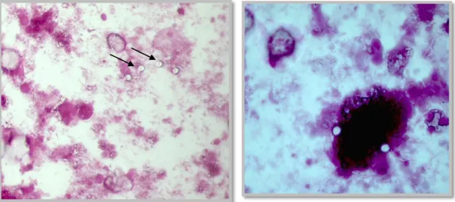
After concentrating the sample, staining methods can be applied to help the differentiation, those include the Ziehl-Neelsen technique (acid-fast stain, as cryptosporidium is an acid-fast organism), safranin-methylene blue stain, Kinyoun stain and DMSO-carbol fuchsin stain. All of these methods stain the oocysts red with a refractile area surrounding them and counter stains the background to facilitate the identification. These stains are quite helpful, but they are very time-consuming (Fayer et al. 2000; Rommel et al. 2000; Tzipori & Ward, 2002). The oocysts are seen as circular structures, 3 to 5 µm in size, often with 2-4 sporozoite nuclei in their interior, and can be easily differentiated from other microorganisms and fecal compounds (Rommel et al. 2000; Tzipori & Ward, 2002; O’Handley & Olson, 2006).
Negative staining with nigrosin, light green merbromide and malachite green, which stain all structures in the background except for the oocysts can be used but are not as trustworthy (Fayer et al. 2000).
As the oocysts often float, they will be lodged just under the coverslip of the microscope slide, in a high focal plane which, when focusing on them, will let other parasites (such as Eimeria) appear blurry (O’Handley & Olson, 2006).
Postmortem analysis of fresh necropsy tissue samples after fixation with methanol and staining of dab preparations with Giemsa (10% solution, 30 minutes staining) or of histological sections with Haematoxillin-Eosin, are two aditional diagnostic methods (Rommel et al. 2000).
20
Histopathologic changes include atrophy and fusion of intestinal villi, especially in the distal jejunum and ileum (Laurent et al. 1999; Rommel et al. 2000; Radostits et al. 2007). Villi might be shorter, wider, blunt and might even appear fused. Crypts are hyperplastic and an inflammatory cell infiltrate of lymphoid cells, macrophages and neutrophils in the lamina propria is present (Laurent et al. 1999, Radostits et al. 2007). In the epithelial membrane, many parasitic forms are embedded (Radostits et al. 2007).
Immunological methods used in the identification of this parasite include: polyclonal fluorescent antibody tests, latex agglutination reactions, IF with monoclonal antibodies, ELISA (enzyme linked), reverse passive haemagglutination (RPH), immunocromatography, and several others (Fayer et al. 2000). These immunological methods may be quite accurate, but they are not routinely used as they are linked to rather high costs and because they can only be performed in specialized laboratories (Tzipori & Ward, 2002; O’Handley & Olson, 2006).
Coproantigen-based immunoessay kits (ELISA) can be used for a more sensitive and specific diagnosis of cryptosporidiosis. Their high price and low availability are factors limiting their application in routine cryptosporidiosis diagnosis, making them only useful in human cryptosporidiosis cases (Garcia & Shimizu, 1997; Thompson et al. 2008).
PCR and other genetic techniques (like the analysis of 18S rDNA, hsp70 and Acetyl- CoA synthethase) can be used to differentiate species of Cryptosporidium (which can not be attained by staining and microscopic observation) and bring great value in an epidemiological matter (determining of the source of a potentially zoonotic case) (Tzipori & Ward, 2002; Divers & Peek, 2008). PCR is a highly sensitive, accurate and quick method, however the number of false positives is rather high and it can be influenced by environmental factors (contaminants) (Fayer et al. 2000).
Viability and infectivity studies are possible to be carried out. Some examples of types of coloration include the vital dyes: propidium iodide and 4,6, diamino- 2´- phenylindole (DAPI). Viability can be also tested by fluorescent in situ hybridization (FISH) and cell culture followed by RT-PCR (Fayer et al. 2000).
As it was already mentioned, the infections are usually mixed, therefore, if the presence of a C. parvum infection is confirmed, diagnostic tests to determine bacterial or viral infections should be carried out (Divers & Peek, 2008).
In vitro studies of the parasite’s life cycle show many restrictions in cell cultures. It usually has to involve several passages in hosts (mainly calves, due to their high susceptibility) (Fayer et al. 1998; Tzipori & Ward, 2002; O’Hara & Chen, 2011). Culturing was attempted in epithelial cell cultures (like HCT-8, a human ileocecal adenocarcinoma cell line), intestinal xenografts and by infection of animal models (e. g. mice) (Laurent et al. 1999).