1
Relatório Final de Estágio
Mestrado Integrado em Medicina Veterinária
AUTOLOGOUS STEM CELL THERAPY:
APPLICATION IN CANINE OSTEOARTHRITIS
Maria Manuel Ribeiro Martins Garcia Afonso
Orientador:
Professor Assistente Leandro da Silva Gardel Co-Orientador:
Professora Doutora Maria Beatriz Beça Gonçalves Porto e Vasconcelos Professor Doutor João Paulo Vilas-Boas Soares Campos
2
Relatório Final de Estágio
Mestrado Integrado em Medicina Veterinária
AUTOLOGOUS STEM CELL THERAPY:
APPLICATION IN CANINE OSTEOARTHRITIS
Maria Manuel Ribeiro Martins Garcia Afonso
Orientador:
Professor Assistente Leandro da Silva Gardel Co-Orientador:
Professora Doutora Maria Beatriz Beça Gonçalves Porto e Vasconcelos Professor Doutor João Paulo Vilas-Boas Soares Campos
iii Abstract
Osteoarthritis (OA) is a chronic pathology that severely decreases the life quality of affected dogs and is thought to affect up to 20% of the canine population. Current therapies are not successful in attaining cure and most of them also fail in slowing down the progression of lesions. Moreover, most drugs currently used for pain management in these patients have adverse systemic effects associated with their long-term administration. Stem Cell Therapy (SCT) has recently been identified as a possible alternative or adjunctive treatment for canine OA. This research was a preliminary study that aimed to objectively evaluate the outcome of autologous SCT in a group of dogs diagnosed with clinical OA and was divided in three parts. Firstly, we set out to identify which tissue source (bone marrow or adipose tissue) was more advantageous for the obtention of MSCs for posterior culture, expansion and differentiation into chondroblasts. Secondly, we proposed to evaluate the outcome of the autologous intra-articular application of these cells in the affected joints of the study sample. In order to do this, a new protocol for simple kinematic analysis was developed and owner questionnaires were carried out. Finally, a technique for cytogenetic analysis was optimized for future application in the chromosomic stability evaluation of stem cells in culture.
The results obtained after these four months of study suggest that SCT was successful in improving comfort and life quality in the animals who participated in the project and that more research should be carried out in this area in order to fully understand the potential of the application of stem cells in the treatment of canine OA.
Keywords: Regenerative Medicine, Autologous stem cell therapy, Canine osteoarthritis, Kinematic gait analysis, Cytogenetics.
iv Resumo
A osteoartrite (OA) é uma patologia crónica e irreversível que afeta cerca de 20% da população canina, levando a uma considerável diminuição da qualidade de vida dos animais afetados. As opções atualmente disponíveis para o tratamento da OA, além de não permitirem a sua cura, têm pouco efeito na diminuição da progressão da patologia. Além disto, a maior parte dos fármacos utilizados para o controlo de dor nestes animais tem efeitos sistémicos adversos associados à sua administração a longo prazo. A Terapia Celular com Células Estaminais foi recentemente apontada como uma possível alternativa ou terapia coadjuvante para da OA. Este estudo preliminar, dividido em três partes, teve como finalidade avaliar objetivamente o efeito da Terapia Celular com Células Estaminais de origem autóloga num grupo de cães diagnosticados com OA. Em primeiro lugar, investigou-se qual o melhor tecido (medula óssea ou tecido adiposo) para a obtenção de células estaminais mesenquimatosas e posterior expansão e diferenciação em condroblastos. Propusemo-nos ainda avaliar o resultado da aplicação destas células nas articulações osteoartríticas dos animais estudados, através de questionários aos proprietários e de um protocolo simplificado de análise cinemática desenvolvido durante o período do estudo. Por último, otimizou-se uma técnica de análise citogenética para futura avaliação da estabilidade cromossómica de células estaminais em cultura.
Os resultados obtidos durante quatro meses de estudo sugerem que a Terapia Celular com Células Estaminais levou a melhorias significativas no que diz respeito ao conforto e qualidade de vida dos animais participantes e que esta abordagem deve ser investigada mais aprofundadamente, a fim de compreender plenamente o seu potencial no tratamento da OA canina.
Palavras-chave: Medicina Regenerativa; Terapia com células estaminais autólogas; Osteoartrite canina; Análise cinemática do movimento; Citogenética.
v Résumé
La osteoarthrite (AO) est une maladie chronique et irréversible, qui touche environ 20% de la population canine, conduisant à une réduction significative de la qualité de vie des animaux qui en sont atteints. Les options actuellement disponibles pour le traitement de la AO, au delà de ne pas permettre leur guérison, ont peu d’effet dans la réduction de la progression de la maladie. En outre, la majorité des médicaments utilisés pour contrôler la douleur chez ces animaux ont des effets systémiques indisérables liés à leur administration à long terme. La Thérapie Cellulaire avec des Cellules Souches a récemment été suggérée comme une alternative possible ou traitement adjuvant pour la gestion de la OA. Cette étude préliminaire, divisé en trois parties, a eu comme but évaluer objectivement l’effet de la Thérapie Cellulaire avec des Cellules Souches d’origine autologue chez un groupe de chiens diagnostiqués avec la OA. Premièrement, on a enquêté sur quel serait le meilleur tissu (moelle osseuse ou tissu adipeux) pour l’obtention de cellules souches mésenchymateuses et ultérieure expansion et différenciation en chondroblastes. Nous nous sommes proposés, encore, d’évaluer le résultat de l’application de ces cellules dans les articulations osteoarthritiques des animaux étudiés à travers les questionnaires addressés aux propriétaries et un protocole simplifié de l’analyse cinématique, developpé au cours de la période d’étude. Finalement, on a optimisé une technique d’analyse cytogénétique pour une future évaluation de la stabilité chromosomique des cellules souches en culture.
Les résultats obtenus pendant quatre mois d’étude suggèrent que la Thérapie Cellulaire avec des Cellules Souches a conduit à une amélioration significative en ce qui concerne le confort et la qualité de vie des animaux participants et que cette approche devrait être étudiée d’une façon plus profonde, à fin de mieux comprendre son potentiel dans le traitement de la OA canine.
Mots-clés: Médecine régénérative, Osteoarthrite canine, Thérapie cellulaire avec des cellules souches d’origine autologue, analyse cinématique, cytogénétique.
vi Acknowledgements
I have come to realize that this chapter was the hardest to write as it was immensely difficult to put into words how thankful I feel towards the people who, in some way, have made this project a reality.
First and foremost, I want to say that it was an immense honor to have been guided by Professor Leandro Gardel. I want to thank him for everything he has taught me, for trusting in my capacities and for always being so helpful during all the phases of this project. I will always see him as a mentor and an example to follow.
I want to show my deepest gratitude to Professor Beatriz Porto, who has welcomed me in her laboratory. I want to thank her for her help and enthusiasm, and for having opened my eyes to the immense unexplored realms of cytogenetic research in veterinary medicine.
It is a pleasure to thank Professor João Paulo Vilas-Boas for all the detailed explanations and advice on a subject I had never encountered before. This project would not have been possible without his help.
I also want to thank Rosa Sousa, for being such a friendly, welcoming person and for the patience with which she has answered all my questions and helped in this project. To her, Lara Andrade and Filipa Ponte, I want to say that I hope that they never change who they are and that it has been an utmost pleasure to meet them all.
I am indebted to Sofia Abreu and Marcelo Castro for everything they have put into this project. I am certain that, without their help, I would not have been able to do this.
I am very grateful to Professor Augusto de Matos for having allowed me to carry out an important part of this study at the UP VET facilities.
I also want to thank Dr. Joana Santos and Dr. Jorge Ribeiro for always explaining everything calmly and in detail. I want to be just like them when I grow up.
I want to show my gratitude to Carla Abreu, Verónica Santos, “Sr. Frias” and “Dª Vitória” for their friendship and valuable help. I also thank Dr. Andreia Santos and Dr. Rui Ferreira for all the pieces of advice they have given me.
I wish to thank Professor Pablo Payo for the precious suggestions he has offered. It was an honor to be taught by someone who is as focused on pedagogy and altruistic when passing on knowledge, as he is.
I also have to thank all the professors who have taught me during my university years. My most special thanks go to Professor Graça Lopes, Professor Paula Ferreira, Professor António Rocha, Professor Ana Lúcia Luís and Professor Miguel Faria.
vii
I thank the maintenance team of the Abel Salazar Biomedical Sciences Institute for helping with the construction of the calibrator.
I cannot put into words how thankful I am to the owners of “Aline”, “Baltazzar”, “Cuca”, “Cairo”, “Java”, “King”, and “Super Bock”. The love that these owners show towards their pets is what makes my world go round.
I also take this time to thank all my classmates and friends. Thank you for all the memories.
I thank Pedro for being so understanding and for all the times he has helped me during the past few years.
Thanks to all my family for being so present. I am deeply grateful to my parents for trusting me and allowing me to pursue my dream. This work is dedicated to my Mom who has never stopped believing in me; she is my greatest friend and the love of my life.
I cannot finish these acknowledgements without thanking my “siblings”, Valentim and Juliana. They are the greatest dogs on earth. I also want to honor Fiona, who was taken away from us far too soon. I bet she is proudly wagging her tail in dog’s heaven. It is because of all the animals that have touched my life that I feel motivated to try and make a difference in my future career as a veterinary surgeon.
Last but not least, the biggest “Thank You” goes to Morris Animal Foundation for believing in me and Professor Leandro, and funding our project.
viii Contents
Abbreviation list
I. State of the Art
1. Regenerative Medicine 1 1.1. Stem Cells 1
1.1.1 Embryonic Stem Cells 1
1.1.2 Induced Pluripotent Stem Cells 2
1.1.3 Hematopoietic Stem Cells 2
1.1.4 Mesenchymal Stem Cells 2
1.2 Clinical applications of stem cell therapy in veterinary medicine 5 1.2.1 Cytogenetic as a Tool to Analyze Stem Cell Passage Safety 5
2. Canine Osteoarthritis 5
2.1 Patophysiology 6
2.2 Diagnosis 7
2.3 Treatment 7
2.3.1 Stem Cell Therapy for Canine Osteoarthritis 9
2.3.2 Evaluation of Therapy Outcome in Osteoarthritic Dogs 9
II. Objectives 11
III. Materials and Methods 11
1. Study Population 11
2. Sample Collection, Processing and Mesenchymal Stem Cell Isolation 12
3. Expansion of Mesenchymal Stem Cells 12
4. Chondrogenic Pre-differentiation of Mesenchymal Stem Cells 13
5. Cell Application 13
6. Owner Questionnaires 13
7. Canine Gait Analysis 14
8. Cytogenetic Analysis 15
IV. Results 16
1. MSCs Source Selection 16
2. AD-MSCs derived chondroblasts application and SCT outcome analysis 16
3. Cytogenetic Analysis 18
V. Discussion 18
VI. Conclusions and Future Perspectives 22
VII. References 24
ix Abbreviations List
AD-MSCs - Adipose Tissue Derived Mesenchymal Stem Cells BM – Bone Marrow
BM-MSCs - Bone Marrow Derived Mesenchymal Stem Cells BMP-4 - Bone Morphogenetic Protein-4
BPM – Beats per minute
CBPI ®- Canine Brief Pain Inventory® CCLR – Cranial Cruciate Ligament Rupture CD - Cluster of Differentiation
CFR – Colony Formation Rate Cm - Centimeter
Cm2 – Square Centimeter
DMEM - Dulbecco’s Modified Eagle Medium DMOADs - Disease-Modifying Osteoarthritis Drugs e.g. – exempli gratia
ESCs - Embryonic Stem Cells FBS - Fetal Bovine Serum GAGs - Glycosaminoglycans HSCs - Hematopoietical Stem Cells Hz - Hertz
ICC – Intraclass Correlation Coefficient IL-1 – Interleukin-1
IL-6 – Interleukin-6
iPSCs - Induced Pluripotent Stem Cells KCl – Potassium chloride
Kg - kilogram mL - Mililiter
MMPs - Matrix Metalloproteinases m/s – Meters per second
x MSC - Mesenchymal Stem Cells
n – Number
NSAIDs - Non-Steroidal Anti-Inflammatory Drugs OA - Osteoarthritis
PBS - Phosphate Buffer Saline PGE2 - Prostaglandin E2 RM - Regenerative Medicine RPM - Rotations per minute SCT - Stem Cell Therapy
TGF-β1 - Transforming Growth Factor-β1 TIMPs - Tissue Inhibitors of Metalloproteinases TNF-α - Tumor Necrosis Factor-α
TTO - Triple Tibial Osteotomy UC - Umbilical Cord
UCBSCs - Umbilical Cord Blood Stem Cells UCSC - Umbilical Cord Stem Cells
ºC - degree Celsius µL - Microlitre % - Percentage = - Equal
1
I. State of the Art
1. Regenerative Medicine The concept of bodily regeneration exists since ancient times. In Greek mythology, Zeus
punished the Titan Prometheus for offering fire to mankind by chaining him to a rock and sending an eagle to eat is liver day after day. However, the liver self-regenerated everyday allowing him to survive. Nowadays, regeneration is the term used to describe the process where lost specialized tissue is replaced by new tissue by means of proliferation of undamaged cells (Mason & Dunnill 2009) and Regenerative Medicine (RM) is a branch of Medicine that focus on creating live functional tissues with the objective of repairing or replacing damaged or lost tissue. The approaches used in RM, often aim to stimulate and support the self-healing capacity of the organism. Although this a very broad field, RM researchers largely focus on exploiting the power that stem cells have to repair and regenerate tissue (Glotzbach et al. 2011).
1.1. Stem Cells The term “stem cell” refers to a cell or a population of cells which are undifferentiated, have an extensive ability to proliferate, have the capacity to self-renew themselves and the potential to differentiate into different cell types. They can be categorized based on their potency and origin (Glotzbach et al. 2011). In what concerns their potency, stem cells might be:
- Totipotent: These cells are totally undifferentiated and might originate any mature cell lineage that exists in a living organism as well as the embryonic annexes that are necessary for its intra-uterine growth, and can only be obtained from an embryo up until to the point where it is constituted by sixteen cells (Regateiro et al. 2005).
- Pluripotent: This term refers to cells that can differentiate into cell types from the three germ layers (ectoderm, mesoderm and endoderm) (Glotzbach et al. 2011). Embryonic Stem Cells (ESCs) and Induced Pluripotent Stem Cells (iPSCs) fall into this category.
- Multipotent: Stem cells belonging to this category can differentiate into various different cell types but only within one of the three germ layers. Mesenchymal Stem Cells (MSCs) and Hematopoietic Stem Cells (HSCs) are examples of multipotent cells found in adult mammals (Glotzbach et al. 2011).
1.1.1 Embryonic Stem Cells ESCs are derived from the inner cell mass of the pre-implantation blastocyst (Friel et al. 2005) and have the ability to differentiate into a variety of cell lineages of all three germ layers
2
(Gorba & Allsop 2003; Yang et al. 2011). These cells are predisposed to form teratomas in the site of application (Li et al. 2010) and there are many ethical concerns associated with their use.
1.1.2 Induced Pluripotent Stem Cells
Somatic cells which were reprogrammed into a pluripotent state are denominated Induced Pluripotent Stem Cells (iPSCs) (O’Malley et al. 2009). The use of these cells avoids ethical concerns associated with the use of embryonic or fetal material (Nsair & McLellan 2011). They can be obtained from a great variety of somatic tissues and share many characteristics of ESCs such as the capacity for in vitro self-renewal and pluripotency (Li et al. 2010). In order to obtain iPSCs, most researchers use the integration of viral vectors, which makes them clinically unviable (Glotzbach et al. 2011). Furthermore, iPSCs tend to differentiate incompletely and have a predisposition to form teratomas, even greater than ESCs (Li et al. 2010). For these reasons there is much more work to be done before clinical application is possible (Glotzbach et al. 2011).
1.1.3 Hematopoietic Stem Cells
HSCs are able to generate all cell types of the hematopoietic system (Glotzbach et al. 2011). In adult mammals, HSCs are quiescent at most times. However, during hematopoietic stress, they respond rapidly in order to regenerate the damaged hematopoietic system (Renström et al. 2010). Although there is not currently a great clinical value for HSCs in veterinary medicine, the utilization of HSCs in human medicine has become routine in many disorders such as leukaemia (Gattegno-Ho et al. 2012).
1.1.4 Mesenchymal Stem Cells
The term Mesenchymal Stem Cells (MSCs) was first used by Caplan in 1991 to describe adult progenitor cells that could be isolated, cultured and expanded from human bone marrow (Caplan 1991). More recently, it has been discovered that these cells can be isolated from almost every tissue in the human body. For this reason, authors also started using the term MSCs to describe adult multipotent stem cells obtained from other sources (Caplan 2010). These cells have emerged as potential sources for tissue regeneration (Pourrajab et al. 2011). They have great tropism for the injured site and participate in tissue regeneration by managing the regenerative process by two mechanisms. In one hand, they differentiate into specific cell types that are necessary in the damaged tissue and contribute to the production of matrix. On the other
3
hand, MSCs produce bioactive substances (growth factors, anti-apoptotic factors, chemotactic agents) that affect the regeneration process by producing anabolic effects, stimulating neovascularization, recruiting additional stem cells and promoting anti-inflammatory effects (by secreting great levels of immunomodulatory and trophic agents) (Caplan 2010; Ribitsch et al. 2010). Thence, because these effects are not directly related to the fact that MSCs are able to differentiate into different cell types, Caplan has proposed a new denomination for the same cells he had first baptized two decades ago. Thus, MSCs would stand for Medicinal Signaling Cells, focusing the name on the therapeutical effects rather than on their stemness characteristics (Caplan 2010).
MSCs can be isolated, expanded and induced to differentiate in vitro and present a typical spindle shape (fibroblast-like). Due to the lack of specific markers for MSCs, they are identified by their ability to adhere to plastic polystyrene flasks when cultured, by being able to differentiate into cells from various lineages and, in the case of humans, through the identification of typical cell markers (CD 105+, CD 73+, CD 90+, CD34-, CD45-). In clinical veterinary medicine, however, because the most common cell surface markers do not cross react with animal cells, characterization of stem cells is more difficult and adherence to plastic and multilineage differentiation potential are the characteristics commonly used to identify MSCs (Ribitsch et al. 2010).MSCs are considered non-tumorogenic in comparison with ESCs (Li et al. 2010; Gattegno-Ho et al. 2012) and have shown a great potential for stem cell-based therapies (Glotzbach et al. 2011; Gattegno-Ho et al. 2012).
a) Bone Marrow Derived Mesenchymal Stem Cells (BM-MSCs)
BM-MSCs are capable of differentiation into a variety of lineages and have been described in various animal species such as cats (Martin et al. 2002; Gardel et al. 2009), dogs (Csaki et al. 2007), pigs (Ringe et al. 2002), horses (Fortier et al. 1998), goats (Eslaminejad et al. 2009) and cattle (Bosnakovski et al. 2005). In the past, it has been reported that bone marrow (BM) was the richest source of stem cells in mammals. However, more recently, researchers concluded that, in rats and humans, the colony formation rate (CFR) for BM-MSCs is lower than the CFR for MSCs derived from other tissues (Gattegno-Ho et al. 2012).In fact, BM-MSCs only represent about 1 in 10x104 cells in the total BM aspirate in humans, 1 in 2,5x10⁴ in dogs, 1 in 3,8x10⁵ in cats and 1 in 4.2x10³ in horses (Vidal et al. 2006). In addition, BM collection is a painful procedure, involves animal sedation, and there is some risk of hemorrhage on the collection site as well as of infection (Giovannini et al. 2007). Nevertheless, BM-MSCs can be
4
cultured and passaged many times over a long period of time and are able to differentiate into bone, fat, cartilage and muscle lineages ( Ribitsch et al. 2010).
b) Adipose Tissue Derived Mesenchymal Stem Cells (AD-MSCs)
Fat is a donor tissue which exists in much more abundance and is more easily accessible than bone marrow. The MSCs rate in the nucleated cell fraction of fat tissue is low and culture purification and expansion is still necessary in order to obtain a substantial MSCs yield. However, recent studies comparing MSCs derived from fat and bone have shown that AD-MSCs present higher proliferation rates, a shorter population doubling time and migrate more rapidly into an wound area when compared to BM-MSCs. Additionally, fat tissue also appears to have a higher frequency of stem cells than bone marrow (Ribitsch et al. 2010). These cells exhibit a stable growth and proliferation rate and are able to differentiate in vitro into osteogenic, chondrogenic, adipogenic, myogenic and neurogenic lineages (Zuk et al. 2001).
c) Tissue Specific Stem Cells
In addition to the relatively undifferentiated populations of MSCs present in adult mammals, many other tissues and organs have pools of tissue-specific stem cells. The regenerative competence of these cells is very variable and this determines the regenerative capacity of each organ system (Glotzbach et al. 2011).In 2006, a study conducted by Meirelles et al. has shown that long-term MSCs cultures could be established from the brain, liver, kidney, lung, muscle, thymus, pancreas and from large and small blood vessels (Meirelles et al. 2006). Other studies also show that tissue specific stem cells exist in synovial membranes (de Bari et al. 2001), the periodontal ligament (Warhonowicz et al. 2006) and hair follicles (Waters et al. 2007).
d) Umbilical Cord Stem Cells (UCSCs)
The umbilical cord (UC) matrix or Wharton’s jelly of humans and animals is a promising source of MSCs with high proliferation potential and a higher yield of stem cells than bone marrow (Simões et al. 2009).
5 e) Umbilical Cord Blood Stem Cells (UCBSCs)
UCBSCs are proven to differentiate into cells from mesodermal and endodermal origin, which makes them slightly different from other MSCs (Ribitsch et al. 2010).
1.2 Clinical applications of stem cell therapy in veterinary medicine
Stem cell therapy (SCT) in Veterinary Medicine became a clinical reality in the past decade. SCT has been used to treat tendon lesions in horses (Richardson et al. 2007) and for bone repair and regeneration in dogs (Bruder 1998), cats (Gardel et al. 2009) and horses (McDuffee 2009). In respect to neurological disorders, improvement has been reported in the locomotive function of dogs with spinal cord injuries after intrathecal transplantation of autologous BM-MSCs (Adel & Gabr 2007).Moreover, researchers have also been studying the use of SCT for the treatment of osteoarthritis (OA) in horses (Frisbie et al. 2009), as well as in dogs (Guercio et al. 2011) and lagomorphs (Oliveira et al. 2010; Toghraie et al. 2011). Research efforts are also being put into developing clinical applications for other disorders such ascardiac disorders (Zimmet & Hare 2005), skin regeneration after burns (Mansilla et al. 2010), among many others. All these accounts make clear that RM in the field of Veterinary Medicine is growing exponentially. However, current research still does not give information about the optimal tissue source, cell concentration or the best mean to deliver the cells to the patient and the injured area (Ribitsch et al. 2010).
1.2.1 Cytogenetic as a Tool to Analyze Stem Cell Passage Safety
Like previously mentioned, RM and SCT are important emerging clinical disciplines. The use of these cells in animals, as well as in humans, requires a guarantee of exhaustive control in order to assure quality control, safety and traceability. Chromosomal abnormalities and instability are potential weaknesses in therapeutic applications of SCT and that is why the introduction, optimization and validation of cytogenetic analysis is of great importance to determine the chromosomal stability of stem cells in SCT and stem cell research(Catalina et al. 2007).
2. Canine Osteoarthritis
Osteoarthritis (OA) affects up to 20% of the canine population (Hay et al. 1997) and isa slowly progressive and irreversible disorder, characterized by intermittent inflammatory episodes (Fox & Millis 2010; Ribitsch et al. 2010). Patients with OA have restricted activity, pain,
6
discomfort, and decreased quality of life. Some factors have to be taken into account in what concerns its development, which include, but are not limited to:
- Size: Large breed dogs make up to 45% of the patients suffering from canine OA and 50% of the latter are giant breed dogs.
- Weight: Studies suggest obesity as a risk factor for the development of OA in dogs. - Genetic predisposition: Some breeds, such as the Labrador Retriever and the German Shepherd have exhibited a predisposition for the development of OA as well as other articular disorders.
- Surgery: Articular surgery favours the onset of OA (Mele 2007).
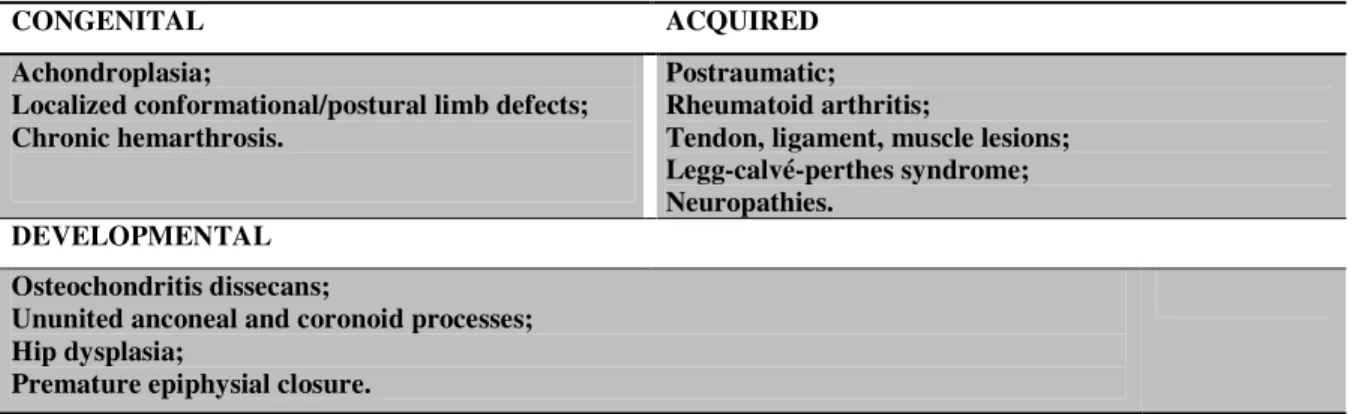
The most common form of OA in dogs is secondary and arises from known conditions (Table I) that affect the joint and its supporting structures (Piermattei et al. 2006).
CONGENITAL ACQUIRED
Achondroplasia;
Localized conformational/postural limb defects; Chronic hemarthrosis.
Postraumatic; Rheumatoid arthritis;
Tendon, ligament, muscle lesions; Legg-calvé-perthes syndrome; Neuropathies.
DEVELOPMENTAL Osteochondritis dissecans;
Ununited anconeal and coronoid processes; Hip dysplasia;
Premature epiphysial closure.
Table I. Examples of conditions predisposing to secondary OA
2.1 Pathophysiology
Osteoarthritis is a very complex pathology and the reasons for its development are not yet fully understood (Chen & Tua 2008). Inflammation plays an important role in OA: inflammatory cytokines such as Interleukin-1 (IL-1), Interleukin-6 (IL-6) and Tumor Necrosis Factor-α (TNF- α) are released from the chondrocyte (responsible for the production of extracellular matrix constituents and matrix metalloproteinases - MMPs) and the synovial cells, which will result in the release of prostaglandins such as prostaglandin E2 (PGE2). This will lead to the phenotypic alteration of chondrocytes which, instead of producing collagen type II, will start producing collagen type I and III. Additionally, the production of MMPs, which are degradative enzymes responsible for matrix turnover, will be increased, and the production of proteoglycans will decline resulting in a significant alteration of the extracellular matrix and consequent articular degeneration. Moreover, OA is also associated with oxidative stress and chondrocyte-produced reactive oxygen species further contribute directly to cartilage degradation as well as indirectly,
7
by up-regulating MMPs genetic expression and decreasing the production of tissue inhibitors of metalloproteinases (TIMPs)(Konttinen et al. 1994; Laflamme 2007).
While articular cartilage degradation is the “hallmark” of the disease, OA is a condition that affects the entire joint and its supporting structures. Subchondral bone sclerosis has been referred in several studies as a pathological finding of great importance in OA (Kuroki et al. 2011) and changes in the synovium and ligaments in patients with OA are also detectable at an early stage of the disease. Being a chronic disorder, OA frequently results in osteophyte and entesiophyte formation, synovial membrane hyperplasia with infiltration of inflammatory cells and muscle and ligament weakness (Fox & Millis 2010).
2.2 Diagnosis
A proper diagnosis depends on a complete anamnesis and a full assessment of the patient, commonly comprising a thorough physical, orthopedic, and neurologic examination and radiographies of affected areas. Clinicopathological exams might also be performed such as hematology and serum chemistry and synovial fluid analysis. Other less common complementary exams include advanced imaging (computed tomography, magnetic resonance imaging and nuclear scintigraphy), arthroscopy, electromyography and advanced gait analysis (Fox & Millis 2010). Common findings upon examination include musculoskeletal pain and lameness, crepitation, stiffness, bony changes in and around the joint in radiographs and signs that indicate the primary condition (for example, radiographic signs in cases of developmental pathologies such as hip or elbow dysplasia, positive drawer test in cases of Cranial Cruciate Ligament Rupture – CCLR, etc.).
2.3 Treatment
OA is a lifelong disease which can be managed but not cured. The approach should be patient oriented as it depends on the severity and location of the lesions, and the objectives of treatment for OA are multifaceted: reducing pain, lameness and discomfort, decreasing the progression of the disease, promoting the reparation of damaged tissue, and improving quality of life (Roudebush 2006). This management might be divided in two categories:
- Surgical: Surgical treatment for osteoarthritis should be considered when the primary disorder has a surgical solution (e.g.: CCLR, hip dysplasia and patellar luxation) or when pain or function is not improved by other conservative methods. The procedures in the latter case include debridement of osteophytes and joint surfaces, muscle release, arthrodesis, arthroplasty,
8
pseudoarthrosis, osteotomy, neurectomy and limb amputation (Piermattei et al. 2006). The description of these techniques and those for primary conditions causing OA are beyond the scope of this report.
- Medical: Successful medical management of OA requires a multimodal approach (Scott 2007). Current conventional treatment modalities may include a combination of anti-inflammatory and analgesic medications, disease-modifying osteoarthritis drugs (DMOADs), physical therapy and weight loss (Fox 2006; Roudebush 2006). Non-steroidal anti-inflammatory drugs (NSAIDs) are the most common class of drugs currently used in the short and long-term management of canine OA (Fox 2006; Scott 2007). These drugs produce effects primarily by the inhibition of cyclooxygenases in the arachidonic acid pathway. In what concerns clinical efficacy, NSAIDs have been proven to provide a rapid relief of symptoms related to inflammation, granting a medium to high level of comfort to patients (Roudebush 2006; Aragon et al. 2007). However, this class of drugs can cause side effects related to the gastrointestinal, hepatic, renal and hematopoietic systems and does not have any effect on slowing the progression of the disease (Roudebush 2006; Scott 2007). Analgesics, like tramadol, can also be used as a pain management drug. Tramadol is a synthetic opioid drug that acts on serotonin and adrenergic receptors and is useful in acute flares of arthritis pain. However, it can cause interactions with other medications and some patients exhibit opiate sensitivity, and its bioavailability and half-life are variable across individuals(Rychel 2010).In addition, DMOADs or chondroprotectants are a popular adjunctive treatment but there is little evidence if any of these compounds is actually able to restore or preserve articular cartilage. In their composition, these products often include precursors of glycosamynoglycans (GAGs) and GAGs usually found in in the extracellular matrix of articular cartilage, such as glucosamine and chondroitin sulphate, respectively (Scott 2007).
Physical therapy is also recognized as an important component of the management of the disease and helps combating muscle atrophy, maintaining the compliance of periarticular soft tissues and diminishing the risk of additional joint injury (Fox 2006). Additionally, weight loss, although not an actual therapy, is vital in patients with canine OA because obesity contributes to a chronic state of systemic inflammation and excessive weight will put extra stress on already sore joints (Rychel 2010).
Other adjunctive and alternative treatments include, but are not limited to, the supplementation with omega-3 fatty acids that have beneficial effects in states of chronic inflammation (Laflamme 2007; Rychel 2010), low-level laser therapy which is thought to
9
decrease muscle spasms and pain in arthritic conditions (Hegedus et al. 2009), and stem cell therapy which is currently being used with successful results (Fox & Millis 2010; Rychel 2010). .
2.3.1 Stem Cell Therapy for Canine Osteoarthritis
Stem cell therapy is already being commercialized and applied in dogs with OA all around the world. There is the possibility of transplanting undifferentiated MSCs or to differentiate them in vitro for posterior application. In both cases, harvested cells can be suspended in liquid solutions and injected into the affected joint which is the most practical mode of application (Ribitsch et al. 2010).
A few reports exist about the use of MSCs to treat canine osteoarthritis. In one report, 18 dogs with coxofemoral joint OA were treated with an intra-articular injection of AD-MSCs (obtained by enzymatic digestion of fat tissue without in vitro culture and expansion) and statistically significant improvement in lameness was demonstrated when compared with a control group (Black et al. 2007). Other report states that 14 dogs with elbow OA, also treated with the same technique, improved in lameness score, range of motion and pain on manipulation when compared with baseline values (Black et al. 2008). A more recent study conducted in 4 dogs with lameness associated with OA of the humeroradial joints has also shown positive results after application of cultured and expanded AD-MSCs in conjunction with platelet rich plasma or hyaluronic acid. These animals have shown improvements in functional disability, lameness at trot and in pain on manipulation of the joints (Guercio et al. 2011). Although the referred papers only evaluate AD-MSCs therapy for canine OA, BM-MSCs can also be used as an alternative therapy (Guercio et al. 2011).
2.3.2 Evaluation of Therapy Outcome in Osteoarthritic Dogs
Evaluation of canine OA is challenging because of the chronic, slowly progressive nature of the disease and the fact that most therapies might only have slightly observable effects (Innes 2010). In order to evaluate the efficacy of treatments and interventions designed to reduce the disability associated with orthopedic disease, defined objective measures of outcome are essential. Traditionally, the outcome after surgical or medical interventions is defined by clinical assessment. However, especially in cases where alternative or new therapies are used, the subjectivity of clinical observation is a major drawback. This is mostly due to the fact that, after a treatment, there is an inherent supposition that lameness and pain will improve and this form of bias is very difficult to eliminate (Sanchez-Bustinduy 2009).
10
Owner-administered questionnaires have surged recently in veterinary literature. The Canine Brief Pain Inventory© (CBPI©), developed by the School of Veterinary Medicine - University of Pennsylvania, includes semi-objective ratings of parameters related to pain and function on a discontinuous ordinal scale and allows owners to quantify the severity and impact of arthritic pain in dogs. It has already been evaluated for content validity, reliability and responsiveness and is frequently used for OA pain assessment (Brown et al. 2008; Innes 2010). However, the human ability to perceive the details of the canine gait cycle is very limited and during a subjective analysis only a few variables can be evaluated.
Biomechanics studies the mechanical functionality of living beings and the factors that affect their performance (Vilas-Boas 2001). Modern biomechanical analysis, such as kinematic and kinetic analysis systems which are able to capture, analyze and store hundreds of observations and measurements, are of great value for the evaluation of the canine gait and its pathological alterations. On the one hand, kinematic analysis studies the motion of objects by evaluating linear and angular velocities and accelerations, as well as positions and trajectories. Three-dimensional kinematic analysis allows a more comprehensive analysis of movement. However, these systems are expensive and complex. In contrast, two-dimensional computer assisted videographic gait analysis systems are less expensive and, being easier to use, provide the most relevant data for general practicioners. Kinetic analysis systems, on the other hand, allow quantification of forces. Most of these systems are ground-based kinetic systems (e.g.: force plates) that measure ground reaction forces that result from the paw impact during the stance phase of gait. Kinematic and kinetic analysis are both considered useful tools in veterinary medicine (Gillette & Angle 2008). These techniques have been used to study normal canine gait in healthy dogs (Kapatkin et al. 2007; Mölsä et al. 2010) and in the study of various musculoskeletal disorders in the same species, such as CCLR (Sanchez-Bustinduy et al. 2009; Bödekker et al. 2012) and coxofemoral joint laxity (Lopez et al. 2006), and have also been used in the assessment of NSAIDs efficacy (Gordon-Evans et al. 2011). Despite of all the efforts being put into gait analysis in veterinary medicine, a lot still has to be done in order to determine which techniques are more useful in each situation and to make them techniques widely available.
11 II. Objectives
The aim of this project is to study stem cell therapy as an alternative for the management of canine OA. For this purpose, three main objectives were defined:
I. To analyze and compare bone marrow and adipose tissue as sources of MSCs and select the best source for cellular therapy in the case of canine OA.
II. To apply MSCs with chondrogenic differentiation in the affected joints of canine patients suffering from OA and evaluate the outcome of such application by recurring to owner questionnaires and kinematic gait analysis.
III. To carry out a pilot study of cytogenetic analysis for evaluation of chromosomic stability in future SCT projects, at each cell culture passage and before cellular application into the patient.
III. Materials and Methods 1. Study Population
Seven dogs, outpatients at the “UPVet” (veterinary clinic of the Abel Salazar Biomedical Sciences Institute – University of Porto, Portugal), with ages between 1 and 10 years old (mean age of 5.2 years) and body mass ranging from 28 to 42 kg (mean of 33.1 kg), were initially recruited for this study. All the animals had a medical history of lameness associated with osteoarthritis, with pain on passive manipulation of the affected joints (Appendix I). Five of the animals also had radiographic signs of osteoarthritis (Appendix II).
Before enrollment, all animals underwent routine clinical chemistry (urea, creatinine, alanine transaminase, alkaline phosphatase, glucose, total proteins, globulins, albumin/globulin ratio, amilase) and hematologic evaluation (complete cell count) to ensure overall health. Dogs with concurrent systemic disease that could possibly interfere with the interpretation of treatment effects were excluded from the study. In addition, to be eligible, animals also had to be cared for by attentive owners who had previously signed an informed consent form and agreed to attend a scheduled set of appointments at the “UPVet” and to observe their dog throughout the entire study period. All the participants were deemed healthy and owners were required to stop administering any pain medication, including NSAIDs and analgesics, and chondroprotectants to the dogs at least 15 days before the beginning of the study.
An 8th dog (cross bred), chosen randomly among the surgical outpatients at the “UPVet” clinic, was chosen to undergo an adipose tissue collection (during a previously scheduled surgery) for posterior cytogenetic analysis of isolated stem cells.
12
2. Sample Collection, Processing and Stem Cell Isolation
The bone marrow and adipose tissue samples were collected either during pre-scheduled surgery or when dogs were sedated with medetomidine (Domitor®, Pfizer) (0.3mL/10kg of body weight) for radiological exams.
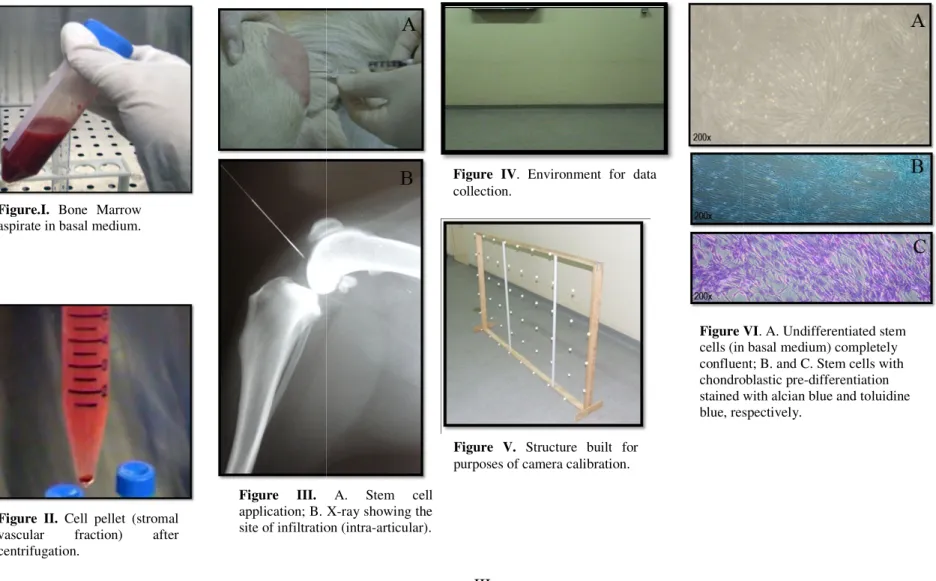
Bone Marrow: A biopsy needle (T-LokTM) accoplated to a 20mL syringe containing heparin (1ml) was used to harvest 10-15 mL of BM from the femoral medullar cavity of two patients (“Cairo” and “Java”). All products (reagents and media) for BM-MSCs culture were purchased from Sigma-Aldrich (USA), unless stated otherwise. The collected sample was mixed in a BD FalconTM tube (25mL) with 10 ml of basal cell culture medium liquid solution (Appendix III - Figure I) comprised by a mixture of Dulbecco’s Modified Eagle Medium (DMEM), Fetal Bovine Serum (FBS), Penicillin-Streptomycin and sodium bicarbonate. The obtained solution was immediately transported to the cytogenetic laboratory. Afterwards, the sample was centrifuged at 1200 rpm during 5 minutes and the obtained pellet was re-suspended in basal medium and seeded in BD FalconTM polystyrene cell culture flasks of 25cm2.
Adipose Tissue: A sample of approximately 5g of adipose tissue was extracted from subcutaneous fat of five patients (“Aline”, “Baltazzar”, “Cuca”, “King” and “Super Bock”) by performing a small surgical incision of 2 cm (posteriorly sutured with a non-absorbable simple interrupted suture) in the abdominal region of the dogs. All products (reagents and media) for AD-MSCs culture were purchased from Sigma-Aldrich (USA), unless stated otherwise. Each sample was then mixed in a BD FalconTM tube (25mL) with 20 mL of a Phosphate Buffer Saline (PBS) solution. Afterwards, the adipose tissue was taken to the laboratory where it was washed with more PBS and minced. It was then enzymatically digested in a 0.1% collagenase type II solution for 45 min at 37ºC. Finally, the solution was centrifuged at 1200 rpm during 5 minutes and the obtained cell pellet (stromal vascular fraction) (Appendix III - Figure II) was re-suspended in basal medium and seeded in BD FalconTM polystyrene cell culture flasks of 25cm2.
3. Expansion of Mesenchymal Stem Cells
Medium changes occurred 24 hours after cell culture and, after that, each three days or as necessary. When the confluence inside the flask reached 80-90%, the cells were enzymatically lifted with 3 to 6 mL of trypsin which was posteriorly neutralized with basal medium. The solution containing the cells was centrifuged (1200rpm during 5 minutes) and the obtained pellet was re-suspensed in basal medium and passaged into 175cm2 polystyrene culture flasks. This process was similar for BM-MSCs and AD-MSCs.
13
4. Chondrogenic Pre-Differentiation of Stem Cells
After approximately one week of culture, if the cells were more than 90% confluent, another passage was carried out. In order to induce the MSCs differentiation into chondroblasts cells where cultured in a chondrogenic medium solution comprised by DMEM, sodium pyruvate, ascorbate-2-phosphate, proline, ITS Liquid Media Supplement (composed of recombinant human insulin, human transferring and sodium selenite) and Penicillin-Streptomycin. Cells continued to be expanded and when the flasks reached 80-90% of confluence trypsinization was carried out and cells where counted in a Neubauer chamber. Passages were carried out until a number of approximately 10x106 cells was obtained. A small amount of cells was also cultured in a BD FalconTM 6-well Multiwell plate. The cells contained in each well were posteriorly fixated in formaldehyde (4%) and stained histologically with alcian blue and toluidine (common cartilage stains) used to highlight the presence of GAGs.
5. Cell Application
Preparation of cells for application: A cell suspension of 10x106 cells was prepared for each dog. In order to do this cells were lifted enzymatically with 3 to 6 mL of trypsin after reaching, at least 80% of confluence in 175 cm2 culture flasks. The trypsine was neutralized and the obtained solution was centrifuged (1200 rpm during 5 minutes). The supernatant was then removed and the cells where suspended in sterile PBS. This process (centrifugation and re-suspension in PBS) was repeated three times in order to ensure that all trypsin and culture medium was removed.
Application: The application site was tricotomized, prepared aseptically, and the solution containing the cell suspension was injected intra-articularly in the osteoarthritic joints of five dogs (“Aline”, “Baltazzar”, “Cuca”, “King”, “Super Bock”) (Appendix III- Figure III). In the case of “Aline”, who underwent a Triple Tibial Osteotomy (TTO) due to a CCLR, cells were applied two weeks after surgery. Animals were previously sedated with medetomidine (Domitor®, Pfizer) (0,3mL/10kg of body weight) and a local topical anesthetic (Lidocaine 1%, Labesfal®) was applied.
6. Owner Questionnaires
Owners completed the Canine Brief Pain Inventory© (CBPI©) (Appendix IV), before the intra-articular application of the cells in the affected joints of their animals and one month after the application. In the case of “Aline”, the CBPI was answered before surgery, two weeks after
14
surgery (when the cell therapy took place) and one month after stem cell application. This questionnaire has 11 questions and is divided in three sections: question 1 to 4 relates to pain, question 6 to 10 relates to function and question 11 to overall impression (quality of life). Questions 1 to 10 are scored from 0 to 10 being that 0 means that there is no pain/interference in function and 10 stands for maximum pain/interference with function. The average values for the two first sections were calculated in order to give a pain and function score to each animal. This methodology has already been applied in previous studies (Brown et al. 2008).
7. Canine Gait Analysis
Kinematic data collection: Dogs were, initially, familiarized to walk on a leash on a room with 6 meters in length (Appendix III - Figure IV). The animal handler had to walk to the beat of a metronome set to 80 beats per minute (bpm). At least ten trajectories (from one end of the room to the opposite end) were recorded, immediately before and one month after the cell application, using a video camera (Sony® Handycam DCR-HC46E, shutter speed: 1/2-1/3500) with a recording frequency of 25 Hz. The same method was used for “Aline” but, in her case, filming was carried out before surgery, two weeks after surgery and one month after SCT. In each animal, the affected joint was on the same plan as the camera (parallel). A two-dimensional grid (160 cm in length and 100 cm in height, divided in squares with sides measuring 20 cm) was built for purposes of camera calibration (Appendix III - Figure V).
Kinematic and Statistic Data Analysis: After videotaping the animals, the obtained data were transferred to a computer in order to be analyzed. The software used for kinematic analysis was the Dvideow (Unicamp, São Paulo, Brazil). More detailed specifications about this software are available in the literature (Figueroa et al. 2003). From all the trajectories recorded per animal pre and post-application, aberrant strides were eliminated and three normal strides were randomly selected for analysis.
Two of the analyzed variables, stride length and paw velocity, were chosen based on a study conducted in 2009 by Sanchez-Bustinduy et al. that has concluded that these are the most reliable and not susceptible to systematic alterations variables for stifle joint dynamics study after CCLR treatment procedures and that they decrease in pelvic limb lameness of any sort. The same authors also imply that these variables decrease in thoracic limb lameness and that they are a reliable option to evaluate treatment outcomes (Sanchez-Bustinduy et al. 2009). The points necessary to calculate stride length and paw velocity for each stride were the 5th metatarsus position at paw take off and the 5th metatarsus position when the paw landed. Since the dogs
15
showed discomfort and abnormal gait when a retro-reflective marker was placed on the 5th metatarsus it was decided that no marker would be attached to the animals and that the points would be digitalized manually on the software. The stride length for each dog was normalized with the distance from the floor to the withers, since this is a very common measurement of dog height used, for example, in official breed standards. Dog velocity was also calculated in order to ensure that it was not influencing paw velocity. The digitalized points for this variable were the withers position at the starting frame used for analysis of each of the other variables and the same point 10 frames after that.
The repeatability of the analyzed variables and kinematic method was obtained by calculation of the Intraclass Correlation Coefficient (ICC). Additionally, despite the fact that the study sample is small and very heterogeneous in what concerns age, breed, sex, type of lesion, chronicity and severity of OA, the Wilcoxon signed-rank test for statistical analysis of non-parametric (distribution free) samples was used, with 95% of confidence, in order to compare variables before and after cell application. The statistical analysis was carried out on SPSS® (version 19.0) for Microsoft® Windows®.
8. Cytogenetic Analysis
Before initiating the cellular processing in order to perform a cytogenetic analysis, the cell culture was observed under the microscope to ensure that there was sufficient cellularity. The culture medium was then replaced by new, pre-heated at 37ºC, basal medium.
Metaphase Arrest: In order to arrest chromosomes in metaphase 100 µL of KaryoMax® ColcemidTM (Gibco) were added to the stem cell culture per each 5 mL of basal medium 3 hours before harvesting the cells.
Cell harvest and fixation: Cells were lifted enzymatically using trypsin which was neutralized, with basal medium, after two minutes of action. The obtained solution was aspirated from the flask and transferred to BD FalconTM tubes which were then centrifuged at 1500 rpm during 10 minutes. After that, 8mL of hypotonic solute (0,05M KCl solution and fetal bovine serum in a relation of 8:1) pre-heated at 37ºC were added while gently agitating the tubes in a vortex. The tubes were placed in a cell culture chamber at a temperature of 37ºC during 35 minutes. Afterwards, they were centrifuged again (1500 rpm for 10 minutes) and the supernatant was rejected. At this time, the cells were fixated 2 times in a 6:1 methanol:acetic acid solution and 2 times in an 3:1 methanol:acetic acid solution. The supernatant was rejected and the cells were spread on microscope slides.
16
Staining: All the slides were stained using a 4% Giemsa solution with PBS which was in contact with the slides for 6 minutes and then rinsed with distilled water.
Chromossome analysis: The canine species (Canis familiaris) karyotype has a total of 78 chromosomes: 39 homologous pairs, in which 38 are autosomal and one corresponds to the sex chromosomes. All the chromosomes are acrocentric with the exception of the X chromosome, which is sub-metacentric. The obtained slides were analyzed and at least 50 metaphases were scored. Aneuploidy and chromosome instability (chromosome breaks) was evaluated.
IV. Results
1. MSCs Source Selection
Bone marrow aspirate was successfully collected from “Cairo” (15 mL) and “Java” (10mL). The presence of stem cells in culture was verified by microscopic observation of spindle shaped cells adhered to the polystyrene culture flask. Cells were cultured during a considerable period of time (approximately 45 days) without chondrogenic differentiation. However, when the basal medium was substituted with chondrogenic medium to induce chondroblastic differentiation, the number of cells started to decrease after two weeks of culture in this new medium and the cells ultimately died. “Cairo” was posteriorly removed from the study due to post-surgical complications and “Java” has not received any SCT yet.
Adipose tissue was easily collected from the remaining dogs and the procedures to obtain the stromal vascular fraction for posterior stem cell culture were successfully performed. AD-MSCs started being observed after two days of culture and, within one week, the cells where 90% confluent for all the animals. The chondrogenic medium started being used after the second passage and, in two to three weeks, a cell count higher than 10x106 was obtained in all cases and cells stained positive for alcian blue and toluidine blue (Appendix III - Figure VI)
Adipose tissue was selected as the source for stem cell therapy for “Aline”, “Baltazzar”, “Cuca”, “King” and “Super Bock”.
2. AD-MSCs derived chondroblasts application and SCT outcome analysis
The intra-articular application of cells was performed without any difficulties and none of the owners described any kind of adverse reaction to the therapy. Clinically the animals showed improvement after SCT on lameness and pain and “Cuca” began to support the affected limb on the ground with weight bearing.
17
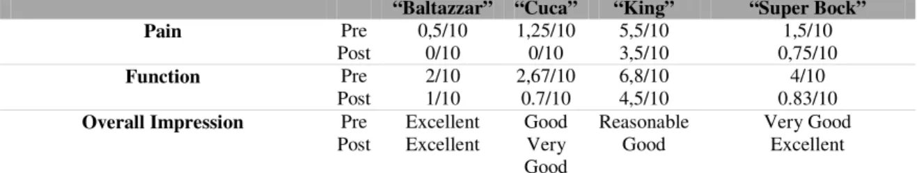
Owner questionnaires: All owners reported improvement after stem cell therapy (Appendix V) which means that the scores for each section (“pain” and “function”) showed a decrease (Table II). In the case of “Aline” there was first an increase in pain and function scores and overall impression also worsened. However, after SCT the scores decreased to a level lower that before surgery (Table III).
Table II. CBPI© scores for “Baltazzar”, “Cuca”, “King” and “Super Bock”, pre and post cell therapy.
“Aline” Pre-Surgery Post-Surgery (2 weeks) Post-Cell therapy
Pain 6.5/10 7.8/10 1.5/10
Function 6.8/10 9/10 3/10
Overall impression Reasonable Bad Reasonable
Table III. CBPI© scores for “Aline” during the various phases of study.
Kinematic and statistical analysis: After calculation of the ICC (Table IV), which was revealed to range from good to excellent, it was decided that mean values for each variable and individual animal would be used for result analysis.
Paw Velocity Stride Length Gait Velocity
ICC 0.69 0.91 0.85
Table IV. Intraclass Correlation Coefficient values for each calculated variable.
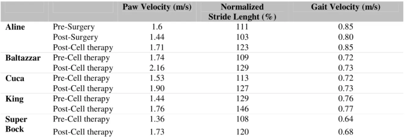
All animals have shown an increase in paw velocity and stride length after stem cell therapy (Table V). In the case of “Aline”, where SCT was only administered after a surgical intervention, a 0.16 m/s reduction in average paw velocity and an 8% reduction in average normalized stride length were observed two weeks after surgery. However, after stem cell therapy, these variables increased in 0.27m/s and 20%, respectively. As for the other animals, which only have had two data collections (pre and one month post-SCT), a consistent increase in both variables was observed. “Baltazzar”, “Cuca”, “King” and “Super Bock” have had 0.42 m/s, 0.37m/s, 0.32 m/s and 0.37m/s increases in average paw velocity and 20%, 14%, 17% and 12% increases in average stride length, respectively. Gait velocity was also calculated for all animals and the obtained values between pre and post-cell therapy were very similar.
“Baltazzar” “Cuca” “King” “Super Bock”
Pain Pre 0,5/10 1,25/10 5,5/10 1,5/10
Post 0/10 0/10 3,5/10 0,75/10
Function Pre 2/10 2,67/10 6,8/10 4/10
Post 1/10 0.7/10 4,5/10 0.83/10
Overall Impression Pre Excellent Good Reasonable Very Good Post Excellent Very
18
Table V. Average values of kinematic variables, pre and post cell therapy, for the animals treated with SCT. After analyzing the results for “Baltazzar”, “Cuca”, “King” and “Super Bock” and observing that paw velocity and stride length had consistently increased for all them, the Wilcoxon signed-rank test was carried out.The results for this test showed that the differences between pre-SCT and post-SCT values for normalized stride length and paw velocity where statistically different and that the values for gait velocity before and after stem cell application did not show a statistical difference (Appendix VI, Appendix VII).
3. Cytogenetic Analysis
Cytogenetic analysis was carried out for the fifth passage of an AD-MSCs cell culture in order to analyze the efficacy of the described protocol (Table VI).
AD-MSCs (Dog A) Passage 5
N % aneuploidy % poliploidy nº breaks/cell % cell with breaks
74.00 17.57 8.11 0.08 8.11
Table VI. Results for the conducted cytogenetic analysis; n stands for the number of analyzed metaphases.
V. Discussion
As it was previously stated, this project had three main objectives which will be discussed separately:
Objective I - To analyze and compare bone marrow and adipose tissue as sources of MSCs and select the best source for cellular therapy in the case of canine OA: Our results indicate that adipose tissue is a better choice for stem cell therapy than bone marrow. Adipose tissue was easier to collect than bone marrow and AD-MSCs have expanded considerably faster than BM-MSCs. This corroborates what has been described in the literature (Ribitsch et al. 2010). In addition, we were successful in inducing chondroblastic pre-differentiation to AD-MSCs but not to BM-AD-MSCs. There might be two explanations for this. On one hand, the used
Paw Velocity (m/s) Normalized Stride Lenght (%)
Gait Velocity (m/s)
Aline Pre-Surgery 1.6 111 0.85
Post-Surgery 1.44 103 0.80
Post-Cell therapy 1.71 123 0.85
Baltazzar Pre-Cell therapy 1.74 109 0.72
Post-Cell therapy 2.16 129 0.73
Cuca Pre-Cell therapy 1.53 113 0.72
Post-Cell therapy 1.90 127 0.73
King Pre-Cell therapy 1.44 129 0.76
Post-Cell therapy 1.76 146 0.77
Super Bock
Pre-Cell therapy 1.36 108 0.64
19
chondrogenic medium might not be the best for these cells. Despite the fact that the medium contained substances which have been proven to enhance chondrogenic differentiation (Phornphuktul & Grupposo 2006; Altaf et al. 2006; Roy et al. 2010; Solchaga et al. 2011), some substances that have also been described as important for chondrogenesis in BM-MSCs such as, for example, Transforming Growth Factor-β1 (TGF-β1) (Ribistch et al. 2010; Solchaga et al. 2011) and Bone Morphogenetic Protein-4 (BMP-4) (Kuroda et al. 2006) were not used due to financial constraints. On another perspective, it seems that BM-MSCs have a better chondrogenic potential than AD-MSCs (Sakaguchi et al. 2005). If, in BM-MSCs cultures, the cells had differentiated into chondrocytes the expansion would have happened at a slow rate. Additionally, it is has also been described that after expansion in monolayers differentiation is usually problematic, especially in cases when cells have derived from geriatric patients (which is the case of “Java”) (Salgado et al. 2006). This could explain the drawback experienced in the cases where BM-MSCs were cultured. Nevertheless, more studies need to be conducted in order to draw definite conclusions.
Objective II - To apply MSCs with chondrogenic differentiation in the affected joints of canine patients suffering from OA and evaluate the outcome of such application by recurring to owner questionnaires and kinematic gait analysis: Stem cell therapy was successfully admininistered to five patients (“Aline”, “Baltazzar”, “Cuca”, “King” and “Super Bock”). Clinically, the animals showed improvement on lameness and overall comfort. However, this is not sufficient to state that SCT was a success.
In what concerns kinematic analysis, the results obtained show that there was an increase, for all cases, in the values of average stride length (increases ranging from 12% to 20%) and paw velocity (increases ranging from 0,32m/s to 0,42m/s) one month after SCT. Assuming that there is direct relationship between the decrease in these variables and lameness (Sanchez-Bustinduy et al. 2009), this suggests that the treatment was effective in reducing lameness (and, probably, pain) and that this led to an improvement in the studied variables. In the particular case of “Aline”, the cell application was performed two weeks after surgery in an attempt to accelerate recovery from the procedure. The results show that, two weeks after surgery stride length and paw velocity are reduced when compared to the pre-surgery values. This might be related, to the fact that the used surgical technique, the Triple Tibial Osteotomy (TTO), is an invasive and somehow aggressive technique and that a strong inflammatory response was taking place (with swelling, increased local temperature, redness and increased pain). In one report it was stated that, usually, animals exhibit a normal gait in 4 to 5 weeks following the procedure (Properzi
20
2010). Therefore, we cannot conclude with certainty that SCT, in this case, was beneficial for the animal. Nevertheless, since the recovery was very difficult at the beginning (before SCT) and, that in the weeks following the cell application, “Aline” has improved significantly, clinically and also in relation to average stride length and paw velocity (which increased in relation to the pre-surgery measurements), we believe that the cell application was of importance in the clinical improvements observed. In the case of “Baltazzar” the improvement in both, stride length and paw velocity, was more notorious than in the other animals. We believe that this is due to the fact that this is the younger animal of the sample, with a less chronic process and, therefore, the effect of SCT was more substantial. On the other hand, “King” has shown the smallest improvement in paw velocity (0,32m/s) which might be related to the severity and chronicity of the pathology since this animal has been suffering from a severe shoulder osteochondritis dissecans for almost 3 years. This also indicates that more studies should be performed in order to understand if there is an inverse relation between the chronicity/severity of osteoarthritis and the efficacy of SCT. It is also interesting to analyze the results obtained in the cases of “Cuca” and “Super Bock”. These animals were the only participants in the study who received cell therapy, that did not show radiographic signs of osteoarthritis and the increases in stride length were smaller than those for the remaining animals. However, the increases in paw velocity were similar to those of “Baltazzar”. The smaller increase in stride length might be associated to the osteoarthritic process in these animals not being as severe as in the cases of “King” and “Baltazzar” but we should also keep in mind that these animals have OA in their thoracic limbs, while “Cuca” and “Super Bock” have knee OA associated with a previously treated CCLR and therefore, the results might not be directly comparable. Secondly, by observing that while stride length did not improve as much as on the other two cases but that paw velocity shows good improvement might be related to the particular action of stem cells. It could be hypothesized that stem cells, by their antialgic action, help in increasing activity levels and, therefore, paw velocity but that do not act as well in regenerating knee function which would explain the fact that movement is more limited and stride length did not improve as much when compared to the other animals. This would need to be further investigated and, again, one should not forget that this sample is very heterogeneous and that there is not any kinematic data prior to the onset of OA, which makes it impossible to know what the normal values would be for the analyzed variables.
The Wilcoxon signed-rank test that was performed for the results of the kinematic variables obtained in this study has further shown that the results are valid and that SCT has