UNIVERSIDADE DE LISBOA Faculdade de Medicina
Sleep, Sleep-disordered-breathing, Cognition and Prematurity
Yu-Shu Huang
Orientadores: Prof. Doutora Maria Teresa de Aguiar dos Santos Paiva Prof. Doutor Christian Guilleminault
Tese especialmente elaborada para obtenção do grau de Doutor em Medicina, especialidade em Psiquiatria e Saúde Mental
Júri:
- Presidente: Doutor José Augusto Gamito Melo Cristino, Professor Catedrático e Presidente do Conselho Científico da Faculdade de Medicina da Universidade de Lisboa
Vogais:
- Professor Christian Guilleminault, Professor of Psychiatry and Behavioral Sciences da Stanford University School of Medicine, EUA; (co-orientador)
- Doutora Maria Hercília Ferreira Guimarães Pereira Areias, Professora Catedrática Convidada da Faculdade de Medicina da Universidade do Porto
- Doutora Helena Cristina de Matos Canhão, Professora Catedrática Convidada da Faculdade de Ciências Médicas da Universidade Nova de Lisboa
- Doutora Maria do Céu Lourinho Soares Machado, Professora Catedrática Convidada da Faculdade de Medicina da Universidade de Lisboa
- Doutora Maria Isabel Segurado Pavão Martins Catarino Petiz, Professora Associada com Agregação da Faculdade de Medicina da Universidade de Lisboa
- Doutora Maria Cristina de Brito Eusébio Bárbara Prista Caetano, Professora Associada Convidada com Agregação da Faculdade de Medicina da Universidade de Lisboa - Doutor André Laboreiro Ferreira Mendes da Graça, Professor Auxiliar Convidado da
Faculdade de Medicina da Universidade de Lisboa 2017
1
Dedication:
This work is dedicated to my parents who believe that I should do what I wanted and could do it. It is also dedicated to my daughter Lulu Pei-Ju Huang who accepted my work in the evenings and nights and my absences while growing-up performing my duties and who accepted the demands placed on a woman academic physician wanting to pursue a career in her field.
Acknowledgements and Thanks
To Professor Christian Guilleminault who was very much involved in helping the completion of the presented studies and the write-up as articles. Under his supervision, the task could be completed successfully.
To Professor Teresa Paiva: Without her help and encouragement this work would not be presented.
To Professor Chih-Ken Chen and Professor Chia-Hsiang Chen who mentored my psychiatric training and train me in research.
To Professor Fang-Ming Hwang who taught me statistics.
My thanks also to the surgeon-specialists and neonatalogists that continuously collaborated with me on my studies on children:
* Professor Hsueh-Yu Li, Chair of the department of Oto-Rhino-Laryngology, Chang Gung Memorial Hospital and Medical College,Taoyuan, Taiwan
* Dr Cheng-Hui Lin, Assistant Professor, Cranio-Facial Surgical Division, Chang Gung Memorial Hospital and Medical College, Taoyuan, Taiwan
* Dr Jen-Fu Hsu, Associate Division of Pediatric Neonatology, Department of Pediatrics, Chang Gung Memorial Hospital andMedical College, Taoyuan, Taiwan
2
Índex
l I-1. Summary:... 4
l I-2. Sumário: ... 9
l II. Introduction ... 14
II-1. Background and Rationale ... 14
II-2. Study Purpose ... 18
l III. Literature Review ... 20
III-1. Pediatric Sleep-Disordered-Breathing (SDB) ... 20
III-2. Obesity and SDB ... 21
III-3. Non-overweight children and SDB: Why does the upper airway collapse during sleep in non-overweight children? ... 22
l Monkey Experimental Investigation ... 22
l Cranio-facial Growth ... 23
l Nasal Breathing and Mouth Breathing ... 24
l Other early-in-life functions involving the oral cavity... 25
l Short lingual frenulum and oral-facial development ... 26
III-4. Neurodevelopmental dysfunctions in prematurely born neonates ... 27
III-5. Neurocognitive function and pediatric SDB ... 27
(1). Pediatric OSA and inflammatory cytokines ... 28
(2). The pro-inflammatory cytokines IL-17 and IL-23 ... 28
l IV. Methodology ... 30
IV-1. Study Process: ... 30
IV-2. Framework: ... 32
IV-3. Study Flux diagram: ... 33
IV-4. Research Instruments: ... 33
(1). Infant Polysomnography: Development of a Pediatric Sleep Lab in Taiwan... 33
(2). Actigraphy: ... 34
(3). Development of Chinese-version of Infant Sleep Questionnaire: “Brief Infant Sleep Questionnaire-Chinese version,” (CBISQ): (published article # 1) ... 35
(4). Development of Chinese-version Pediatric Obstructive Sleep Apnea Questionnaire: Chinese version OSA-18 (published article # 2) ... 36
3
(5). Neurodevelopmental assessments: ... 38
IV-4. Analytical Methods ... 38
l V. Results ... 40
V-1. Development of the “ Brief Infant Sleep Questionnaire-Chinese version” and investigation of “ Sleep and breathing” in premature infants at 6 months post-natal age ( # 1 article was published in BMC Pediatrics, 2014 ) ... 40
V-2. Minor neurodevelopmental dysfunctions at age 6 months in prematurely born neonates (#2 article published in Early Human Development,2013) ... 46
V-3. Sleep-disordered breathing, Craniofacial development and Neurodevelopment in Premature Infants: A 2 years following Study (This article is going to be submitted/ unpublished) ... 54
V-4. Development of Chinese-version Pediatric Obstructive Sleep Apnea Questionnaire( OSA-18) and To find the common symptoms of Pediatric OSA in Taiwan ( # 4 article was published in Psychiatry and Clinical Neurosciences, 2016) ... 62
V-5. Sleep-Disordered Breathing and oral-facial development in the premature infant (# 5 article was published in Frontier in Neurology, 2013) ... 68
V-6. Short lingual frenulum, mouth breathing and abnormal oral facial growth (# 6 article was published in Int J Pediatr Res ,2015) ... 78
V-7 Inflammation, Cytokines, Neurocognition and Pediatric sleep-disordered-breathing ( # 7 article was published in Medicine J, 2016) ... 84
l VI. Discussion ... 93
VI-1. Sleep and breathing in premature infants ... 93
VI-2. Neurodevelopmental dysfunctions in prematurely born neonates ... 94
VI-3. Sleep-Disordered Breathing and oral-facial development in the premature infant ... 96
VI-4. Inflammation, Neurocognition and Pediatric sleep-disordered-breathing ... 97
l VII. Conclusion and Recommendations ... 99
l VIII. Bibliography ... 101
l VIII. Articles List... 110
4 l I-1. Summary:
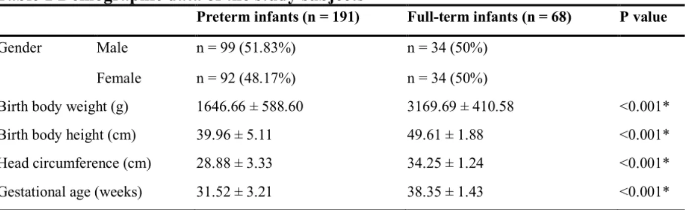
Prematurity leads to many handicaps, some of them are only recognized later in life and may impact the individuals for the rest of their life. The delivery date compared to the full term delivery time will be a measure of “indication of risks” of post-natal handicap risk, but this is only one of the many measurements that can be looked at. Many studies have investigated development of premature infants, based on different criteria at entry in the considered study. Our investigations are only a limited contribution to the investigation of premature infants. We included infants born as young as 24 weeks of gestational-age [GA] but none of the infants had major neurological syndromes recognized at birth. “Normal infants” defined as infants with more than 37 weeks of GA, birth-weight >2500g and absence of any indication of health problems born in the same hospital maternity at same time as premature infants were also recruited to serve as normal controls.
As most newborn infants spend a large amount of time asleep, all the presented studies include investigation of sleep, and once sleep-time occurred mostly during the nocturnal period, it focused on the polygraphic monitoring of the nocturnal sleep.
The premature cohort study was a longitudinal study and parents who signed informed consent approved by the Chang Gung Hospital and Medical College Ethic Committee, were asked to come back on a yearly basis for at least 5 years. This is an on-going study and not every child has been followed for such time. Furthermore, as in any longitudinal study, loss of patients occurred as parents did not bring children back. At entry 400 parents signed the informed consent, currently at 5 years follow-up 150 children have ended the follow-up period and about 215 at 4 years. The sample is a non-random convenience sample of children selected based on the parents` willingness to participate in the protocol and obtained with the help of neonatologist physicians in our NICUs.
Most of the studies presented in the thesis come-out of this longitudinal study. The studies asked specific questions, particularly looking at development of abnormal obstructive breathing during sleep. But some of our studies looked also at children of older age as some of the findings that we observed in our premature cohort needed a different investigative approach, and prior validation on older children-premature and in non-premature infants.
We have included these studies in our narrative as they become part of our research, and they are part of a general research program on “sleep- breathing-and-cognition” in children.
5 The thesis has the following organization:
1. Summary in English and Portuguese
The preamble explains major sleep problems of premature infants and the work done 2. Introduction with background and objectives as follows:
(1) the understanding of the development of abnormal upper airway in premature infants and abnormal breathing during sleep.
(2) the understanding the craniofacial development and impact on occurrence of pediatric SDB.
(3) to evaluate the differences in development between premature and full-term infants.
(4) finally investigating, from our cohort-data, the relationship between the SDB and the developmental problems that we could observed in our premature infants.
To perform these tasks she had to:
(1) Develop an “infant sleep questionnaires” in Chinese version to use through our studies
(2) Develop pediatric obstructive sleep apnea questionnaires in Chinese version to use in our study.
(3) To investigate the possible actions of pediatric SDB on neurocognitive dysfunctions in our children and is behind the protocols presented in our studies.
(4) To develop the biomarkers for diagnosis and selection for treatment of pediatric SDB
(5) To improve treatments of pediatric OSA.
3. Literature review including Sleep disordered breathing in infants and children, the problem of obesity, the issues related to craniofacial development, type of breathing, facial configuration and the lingual frenulum, the pro inflammatory cytokines, neurodevelopmental and neurocognitive dysfunctions of SDB in infants and children.
6
4. The methodology used which includes the set-up of the Sleep Lab, the multidisciplinary team and the development of Chinese-version of Infant Sleep Questionnaire: “Brief Infant Sleep Questionnaire-Chinese version”
5. The results which are based on the published articles
6. The discussion in which she integrates the achieved results in the international literature. The following research projects were undertaken:
Study #1The Chinese version of the obstructive-sleep-apnea questionnaire-18
Our first study involved defining another tool for our research considering the parents’ reluctance to come back, year after year to the sleep-laboratory for follow-up investigation. Not to have too many “absence of data”, we had to validate another tool: a questionnaire that parents would be willing to fill- in, even at home if necessary. This validation involved monitoring of children during sleep.
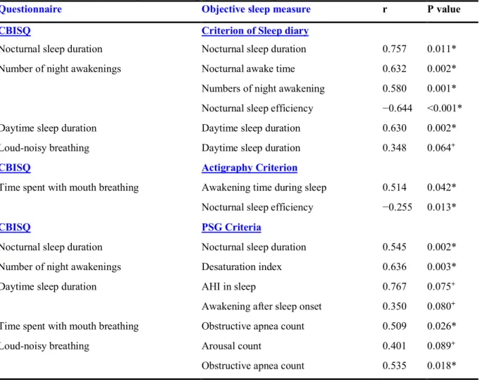
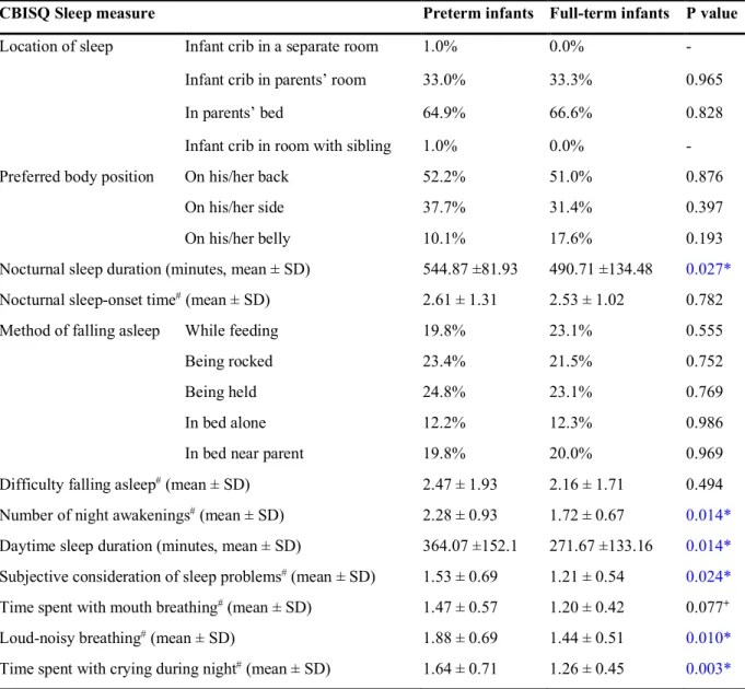
Our study validate the CBISQ, but it is also the first study targeting the difference between premature and full-term infants through a reliable and valid screening questionnaire.
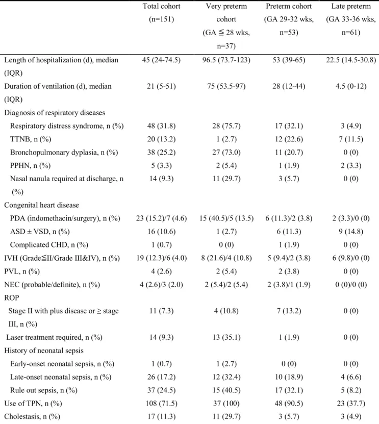
Study #2 - Sleep and Breathing in premature infants at 6 months post-natal age –
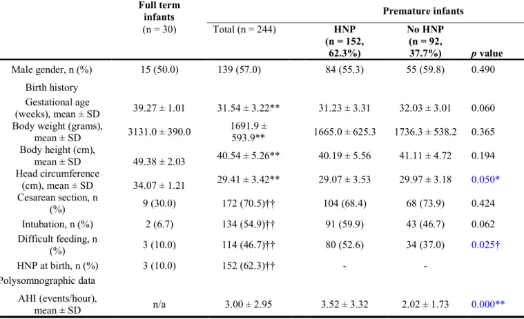
Sleep and sleep breathing disturbances were investigated in premature infants at 6 months of age.
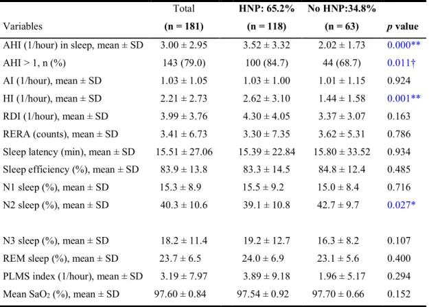
Premature infants are presenting with a more narrow and high-arched palate, more SDB-related sleep problems, and more neurodevelopmental deficits than normal full-term infants.
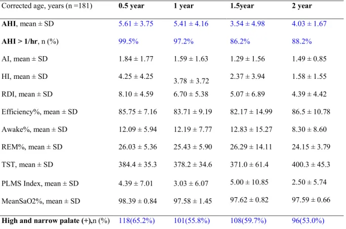
Till 2 years of age, premature infants with narrow and high-arched palate have more SDB-related sleep problems and more neurodevelopmental deficits than those without.
These findings support our hypothesis that high and narrow-arched palate in premature infants plays a role in the development of sleep problems and neurodevelopmental delays.
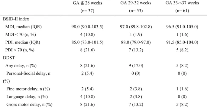
Study #3 - Early detection of minor neurodevelopmental dysfunctions at age 6 months in prematurely born neonates.
The infants involved in our longitudinal studies were selected as free of clear neurological signs and symptoms. However, these children are considered to have clear neurological risks. We
7
investigated such possibility assessing subtle neuromotor dysfunctions, such as difficulties with gross motor skill, social contact, or learning.
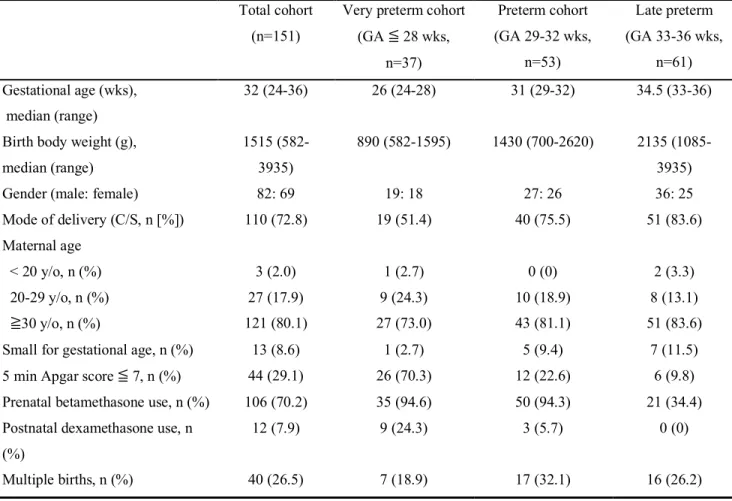
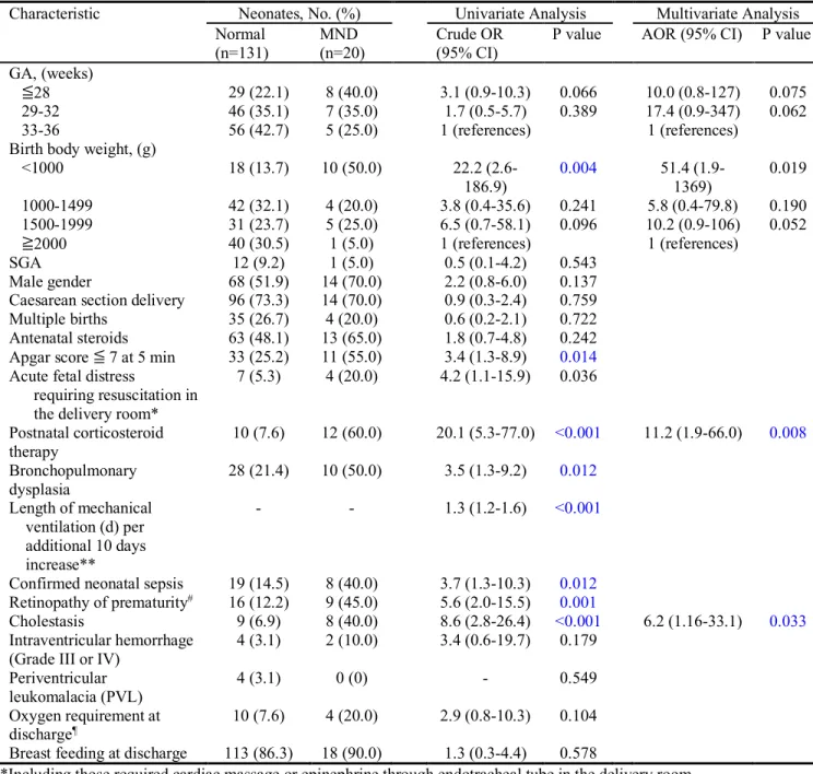
Premature neonates, even those born at 33 to 36 weeks, are found to have MNDs (Minor Neurodevelopmental Dysfunction) as early as 6 months corrected age by BSID-II and DDST, with risk increasing as gestation decreases.
Moreover, we used multivariate logistic regression to evaluate the relationships between neonatal factors and the presence of MNDs. After multivariate logistic regression adjusted for GA and BBW (Birth Body Weight), MND was independently associated with postnatal corticosteroid use and cholestasis. Besides, neonates with BBW less than 1000g were significantly associated with MNDs when compared with those with BBW more than 2000g. Study#4 Pediatric Obstructive Sleep Apnea and the critical role of oral-facial growth (a review of our evidences placed in an historical context)
We looked to the critical role of facial growth in the development of SDB.
Currently at 4 years of age, 77% of our premature infants have OSA and a high and narrow hard palate. There is a relationship between gestational age(GA) at birth and presence of problems which increase with lower GA at birth. But, this is not the only factor. As found, children with normal palate and AHI may progressively deteriorate during the first post-natal months. Preliminary data indicate that normal functioning of suction, swallowing, mastication and nasal breathing post-birth are key factors in the switch overtime to abnormal breathing and high and narrow hard palate.
Study #5 Short nasal lingual frenulum, mouth breathing and abnormal oral facial growth. This study was performed to investigate the role of abnormal oral facial functioning in children and development of OSA.
Short lingual frenulum may lead to abnormal orofacial growth early in life, a risk factor for development of SDB.
Careful surveillance for abnormal breathing during sleep should occur in the presence of short lingual frenulum.
8
Study #6 Inflammation, cytokines and mouth breathing
From of our longitudinal study on premature children we had found out that mouth breathing was frequent and that tonsils were not initially enlarged in children that developed OSA, but become enlarged. We had shown that children with enlarged tonsils and adenoids (T&A) treated with adenotonsillectomy present relapse of OSA within 3 years. We questioned if inflammatory factors could not play a role in the development of OSA and if persistence of inflammatory factors could not be involved in relapse of OSA. This study is only the first step in our investigation but it shows that specific inflammatory factors, currently very much looked at: ie interleukines(IL) 17 and 23 are clearly abnormal in OSA children, supporting our hypothesis and opening the field for further studies. We do not have post-surgery results as the study takes time to be completed, but we demonstrated the validity of our hypothesis and the need to pursue this line of research.
By Regression analysis the following conclusions were achieved: significant relationships between pro-inflammatory cytokines and PSG scores with higher AHI score and OSA severity, such as HS-CRP (High Sensitivity- C Reactive Protein) (β=0.390, P<0.05) and IL-17(β=0. 329, P<0.05. Higher AI “influenced” serum levels of HS-CRP suggesting an impact of inflammatory cytokines on soft tissues hypertrophy. Higher serum levels of IL-23 (β=0.403, P<0.05) were “influenced” by higher AI. Lower mean SaO2 (O2 saturation) “influenced” IL10 level (β=
-0.567, P<0.01), and higher serum levels of TNF-α (tumor necrosis factor alpha) and IL-1β were “influenced” by higher diastolic pressure (β=0.469 and 0.659, P<0.01). There was a significant relationship between lower performances of CPT test and pro-inflammatory cytokines. Significant Spearman’s correlation factors between pro-inflammatory cytokines and clinical findings such as asthma and IL-6 (ρ= 0.261, P=0.026*); allergic rhinitis and HS-CRP (ρ= 0.280, P=0.022*), IL-6(ρ= 0.299, P=0.01*) and IL-10(ρ= -0.265, P=0.023*) ; Tonsil hypertrophy and HS-CRP(ρ= 0.244, P=0.046*) ; Adenoid hypertrophy and IL-6 (ρ=0.232, P=0.048*) were observed.
9
l I-2. Sumário:
A prematuridade leva a muitas deficiências, algumas das quais são apenas reconhecidas mais tarde, podendo afetar os indivíduos para o resto das suas vidas. À data do parto, a comparação com o tempo de gestação duma criança prematura com o de uma criança de termo será uma medida de "indicação de riscos", nomeadamente de risco de deficiência pós-natal. Muitos estudos têm investigado o desenvolvimento do bebé prematuro, com base em critérios diferentes. As nossas investigações são apenas uma contribuição limitada à investigação de bebés prematuros. Incluímos bebês nascidos com apenas 24 semanas de idade gestacional (IG), mas nenhuma das crianças incluídas tinha síndromes neurológicas graves reconhecidas na altura do nascimento. As "Crianças normais" foram definidas como tendo mais de 37 semanas de IG, peso ao nascer > 2500g e ausência de qualquer indicação de problemas de saúde, e foram recrutadas para servir como controlos normais.
Como a maioria dos recém-nascidos passa uma grande quantidade de tempo dormindo, todos os estudos apresentados incluem a investigação de sono durante o período noturno, designadamente registo poligráfico do sono noturno.
O estudo de coorte de prematuros foi um estudo longitudinal e os pais que assinaram o consentimento livre e esclarecido aprovado pela Comissão de ética da Faculdade de Medicina e do Chang Gung Hospital, foram convidados a voltar anualmente, pelo menos durante 5 anos. Este é um estudo que continua a decorrer e nem todas as crianças foram seguidas por este período de tempo. Além disso, como em qualquer estudo longitudinal, a perda de pacientes ocorreu quando os pais não trouxeram as crianças para re-observação. Na entrada 400 pais assinaram o consentimento informado, atualmente há follow-up de 5 anos para 150 crianças e cerca de 215 têm follow-up aos 4 anos. A coorte resulta de uma amostra de conveniência não-aleatória das crianças selecionadas com base na disponibilidade dos pais para participar do protocolo, os quais foram obtidos com a ajuda de médicos neonatologistas no nosso Hospital. A maioria dos estudos apresentados na tese resultam deste estudo longitudinal. Também foram feitos estudos com perguntas específicas, particularmente os que se referem ao desenvolvimento de disfunção respiratória de tipo obstrutivo durante o sono. Mas alguns dos estudos também olharam para as crianças de idade mais avançada, no sentido de validar algumas das conclusões
10
que observámos na nossa coorte prematura. Estes estudos são parte de um programa de investigação sobre "sono-respiração-e-cognição" em crianças.
A tese tem a seguinte organização: 1. Sumário em Inglês e Português 2. Preâmbulo
3. Introdução com justificativo e objetivos:
(1) compreensão do desenvolvimento anormal das vias aéreas superiores em recém-nascidos prematuros e da respiração anormal durante o sono.
(2) compreensão do desenvolvimento craniofacial e seu impacto na ocorrência de apneia pediátrica.
(3) avaliação da diferença no desenvolvimento de recém-nascidos de termo e prematuros.
(4) investigação, na coorte de dados, a relação entre a disfunção respiratória do sono e os problemas do desenvolvimento em prematuros.
Para executar essas tarefas, teve que:
(1) Desenvolver um "questionário de sono infantil" em versão chinesa
(2) Desenvolver um questionário para apneia obstrutiva pediátrica do sono em versão chinesa.
(3) Investigar os efeitos da apneia pediátrica em disfunções neurocognitivos (4) Identificar biomarcadores para diagnóstico e seleção do tratamento da SAOS
(Síndrome da Apneia Obstrutiva do Sono) pediátrica (5) Melhorar os tratamentos de SAOS pediátrica.
4. Revisão de literatura, incluindo disfunção respiratória do sono em bebês e crianças, o problema da obesidade, os problemas relacionados ao desenvolvimento craniofacial, tipo de respiração, configuração facial e o freio lingual, o citoquinas pro-inflamatórias, desenvolvimento neurológico e neurocognitivo, e disfunções da SAOS em bebês e crianças.
5. A metodologia utilizada, incluiu a estruturação de um laboratório de sono, uma equipa multidisciplinar e a validação de questionários em chinês (Questionário Breve de Sono do Recém-nascido.
11
6. Os resultados que foram publicados em revistas indexadas
7. A discussão que integra os resultados obtidos com os da literatura internacional. As investigações feitas foram as seguintes:
Estudo # 1 - Versão chinesa do questionário---apneia obstrutiva 18
Desenvolvemos uma ferramenta para nossa pesquisa. Considerando a relutância dos pais em voltar, ano após ano ao laboratório de sono, e para evitar a "ausência de dados", tivemos que validar uma outra ferramenta: um questionário que os pais estariam dispostos a preencher, mesmo em casa, se necessário. Esta validação implicava o acompanhamento das crianças durante o sono.
O nosso estudo validou o CBISQ, (Chinese Brief Infant Sleep Questionnaire) e é o primeiro estudo que avalia a diferença entre crianças prematuras e de termo, através de um questionário de triagem fiável e válido.
Estudo #2 - Sono e respiração em bebés prematuros com 6 meses de idade post-natal – O sono e os distúrbios respiratórios do sono foram investigados em recém-nascidos prematuros com 6 meses de idade.
Os bebés prematuros que apresentam um palato mais estreito e mais arqueado, têm mais problemas de sono relacionados com a respiração e mais déficits de desenvolvimento neurológico do que os recém-nascidos de termo.
Estes resultados suportam a nossa hipótese que o palato estreito e arqueado em bebés prematuros desempenha um papel no desenvolvimento de problemas de sono e atrasos de desenvolvimento neurológico.
Estudo #3- Deteção precoce de disfunções minor do neurodesenvolvimento (DMND) aos 6 meses de idade em recém-nascidos prematuros.
As crianças envolvidas em nossos estudos longitudinais foram selecionadas como não tendo sinais e sintomas neurológicos na altura do nascimento. No entanto, estas crianças são consideradas ter riscos neurológicos claros. Nós investigámos esta possibilidade avaliando disfunções subtis, tais como motilidade fina, contato social, ou de aprendizagem.
12
Prematuros, mesmo aqueles que nasceram com 33 a 36 semanas, são encontrados para ter DMND logo aos 6 meses corrigidos idade; o risco aumenta com a diminuição da idade de gestação
Usando a regressão logística multivariada para avaliar as relações entre fatores Neonatais e a presença de DMND, verificou-se, após ajustamento para IG e PN (Peso à Nascença), que a presença de DMND tinha uma associação independente com o uso de corticosteroides no período pós-natal e a presença de colestase. Além disso, recém-nascidos com PN inferior a 1000g tiveram significativamente mais DMND quando comparados com aqueles com PN superior a 2000 g.
Estudo #4 Papel crítico de crescimento oral-facial na apneia obstrutiva do sono pediátrica (uma revisão das nossas evidências num contexto histórico)
Olhámos para o papel crítico de crescimento facial no desenvolvimento da SAOS
Aos 4 anos de idade, 77% dos nossos bebés prematuros têm SAOS e um palato duro alto e estreito. Existe uma relação entre a idade gestacional e presença de problemas Crianças com o palato normal e IAH (Índice de Apneia Hipopneia) podem deteriorar progressivamente durante os primeiros meses pós-parto. Dados preliminares indicam que o normal funcionamento da sucção, deglutição, mastigação e respiração nasal são fatores-chave na prorrogação respiração anormal.
Estudo n º 5 Freio lingual, respiração bucal e crescimento oro-facial anormal. Este estudo foi realizado para investigar o papel do funcionamento orofacial no desenvolvimento de SAOS Um freio lingual curto pode levar a um crescimento orofacial anormal no início da vida, sendo um fator de risco para o desenvolvimento de DRS (Distúrbio Respiratório do Sono). A vigilância cuidadosa da respiração durante o sono deve ocorrer nestas condições
Estudo #6, Inflamação, citocinas e respiração oral
A partir de nosso estudo longitudinal em crianças prematuras descobrimos que a respiração oral era frequente e que as amígdalas não estavam necessariamente aumentadas em crianças que desenvolveram SAOS. Crianças com hipertrofia das amígdalas e adenoides apresentavam recidivas de SAOS após adenoamigdalectomia ao fim de 3 anos. Questionámos se fatores
13
inflamatórios poderiam ter um papel no desenvolvimento da SAOS e se a persistência de fatores inflamatórios poderia estar envolvida na recaída. Este estudo é apenas o primeiro passo em nossa investigação, mas mostra que fatores inflamatórios específicos, as interleucinas 17 e 23 estão claramente anormais em crianças com SAOS. Não temos ainda resultados post cirurgia, mas demonstramos a validade da nossa hipótese e a necessidade de prosseguir nesta linha de pesquisa. Por análise de regressão, as conclusões foram: há uma relação significativa entre citocinas pró-inflamatórias e valores da PSG, designadamente de IAH elevado e severidade da SAOS, tais como PCR-as (Proteína C reativa de alta sensibilidade) (β = 0.390, P < 0,05) e IL-17(β=0. 329, P<0.05). IA (Índice de Apneia) e níveis séricos de PCR mais elevados-sugerem um impacto de citocinas inflamatórias na hipertrofia de tecidos moles. Níveis séricos de IL-23 mais elevados (β = 0.403, P < 0,05) associaram-se a um IA mais elevado. SatO2 médias (Saturações médias de
O2) baixas associaram-se a IL-10 (β =-0.567, P < 0,01), e níveis mais elevados de FNT-α (Fator de necrose tumoral alfa) e IL-1 β foram "associaram-se" a pressão diastólica mais elevada (β = 0.469 e 0.659, P < 0,01). As citocinas pró-inflamatórias correlacionaram-se positivamente com dados clínicos tais como asma e IL-6 (ρ = 0,261, P = 0,026); rinite alérgica e PCR-as (ρ= 0.280, P=0.022*), IL-6(ρ= 0.299, P=0.01*) e IL-10(ρ=-0.265, P=0.023*); Hipertrofia de amígdalas e PCR-as (ρ= 0.244, P=0.046); Hipertrofia dos adenoides e IL-6 (ρ = 0.232, P = 0,048*).
14
l II. Introduction
Sleep is essential to human life and developmentally involves both physiologic and mental processes. During infancy, humans spend a majority of time asleep. Sleep is recognized not only as a resting state, but also as a state of intense brain development during which neurotransmitters, specific for each sleep stage, impact brain maturation. Therefore, we will discuss some important issues for pediatric sleep especially pediatric sleep-disordered breathing in the early years of life in our thesis.
II-1. Background and Rationale
Abnormal breathing during sleep in children may affect up to 7 to 10% of pre-pubertal children [1]. The consequences of sleep disordered breathing-SDB- are not only tiredness, fatigue, but the syndrome may also lead to cardio-vascular and metabolic changes. In children, behavioral problems are also common with daytime hyperactivity, inattention, difficulties in learning, aggressiveness against peers, and at night nocturnal disrupted sleep, parasomnias- more particularly night terrors and sleepwalking-, bruxism and even insomnia [2]. Persistence of obstructive sleep apnea-OSA- in children after adenotonsillectomy has been reported over the years by different groups [1-5]. In one of our studies [6], four factors were identified as significantly related to persistence of SDB after adenotonsillectomy: 1) enlarged nasal inferior turbinates; 2) nasal septal deviation; 3) retro-placement of the mandible; and 4) a Mallampati-scale grade 3 or 4 airway [7]. Mallampati-scale scores of 3 and 4 are associated with risk for difficulty with intubation and may also indicate a narrow upper airway (see figure1). Anatomically small upper airway was similarly suggested as a cause for persistent SDB by
15
Tauman et al [2]. Studies have suggested that children with SDB and residual problems after adenotonsillectomy, had certain facial features that were hypothesized to play a role in the development of OSA in children. These facial features involve the nasomaxillary complex and the mandible [6,8]. Based on this hypothesis, some patients have undergone orthodontic-treatments such as rapid maxillary expansion or bimandibular expansion that targeted widening of the maxilla and mandible respectively; these approaches have been successful to some degree in eliminating residual obstructive sleep apnea in children [8-12]. But the factors behind the findings of anatomical involvement in development of SDB are still very much unknown. It has been shown that SDB and OSA apnea may be seen in different family members and a genetic
influence behind occurrence of these syndromes has been suggested [13-18]. Environmental factors are also suspected: experimental studies on new-born monkeys
[19-22] have shown that creation at birth of increase in nasal resistance by placing a ligature restricting the size of the nares results in concomitant mouth breathing, increased facial height, abnormal maxillary and mandibular development with mandibular retrusion and abnormal mandibular growth, and secondary small upper airway. In children, nasal allergies leading to nasal turbinate enlargement, deviated nasal septum, enlarged adeno-tonsils have similar effects emphasizing the role of environmental factors in the development of small upper airway leading to SDB [23,24]. A recent retrospective study performed at Stanford University on 400 children showed that 373 (93.3%) had craniofacial features considered to be risk-factors for SDB, including small mandible and/or high and narrow hard palate associated with a narrow nasomaxillary complex [25]. It seems also that premature infants are at greater risk to present these anatomical risks factors [25] and to develop SDB with its behavioral consequences.
16
Sleep is important for pediatric development: Premature neonates bear less mature organs and higher incidence of multiple morbidities, as well as poorer developmental outcomes later during their infancy and childhood. SDB encompasses varieties of respiratory disorders that occurs or are exacerbated exclusively during sleep; and the breathing event may be defined as central, obstructive, or mixed based on polysomnographic recording. Conditions that disrupt respiration and sleep in premature neonates and infants include apnea of prematurity, central apnea, bronchopulmonary dysplasia, etc.
Obstructive SDB is characterized by partial or complete upper airway obstruction and includes a spectrum of conditions ranging from primary snoring (PS) to upper airway resistance syndrome (UARS) to evident apneas with repeated arousals and intermittent hypoxia, as seen in obstructive sleep apnea syndrome (OSAs). Since the first identification of sleep apnea in children by Guilleminault et al. in 1976 [26], SDB and OSA have been found to be associated with cardiovascular and metabolic (e.g., hypertension) [27], growth (e.g., failure to thrive) [28], neurocognitive (e.g., low academic performance) [29], and neurobehavioral (e.g., inattention, hyperactivity, impulsivity, aggressivity, and poor executive functions, communication, and adaptive skills) [30,31] morbidities during childhood or adolescence. Even primary snoring itself, without other symptoms or polysomnographic findings, has been shown to be associated with adverse neurocognitive and neurobehavioral outcomes as well as cardiovascular sequelae that are linked with systemic inflammation [31]. Recently, preterm birth has been recognized as a risk factor for both sleep disordered breathing (group-age 8 – 11) and obstructive sleep apnea (group-age 2.5 – 6) in prepubertal children [32,33].
17
Anatomical factors contributing to upper airway obstruction include: nasal septum deviation, allergic rhinitis and chronic nasal obstruction, craniofacial anomalies, adenotonsillar hypertrophy, obesity, cleft palate following pharyngeal flap surgery, etc.
Adenotonsillar hypertrophy is the first-line treatment target for childhood SDB and OSA. However, craniofacial anomalies, instead of adenotonsillar hypertrophy, are important factors that caused of OSA in infants [33-35]. Craniofacial anomalies are hypoplasia or displacement of the maxilla or mandible, such as midface hypoplasia, micrognathia (small mandible), and glossoptosis (posterior tongue displacement), which commonly contribute to OSA in infants with Down syndrome [36] or Pierre Robin sequence [37] as examples. High and narrow-arched palate associated with narrow nasomaxillary complex is also an important craniofacial abnormality that gives rise to OSA [25,38], as seen in Apert syndrome [38] Reduced nasal breathing is accompanied by open mouthed breathing, which exposes the tonsils to abnormal stimulation and subsequently leads to their local inflammation and hypertrophy as hypothesized. Furthermore, mouth breathing and abnormal tongue positioning may give rise to impairment of maxillomandibular growth [38,39].
The first 4 – 6 years of life are critical for maxillomandibular growth as 60% of the adult face is built during that period [39]. The consequent impairment of maxillomandibular growth further increases the risk of SDB.
In 1998, Gozal first reported high prevalence of sleep disordered breathing (18.1%) among 279 low performance (< 10 percentile) first-grade elementary school children [29]. To date, there is an abundant literature demonstrating the association of pediatric SDB and OSA with cognitive (mental) and behavioral problems [30,40-42]. But how sleep problems and early
18
developmental deficits interact in premature infants and young toddlers is still not extensively studied. We hypothesize that abnormal craniofacial development and narrow-high -arched palate in premature infants may play a major role in the development of SDB and may subsequently worsen any neurodevelopmental deficits. In this prospective study, we focused on the craniofacial development, presence of sleep problems, and neurodevelopment, trying to note the evolution of these three factors and their interaction during the post-natal growth of premature individuals.
II-2. Study Purpose Our research aims at: 1. Contributing to:
(1) the understanding the development of abnormal upper airway in premature infants and abnormal breathing during sleep.
(2) the understanding the craniofacial development and impact on occurrence of pediatric SDB.
(3) evaluate the difference in development of full-term infants versus premature individuals.
(4) finally investigating, from our cohort-data, the relationship between the SDB and the developmental problems that we could observe in our premature infants.
2. To perform these tasks we had firstly to:
(1) Develop an “infant sleep questionnaires” in Chinese version to use through our studies
(2) Develop pediatric obstructive sleep apnea questionnaires in Chinese version to use in our study.
(3) Our ultimate goal was to investigate the possible actions of pediatric SDB on neurocognitive dysfunctions in our children and is behind the protocols presented in our studies. Based on our findings we hope:
19
(4) To develop the biomarker for diagnosis and selection of treatment of pediatric SDB
20
l III. Literature Review
III-1. Pediatric Sleep-Disordered-Breathing (SDB)
Pediatric obstructive sleep apnea (OSA) was initially described in 1976 [26], and in 1981 Guilleminault et al published a review of 50 pediatric patients [43] emphasizing that pediatric OSA was different from the clinical presentation reported in adults. The authors emphasized that these children had more disturbed nocturnal sleep than excessive daytime sleepiness, and presented more behavioral problems, particularly school problems related to attention deficit, poor school performance, hyperactivity, all symptoms classified as “attention-deficit-hyperactivity syndrome”, nocturnal enuresis, sleep-terrors, sleep-walking, confusional arousals: symptoms classified as “NREM hypersomnias”, depression, insomnia and psychiatric problems. Cardiology-related symptoms were infrequent but tachybradycardia was regularly noted. Adenotonsillectomy (T&A) was performed and was successful in some but not all children, as shown well by follow-up studies; finally, a small group of children presented an abnormal weight increase post-T&A. These children presented apnea and hypopneas closely following the current polysomnographic definition.
However, a year later, Guilleminault et al published a new report indicating that children may present the same chronic symptoms, but polysomnographic investigations performed with these children using esophageal pressure manometry showed absence of apnea and hypopnea, but presence of abnormal upper airway (UA) resistance with snoring of variable intensity [44]. In 1982, many of the features presented today in reports on pediatric SDB were already clearly indicated but some of the raised issues still need further research, including recurrence post-T&A, weight increase also post-T&A and the issue of having “sleep-disordered-breathing“ with similar complaints, symptoms and clinical findings at evaluation associated with and without snoring with very different patterns of abnormal breathing at the PSG evaluation.
In the 1990s, the obesity epidemic started in the industrialized world and added a level of complexity. Two different syndromes were observed: a) obesity per se could lead to the same complaints and symptoms as OSA syndrome in a normal-weight child; b) obesity could lead to the development of OSA as a co-morbidity due to the deposit of fat in the tongue tissues and other UA muscles. The obese presentation could lead to a “chest-bellow syndrome” when supine, related to the abdominal fat deposit, and it could worsen the symptoms seen in a slim OSA child.
21
To attribute to obesity and OSA their respective responsibilities in the clinical presentation was difficult, particularly due to the fact that the children were often not seen early at time of development of the health problem but only after several years of evolution.
III-2. Obesity and SDB
Obesity is a complex disorder leading to worsening supine ventilation secondary to restrictive chest-bellows syndrome [45]. Obesity also leads to progressive fatty infiltration of the neck and UA. MRI studies have shown that a progressive fatty infiltration of the hyoid and genio-glossal muscles occurs along with dissociation of muscle fibers with fat cells [46]. Certain ethnicities, particularly African-American children, have a stronger association between obesity and SDB [47].
Obesity is associated with a progressive dysfunction of the adipocytes. Pre-adipocytes differentiate into mature adipocytes and form adipose tissue in response to a positive energy balance. Adipose tissue not only stores energy, but also acts as a dynamic endocrine organ, vital for hormone and cytokine (adipokine) secretion. White adipose tissue (WAT), located in abdominal and subcutaneous deposits in mammals, performs the majority of energy storage and adipokine secretion [48]. Brown adipose tissue (BAT) mediates the non-shivering thermogenesis, well known to protect infants from cold exposure. Genetics play a role in the control and development of WAT and BAT.
Dysfunction of adipocytes leads to stimulation of adipokines, particularly TNF-alpha and interleukins 6 and 1. These defects lead to pivotal inflammatory responses, both local and general, in addition to abnormal secretion of peptides found not only in the adipocyte, but also in the gut and brain. Peptides such as leptin, adinopectin, obesin, etc., are involved, and dysfunction of the adipocytes leads to leptin resistance and ghrelin dysfunction. These two peptides are crucial to food intake, insulin resistance, and dysregulation of glucose and lipid control [48]. Overweight and obese individuals, with or without SDB, will develop these dysfunctions. The consequences of these abnormalities affect the cardiovascular, respiratory, metabolic, and cerebral systems. Sleep fragmentation, which occurs with abnormal breathing, will cause changes in metabolic controls in part through the process of epigenetics, by which environmental events trigger a genetic cascade that would not have otherwise occurred. Obesity
22
along with fatty infiltration of the UA will always lead to SDB from simple flow limitation to frank OSA.
III-3. Non-overweight children and SDB: Why does the upper airway collapse during sleep in non-overweight children?
The upper airway (UA) is a collapsible tube and the muscles that constitute its borders are inserted on bones that are part of the oral facial region. The muscles forming the limits of the UA are controlled by reflexes, and the reflex-loops call upon sensory receptors, sensory nerve-fibers, brainstem neurons integrators, and a motor loop to act on these muscles. During sleep, it was shown that many of these reflexes are attenuated or even non-functional at times, particularly during Rapid-Eye-Movement (REM) sleep. This leads to an increase in the risk of collapse during sleep as compared to wakefulness. Studies looking at the laws of physics that govern the airflow in the UA have determined that fluid-dynamics-physic-laws can be applied to investigate the changes in UA airflow [49].
One of the features impacting the UA is its dynamic airway collapsibility. The abnormal collapsibility in both children and adults has been related to the different stages of sleep, which cause fundamental modifications to the pharyngeal muscle tone and reflex responses. Other factors have also been considered, including one’s position during sleep.
Given that sleep usually occurs in a recumbent position, both intrinsic and extrinsic factors affect its collapsibility: The upper airway (UA) has an intrinsic collapsibility that is studied via evaluation of the “critical pressure”, [50,51] while extrinsic factors may lead to increased overall collapsibility.
Three external factors that impact the retropalatal and retroglossal space of the UA have been firmly established: (a) UA fat deposit, (b) non-fat related hypertrophy of UA tissues in which chronic inflammation is a participant, and (c) craniofacial features impacting UA size, and possibly related to genetic and environmental factors.
l Monkey Experimental Investigation
Historically, orthodontists performed fundamental experimental studies in the 1980s on new-born Rhesus monkeys [20,22,52]. These experimenters placed a soft hollow cone silicon plug filling the nares and held in position by a silk ligature. The emphasis of the study was on the
23
orthodontic changes, and sleep was not monitored. However, it became evident that the great increase in nasal resistance had a dramatic impact on the naso-maxillary and mandibular skeleton leading to a halt in the growth and development of abnormal maxilla and mandible, and to adaptive changes in soft tissues that were associated with deviation in jaw posture and tongue activity. Systematic recording of orofacial muscles, including the genio-glossus and genio-hyoid muscles, demonstrated that such abnormal nasal resistance led to abnormal electro-myographic (EMG) activity with induction of an abnormal rhythmic discharge pattern compared to control animals. This pattern was slowly reversible once the nasal resistance was eliminated. The experiment showed that in these growing monkeys, the nose is progressively occluded with increase in nasal resistance, and an adverse effect is seen on the morphology of maxilla and mandible. Moreover, this adverse effect is associated with changes in the EMG activity of orofacial muscles [20,22,52].
l Cranio-facial Growth
The earliest form of the face appears in the fourth week of life of fetal development. Migration of cranial neural crest cells into developing facial prominences is an important step in fetal development and the family of Homeobox or HOX genes (n=39) play a major role in the development of the end tissue [38]. By the ninth week of fetal development, the initial cartilaginous facial skeleton is well established and by the twelfth week of fetal growth, areas of ossification appear and bone rapidly replace the cartilaginous template forming the early cranial base. At the same time, the bones of the cranial vault and of the mandible and maxilla develop through intramembranous ossification [53,54]. Post-natal development is rapid. The head that represent nearly a quarter of the child’s length at birth decreases to about 12% at adulthood. 60% of the adult face is developed by 6 years of age, with maximum growth between birth and 2 years of age.
During infancy and early childhood, the cranial base increases in length through endochondral ossification that occurs at important growth sites called synchondroses (growth centers). Two of these growth centers, the” intermaxillary synchondrosis” and “alveolo-dental ligament”, are active until close to the end of puberty. The growth of the cranial-base is the initial engine of the facial growth through enchondral ossification. The maxilla and mandible are pulled down and forward by the soft tissues on which they are attached. However, if the maxilla benefits
24
from growth at the mid-palatal suture and from growth of the alveolar process that accompanies tooth eruption, the mandible lacks an open suture and grows mostly through enchondral ossification at the condyles. The dental-alveolar structure develops with eruption of the teeth, and the maintenance of the occlusal contact is an important element related to vertical ramus growth [55-57].
l Nasal Breathing and Mouth Breathing
At birth, an infant is an obligatory nose breather. Beginning in the 1960s, important attention was given to the development of nasal breathing early in life. Planas [58] indicated that the normal airflow through the nose can be considered a “praxia” that develops very early in life. That is, nasal ventilation provides direct feedback on the thoracic ventilatory movements. The nasal airflow-thoracic-ventilatory movements involved a complex series of reflexes with engraving in the motor cortex. The lack of normal nasal breathing coupled with thoraco-abdominal ventilation leads to deficiencies in the development of normal breathing and lack of learning to adjust between the amplitude of the thoraco-abdominal ventilatory movements and the nasal resistance. It was found that normal nasal ventilation is critical for the normal development of the sinuses. But investigation of a deviated septum or presence of enlarged adenoids close from birth showed that nasal breathing has an impact on skeletal oral-facial development [59-63]. This is particularly important because, as mentioned above, the face grows extensively between birth and 2 years of age.
The skeletal changes noted involved the anterior part of the nasal fossae. In this anterior portion, a slight elevation of the floor of the nasal cavity was found to be related to normative bone resorption and an abnormal narrowness to the transversal lower part of the pyriform aperture, which impacts the anterior part of the maxilla. This impact on the maxilla, in turn, leads to a malposition of the superior incisive, and such malposition has a negative feedback on the growth of the anterior part of the maxilla and the nasal fossae. Abnormal nasal flow also leads to a dysfunction of the deciduous canine that may lead to a malposition of the permanent canines. The skeletal changes also involve the posterior part of the nasal fossae. The impairment of the nasal flow impacts the normal periosteum resorption and this absence of resorption limits the normal lowering of the inferior part of the nasal fossae. Finally, abnormal nasal flow also leads to a narrowness of the transversal section of the nasal fossae and to an abnormal sagittal growth
25
of the maxilla (i.e., an inconstant impairment of the development and expansion of the maxillary sinuses). The narrowness of this transversal section may have an impact on the normal development of the 3rd molar later on [59-63].
Abnormal nasal airflow was shown to affect the palate and its maxillary-alveolo-dental development [59,62,63]. The development of the palate is impacted in 3 dimensions:
First, there is an abnormal vertical development with appearance of a high, ogival vault. Secondly, a narrowness occurs whereby the palate forms an extreme V-shape and the narrowness involves both the part of the palate at the level of the nasal fossae and the lower part located under the sinus. It was noted that if the nasal obstruction is predominant on one side, there is an asymmetry of the palate vault with deviation of the ogival arch toward the hypoplastic nasal fossa. Such changes have an impact on teeth orientation: with an oblique teeth direction on one side with the least impairment and a vertical development on the other side [59].
Finally, there is a sagittal impact leading to development of a small maxilla. Such changes interfere, as mentioned above, with the maxillary dental arch growth, which will disturb the mandibular dental arch development secondarily. In particular, the changes lead to the disappearance of the diastasis (or interspace) between the deciduous incisive teeth, which in turn interferes with the placement of the permanent teeth.
l Other early-in-life functions involving the oral cavity
Nasal breathing is not the only function that has a very important role on the oral-facial development. Coordination between nasal breathing and sucking must also develop very early in life. This is very apparent with breastfeeding, but also necessary with bottle-feeding. Sucking and swallowing are very coordinated activity that starts during the last trimester of gestation, and appropriate nasal breathing is important for these activities. Such coordinated actions (e.g., breathing and sucking) play a role in the stimulation of the structures involved in maxillary growth early in life. Mastication between 6 to 12 months of age is an added stimulus for such growth and involve a cortico-geniculum pathway and development of “active swallowing” on the top of the “swallowing reflex” involving only brain-stem neuronal networks. Anomalies in these functions will increase the risk of abnormal development of the bone structures supporting the UA leading to an increased risk of collapsibility of the UA during sleep [64].
26
To maintain a UA lumen that avoids the risk of collapsibility during sleep means appropriate orofacial development during childhood.
We question if specific risks factors for the occurrence of SDB exist early during postnatal development, and if these risk factors can be identified in children developing OSA.
l Short lingual frenulum and oral-facial development
We have already reported several factors that impact normal growth of the oral-facial structures leading to the development of OSAS in both children and adults. Another risk factor which has not yet been linked to the development of OSAS is “a short lingual frenulum “. Normally at birth, the tongue is placed high in the palate, and its continuous activity related to sucking, swallowing and masticating induces stimulation of the intermaxillary synchondrosis [65], which is active until 13–15 years of age, leading to normal oral-facial growth. Normal nasal breathing is associated with this tongue position. A short lingual frenulum has been associated with sucking and swallowing difficulties early in life, leading to “clipping” of the frenulum in the newborn [66-69]. In older children speech difficulties have been related to an untreated short frenulum [69-71]. It was also shown to lead to mouth breathing with modification of the position of the tongue and secondary orthodontic impacts resulting in an anterior and posterior crossbite, a disproportionate growth of the mandible and an abnormal growth of the maxilla [69,71,72]. All these anatomical changes impact the size of the upper airway and increase the risk of its collapse during sleep. But the association between a short lingual frenulum and OSAS is currently often unrecognized. “Clipping” of the short lingual frenulum is still proposed when difficulties are recognized during very early infancy, but if a simple clipping is performed after the first few months of life, the long-term results are reported as unpredictable, with persistence of an abnormal short lingual frenulum due to fibrosis occurring on the clipped abnormal vestigial tissue. [Our study presented here, investigated the association between a short lingual frenulum and OSAS in children and results of limited treatment.]
In summary: To maintain a UA lumen that avoids the risk of collapsibility during sleep means appropriate orofacial development during childhood. Moreover, early recognitions of these factors may potentially lead to early interventions aimed at preventing OSA from occurring.
27
III-4. Neurodevelopmental dysfunctions in prematurely born neonates
The prevalence and risk factors of neurodevelopmental sequelae, such as cerebral palsy, kernicterus, hearing loss, and cognitive deficiencies are the major topics of premature infants [73-75]. However, prematurely born neonates without major neurological deficits have been proven to be at higher risk of developing subtle neuromotor dysfunctions, such as difficulties with gross motor skill, social contact, or learning [76]. The frequency of these minor neurodevelopmental dysfunctions (MNDs) is usually assessed in early childhood (age 2-6 years) [73,75-77], and assessment of the quality of general movement during this period is found to be a powerful instrument to predict later neurological and behavioral developmental difficulties at school age [78-80]. Given the fact that the most common disability of premature infants at 2 years of age is developmental and cognitive impairments and these assume great significance at their school years, it is imperative to understand the prevalence and risk factors of these disabilities. Besides, few studies have provided data related to neurodevelopmental outcomes of late-preterm infants [81-83], and it is unknown whether these MNDs can be detected at earlier age of young infancy.
The aim our present study was to determine the prevalence of MNDs and cognitive and motor functions at 6 months corrected age in a cohort of premature neonates, and also to investigate which neonatal factors are associated with MNDs, and to figure out if MNDs in these high-risk neonates is associated with pediatric SDB.
III-5. Neurocognitive function and pediatric SDB
OSA not only affects cardiovascular functions and growth problem but also causes behavioral and cognitive dysfunction in children [84]. But the mechanisms involved are still unknown. OSA syndrome affects the sleep and neurocognitive functioning of children [44,85-87] including symptoms of attention deficit/hyperactivity disorder [44,87]. Pediatric OSA results in long-term effects on children’s health and development [88-90]. The factors involved in the decrease in cognition, learning and memory are still incompletely chartered. In our study we investigated some of the pathophysiology of the cause and the effect of cognitive dysfunction and pediatric SDB.
28 (1). Pediatric OSA and inflammatory cytokines
There is an interaction between OSA and chronic diseases [91-93]. The most acceptable hypothesis associates occurrence of chronic systemic inflammation with OSA [94-96]. Increase in pro-inflammatory cytokines (C reactive protein (CRP), tumor-necrotic-factor (TNF-α), interleukines (IL-6, and IL-10) in adult OSA patients and high-specific C reactive-protein (HS-CRP) in pediatric OSA patients) supports this hypothesis [96-98], with a possible association between the apnea-hypopnea-index (AHI) and inflammatory cytokine levels. The inflammatory responses may be reversed after OSA treatment [99,100]. In the recent past, advances in our understanding of the precursors of some of the measured cytokines have occurred. Also very recently, the discovery of functional lymphatic vessels lining the dural sinuses and expressing the molecular hallmarks of lymphatic endothelial cells and carrying fluid and immune cells from the cerebrospinal fluid with connection to the cervical lymphatic nodes, has been reported [101]. (2). The pro-inflammatory cytokines IL-17 and IL-23
The pro-inflammatory cytokines IL-17 and IL-23 have been recently emphasized. IL-17 is a pro-inflammatory cytokine secreted predominantly by T helper 17 cells (TH 17) and various cells including innate immune cells and non-immune cells [98]. It is referred to as IL-17A as it is a member of the IL-17 family [102]. The IL-17-producing cells secrete IL-17A and another family member, IL-17F, under the stimulation of cytokines such as IL-1, IL-6, and IL-23 secreted by antigen-presenting-cells (APC) in response to antigen stimulation [102,103]. The interaction is as follow: IL-17A and IL-17F form homodimers or heretodimers that bind to the IL-17 receptor complex on inflammation-related cells such as macrophages, epithelial cells and endothelial cells [104,105]. The activated inflammatory cells produce various cytokines including IL-1, IL-6 and TNF-α. The stimulation of these cytokines and inflammatory cells leads to inflammatory responses such as neutrophil recruitment, tissue destruction and neovascularization. The overreacted immune responses resulted in autoimmune diseases and allergy. During inflammation, expression of IL-17 and IL-17F is upregulated [104,105], with expression of high levels of IL-17 in patients with severe allergy, chronic inflammatory diseases and autoimmune diseases [105,106]. IL-17 also takes part in neutrophilic inflammation in the
29
respiratory system [106,107], and leading to chronic inflammation of the airway [107]; as example there is high expression level of IL-17F in asthma [108]. IL-17 has been linked to adult OSA: there is an up-regulated Th17/T-regulatory –Treg-cell ratio, and an overexpression of IL-6 and IL-17 in plasma cytokine suggesting that the imbalance of Th17/Treg and the microenvironment created by over-secreted pro-inflammatory cytokines contribute to the development of OSA [109]. In OSA children, cytokine profile obtained from tonsils shows high levels of IL-1b, IL-10 and IL-17A production, indicating a T cell activation in response to local inflammation [110].
IL-23, is a cytokine with immunomodulatory effects [111]. It acts on memory-cluster – designation-4(+) T-cells, activates the transcription activator, and stimulates the production of interferon-gamma [112,113]. Studies showed that TH17 cells can be regulated by IL-23 [114]. Factors leading to cognitive changes in children with OSA are still subject of research: sleep fragmentation, hypoxemia, hypercapnia, change in cerebral-blood-flow may be involved, Inflammatory cytokines may also play a role.
We investigated interleukins 17 and 23 and cognition changes in children, we hypothesized that chronic inflammation not only causes cardiovascular diseases in pediatric OSA patient, but also affect cognitive functions and we wondered if a correlation between psychometric test and these cytokines could be shown [98]. A previous study had found a relationship between abnormal level of C-reactive protein and cognitive dysfunction in school age children but investigation of interleukins 17 and 23 will give a much more important view on the inflammatory status present in children with OSA and potential correlations with specific cognitive testing.
We prospectively examined whether the plasma levels of the inflammatory cytokines are altered in children with pediatric OSA related to enlarged T&A and we simultaneously surveyed the changes of neurocognitive tests: We investigated the potential relationship between increase in inflammatory cytokines and neurocognitive functions investigated by psychometric tests, correlating the level of CRP, TNF-α, IL-1, IL-2, IL-6, Il-10, IL-17 and IL-23 with polysomnogram-PSG- results and neurocognitive test findings.
30
l
IV. Methodology
IV-1. Study Process: Ethics:
The study was approved by the Chang Gung Hospital and Medical College Ethic Committee. All parents of the included infants signed an informed consent.
Subjects:
(1) Number of subjects: A total of 400 infants (300 preterm and 100 full term infants) were eligible for inclusion in this study. The study included in the conditions of a total of 300 premature infants. (Effect = 0.25, a = 0.05, power = 0.95, total sample size = about 180. Considering the long-term drop-out rate is about 40%, so 300 premature infants were included). (2) Inclusion criteria:
(a). Study group A: All neonates born in our hospital before 37 completed gestational weeks, without presentation of exclusion criteria (temporary intubation of premature children can still be included in the study) comprised the preterm group.
(b). This group of premature babies will be divided into two groups according to the “high and narrow hard palate”, one group is "high and narrow hard palate" group A1, the other group is no "high and narrow hard jaw" group A2.
(c). Normal full term group: We also included 100 neonates that delivered at 37 to 40 weeks of gestational age with birth body weight of more than 2500 grams, without presentation of exclusion criteria comprised the normal “full-term-infants group”.
(d). All parents had to sign consent form during the study period. (3) Exclusion criteria:
(a). The neonates with severe physical impairments (such as severe congenital heart diseases, DiGeorge syndrome, congenital hydrocephalus and kernicterus) due to perinatal insults or hypoxic ischemic encephalopathy.
(b). We also excluded bronchopulmonary dysplasia or requirement of oxygen support (nasal canula) after discharge.
(c). Neonates with confirmed severe congenital malformations were excluded.
(e). Parents that cannot sign the informed consent and cannot be involved in systematic following.
31
(f). Subjects judged by the investigator to be inappropriate as a subject of the study. Duration: 2009.01.09 to 2016.01.09.
Procedure:
(1). Visit 1: Infants who meet the inclusion criteria had an oral photography within 48 hours of birth (included the photo of upper hard and soft jaw, tongue and uvula part). Physical (PE) and neurological (NE) examination was performed at the same time by experienced neonatal pediatrician.
(2). Visit 2: The same procedure for photography, PE and NE was followed at 3 months after birth. In the same time Parents were asked to complete the Children's Sleep Questionnaires. (3). Visit 3 to 14: The same procedure of photography, PE and NE was followed at
6 months, 12 months, 18 months, 24 months, 30 months, 36 months, 42 months, 48 months, 54 months, 60 months, 66 months and 72 months after birth.
Parents were asked to complete the Children's Sleep Questionnaires, Sleep Log and developmental, emotional and behavioral questionnaires. In the same time, experienced pediatric psychiatrist routinely performed the developmental assessment (such as DDST), and pediatric psychologists performed Bayley test (or WPPSI and WISC test) every year.
(4). Sleep assessment: sleep focused interview, portable actigraphy-watch, night polysomnography (PSG) were performed every year.
(5). Craniofacial 3D-CT or cephalometric X ray was performed at the age of four, five and six years old.
(6). The collected craniofacial and oral pictures were sent to Stanford Sleep Center and the Department of Craniofacial Surgery (in a blind) to score blindly the palate and craniofacial profile.
32 IV-2. Framework:
2009-2015 infants
Neonatal department of Children Hospital
(Medical center)
N=100
Normal full term
N=300
premature infants (gestational age < 37
weeks) admitted to our neonatal intensive
care unit (NICU)
PE. NE. Photograph. Questionnaires
premature infants wereexcluded( incl uding severe hypoxic ischemic encephalopath y, Down syndrome, DiGeorge syndrome, spina bifida, congenital hydrocephalu s, and kernicterus)
PE, NE, Photograph,
Questionnaires, Actiwatch,
PSG, Bayley test
PE, NE, Photograph,
Questionnaires, Actiwatch, PSG,
Bayley test
Statistical analysis
3
months
3
months
6
months
6
months
12 18 24 30 36 42
48 54 60 66 and
72months
PE, NE, Photograph, Questionnaires,
Actiwatch, PSG, Bayley test or IQ test,3D-CT
or cephalometric X ray
33 IV-3. Study Flux diagram:
The following diagram shows the number of children, both included and lost for follow up in each evaluation time point
T0m T6m T12m T 24m T 36m T 48m T 60m T72m
n=392 n=367 n=334 n=290 n=258 n=215 on going loss =25 loss=58 loss=102 loss=134 loss=177
IV-4. Research Instruments:
(1). Infant Polysomnography: Development of a Pediatric Sleep Lab in Taiwan
All our studies involved monitoring of sleep, a particularly arduous task the younger and the more premature the child is, and the presented data took several years to collect and to process, and implied the development from scratch of a Pediatric Sleep Lab in Taiwan.
The recording protocol was the same over-time:
Monitoring of sleep means that polysomnography –PSG-was systematically performed in the laboratory.
PSG was always performed following the recommendations of the American Academy of Sleep Medicine-AASM [115]. The following variables were systematically monitored: 4 EEG leads referred to the contra-lateral mastoid: F1-M2, C3-M2, C4-M1, O1-M2 (2 extra channels were placed- F2-M1 and O2-M1- to be able to monitor these brain-regions if the initial recordings showed technical problems during the long-sleep related-recording) 2 eye-leads (E1-M2, E2-M1) 3 chin-EMG (right, central and left) with possibility of monitoring chin EMG between 2 of these electrodes. We also monitored leg (right and left) muscle contractions (tibial anterior EMG) and intercostal/diaphragmatic activity. Respiration was monitored using nasal cannula-pressure transducer, mouth thermistor, thoracic and abdominal respiratory inductive plethysmography
34
bands, finger-oximetry, and a neck sound recording device. Children were continuously video monitored during the recording; mother were always present during recordings.
Example of a 30 seconds of a PSG recording during stage 2 NREM sleep in a snoring child with a montage as indicated above.
(2). Actigraphy:
All infants were set up with an actigraph (Philips Respironics actiwatch 2, with a small size well-suited for use with younger subjects or those sensitive to wrist-worn devices) on the left leg of the non-dominant side. The equipment measured body movements and light exposure. It was placed on the infant at the time of the visits and kept for 7 days, and was analyzed with commercially available software with one point every 2 minutes and indicated activity/non-activity. The equipment was correlated with a log simultaneously kept by care-givers. Therefore, actigraphy data can be analyzed in many ways including estimation of PSG endpoints such as: