UNIVERSIDADE DE LISBOA
FACULDADE DE CIÊNCIAS
DEPARTAMENTO DE BIOLOGIA ANIMAL
The role of pericytes in the generation of hematopoietic stem
cells in the mouse embryo
Telma Filipa da Cruz Ventura
Mestrado de Bilogia Evolutiva e do Desenvolvimento
Dissertação orientada por:
Dr. Gabriela Rodrigues, Universidade de Lisboa
Dr. Mihaela Crisan, Universidade de Edimburgo
i
Acknowledgements
For sure that I would not be here, overcoming this milestone, without the exceptional guidance and support of everyone around me. I would like to thank those who, during these two years of Masters, gave me everything that I needed and more.
First, I would like to thank Mihaela Crisan for giving me the opportunity to work in her lab for this past year and develop this project that, since the beginning, became my precious baby. I also want to thank her for all the advices, teaching and help that made me get through some doubts and hesitations.
Second, I would like to thank Diana, without her tutoring and assistance I would not be able to become as autonomous and independent in the lab as I did. I still have a long way to go, but it was a good start thanks to her. Also, for being the Portuguese support in the lab, of course. It was always good to feel a little bit of home while working.
I would like to thank Zaniah, David and Victoria as well, for their care and for welcoming me in the group. To all the people from the SCRM facilities, Fiona Rossi in Flow Cytometry and Bertrand Vernay in Imaging Facility and all technicians from SCRM animal facility, as well as Wilfred and Harmen for helping with RNA sequencing. It was great to be part of this team! Thank you to all my professors of the Masters. Last year was a great year, I felt that all the classes matter and because of them this year, I did not feel lost. Every subject matter and had its relevance for this project. I am glad that I integrate this Master Program. I will take all this knowledge with me on my professional life in science.
Thanks to all the guys in Peault’s group for all their suggestions and tips, I will have them in account in the future.
Now, I would like thank Catarina, Livia and Matteo. They were an essential key in this journey. During this year, along with Ludovica and Simone, they were, without a doubt, the closest thing to a family: the Southern Europeans with all our food, good mood and happiness. I will never forget their support and friendship.
To Sara, Virginia and Ines: the magnificent. I miss you. To handle this process without you around this time was more difficult than I expected. Next time we all have to go to the same country!
To Ana, Helena and Susana. Thank you for coming to visit me and for always being around. Your support and understanding are inestimable. I miss you all.
At last, thank you to my wonderful family! Thank you to my brother Vasco and my sister Carolina that are always present, even if miles away. Thank you to my parents that are always, always on my side. You are the ones who made it possible. Everything. I am grateful every day for your effort to give us all the opportunity of a better future. I love you and I miss you all every day.
ii
Abstract
Hematopoiesis is a complex process that gives rise to hematopoietic stem cells (HSCs) and progenitors responsible for the production of all blood cells in the organism. This process is defined by two major waves: primitive and definitive haematopoiesis. In the primitive haematopoiesis, the yolk sac gives rise to erythrocytes, macrophages and megakaryocytes during early embryonic development. This wave is transitory and contribute to the survival of the early embryo. In the definitive haematopoiesis that occurs in the mid-gestation of mouse embryos hematopoietic stem cells are generated from hemogenic endothelial cells found in the aortic wall. These are adult-type HSCs, that are multilineage, are able to reconstitute hematopoietic system of both primary and secondary host irradiated mice upon transplantation in vivo. It is unclear how these HSCs born in the aorta. The microenvironment surrounding the dorsal aorta was shown to influence their birth. However, what are the exact cell types involved in this process remains unclear. We here found that the dorsal aorta is surrounded by three layers of perivascular cells, including pericytes, that are phenotypically and genetically distinct. Recent work showed that pericytes support adult HSCs but whether they also support their birth is not known. The proximal layers express PDGFRβ, known to be involved in pericyte recruitment and to control smooth muscle cell proliferation. Our general hypothesis is that PDGFRβ is required to generate HSCs in the mouse embryo. To address this, we used the PDGFRβ knock-out mouse model. The deletion of this receptor in mice leads to a reduced number of pericytes and vascular smooth muscle cells in the developing embryo following by death before birth. In this study, we performed in vitro and in vivo hematopoietic assays, as well as flow cytometry and immunohistochemistry to characterise wild-type and PDGFRβ mutant embryos at the time of HSC generation. Our preliminary data indicate that both hematopoietic progenitor and stem cell activities are impaired in mutant embryos. Blood vessel integrity is not affected at this stage. However, we found that the percentage of hemogenic endothelial cells, which are HSC precursors significantly decrease in the mutant embryos. Altogether, my data demonstrate that PDGFRβ signalling is required to generate the first HSCs in the mouse embryo. This study is of high importance for the understanding of how hematopoietic cells are generated in vivo and for the identification of the key cells in its surrounding environment responsible for its production, so that this process can be transposed into in vitro assays for its production.
Key words
iii
Resumo
Hematopoiese é o processo responsável pela formação e maturação de células sanguíneas no organismo desde o desenvolvimento embrionário e durante toda a sua vida. Células estaminais hematopoéticas (HSCs) são primeiro geradas no embrião. Estas células apresentam três propriedades principais: 1) são multipotentes e dão origem a todos os tipos de células do sangue, 2) reconstituem o sistema hematopoiético a longo-termo quando transplantadas in vivo em recipientes irradiados e 3) têm capacidade de autorrenovação. Assim, HSCs irão dar origem a células-filhas idênticas a si mesmas e a progenitores, que irão diferenciar-se em células sanguíneas adultas.
Em vertebrados, a hematopoiese embrionária é caracterizada por duas fases principais: primitiva e definitiva. Hematopoiese primitiva ocorre no embrião no sétimo dia de desenvolvimento (E7) onde células sanguíneas, macrófagos e megacariócitos emergem do saco amniótico e entram em circulação. Estas células são imaturas e com um tempo de vida curto, não sendo capazes de sustentar a contínua produção de células hematopoiéticas. No dia E10.5 do desenvolvimento HSCs são geradas na região da aorta-gonadas-mesonefros (AGM), definindo o início da hematopoiese definitiva. Aqui, HSCs resultantes têm características de células adultas com as propriedades definidas anteriormente. Estas são produzidas a partir da diferenciação de células endoteliais com potencial hematopoiético (HECs) na parte ventral da aorta dorsal e formam aglomerados celulares (clusters) intra-aórticos. Contudo, os mecanismos que levam a esta formação estão por esclarecer.
Estudos recentes mostram que a interação entre células endoteliais e o mesenquima adjacente controla o comportamento de HECs em embriões de galinha aquando do aparecimento de HSCs. Estudos do nosso laboratório e outros mostram que o microambiente tem um papel importante na formação de HSCs; contudo, ainda não se sabe que células desempenham este papel, nem que sinais estão envolvidos. Vários estudos mostram que na medula óssea adulta, células do estroma, e em particular pericitos, têm um papel crucial no suporte e manutenção de HSCs, mas nada se sabe sobre o papel de pericitos em tecidos hematopoiéticos em embriões de ratinhos. A fim de abordar esta questão, neste projeto foi utilizado um modelo de ratinho onde PDGFRβ é eliminado (PDGFRβ-KO). Para
entender se o knock-out do sinal de PDGFRβ em embriões E11 afeta o aparecimento de progenitores hematopoiéticos e células estaminais, ensaios in vitro e in vivo, assim como análises de citometria de fluxo e técnicas de imunohistoquímica foram feitos ao longo do estudo realizado.
Primeiro, através de imunohistoquímica, foram identificadas quais as células que compõem o microambiente envolvente da aorta. Três camadas celulares principais foram identificadas devido aos seus distintos fenótipos. A primeira camada é composta por pericitos, que se encontram adjacentes ao endotélio da aorta e embebidos na membrana basal (Coll IV+), estes foram definidos como CD31-C-kit-CD45-NG2+PDGFRβ+. À volta destas células estão células PDGFRβ+ (nomeadas de sub-pericitos), CD31-C-kit-CD45-NG2-PDGFRβ+, que são por sua vez rodeadas por outras células mesenquimais do estroma (CD31-C-kit-CD45-NG2 -PDGFRβ‾). Estas células foram isoladas por FACS (Fluorescence-activated Cell Sorting), e o RNA foi sequenciado, para descrever os seus perfis genéticos.
A ausência da expressão de PDGFRβ é confirmada por citometria de fluxo e por imunohistoquímica nos embriões mutantes, validando assim o modelo utilizado. Ensaios hematopoiéticos in vivo e in vitro foram feitos para testar a ausência deste fator de crescimento na atividade hematopoiética. Com estes ensaios foi possível verificar que progenitores hematopoiéticos são afetados na região da AGM. Em contraste, o número de células progenitoras noutros órgãos hematopoiéticos como a placenta, o saco amniótico, cabeça e fígado fetal não são afetados, sugerindo que o papel de PDGFRβ na produção de progenitores hematopoiéticos é dependente do tecido.
iv De seguida quisemos saber se células estaminais hematopoiéticas também eram afetadas na ausência de PDGFRβ. Para tal, foram feitos ensaios in vivo, onde a região da AGM de E11 embriões, tanto selvagens (wild-type, WT) como de mutantes, foram injetadas em ratinhos recetores que foram irradiados sub-letalmente. Os resultados preliminares obtidos mostram que a formação de HSCs é dependente de PDGFRβ, já que não há reconstituição do sistema hematopoiético de ratinhos hospedeiros aquando do transplante de AGM mutantes, em comparação com os controlos WT positivos.
A questão que surge após a análise destes resultados é se o decréscimo do número de progenitores e a ausência de HSCs se deve a alterações da aorta dorsal, visto que os pericitos desempenham um papel importante na integridade dos vasos sanguíneos. Com o objetivo de esclarecer esta questão, foi feito um three-dimensional whole mount immunostaining, técnica de imunohistoquímica onde todo o embrião é corado e é possível visualizar toda a região da aorta nos embriões. Em mutantes de PDGFRβ verifica-se que não há alterações na morfologia da aorta, e os clusters intra-aórticos hematopoiéticos não são afetados em comparação com embriões WT. Por citometria de fluxo, estes resultados são confirmados: as percentagens de células endoteliais (CD31+) e de clusters hematopoiéticos (C-kit+CD31+) não alteram em embriões mutantes, confirmando que a integridade da dorsal aorta não é afetada com a eliminação de PDGFRβ neste estádio de desenvolvimento, entre E10.5-11.
Um estudo feito por Richard et al. em 2013, demonstra que a comunicação entre células endoteliais e mesenquimais adjacentes em embriões de galinha, controlam a expressão de Runx1, um fator de transcrição necessário para gerar HSCs no embrião, expresso por HECs. Para testar se a expressão de Runx1 é afetada pela ausência de PDGFRβ, ratinhos PDGFRβ-KO foram cruzados com ratinhos que expressam Runx1-GFP, a fim de detetar GFP nos embriões aquando da expressão de Runx1. Por citometria de fluxo, os resultados obtidos mostram que em embriões onde PDGFRβ foi eliminado as percentagens de HECs decrescem significativamente em comparação com embriões WT e heterozigóticos, sugerindo que a sinalização através de PDGFRβ tem um papel no controlo do aparecimento de células estaminais hematopoiéticas num estádio de desenvolvimento anterior a estas serem produzidas.
Ainda fica por esclarecer se células PDGFRβ+ são células do nicho ou precursores hematopoiéticos. Dados preliminares obtidos no laboratório mostram que células perivasculares PDGFRβ+ isoladas da aorta embriónica têm potencial hematopoiético in vitro. Trabalhos em curso visam testar se células provenientes de células PDGFRβ+ dão origem a células hematopoiéticas, a fim de concluir qual o papel destas como células no nicho. Será também importante confirmar quais das células que exprimem PDGFRβ, pericitos, sub-pericitos e/ou estroma, são responsáveis pelo efeito observado na atividade hematopoiética. Co-culturas com cada uma destas três populações perivasculares, em conjunto com células endoteliais, vão determinar quais as células capazes de potenciar a formação de células hematopoiéticas.
Palavras-chave
PDGFRβ, célula estaminal hematopoiética, célula endothelial hemogénica, nicho, embrião de ratinho
v
Index
Acknowledgements.………...……….………. i Abstract………..………...………….……….. ii Key words………...……….……… ii Resumo..………..………..………...…… iii Palavras-chave………..…... iv Index……….……… vTables and Figure index……….………..……….…… vi
Abbreviations……….………...…… vii
Introduction……….…. 1
Hypothesis and Aims ……… 3
Materials and Methods……….……….…… 3
Results……….………. 6 Discussion………...……….………….…… 14 Future directions……….. 15 Conclusions………. 15 References………..……….……...….. 16 Supplementary Material………...……….…..…….. 21
vi
Tables and Figures Index
Figure 1. E11 AGM is surrounded by distinct perivascular cell subsets………..…...…….. 7
Figure 2. Purified perivascular cells from E11 WT AGM show different gene expression.. 8
Figure 3. AGM derived hematopoietic progenitor numbers are affected in PDGFRβ
-/-embryos ……….…. 10
Figure 4. PDGFRβ is required to generate HSCs in the AGM in vivo…..……… 11 Figure 5. Blood vessel integrity and intra-aortic hematopoietic clusters (IAHCs) are not
affected in PDGFRβ-/- embryos ………..…... 12
Figure 6. Hemogenic endothelial cell frequency significantly decrease in the absence of
PDGFRβ……….. 14
Supplementary Table 1. Phenotype of PDGFRβ-/- mutant embryos ……….. 21
Supplementary Table 2. Primers used to genotype both KO and
PDGFRβ-KO Runx1GFP mouse lines……….... 21
Supplementary Figure 1. Immunohistochemistry of E11 PDGFRβ WT(+/+) and KO(-/-)
embryos ……….. 22
Supplementary Figure 2. E11 KO AGM immunohistochemistry.…..………... 23
Supplementary Figure 3. Hematopoietic progenitor numbers are not affected in others
hematopoietic organs in E11 PDGFRβ mutant (+/- and -/-) embryos ………..……… 24
Supplementary Figure 4. Flow cytometry peripheral blood analysis gating………... 24
Supplementary Figure 5. Confocal images of a 3D whole mount immunostained WT
(+/+) and KO (-/-) E10.5 dorsal aorta……...……….. 25
Supplementary Figure 6. Flow cytometry analysis of single markers in PDGFRβ-KO
E11 AGMs……….. 25
Supplementary Figure 7. Gating in flow cytometry analysis………….………. 26
vii
Abbreviations
3D Three Dimensional
αSMA Alpha Smooth Muscle Actin
AGM Aorta-Gonad-Mesonephros
BABB Benzyl Alcohol Benzyl Benzoate
BM Bone Marrow
BSA Bovine Serum Albumin
CFU-C Colonies Forming Unit-Culture
CO2 Carbon Dioxide
Coll IV Collagen type IV
DAPI 4′,6-Diamidino-2-Phenylindole
DNA Deoxyribonucleic Acid
EC Endothelial Cell
EDTA Ethylene Diamine Tetraacetic Acid
ee Embryo Equivalent
ES Embryonic Stem Cells
EtOH Ethanol
FCS Fetal Calf Serum
FL Fetal Liver
FMO Fluorochrome Minus One
GFP Green Fluorescence Protein
Gy Gray
Het Heterozygous
Homo Homozygous
HPSCs Hematopoietic Progenitors and Stem Cells
HSCs Hematopoietic Stem Cells
IHC Immunohistochemistry
KO Knock Out
MetOH Methanol
MSC Mesenchymal Stem/Stromal Cell
OCT Optimal Cutting Temperature Compound
ON Overnight
PB Peripheral Blood
PBS Phosphate-Buffered Saline
PBS-MT Phosphate-Buffered Saline-Milk-TritonX
PC Perivascular Cell
PCR Polymerase Chain Reaction
PDGFR Platelet Derived Growth Factor Receptor
PFA Paraformaldehyde
Pl Placenta
PS Penicillin/Streptomycin
RPM Rotations Per Minute
Runx1 Runt Related Transcription Factor1
SP Somite Pairs
WT Wild Type
Introduction
Hematopoiesis is the process responsible for the generation and maturation of blood cells in the organism (Jagannathan-Bogdan and Zon, 2013). Hematopoietic stem cells (HSCs) are first generated in the embryo. These cells have three major characteristics: 1) they are multipotent and give rise to all blood cell types, 2) they reconstitute the hematopoietic system at long-term when transplanted in vivo into irradiated recipients and 3) they self-renew (Cumano and Godin, 2007). Thus, HSCs give rise to daughter cells that are identical to themselves and to progenitors, that are committed cells that differentiate toward mature blood cells including T and B lymphocytes, plasma cells, erythrocytes, megakaryocytes, platelets, eosinophil, macrophages and basophil cells (Cumano and Godin, 2007; Metcalf, 2007).
In vertebrates, embryonic hematopoiesis is characterised by two major waves: primitive and definitive (Palis et al., 1999). The primitive hematopoiesis occurs at embryonic day (E)7 where blood cells emerge from the yolk sac (YS) and enter the circulation. Primitive erythrocytes, macrophages and megakaryocyte progenitors arise from the first hematopoietic wave (Kingsley et al., 2004, 2006). These cells are immature and with short lifespan (Metcalf 2007; Lassila et al. 1982). However, they have an important role, providing the growing embryo with oxygen and phagocytosis during tissue remodelling processes (Kauts, Vink and Dzierzak, 2016). At E10.5 the first hematopoietic stem cells are generated in the aorta-gonads-mesonephros (AGM) (Medvinsky et al., 1993; Müller et al., 1994; Medvinsky and Dzierzak, 1996), setting the beginning of the definitive hematopoiesis (Godin, Dieterlen-Lièvre and Cumano, 1995; Travnickova et al., 2015). Arising HSCs have adult-like features (Godin, Dieterlen-Lièvre and Cumano, 1995; Godin et al., 1999). They originate from hemogenic endothelial cells from the ventral aspect of the dorsal aorta and form intra-aortic clusters (Kauts, Vink and Dzierzak, 2016). Hemogenic endothelial cells and HSCs share markers that include VE Cadherin, C-kit and CD31 (Oberlin et al., 2002; Jaffredo et al., 2005; Bruijn and Dzierzak, 2017a). This was further confirmed in 2010 by several groups using time lapse confocal imaging of a E10.5 dorsal aorta, where the transition from endothelial cells to hematopoietic cells was visualised in vivo (Boisset et al., 2010; Kauts, Vink and Dzierzak, 2016). The AGM is not the only hematopoietic site from which HSCs arise. Whilst discrepancies exist as to whether the YS can generate adult-type HSCs (Cumano et al. 1996; Godin et al. 1995; Yokota et al. 2006, Rodriguez-Fraticelli et al. 2018), both placenta (Gekas et al., 2005; Ottersbach and Dzierzak, 2005) and the embryonic head (Li et al. 2012)can also generate HSCs. Additionally, HSCs can be found in the fetal liver from E11 on (Frame, McGrath and Palis, 2013). Indeed, HSCs from all hematopoietic sites migrate to the liver where they highly expand (Ema and Nakauchi, 2000; Dzierzak and Bigas, 2018). Before birth, HSCs migrate to the bone marrow (Mendelson and Frenette, 2014; Crisan and Dzierzak, 2016), where are maintained throughout the organism life time.
Hematopoiesis is controlled by both intrinsic and extrinsic factors (Kaimakis et al, 2013). As intrinsic factors, Scl, Gata2 and Runx1 are essential transcription factors in the early embryo. Deficiency in one of those intrinsic factors leads to early embryonic lethality and severe anaemia. Runx1 is part of the family of DNA binding proteins, including also Runx2 and Runx3, where the dimerization with Cbfβ increases their DNA affinity. This factor has a pivotal role in the generation of the HSC and hematopoietic progenitor cells (Bruijn & Dzierzak 2017). Deficiency of this factor results in the complete absence of the definitive hematopoietic wave in the yolk sac, fetal liver and AGM at mid stage of development, as well as the complete lacking of hematopoietic clusters (North et al., 1999; Yokomizo et al., 2001; Chen et al., 2009). Additionally,the expression of Runx1 one day before the emergence of HSCs and progenitor cells in the aorta, suggest its crucial role in the endothelial to hematopoietic transition. However,
2 this process cannot be justified by the only presence of transcription factors at the time of HSC generation. Other cells must indirectly guide the HSC fate by releasing extrinsic factors.
The aorta surrounding microenvironment has been an extensive matter of study by our lab and others, to identify and describe extrinsic factors that influence HSC generation (Oostendorp et al., 2002; Mendes, Robin and Dzierzak, 2005; Durand, Robin and Dzierzak, 2006; Durand et al., 2007; Peeters et al., 2009; Crisan et al., 2015, 2016; Monteiro et al., 2016). In the embryo, hematopoiesis is controlled by TGFβ, BMP, Hedgehog, Wnt and Notch signalling pathways. Since HSCs are born exclusively in the ventral part of the dorsal aorta, it is thought that the adjacent mesenchyme is responsible for the production of signalling molecules that will have a direct or indirect impact in its emergence (Cai et al., 2000). However, what are the particular cells in its surrounding environment responsible for this process remains unclear.
Proximal to endothelial cells are found cells called pericytes. Pericytes were first described by Charles Rouget in 1883 (Rouget, 1883). They surround capillaries and microvessels and are in direct contact with endothelial cells with whom they share the basement membrane (Allt and Lawrenson, 2001). They are morphologically, phenotypically and functionally heterogeneous (Allt and Lawrenson, 2001; Dias Moura Prazeres et al., 2017; Sá da Bandeira, Casamitjana and Crisan, 2017). The main characteristic attributed to these cells is their contractile property (Sims, 1986), however, pericytes are also tightly correlated with microvessel architectural features, having an important role in vascular permeability and integrity (Mathiisen et al., 2010), regulating capillary barriers and diameter (Hellström et al., 2001), blood flow (Pallone and Silldorff, 2001) and endothelial proliferation (Armulik, Abramsson and Betsholtz, 2005). Importantly, our lab has previously demonstrated that pericytes give rise to mesenchymal stem cells (Crisan et al., 2008) known to support HSCs in adult.
There are several molecular markers’ combinations that can be used to determine pericytes phenotypically (Armulik, Abramsson and Betsholtz, 2005; Krueger and Bechmann, 2010; Armulik, Genové and Betsholtz, 2011). The most common makers are NG2 (Huang et al., 2010), CD146 (Crisan et al., 2008), αSMA (Armulik, Genové and Betsholtz, 2011) and PDGFRβ (Lindahl et al., 1997). The presence of PDGFRβ on these cells is important for their recruitment during development. Indeed, pericytes are recruited and stimulated to proliferate through PDGFB paracrine signalling released by endothelial cells (Lindahl et al., 1997). This process is disrupted in the complete absence of the PDGFRβ or PDGFB leading to a severe phenotype and death at later stages (Hellström et al. 1999; Soriano 1994).
Lately, pericytes have been shown to support adult HSCs (Sá da Bandeira et al, 2017). However, their roles during development have been not questioned yet. Whether pericytes influence the generation of HSCs or their maintenance was never documented. To address this question, I used the PDGFRβ germline knock out mouse model described by Soriano in 1994. These mice die between E17.5 and birth due to severe complications (Table 1). To understand whether the knocked out of PDGFRβ signalling in E11 embryos affects the generation of hematopoietic progenitors and stem cells, in vitro and in vivo assays, along with analysis in flow cytometry and immunohistochemistry were performed in this project. By flow cytometry and immunohistochemistry, it is possible to confirm our mouse model, where the absence of PDGFRβ expression is noticeable. We found that the numbers of hematopoietic progenitors are affected in the AGM. PDGFRβ KO AGMs injected in sub-lethal irradiated recipients do not reconstitute the host hematopoietic system, in comparison with positive control, WT AGMs, although this need to be further confirmed with more mice. Interestingly, hemogenic endothelial cell percentages significantly decrease in the PDGFRβ KO AGMs. More in vitro and in vivo
3 experiments are ongoing in order to understand the effect that PDGFRβ signalling has on the hematopoietic stem cell activity.
Hypothesis and Aims
In this project it is hypothesised that pericytes, along with others PDGFRβ+ perivascular cells are required to generate hematopoietic stem cells in the mouse embryo.
To study this hypothesis different aims were addressed:
1. Characterise dorsal aorta surrounding’ microenvironment;
2. Investigate whether hematopoietic progenitors are affected in mutant mice; 3. Test whether PDGFRβ is required to generate long-term HSCs;
4. Investigate if blood vessel integrity is impaired in mutant embryos; 5. Characterise hemogenic endothelial cells frequency.
Materials and Methods
Model organism
Mice used for breeding in all experiments were maintained in animal facilities at the University of Edinburgh in conformity with UK Home Office Regulations. Mus musculus embryos, at developmental day (E)10 (30-38 somite pairs) and E11 (41-45 somite pairs) were obtained from intercrossing heterozygous PDGFRβ (PDGFRβ+/-) male and female mice or PDGFRβ +/-Runx1+/GFP with PDGFRβ+/-Runx1GFP/. All experiments were pre-approved under Veterinary Scientific Services Project License from University of Edinburgh according to the ethical guidelines applied at the University.
Genotyping
DNA from all tissues was extracted using DNA Extraction Kit (Sigma). PCR was performed using HotStart Taq Plus Buffer Set Kit (Qiagen), involving different primers for each mouse model used(Table S2). Agarose gel electrophoresis, 1.5% (w/v) agarose (Bioline) with 1x TAE Buffer (Invitrogen), was performed at 115v for 1h30 mins. Wild type, knock-out and GFP bands were revealed due to SYBR® Safe DNA Gel Stain (Invitrogen) and identified in comparison with Easy Ladder (Bioline) - DNA molecular weight marker.
Colonies Forming Unit-Culture assay
Cell suspension from hematopoietic tissues including placenta, yolk sac, head, AGM and fetal liver were obtained after dissection and enzymatic dissociation with collagenase type 1 (Sigma) for 45 min (AGM and head) or 1h15min (Placenta and Yolk sac) in the waterbath at 37C with the exception of the liver which was mechanically disrupted. Dissociated cells were washed with Dulbecco’s PBS buffer (Sigma) enriched with 10% FCS (Life Tech) and 1%PS (Invitrogen) and centrifuged at 2000 rpm for 10 mins at 4C. Cell pellet was resuspended in the same buffer enriched with fetal calf serum and the cells were plated in methylcellulose medium with cytokines (MethoCult - Stem Cell Technology), 1%PS and incubated in small petri dishes for 10 to 12 days at 37C, 5% CO2 and humid atmosphere. Each tissue is seeded in different
proportion: AGM is 0.33 ee/dish (times 3 dishes – all AGM is seeded), placenta, yolk sac and head are seeded in the proportion of 0.33 ee/dish and the fetal liver is seeded at 0.05 ee/dish. After incubation period progenitors’ numbers were counted based on their morphology that allows to distinguish between blast forming unit erythroid progenitors (BFU-E), colony forming unit granulocyte progenitors (CFU-G), macrophages (CFU-M), granulocytes-macrophage progenitors (CFU-GM) and the most immature multipotent hematopoietic progenitors, granulocyte-erythroid-megakaryocyte-macrophage progenitors (GEMM) also called CFU-Mix.
4
Transplantation assay
Donor cells were prepared from E11 AGM of different PDGFRβ genotypes. AGM region was dissected and enzymatic dissociated with collagenase type I, as explained above. Cells were washed, centrifuged at 2000 rpm for 10 mins, resuspended in 300µL of PBS-10%FCS-1%PS and injected intravenously into recipient Ly5.1 Heterozygous (Het) mice. Mice injected are first sub-lethally irradiated. Irradiations are split in two steps of 4.8Gy (9.6Gy in total), using 33% of attenuator, with 3 hours break between them. To avoid stress due to irradiation, 20.000 bone marrow helper cells from Ly5.1 homozygous (Homo) mice where co-transplanted. Following transplantation, recipient mice received antibiotic containing water for 30 days. Short and long-term hematopoietic reconstitution by donor cells was analysed in the peripheral blood, 4 and 16 weeks after transplantation. Peripheral blood was collected from the cheek into tubes with K3 EDTA (Sarstedt) to avoid coagulation. Blood was first treated with red cell lysis buffer - Ammonium Chloride Solution (Stem Cell Technologies) - for 12 mins at room temperature. Cells were then washed and incubated with CD45.1-FITC (BioLegend, clone A20, 1:1000), CD45.2-Pacific Blue (BioLegend, 104, 1:1000), CD4-PE (BioLegend, clone H129.19, 1:5000), CD8a (BioLegend, clone 53-6.7, 1:500), CD11b/Mac-1 – APC (BioLegend. Clone M1/70, 1:1000), CD19 – APC-Cy7 (BioLegend, clone 6D5, 1:1000) and Gr1/Ly-6G/C – PE-Cy7 (BioLegend, clone RB6-8C5, 1:2000). Cells were incubated for 30 mins, at 4C. Flow cytometry analysis was carried in BD LSR Fortessa 4 laser with Diva software (8.0.1). All data were analysed using FlowJo software. Recipient mice with more than 4% donor CD45.2+CD45.1- cells in the peripheral blood reconstitute. Primary reconstituted mice were next tested in secondary transplantation assay. Bone marrow of the reconstituted mice was harvest with PBS-10%FCS-1%PS supplemented with 2mM EDTA (Lonza). Cells were centrifuged at 2000 rpm for 10 mins and resuspended in red cell lysis buffer. After being washed with PBS-10%FCS-1%PS-2mM EDTA and centrifuged, cells were resuspended in the same supplemented buffer and injected intravenously into Ly5.1 Het sub-lethally irradiated mice. The ratio of injection was 1 bone marrow into 2 secondary recipient mice. Peripheral blood analysis was made 4 and 16 weeks after injections.
Flow Cytometry
AGMs were dissected and enzymatic dissociated as described above. Cells were incubated for 30 mins at 4C with anti-NG2-AF488 (Millipore, 1:100), PDGFRβ/CD140b-PE (eBioscience, clone APB5,1:250), CD31– PECy7 (eBioscience, 1:4000), CD45-PerCPCy5.5 (BD Pharmigen, clone 30-F11/RUO,1:400), CD117/C-kit-BV421 (BD Horizon, clone 2B8, 1:500) and PDGFRα/CD140a-APC (BioLegend, 1:100). PDGFRβ-KO Runx1GFP embryos were stained with the same antibodies’ panel, except for NG2 (anti-NG2-Cy3, Millipore, 1:100) – and PDGFRβ (PDGFRβ/CD140b-APC, Bioscience, clone APB5, 1:250). Sytox-7AAD (Invitrogen, 1:10000), was used for cell viability and added before analysis. Flow cytometry analysis was carried using BD LSR Frotessa 4 laser with Diva software (8.0.1). All data were analysed with FlowJo X software. Endothelial cells (CD31+C-kit-CD45-NG2-PDGFRβ-), hematopoietic cells (CD31+C-kit+CD45+NG2-PDGFRβ-), pericytes (CD31-C-kit-CD45-NG2+PDGFRβ+), sub-pericytic layer (CD31-C-kit-CD45-NG2-PDGFRβ+) and other stromal cells (CD31-C-kit-CD45 -NG2-PDGFRβ-) percentages were compared between the wild-type, heterozygous and knock-out AGMs.
Three-dimensional (3D) whole-mount immunostaining
Placenta and yolk sac were removed from E10.5 embryos. The embryonic body was then fixed in 2%PFA for 20 mins and dehydrated in consecutive dilutions of MetOH/PBS (50%, 75% then 100%). Embryos were then stored in 100% methanol at -20C or directly immunostained. Prior to immunostaining, embryos were trimmed – limb buds and body wall removed – exposing the aorta and leaving both umbilical and vitelline arteries intact, as described (Yokomizo et al.,
5 2012). After rehydration in 50% and 75% methanol and washed in PBS (Mili-Q), embryos were blocked with Avidin/Biotin blocking Kit (Invitrogen) followed by protein blocking with PBS-MT 0.2%BSA. For staining, the embryos were incubated sequentially overnight: rat anti mouse C-kit unconjugated (BD Biosciences, clone 2B8, 1:500), CD31 Biotin (BD Pharmigen, clone MEC13.3, 1:500) and mouse anti mouse αSMA–FITC (Sigma, clone 1A, 1:500) were used. AF594 goat anti mouse (Invitrogen, 1:2500); Alexa647 chicken anti rat (Invitrogen, 1:2500) and Streptavidin Cy3 (Sigma, 1:2500), were used as secondary antibodies. Embryos were washed in PBS, dehydrated with methanol and cleared with BABB solution (solution ratio 1:2), as described (Yokomizo & Dzierzak 2010). Samples’ imaging was performed with Leica TCS SP8 Confocal and Digital LightSheet (DLS.). Images were analysed using FIJI 1.51n software.
Immunohistochemistry
E11 embryos, placenta and yolk sac, were harvest and fixed with 2% PFA for 20 mins at 4°C then washed twice with PBS and dehydrated in 20% (w/v) sucrose (Sigma) overnight at 4°C. Tissues were next embedded in OCT compound(CellPath) and frozen using dry ice and 100% EtOH. Sections of 10 µm thickness were cut with the cryostat (ThermoFischer). After fixation with 100% cold MetOH, slides were blocked with Avidin/Biotin Kit (Invitrogen) and protein block (Spring-Bioscience) for 30 mins each. The following unconjugated primary rabbit anti mouse antibodies were used: NG2 (Millipore, ab5320, 1:100); PDGFRβ (Cell Signalling, 3169S, 1:250); PDGFRβ (Abcam, Y92, 1:250); Collagen IV (Bioconnect, 2150-1470, 1:250); as well as the goat anti mouse CD45 (R&D Systems, AF114, 1:50). Conjugated rat anti mouse antibodies such as CD31 Biotin (BD Pharmigen, 553371, 1:50) and CD146 AF488 (Biolegend, 134707, 1:100), as well as the mouse anti mouse αSMA Cy3 (Sigma, C6198.2ml, 1:100) were also tested. With exception of αSMA, that had incubation time of 1hour at RT, all the primary antibodies were incubated overnight at 4˚C and humid atmosphere. The following secondary antibodies were next used: goat anti rabbit AF488 (Invitrogen, A11008, 1:500) and Cy3 (Millipore, AP132C, 1:250); donkey anti goat AF488 (Invitrogen, A-11055 1:500); and Streptavidin Cy3 (Sigma, S6402.1ml, 1:250) and Streptavidin FITC (BD Pharmigen, 554060, 1:100) that were incubated for 1 hour at RT and humid atmosphere. To reveal nuclei, DAPI staining was used on all slides (1:1000, 15 mins, RT and humid atmosphere). Slides were mount with mounting medium – fluoromount - (Southern Biotech) and cover slips. For imaging, Widefield Zeiss observer was used and images were captured with Hamamatsu camera. Images from both wild type and knock out embryos were acquired with the same exposure time. All images were captured with 63x oil immersion lens in tiles 2x2 and Z-stack. Images were then deconvolved with Huygens Professional version 18.04 (Scientific Volume Imaging, The Netherlands, http://svi.nl), stitched and treated in FIJI 1.51n software.
RNA sequencing and analysis
Cells were sorted and collected directly into 20µl of lysis buffer containing Nuclease-free water (Ambion AM9930) 0.2% Triton and 1/20 RNAse inhibitor. Full-length cDNA was generated from 3.4 ul of this cell lysate using the Smarter2 procedure as described (Picelli et al. 2013). Sequencing libraries were generated from 500pg of cDNA with Illumina's Nextera XT sample prep kit (Illumina Inc., U.S.A) and sequenced for single-read 43bp on Illumina HiSeq2000 using the Truseq v3 sequencing chemistry (Illumina Inc., U.S.A). Reads were aligned against the mouse reference genome (mm10) with tophat2 version 2.0.10 (Kim et al., 2013). Gene expression values have been called using Cufflinks (version 2.1.1) (Trapnell et al., 2011).
Statistical Analysis
All data were analysed using one-way ANOVA statistical test, along with Tuckey’s multiple comparison test to compare wild type, heterozygous and knock out embryos.
6
Results
The dorsal aorta is surrounded by phenotypically and genetically distinct perivascular cell layers at the time of hematopoietic stem cell generation
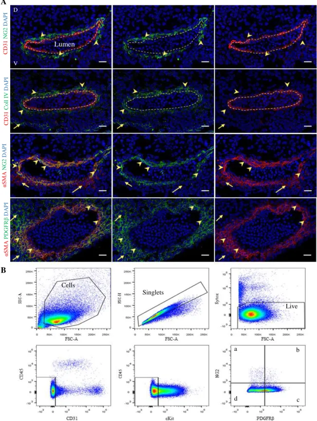
Frozen sections of E11 wild-type (WT) embryos were stained by immunohistochemistry with different marker combinations to identify and characterise perivascular mesenchymal stromal cells (PV-MSCs) that surround the dorsal aorta at the time of hematopoietic stem cell (HSC) generation. The following markers were used to detect perivascular cells - NG2, PDGFRβ and αSMA -, endothelial cells - CD31- and basement membrane - Collagen IV (Figure 1A). We identified the aorta lumen delimited by endothelial cells expressing CD31 (dotted line), and close to the endothelium, NG2+ perivascular cells (arrowheads) surrounding the aorta (first row). In the second row, the basement membrane expressing Collagen IV (arrowheads) surrounds the dorsal aorta. Perivascular cells expressing αSMA are proximal to the aorta (third row). They co-express NG2 (arrow heads). Few αSMA+
cells that do not express NG2 can be also observed (arrows). The fourth combination includes αSMA and PDGFRβ, were cells expressing both markers are identified by the arrowheads; they are further surrounded by cells expressing only PDGFRβ (arrows). Altogether, these data show that pericytes can be identified as being NG2+PDGFRβ+αSMA+. They closely surround CD31+ endothelial cells with whom they share the same basement membrane. This was further confirmed by immunolocalization of both Collagen IV and αSMA (Supplementary Figure 1, second column, arrowheads). Our laboratory and others also identified CD146 as pericyte marker in both human and mouse organs. I tested this marker on frozen sections of E11 AGM and confirmed that indeed, CD146is alsoperivascular in the mouse embryo (Supplementary Figure 1, first column, arrowheads). We next performed immunostaining using the hematopoietic marker CD45. Our data show that CD45+ cells are present in the mesenchyme that surrounds the endothelium (third column, Supplementary Figure 1 arrowheads).
To further characterize and quantify the variety of vascular and perivascular cells that surround the dorsal aorta, we performed flow cytometry analysis using the surface marker combinations we defined by immunohistochemistry. We combined perivascular cell markers (NG2, PDGFRβ), endothelial and hematopoietic cells marker (CD31) and hematopoietic markers (CD45, C-kit). In the Figure 1B we show an example of the gating used to define the perivascular cell subsets. Populations of cells of interest are selected, along with single and viable cells, from those, hematopoietic and endothelial markers are discarded (CD45, C-kit and CD31), remaining perivascular cell markers, where four different populations can be identified: NG2+PDGFRβ- cells, pericytes NG2+PDGFRβ+, sub-pericytes NG2-PDGFRβ+ and other stromal cells NG2-PDGFRβ-. These data clearly demonstrate that perivascular cells are phenotypically distinct.
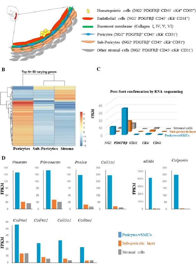
We next wanted to know whether these cells are also genetically different. To address this question, we isolated these perivascular cells based on these markers (Figure 2A) and performed RNA sequencing. We found that the three layers of perivascular cells have distinct transcriptomic profiles (Figure 2B). A post-sorting analysis was made and shows that the distinct cell populations analysed are not contaminated by other markers (Figure 2C). Our data further confirm that our choice of antibodies to isolate these cells is correct. Indeed, the NG2+PDGFRβ+CD31-CD45-C-kit- purified cells are enriched in genes expected to be found in pericytes (Figure 2D). They are known to express genes encoding structural proteins such as the Vimentin and also cell adhesion, growth, migration and differentiation (Fibronectin), along with proteins important for the formation of the extra cellular matrix and the basement membrane (Pcolce and Coll1a1, Col4 a1, Coll4a2, Coll5a1 and Coll6a1). These cells are also enriched in αSMA and Calponin genes involved in the structure and integrity of the blood vessels.
Figure 1. E11 AGM is surrounded by distinct perivascular cell subsets. (A) Immunohistochemistry of E11 WT AGM
(40-42 sp) cryo-sections with different marker combinations, allowing to distinguish different vascular and perivascular cell subsets: CD31 and NG2; CD31 and Coll IV; αSMA and NG2; αSMA and PDGFRβ. DAPI stains all nuclei (blue). In the first two rows CD31+ cells are identified with a dotted line that delimits the dorsal aorta, in order to locate the lumen (N=1). D: Dorsal and V: Ventral side of the embryo. Scale bars: 20µm. (B) Example of a flow cytometry gating strategy used to identify different perivascular cell subsets that surround the wild-type AGM at day 11 of development. Populations of interest were selected in the first gate, following by single cells and live cells. Endothelial and hematopoietic markers were next excluded (CD31-CD45-C-kit-). Within this gate, different cell populations were identified: a) NG2+PDGFRβ-; b)
NG2+PDGFRβ+ c) NG2-PDGFRβ+ and d) NG2-PDGFRβ-. a b c d Cells Singlets Live D V C D 3 1 C o ll I V D A P I Lumen A B C D 3 1 N G 2 D A P I αS M A N G 2 D A P I αS M A PD G FR β D A P I
Figure 2. Purified perivascular cells from E11 WT AGM are phenotypically and genetically distinct. (A)
Schematic representation of part of the dorsal aorta. Hematopoietic and endothelial cells are shown. In contact with endothelial cells are pericytes that are embedded in the basement membrane, followed by a sub-pericytic layer of PDGFRβcells and other stromal cells. (B) Heatmap showing distinct expression patterns of pericytes, sub-pericytic PDGFRβ cells and stroma for top 50 varying genes obtained from RNA sequencing. (C) RNA sequencing showing the separation efficiency of E11 WT AGM sub-populations (D). Selected genes involved in the basement membrane formation are enriched in the pericyte/vascular smooth muscle cell fraction thus validating their cell identity and location (N=1).
A
B C
9
Hematopoietic progenitor activity in the PDGFRβ- / - AGM is impaired
Perivascular cells, proximal to the blood vessel wall, express PDGFRβ. We aim here to investigate whether this receptor plays a role in the aortic haematopoiesis. We used a mouse model where PDGFRβ is knock-out.
First, as proof of the model, immunohistochemistry was performed in sections of PDGFRβ-/- E11 embryos as well as on the PDGFRβ+/+ wild-type control. As it is shown in figure 3A, in the the WT embryos, CD31+ endothelial cells (dotted line) are surrounded by PDGFRβ+ cells (arrows), whilist on the section of the PDGFRβ-KO embryo, PDGFRβ expression is absent. The same pattern can be seen with the immunostaining of αSMA and PDGFRβ in Supplementary Figure 2 (third row). The absence of PDGFRβ was further confirmed by flow cytometry analysis (Figure 3B).
Embryos from this mouse line were harvested and all E11 hematopoietic organs – placenta, yolk sac, head, fetal liver and AGM – were separated (Figure 3C). Two independent experiments were performed. Our data show that in the AGM, the total number of colonies per tissue (CFU-C) is affected, decreasing significantly in PDGFRβ-/- embryos in comparison with
wild type (Figure 3D). There is also a significant decrease between PDGFRβ+/+ and PDGFRβ +/-hematopoietic progenitor numbers. This difference in the total number of progenitors is due to the variation between the number of colonies forming unit of granulocytes (CFU-G) and, more immature progenitors responsible for the formation of granulocytes, erythrocytes, monocytes and megakaryocytes (CFU-GEMM), where AGM PDGFRβ-/- show lower numbers. Other progenitors, such as burst-forming unit erythroid (BFU-E), macrophages (CFU-M) and granulocyte-macrophages (CFU-GM) are not significantly affected at this stage of development. In other hematopoietic organs – placenta, yolk-sac, head and fetal liver - there are no significant differences between the total number CFU-C per tissue, neither between each individual hematopoietic progenitor type at E11 (Supplementary Figure 3) suggesting a tissue-dependent role of PDGFRβ to control hematopoiesis at this developmental stage.
PDGFRβ is required to generate long-term aortic HSCs
To test if the lack of PDGFRβ expression affects the generation of the first hematopoietic stem cells in vivo, E11 AGM cells harversted from wild-type and mutant embryos were injected into sub-lethaly irradiated primary recipient mice. Four month later, reconstituted primary recipient mice were further tested in secondary transplantation (Figure 4A). In both primary and secondary transplantated mice, the peripheral blood is used to confirm the presence or the absence of donor hematopoietic cells that can be identified with CD45.1and CD45.2 antibodies (Supplementary Figure 4). Our data show that, compare to the positive control (PDGFRβ+/+ transplanted AGMs) in which 6 out of 24 mice reconstituted, mice injected with PDGFRβ+/- or PDGFRβ-/- AGMs showed low (1/9) or no reconstitution (0/4) over the long-term (Figure 4B). Data from the peripheral blood analysis of the secondary transplanted recipient mice, show that both WT (3/6) and heterozygous (2/2) derived donor cells self-renew. More experiments are ongoing and are expected to give us insight of the role of these cells in HSC generation in vivo.
10
Figure 3. AGM derived hematopoietic progenitor numbers are affected in PDGFRβ-/- embryos. (A)
Immunohistochemistry of the dorsal aorta from WT and KO E11 embryos. Endothelial cells (CD31+, dotted line) are surrounded by PDGFRβ+
cells (arrows) in WT embryos, while PDGFRβ expression is absent in the KO embryos (N=1). Scale bar: 20μm. (B) Flow cytometry analysis of WT (+/+), heterozygous (+/-) and KO (-/-) AGMs for PDGFRβ. * p <0.05, ****p <0.00001 (N=2, WT=19; Het=21; KO=15, 42-46sp) (C) Schema of Hematopoietic Progenitors assay. AGM is dissected from E11 embryos, digested and cultured in methylcellulose. Each AGM is seeded in three plates with a ratio of 0.33ee/dish. After 10-12 days of incubation at 37ºC, different progenitor colonies can be identified and counted in the microscope by their different morphologic features. (D) Colony number per embryo equivalent (CFU-C/ee) from AGM region is shown, where different colony types were distinguished: BFU-E, burst-forming unit erythroid; CFU-G, (colony forming unit) granulocytes; CFU-M, macrophages; CFU-GM, granulocyte-macrophages; CFU-GEMM, granulocytes, erythroid, macrophages, megakaryocytes. The data represent the mean ± SEM * p <0.05, **p <0.001, ***p <0.0001 (N=2) (WT=10; Het=6; KO=3, 40-42sp).
A D C C D 3 1PD G FR βD A P I PDGFRβ +/+ PDGFRβ -/- B
11
Figure 4. PDGFRβ is required to generate HSCs in the AGM in vivo. (A) Simplified representation of HSCs
transplantation assay. AGM is collected, digested into single cells and injected into a sub-lethal irradiated mouse – primary transplantation. Four and 16 weeks after injections, few drops of peripheral blood (PB) are collected and analysed. Bone marrow of primary reconstituted mice is harvested and injected into two secondary sub-lethal irradiated recipients, from which PB is analysed. Percentage of donor chimerism 4 and 16 weeks after primary (B) and secondary (C) transplantation is analysed by flow cytometry. (B and C) show the percentage donor chimerism found in transplanted mice over total number of mice injected (N=4). Dotted base line represents the limit of reconstitution. Recipients with donor chimerism >4% in the PB are considered reconstituted.
The integrity of the dorsal aorta and intra-aortic hematopoietic clusters are not affected in PDGFRβ- / - AGM
We next wanted to test whether the absence of HSCs in mutant AGMs was due to an abnormal formation of the dorsal aorta. To this end, we analysed the full aorta using three-dimensional (3D) whole mount immunostaining technology on E10.5 old mouse embryos. Endothelial and pericytes/vascular smooth muscle cells were stained with CD31 and αSMA, respectively (Figure 5A). The dorsal aorta, the intersomitic vessels and umbilical and vitelline arteries appear to be normal in both WT and KO embryos as shown by CD31 expression (in the KO embryo vitelline and umbilical arteries are disrupted due to the embryo manipulation during the assay). In cyan, C-kit expressing hematopoietic cells can be identified. Intra-aortic hematopoietic clusters co-express C-kit and CD31 (Supplementary Figure 5) with no clear differences between PDGFRβ+/+ and PDGFRβ-/- embryos. However, we could note that hematopoietic clusters are more dispersed in the WT than in the KO embryos, where they look more compacted between each other.
To support this immunostaing results, we next quantified hematopoietic and vascular cells by flow cytometry and confirmed that the percentage of both endothelial cells and HPSCs does not change in mutant AGM compare to control (Figure 5B). The percentage of cells expressing CD31, CD45, C-kit, NG2, PDGFRα were also analysed (Supplementary Figure 6, 7). We found that, endothelial and hematopoietic cell frequencies are not affected in the mutant AGMs. We next tested whether PDGFRα expression change in these mutants and found that, by flow cytometry, there are no clear differences between the embryos.
A
12
Figure 5. Blood vessel integrity and intra-aortic hematopoietic clusters (IAHCs) are not affected in PDGFRβ-/- embryos. (A) Confocal images of 3D whole mount stained WT (PDGFRβ+/+) and KO (PDGFRβ-/-) E10.5 embryo
(34 sp). This staining includes markers of endothelial and hematopoietic cells (CD31), perivascular vascular smooth muscle cells (αSMA) and hematopoietic cells (C-kit). In both embryos, it is possible to identify the dorsal aorta (DA), umbilical and vitelline arteries (UVA), along with intersomitic vessels. Scale bar 50 µm. (B) Percentage of cell subsets found in the E11 wild-type (+/+), heterozygous (+/-) and PDGFRβ knock out (-/-) AGMs, analysed by flow cytometry. Percentage of endothelial cells is shown. Hematopoietic progenitors and stem cells, pericytes, sub-pericytes and stromal cells were also analysed. The data represent the mean ± SEM of independent embryos tested (WT=21; Het=19; KO=15, N=6). One-way ANOVA along with Tuckey’s multiple comparison test was used. * p <0.05, sp: 42-46. CD31 αSMA C-kit P DGF R β -/ -P DGFR β + /+ A B DA UVA Intersomitic Vessels
13
The frequency of hemogenic endothelial cells is altered in the absence of PDGFRβ
To understand how PDGFRβ control the generation of hematopoietic stem cells, Runx1 signalling was traced in mutant embryos. For that, a double transgenic mice model was created by breeding PDGFRβ-KO mice with mice from Runx1GFP line (Lorsbach et al., 2004) were embryos PDGFRβ-/-, PDGFRβ-/+ and PDGFRβ+/+ expressed Runx1GFP (Figure 6A). Cell populations were selected from single live cells and the gates were defined based on the unstained sample (Supplementary Figure 7). The absence of PDGFRβ by flow cytometry was first confirmed in the KO embryos (Supplementary Figure 8). Additionally, it is also possible to see a major decrease in pericytes population and, in contrast, a significant increase in the stromal cell population (Supplementary Figure 8). By flow cytometry, we found that the percentage of GFP+ (Runx1) cells is not different in mutant embryos although more experiments need to be done (Figure 6A). The same occurs with the population of non-hemogenic endothelial cells and hematopoietic progenitors and stem cells (Figure 6B). However, there is a significant decrease of the rare population of hemogenic endothelial cells, HSC precursors, in PDGFRβ-/- in comparison with wild-type and heterozygous embryos, suggesting a role for PDGFRβ in the HSC fate.
Figure 6. Hemogenic endothelial cell frequency significantly decreases in the absence of PDGFRβ. (A)
PDGFRβ-KO mice were recombined with Runx1GFP mice and double transgenic mice were used to obtain mouse embryos. All embryos were GFP (PDGFRβ +/+, +/- and -/-). GFP+ cell frequency in the AGM is not affected between embryos with different PDGFRβ genotypes. (B) Hemogenic endothelial cell percentage (HEC, NG2 -PDGFRβ-CD45-C-kit-CD31+Runx1+) decreases significantly in PDGFRβ-/- embryos in comparison with PDGFRβ +/-and PDGFRβ+/+ AGM controls. The non-HECs and HPSC or GFP+HPSC numbers are not affected. The data
represent the mean ± SEM (WT=12; Het =25; KO =18, N=7). One-way ANOVA along with Tuckey’s multiple comparison test was made in order to analyse significance between WT, Het and KO embryos. * p <0.05, **p <0.001. sp: 42-46.
14
Discussion
Understanding the native specification of hematopoietic stem cells in vivo, to uncover pivotal signals that might help improve in vitro directed differentiation protocols, has been a long-standing biomedical goal.The current impossibility of specifying true HSCs in vitro suggests that key signals remain unknown. We speculated that such signals might be presented by surrounding niche cells, but no such cells have been defined. In this study, we aimed to reveal the particular cells from the microenvironment that have a supportive role to generate HSCs, either because they are in direct cell contact with hemogenic endothelial cells or may release decisive fate factors. The microenvironment is highly heterogeneous, and we here showed that there are several cell layers adjacent to the dorsal aorta that can be prospectively isolated and analysed. We found that the proximal layers express PDGFRβ and we used a knock-out model where PDGFRβ is absent to test whether this is required to generate HSCs.
Our preliminary data show that at E11, the dorsal aorta is surrounded by three distinct perivascular cell layers that can be purified to homogeneity: - pericytes/vSMCs (vascular smooth muscle cells) that co-express NG2, αSMA and PDGFRβ, but lack CD31 and CD45 expression, - non-pericyte PDGFRβ+cells (we also called them sub-pericytes being adjacent to pericytes) that do not express NG2 or αSMA nor CD31 or CD45 and - stromal cells which do not express any of these markers. Expression of the basal membrane marker collagen type IV confirmed the pericyte status of the mural cells. Importantly, pericytes do not share markers with intra-aortic hematopoietic clusters co-expressing CD31 and C-kit and lacking αSMA. Our RNA sequencing preliminary data further show that pericytes have a distinct expression pattern compared to other two layers and are highly enriched in genes related to basement membrane formation such as vimentin, fibronectin, αSMA, Coll1a1, Coll4a1, Coll4a2, Coll5a1 and Coll6a1 expected to be found in pericytes. Importantly, we also confirmed the presence of other markers for pericytes that are enriched at the RNA level such as CD146 (MCAM) and nestin which were shown to be involved in the HSC niche in adult (Sacchetti et al., 2007; Ding et al., 2012; Corselli et al., 2013; Morrison et al., 2014).
We next checked whether described associated HSC niche related transcripts found to support adult HSCs are also found in the embryonic generating niche. Interestingly, our RNA sequencing data showed that genes such as Jagged-1 (Notch ligand), N-cadherin, CXCL12, VCam-1 and Opn3, implicated in the maintenance of adult HSCs, are also enriched in embryonic aortic pericytes suggesting a predetermined developmental role for these cells to maintain HSCs. Whether these gene expressions are altered in our PDGFRβ-/- embryos need to be tested. However, in these mice, PDGFRβ is absent which pose difficulties to isolate the different cell subsets and thus, these genes could then be tested by in situ hybridisation on fixed sections from KO animals. Our flow cytometric data suggest that NG2 expression is not affected in KO embryos. However, this was analysed in one experiment and thus, this observation requires further confirmation. Using NG2 as only marker for pericytes would not allow to separate all three perivascular fractions. However, it would be interesting to test whether a combination of markers, other than PDGFRβ such as CD146 or PDGFRα in mutant embryos would allow to separate these perivascular layers for further analysis of stromal cells in PDGFRβ KO embryos. Altogether, these data demonstrate that perivascular cells found in the embryonic HSC generating niche are highly heterogeneous and that perivascular cells proximal to endothelial cells are enriched in hematopoietic niche related genes. Whether these cells are HSC supportive is unknown.
Previous in vivo and in vitro studies showed that PDGF family plays an important role in haematopoiesis, enhancing the production of a variety of factors that act directly in the production of hematopoietic progenitor cells. PDGF was shown to be important in erythropoiesis (Dainiak et al., 1983; Delwiche et al., 1985; Sytkowski et al., 1990; Keutzer and Sytkowski, 1995), promoting the in vitro proliferation of erythropoietic progenitors (Keutzer and Sytkowski, 1995) and in the stimulation of primitive hematopoietic progenitors cells in
15 mixed marrow cultures (Michalevicz, Francis and Price, 1985; Michalevicz et al., 1986; Sytkowski et al., 1990).
Importantly, PDGF signalling was also shown to be involved in HSC specification niche in zebrafish model (Damm and Clements, 2017). We tested whether this is the case in the mouse embryo. We found that some hematopoietic progenitor types including the most immature ones were affected. This led us to hypothesise that HSC themselves will be impaired. Indeed, we found that HSCs are completely absent in the AGM of PDGFRβ-/- embryos. This is not due to alteration of the blood vessel structure. Using sophisticated technology to visualise the full aorta by confocal imaging and quantification of vascular cells by flow cytometry, we here demonstrated that the blood vessel is intact. Altogether, these data show that PDGFRβ plays a key role in the generation of the first adult-type HSCs in the mouse embryo in vivo. However, the mechanism remains unclear.
PDGFRβ may be involved during the hemogenic endothelium specification prior to HSC generation or at later stages during endothelial to hematopoietic transition after the hemogenic endothelial cells are already in place. Hemogenic endothelial cell phenotype was defined previously (Chen et al., 2009). They express Runx1 GFP besides other markers. To answer this mechanistic question, we recombined the PDGFRβ mice with Runx1GFP mice. In these mice, when Runx1 is expressed, GFP is also expressed. This allow to compare hemogenic endothelial cell phenotype and number between WT and PDGFRβ mutant embryos. Our data show that in the absence of PDGFRβ, hemogenic endothelial cell number significantly decreases, whilst the non-hemogenic endothelial cell number remains unchanged. There are in vitro studies showing that in the presence or absence of vascular endothelial growth factor (VEGF), cells from the blastocyst form endothelial or hematopoietic cells, respectively (Jaffredo et al., 2005). A different study shows that PDGF family members are closely related with VEGF (Ball et al, 2007). It would be interesting to understand if VEGF regulation is correlated with PDGFRβ and whether this changes in the PDGFRβ mutants. Our data here suggest that PDGFRβ plays a role to control HSCs prior to their birth.
Future directions
It is important to understand the identity, the origin and the biology of PDGFRβ+ cells that are important for HSCs in vivo. Which of the PDGFRβ+ cells are required? Pericytes or sub-pericytes or both? Is the cell contact with endothelial cells required? Which factors do they release? Are they hematopoietic, hematopoietic precursors or are they acting as only niche cells? Ongoing work in the lab aims to address these questions. Preliminary data show that these cells are not hematopoietic. Indeed, purified PDGFRβ+ cells from the AGM do not contain hematopoietic progenitors when seeded in methylcellulose assay. To test whether PDGFRβ+ cells are hematopoietic precursors we will recombine PDGFRβ-Cre transgenic mice with tdTomato floxed mice and sorted Tomato+ cells will be tested in hematopoietic assays. To answer whether PDGFRβ+ cells are niche cells, we will co-culture them with OP9 bone marrow supportive stromal cell line and endothelial cells and test their ability to contribute to HEC maturation toward various hematopoietic lineages both in vivo and in vitro.
Conclusions
In conclusion, our results, although preliminary at this stage, define PDGFRβ signalling as key component of the HSC specification niche in vivo that can be tested in vitro to derive HSCs from hematopoietic and non-hematopoietic cell sources for cell therapy to treat patients with blood diseases.
16
References
Allt, G. and Lawrenson, J. G. (2001) ‘Pericytes: Cell biology and pathology’, Cells Tissues Organs, 169(1), pp. 1–11. doi: 10.1159/000047855.
Armulik, A., Abramsson, A. and Betsholtz, C. (2005) ‘Endothelial/pericyte interactions’, Circulation Research, 97(6), pp. 512–523. doi: 10.1161/01.RES.0000182903.16652.d7.
Armulik, A., Genové, G. and Betsholtz, C. (2011) ‘Pericytes: Developmental, Physiological, and Pathological Perspectives, Problems, and Promises’, Developmental Cell, 21(2), pp. 193– 215. doi: 10.1016/j.devcel.2011.07.001.
Ball, S. G., Shuttleworth, C. A. and Kielty, C. M. (2007) ‘Vascular endothelial growth factor can signal through platelet-derived growth factor receptors’, The Journal of Cell Biology, 177(3), pp. 489–500. doi: 10.1083/jcb.200608093.
Boisset, J. C. et al. (2010) ‘In vivo imaging of haematopoietic cells emerging from the mouse aortic endothelium’, Nature. Nature Publishing Group, 464(7285), pp. 116–120. doi: 10.1038/nature08764.
Bruijn, M. De and Dzierzak, E. (2017a) ‘Runx transcription factors in the development and function of the de fi nitive hematopoietic system’, Blood, 129(15), pp. 2061–2070. doi: 10.1182/blood-2016-12-689109.deletion.
Bruijn, M. De and Dzierzak, E. (2017b) ‘Runx transcription factors in the development and function of the definitive hematopoietic system’, Blood, 129(15), pp. 2061–2070. doi: 10.1182/blood-2016-12-689109.deletion.
Cai, Z. L. et al. (2000) ‘Haploinsufficiency of AML1/CBFA2 affects the temporal and spatial generation of hematopoietic stem cells in the mouse embryo’, Immunity, 13, pp. 423–431.
Chen, M. J. et al. (2009) ‘Runx1 is required for the endothelial to hematopoietic cell trasition but not thereafter’, Nature, 457(7231), pp. 887–891. doi: 10.1038/nature07619.Runx1.
Corselli, M. et al. (2013) ‘Perivascular support of human hematopoietic stem / progenitor cells’, Blood, 121(15), pp. 2891–2901. doi: 10.1182/blood-2012-08-451864.
Crisan, M. et al. (2008) ‘A Perivascular Origin for Mesenchymal Stem Cells in Multiple Human Organs’, Cell Stem Cell, 3(3), pp. 301–313. doi: 10.1016/j.stem.2008.07.003.
Crisan, M. et al. (2015) ‘BMP signalling differentially regulates distinct haematopoietic stem cell types’, Nature Communications. Nature Publishing Group, 6, pp. 1–8. doi: 10.1038/ncomms9040.
Crisan, M. et al. (2016) ‘BMP and Hedgehog Regulate Distinct AGM Hematopoietic Stem Cells Ex Vivo’, Stem Cell Reports. Elsevier Ltd, 6(3), pp. 383–395. doi: 10.1016/j.stemcr.2016.01.016.
Crisan, M. and Dzierzak, E. (2016) ‘The many faces of hematopoietic stem cell heterogeneity’, Development, 144(22), pp. 4195–4195. doi: 10.1242/dev.160812.
Cumano, A., Dieterlen-Lievre, F. and Godin, I. (1996) ‘Lymphoid potential, probed before circulation in mouse, is restricted to caudal intraembryonic splanchnopleura’, Cell, 86(6), pp. 907–916. doi: 10.1016/S0092-8674(00)80166-X.
17 Cumano, A. and Godin, I. (2007) ‘Ontogeny of the Hematopoietic System’, Annual Review of Immunology, 25, pp. 745–785. doi: 10.1146/annurev.immunol.25.022106.141538.
Dainiak, N. et al. (1983) ‘Platelet-derived growth factor promotes proliferation of erythropoietic progenitor cells in vitro.’, The Journal of clinical investigation, 71(5), pp. 1206–14. doi: 10.1172/JCI110869.
Damm, E. W. and Clements, W. K. (2017) ‘Pdgf signalling guides neural crest contribution to the haematopoietic stem cell specification niche’, Nature Cell Biology, 19(5), pp. 457–467. doi: 10.1038/ncb3508.
Delwiche, F. et al. (1985) ‘Platelet-derived growth factor enhances in vitro erythropoiesis via stimulation of mesenchymal cells.’, The Journal of clinical investigation, 76(1), pp. 137–42. doi: 10.1172/JCI111936.
Dias Moura Prazeres, P. H. et al. (2017) ‘Pericytes are heterogeneous in their origin within the same tissue’, Developmental Biology. Elsevier Inc., 427(1), pp. 6–11. doi: 10.1016/j.ydbio.2017.05.001.
Ding, L. et al. (2012) ‘Endothelial and perivascular cells maintain haematopoietic stem cells’, Nature. Nature Publishing Group, 481(7382), pp. 457–462. doi: 10.1038/nature10783.
Durand, C. et al. (2007) ‘Embryonic stromal clones reveal developmental regulators of definitive hematopoietic stem cells.’, Proceedings of the National Academy of Sciences of the United States of America, 104(52), pp. 20838–43. doi: 10.1073/pnas.0706923105.
Durand, C., Robin, C. and Dzierzakt, E. (2006) ‘Mesenchymal lineage potentials of aorta-gonad-mesonephros stromal clones’, Haematologica, 91(9), pp. 1172–1179.
Dzierzak, E. and Bigas, A. (2018) ‘Blood Development: Hematopoietic Stem Cell Dependence and Independence’, Cell Stem Cell. Elsevier Inc., 22(5), pp. 639–651. doi: 10.1016/j.stem.2018.04.015.
Ema, H. and Nakauchi, H. (2000) ‘Expansion of hematopoietic stem cells in the developing liver of a mouse embryo’, Blood, 95(7), pp. 2284–2289.
Frame, J. M., McGrath, K. E. and Palis, J. (2013) ‘Erythro-myeloid progenitors: “Definitive” hematopoiesis in the conceptus prior to the emergence of hematopoietic stem cells’, Blood Cells, Molecules, and Diseases. Elsevier Inc., 51(4), pp. 220–225. doi: 10.1016/j.bcmd.2013.09.006.
Gekas, C. et al. (2005) ‘The placenta is a niche for hematopoietic stem cells’, Developmental Cell, 8(3), pp. 365–375. doi: 10.1016/j.devcel.2004.12.016.
Godin, I. et al. (1999) ‘Stem cell emergence and hemopoietic activity are incompatible in mouse intraembryonic sites.’, The Journal of experimental medicine, 190(1), pp. 43–52. doi: 10.1084/jem.190.1.43.
Godin, I., Dieterlen-Lièvre, F. and Cumano, a (1995) ‘Emergence of multipotent hemopoietic cells in the yolk sac and paraaortic splanchnopleura in mouse embryos, beginning at 8.5 days postcoitus.’, Proceedings of the National Academy of Sciences of the United States of America, 92(3), pp. 773–777. doi: 10.1073/pnas.92.3.773.
Hellström, M. et al. (1999) ‘Role of PDGF-B and PDGFR-beta in recruitment of vascular smooth muscle cells and pericytes during embryonic blood vessel formation in the mouse.’, Development (Cambridge, England), 126(14), pp. 3047–3055.