Inês Tavares Pinto de Sá Pereira
Licenciatura em Biologia
Basement membrane alterarions in
kernicteric brain microvasculature and
pericyte response to bilirubin
Dissertação para obtenção do Grau de Mestre em Genética Molecular e Biomedicina
Orientador: Maria Alexandra de Oliveira Silva Braga Pedreira
de Brito, Professora Auxiliar, Faculdade de Farmácia,
Universidade de Lisboa
Co-orientador: Dora Maria Tuna de Oliveira Brites,
Investigadora Coordenadora e Professora Catedrática
Convidada, Faculdade Farmácia, Universidade de Lisboa
Júri:
Presidente: Prof. Doutora Paula Maria Theriaga Mendes Bernardo Gonçalves
Arguente: Doutora Isa Domingues Serrano Fernandes Vogal: Prof. Doutora Maria Alexandra de Oliveira Silva Braga
Pedreira de Brito
Basement membrane alterations in kernicteric brain microvasculature and periyte response to bilirubin
Copyright Inês Tavares Pinto de Sá Pereira, FCT/UNL, UNL
Two publications are in preparation, ensuing from the work developed in this thesis
Sá Pereira I, Brites D, Brito MA.
Neurovascular unit: a focus on pericytes (Review)
Sá Pereira I, Fernandes A, Palmela I, Brites D, Brito MA.
Unconjugated bilirubin induces apoptotic cell death, cytokine secretion and nitrosative stress in human brain vascular pericytes
ACKNOWLEDGMENTS
Começo por agradecer à Profª Doutora Dora Brites pela possibilidade e oportunidade de realizar este trabalho, o qual me fez iniciar a minha carreira na investigação. Agradeço-lhe também pelo conhecimento, ideias e rigor que me transmitiu ao longo do ano.
À Professora Doutora Alexandra Brito um enorme obrigada pelo constante acompanhamento, por todas as dúvidas tiradas, pelo conhecimento e incentivo que me transmitiu ao longo de todo este ano. Um obrigada pelo o apoio e por nunca me ter deixado desamparada.
I would like to express my gratitude to Professor Eleonora Aronica for the brain sections that sent me whenever necessary. These were essencial for my research work.
Quero agradecer também ao Pedro por toda ajuda que me deu. Obrigada Pedro, pela insistência, por todo o trabalho e tempo que dispensaste e pela boa companhia.
À Adelaide agradeço a constante ajuda, disponibilidade e por me ter introduzido no trabalho de laboratório. Obrigada pela paciência e pelas dúvidas tiradas. És sem dúvida uma grande investigadora, um exemplo a seguir.
À Inês, um obrigada pelas horas no quentinho do fluxo e por toda a ajuda que me deste ao longo do trabalho.
Ao Professor Doutor Rui Silva agradeço a disponibilidade para esclarecer as dúvidas que foram surgindo.
Um beijinho especial para as restantes meninas do grupo. Obrigada pelos sorrisos, pela boa disposição e por todo o apoio que me deram ao longo do ano. Em especial: obrigada Filipa pelo carinho e por seres tão querida e Cátia pelo acompanhamento nesta nossa viagem.
À Ana nem tenho palavras para agradecer. Obrigada por tudo! Tornaste-te na amiga que nunca mais quero perder! Adorei todos os nossos momentos. Obrigada por toda a partilha e pelos momentos de diversão, de loucura e de desespero.Obrigada pelo teu apoio e ajuda essenciais ao meu trabalho. Vou voltar para o porto contigo no coração e com a certeza que este ano foi só o começo.
Ao Mi, agradeço os sorrisos, os “então cala-te” e as conversas. Obrigada pela tua boa companhia.Take my nose that I take yours!
Aos restantes membros do tão refrescante clube chamado cave, agradeço o bom ambiente e as pausas tão saudáveis essenciais ao meu bem-estar e ao meu trabalho.
Quero também agradecer a todos os meus amigos do Porto pelo constante interesse sobre o meu trabalho e pelos poucos mas bons momentos que vivemos aos f.d.s e que me ajudaram sempre a ganhar forças para continuar. Em especial ao Miguel, barboso, um beijinho e um obrigada pelo carinho, apoio e por nunca me deixares ir a baixo.
À minha melhor amiga Isabel agradeço o amor incondicional e a companhia. Obrigada pelo apoio, pelo ombro sempre disponível nos momentos mais difíceis e pela força que sempre me deste e que me ajudou a enfrentar todos os obstáculos.Os momentos foram poucos, mas únicos.
Aos eternos três, Maria, Xano e Loni, agradeço a amizade que nunca falhou. Obrigada pelo constante interesse e pelos momentos que me fortaleceram.
A toda a minha família que eu adoro obrigada pelo apoio constante e pelo incentivo. Avó, obrigada por olhares por mim e por teres sido a minha companhia nos momentos mais solitários.
Agradeço-te também Zé por toda a ajuda e apoio que me deste em Lisboa e por estares sempre disponível para me “aturar”. Um beijinho especial para as minhas irmãs do coração, Ana e Joana. Obrigada pelas várias visitas e pelo carinho que soube sempre tão bem.
ix
ABSTRACT
Kernicterus is a neuropathological condition characterized by deposition of unconjugated bilirubin (UCB) in specific brain regions that can lead to permanent sequelae and death, particularly in premature infants. UCB-induced toxicity has been studied in nerve and glial cells and, more recently, in brain microvascular endothelial cells. However, the effects of UCB on pericytes or on the basement membrane were never reported.
We performed in vitro studies to assess apoptotic death, nitrosative stress and inflammatory reaction elicited by human brain vascular pericytes exposed to UCB. We also assessed the basement membrane component, collagen type IV, in brain sections of cortex, basal nuclei, hippocampus and cerebellum, collected at autopsy of a kernicteric preterm newborn.
Using the pericyte marker, α-smooth muscle actin, we characterized the cells and confirmed the
normal outgrowth towards a typical morphology with long processes. UCB induced an early secretion of interleukin-6, followed by that of vascular endothelial growth factor. mRNA upregulation preceded the secretion and confirmed the precocious profile of IL-6. UCB also caused the release of nitrites, which was maximum at 72 h incubation. The earlier upregulation of endothelial nitric oxide synthase expression confirmed the induction of nitric oxide production by UCB, although not excluding that other isoforms of the enzyme are also involved. Probably as a corolary of all these events, apoptotic cell death occurs in a time- and concentration-dependent manner. Through immunohistochemistry we examined the area occupied and the immunoreactivity of collagen type IV, which were reduced in the kernicterus case as compared with a non-icteric control.
These findings are the first to demonstrate the compromise of pericytes and the impairment of collagen IV by hyperbilirubinemia and raise some basis for creation of possible target-directed therapy against pericyte and basement membrane damages as a result of UCB exposure.
xi
RESUMO
Kernicterus é uma condição neuropatológica caracterizada pela deposição de bilirrubina não conjugada (BNC) em regiões específicas do encéfalo que pode resultar em danos permamentes e na morte, especialmente em recém-nascidos prematuros. A toxicidade da BNC já foi estudada em células nervosas e da glia e, mais recentemente, em células endoteliais da microvasculatura do cérebro. No entanto, os efeitos da BNC em pericitos e na membrana basal nunca foram descritos.
Neste trabalho, avaliámos a morte por apoptose, stress oxidativo e reacção inflamatória de pericitos humanos expostos à BNC. Avaliámos também um dos componentes da membrana basal, o colagénio tipo IV, em cortes humanos de cortex, núcleos da base, hipocampo e cerebelo provinientes de uma autópsia de um recém-nascido permaturo com kernicterus.
Usando α-actina do músculo liso como marcador de pericitos, procedemos à sua caracterização e confirmámos um crescimento normal no sentido de uma morfologia típica com longos prolongamentos. A BNC induziu a secreção precoce de interleucina-6 e mais tardia do factor de crescimento endotelial vascular. A sobrexpressão do mRNA antes da libertação confirmou ambos os perfis de secreção. A BNC levou também à libertação de nitritos, atingindo valores máximos às 72 h de incubação. A sobrexpressão precoce da enzima endotelial de síntese do óxido nítrico comprovou a indução da produção do mesmo. Porém, não podemos excluir o facto de que outras isoformas da enzima poderão estar envolvidas. Provavelmente como uma sequência destes eventos, a morte por apoptose ocorreu de uma maneira dependente do tempo e da concentração. Através de uma análise imunohistoquímica do caso de kernicterus, observámos uma redução da área ocupada e da intensidade do colagénio tipo IV, comparativamente a um caso controlo não ictérico.
Estes resultados são os primeiros a demonstrar o comprometimento dos pericitos e do colagénio tipo IV causado por hiperbilirrubinémia e a levantar algumas bases para a criação de uma possível terapia direccionada a alvos contra danos nos pericitos e na membrana basal resultantes da exposição à BNC.
xiii
TABLE OF CONTENS
ABBREVIATIONS ... xvii
I. Introduction ... 1
1.1. Endothelial cells ... 5
1.2. Basement membrane ... 7
1.3. Neurons ... 10
1.4. Astrocytes ... 10
1.5. Microglia ... 10
2. Pericytes ... 11
2.1. Characteristics of pericytes ... 11
2.2. Functions ... 13
2.2.1. Contribution to BBB properties ... 13
2.2.2. Participation in vascular development ... 15
2.2.3. Contractile function ... 17
2.2.4. Immune and phagocytic function ... 19
2.2.5. Roles on hemostasis ... 20
2.2.6. Multipotent cells ... 20
3. Neurovascular unit pathology ... 21
3.1. Involvement of pericytes in BBB dysfunction ... 21
3.2. Bilirubin neurotoxicity ... 23
3.2.1. Hyperbilirubinemia ... 23
3.2.2. Acute bilirubin encephalopathy vs. kernicterus or chronic bilirubin encephalopathy 23 3.2.3. Effects of unconjugated bilirubin in the neurovascular unit ... 23
4. Aims ... 24
II. Materials and methods ... 25
1. Materials ... 27
2. In vitro studies – pericytes ... 27
2.1. Primary culture ... 27
xiv
2.3. Characterization... 28
2.4. Assessment of apoptosis ... 29
2.5. Quantification of Cytokine Release ... 29
2.6. Measurement of Cytokine mRNA Expression ... 29
2.7. Quantification of Nitrite Levels ... 30
2.8. Evaluation of eNOS Expression ... 30
3. Ex vivo studies – Basement membrane ... 30
3.1. Subjects ... 30
3.2. Immuhistochemistry ... 31
4. Statistical analysis ... 32
III. Results ... 33
1. In vitro studies – Pericytes ... 35
1.1. Characterization... 35
1.2. Assessment of apoptosis ... 35
1.3. Quantification of cytokine release... 35
1.4. Measurement of Cytokine mRNA Expression ... 38
1.5. Quantification of Nitrite Levels and Evaluation of eNOS Expression ... 39
2. Ex vivo studies – Basement membrane ... 41
IV. Discussion ... 45
xv
INDEX OF FIGURES
Figure I. 1: The discovery of the blood-brain barrier. ... 3
Figure I. 2: Scheme of the main roles of the blood-brain barrier. ... 4
Figure I. 3: Schematic representation of the neurovascular unit. ... 5
Figure I. 4: The main characteristics of endotheial cells and their junctional complexes. ... 7
Figure I. 5: Basement membrane of human hippocampus sections. ... 8
Figure I. 6: Simplified representation of the blood-brain barrier focusing on the basement membrane. 9 Figure I. 7: Original draw of pericytes by Rouget in 1873. ... 11
Figure I. 8: Schematic representation of a pericyte ensheathing a blood vessel. ... 12
Figure I. 9: Double labeling immunofluorescence analysis of endothelial cells and pericytes in human hippocampus. ... 13
Figure I. 10: The main features of pericytes that determine the blood-brain barrier properties ... 15
Figure I. 11: Factors and receptors produced by pericytes that contribute to angiogenesis. ... 17
Figure I. 12: Morphological features of human brain perivascular pericytes. ... 18
Figure I. 13: Pericytes properties that turn them into cells with contractile functions. ... 19
Figure I. 14: Characteristics of pericytes that are responsible for their immune and phagocytic role... 19
Figure I. 15: Pericytes have the capacity to regulate blood clotting through a pro-and anti-coagulant activity. ... 20
Figure I. 16: Pericytes can differentiate into osteoblast, chondroblast, fibroblast, adipocytes and smooth muscle cells. ... 21
Figure I. 17: Dysfunction of the blood-brain barrier. ... 22
Figure I. 18: Some effects of unconjugated bilirubin in central nervous system cells. ... 24
Figure II. 1: Schematic representation of the pericytes primary culture and UCB treatment. ... 28
Figure II. 2: ImageJ analysis of vessels stained for collagen type IV. ... 32
Figure III. 1: Characterization of human brain vascular pericytes in primary culture. ... 36
Figure III. 2: Apoptosis of human brain vascular pericytes. ... 37
Figure III. 3: Secretion of VEGF and IL-6 by human brain vascular pericytes exposed to UCB. ... 38
Figure III. 4: mRNA expression of VEGF and IL-6 by human brain vascular pericytes exposed to UCB ... 39
Figure III. 5: Nitrite production by human brain vascular pericytes exposed to UCB. ... 40
Figure III. 6: Expression of eNOS by human brain vascular pericytes exposed to UCB. ... 41
Figure III. 7: Immunohistochemistry for collagen type IV. ... 42
Figure III. 8: Area per vessel occupied by collagen type IV. ... 43
Figure III. 9: Collagen type IV immunostaning intensity. ... 43
xvii
ABBREVIATIONS
α-SMA α-smooth muscle actin
ABC ATP-binding cassette Ang angiopoietin
AJ adherens junction BBB blood-brain barrier
bFGF basic fibroblast growth factor
BM basement membrane
BMVEC brain microvessel endothelial cell BSA bovine serum albumin
CAM cell adhesion molecule CNS central nervous system
DMEM Dulbecco’s modified Eagle’s medium ECM extracellular matrix
ECs endothelial cells
eNOS endothelial nitric oxide synthase FBS fetal bovine serum
Glut-1 glucose transporter-1
HBVP human brain vascular pericytes HSA human serum albumin
IL interleukin
JAM junctional adhesion molecule LPS lipopolysaccharide
MHC major histocompatiblity complex MMP matrix metalloproteinase
MRP multidrug resistance-associated proteins Na-F sodium fluorescein
NG-2 nerve Glial-2 NO nitric oxide
PAI-1 plasminogen activator inhibitor-1 PBS phosphate buffered saline PDGF-β platelet-derived growth factor-β
PDGFR-β platelet-derived growth factor-β receptor
Pe transendothelial permeability coefficient P-gp P-glycoprotein
PN-1 protease nexin-1
RT-PCR real-time Polymerase chain reaction S1P sphingosine-1-phosphate
xviii
TGF-β transforming growth factor-β
TJ tight junction
tPA tissue plasminogen activator UCB unconjugated bilirubin
VEGF vascular endothelial growth factor
3
I.
INTRODUCTION
1. Neurovascular unit
It was in 1885, that Paul Ehrlich injected vital dyes into the circulatory system and observed that all organs except the brain and spinal cord were stained (Ehrlich, 1885). Latter on, Edwin Goldmann, an Ehrlich’s student, injected trypan blue into cerebro-spinal fluid and noticed that it only stained the central nervous system (CNS) (Goldmann, 1913) (Fig. I.1). These evidences pointed to the existence of a barrier separating the CNS and the circulation that was named bluthirnschranke (blood-brain barrier, BBB) by Lewandowsky (1900) who further noticed the absence of pharmacological actions of bile acids and ferrocyanide in the CNS. However, this barrier was related with endothelial cells (ECs) and their tight junctions (TJ) only with the advance of electron microscopy (Reese and Karnovsky, 1967).
Figure I. 1: The discovery of the blood-brain barrier. Paul Ehrlich injected vital dyes into the circulatory system and all organs except the brain and spinal cord were stained. Edwin Goldman injected trypan blue into cerebro-spinal fluid and all central nervous system (CNS) was stained. These evidences led to the discovery of a barrier between CNS and blood.
I. INTRODUCTION
4
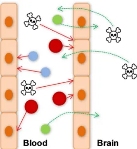
Figure I. 2: Scheme of the main roles of the blood-brain barrier (BBB). TheBBB protects the central nervous system (CNS) from toxic and harmful substances (toxic symbol) and from paracellular diffusion of water-soluble molecules (blue). BBB also limits the entry of cells from the blood (red), allows the uptake of molecules needed for CNS (green) and eliminates harmful compounds from the brain to blood (toxic symbol).
The only regions where there is no BBB are those that regulate autonomic nervous system and endocrine glands of the body since blood vessels allow diffusion of blood-borne molecules across the vessel wall (Ballabh et al., 2004). This barrier is mainly formed by ECs, astrocytes end-feet, basement membrane (BM) and pericytes. All these elements, together with microglia and neurons are part of the functional neurovascular unit (Cardoso et al, 2010). More specifically, a differentiated BBB is composed by the highly specialized ECs, surrounded by a BM in which a large number of pericytes are embedded. These last cells are also covered by the BM, which is ensheathed by astrocytic endfeet (Kim et al., 2008; Engelhardt and Sorokin, 2009) (Fig. I.3).
I. INTRODUCTION
5 Figure I. 3: Schematic representation of the neurovascular unit. EC, endothelial cell; P, pericyte; A, astrocyte endfeet; N, neuron; M, microglia and in grey the basement membrane.
1.1. Endothelial cells
In developed animals like mammals the cerebral endothelium is considered the anatomic basis of the BBB (Hawkins et al., 2006) and its interactions with others brain cells determine and turn possible the correct function of the BBB (Ballabh et al., 2004; Cardoso et al., 2010).
I. INTRODUCTION
6
such as P-glycoprotein (P-gp) and multidrug resistance-associated proteins (MRP), move out of the brain harmful hydrophilic and hydrophobic molecules (Begley, 2004). The expression of P-gp is one of the specialized characteristics of BMVECs (Tatsuta et al., 1992; Tatsuta et al., 1994). Ecs also express enzymes that can modify and change a range of molecules, which otherwise could pass through the BBB and affect neuronal function (Persidsky et al., 2006; Zlokovic, 2008). Enzymes concentration is high in cerebral microvessels and includes γ-glutamyl transpeptidase, alkaline
phosphatase, and aromatic acid decarboxylase (Persidsky et al., 2006). Besides all these, BMVECs also have other active pumps that help in regulation of ions, metabolites and xenobiotics concentrations in the brain (Cardoso et al., 2010).
To eliminate spaces between ECs and prevent free paracellular diffusion of blood-borne substances into the brain parenchymal space, BMVECs of capillaries and postcapillary venules have junctional complexes that include mainly TJs and adherens junctions (AJs). The first are located on the apical region of ECs and AJs are below TJs. TJs proteins include the transmembranar proteins claudins, occludin, junctional adhesion molecules (JAMs) and the cytoplasmic proteins like zonula occludens (ZO). The claudins family include by now 27 members (Mineta et al., 2011), which have 20-27 kDa and four domains. Occludin was the first TJ protein discovered. It has 65 kDa and four domains like claudins but with a different amino acid sequence (Furuse et al., 1993). JAMs are proteins with approximately 40 kDa that have been identified in 1998. They belong to the immunoglobulin superfamily and have a single transmembrane domain (Martin-Padura et al., 1998). There are three JAMs, including JAM-1 predominantly expressed in the brain (Aurrand-Lions et al., 2001) that is involved in cell-to-cell adhesion and takes part in the formation of TJs as an integral membrane protein together with occludin and claudins. Recent data obtained in our lab revealed the presence of another transmembarne TJ protein in BMVECs, which is particularly concentrated at tricellular TJs, though also distributed along bicellular TJs (Mariano et al., unpublished). Finally, cytoplasmic proteins include ZO-1 that ensures the linkage of TJs to the cytoskeleton. AJs include cadherins that have a plasma membrane-spanning domain and a cytoplasmic domain associated with catenins, the other AJs proteins (Vorbrodt and Dobrogowska, 2003). These last make the link between cadherins and actine cytoskeleton (Cardoso et al., 2010). AJs also include nectin-afadin complex where afadin, also known as AF-6, anchors nectins to the cytoskeleton (Niessen, 2007).
I. INTRODUCTION
7 Figure I. 4: The main characteristics of endotheial cells and their junctional complexes. A, transport
systems; B , specifics receptors; C, tight and adherens junctions: 1, actin filament; 2, zonula occludens; 3, claudin; 4, occluding; 5, junctional adhesion molecule; 6, tricelullin; 7, catenins; 8, vascular endothelial cadherin; 9, afadin; 10, nectin.
ECs also adhere to the BM through transmembrane proteins that are classified into three families of cell adhesion molecules (CAMs): selectins, immunoglobulin superfamily and integrins (Cardoso et al., 2010). Thus, these molecules may contribute to BBB integrity (Engelhardt and Sorokin, 2009).
1.2. Basement membrane
I. INTRODUCTION
8
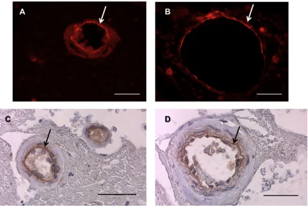
Figure I. 5: Basement membrane (BM) of human hippocampus sections. Arrows point to collagen IV, a main
component of the BM, visible by immunofluorescence (red) (A,B) and imunohistochemistry (brown) (C,D). Scale bars: 40 µm.
The collagen type IV, is a covalently-stabilized network polymer and is one of the proteins most important for structural integrity of small vessels. However, collagen type IV is dispensable for initiation of its assembly during early development (Pöschl et al., 2003). The laminins, with a branched structure (Yurchenco and Patton 2009), present in CNS BM are the laminin α4 and α5 produced by ECs (Sixt et
I. INTRODUCTION
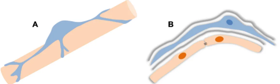
9 Figure I. 6: Simplified representation of the blood-brain barrier (BBB) focusing on the basement
membrane. A, components of the BBB: endothelial cell (EC), basement membrane (gray), astrocyte (A), pericyte (P), neuron (N) and microglia (M). B, the main elements of basement membrane: the collagen type IV (1), laminins (2) that are the principal responsible for the attachment of basement membrane to the cells receptors (3), and the perlecan (4) that is the predominant heparan sulfate proteoglycan in the endothelial cell basement membrane (adapted from Yurchenco and Patton, 2009).
Beyond all these matrix molecules the BM is also composed by matrix adhesion receptors, known as CAMs, as well as signaling proteins, which form an extensive and complex extracellular matrix (ECM) (Carvey et al., 2009). These molecules are expressed in the vascular cells, neurons and supporting glial cells and are important for BBB’s maintenance (del Zoppo et al., 2006). Integrin and dystroglycan families of CAMs are expressed by cells within cerebral microvessels. Integrins participate in mediating endothelial signaling, cell migration and brain capillary tube formation during angiogenesis. These and all CAMs have an essencial role in the maintenance of BBB integrity (Zlokovic, 2008). The receptors on the cell surface, in turn, allow the link between the ECM and the underlying cytoskeleton (Yurchenco and Patton, 2009).
A perfect BM besides provide anchoring and structural integrity to capillaries may also be involved in pericytes function and differentiation. Pericytes encased in the BM or exposed to laminal proteins do not usually differentiate (Dore-Duffy, 2011).
Matrix metalloproteinases (MMPs) have the capacity to digest the BM, consequently reducing anchoring of brain ECs, and to affect TJs integrity that lead to alteration in BBB integrity (Carvey et al., 2009). MMP-2 and MMP-9 are two gelatinases capable of proteolyze some membrane compounds including collagen type IV, glicoptotein like fibronectin and vitronectin, the proteoglycan aggrecan and elastin. MMP-2 can also cleave laminin. Serine proteases, cysteine proteases, and heparanase are other families of proteases that can affect the BM (del Zoppo, 2010).
I. INTRODUCTION
10 .
1.3. Neurons
There are many neurons that directly innervate ECs and astrocytic processes, like noradrenergic (Cohen et al., 1997), serotonergic (Cohen et al., 1996), cholinergic (Tong and Hamel, 1999) and GABA-ergic neurons (Vaucher et al., 2000). Furthermore, there are evidences that neurons can regulate the blood vessel’s function through inducing expression of enzymes unique for ECs in response to metabolic requirements (Tontsch and Bauer, 1991). Thus, neurons may have an important role on the BBB phenotype but little is known about this. On the other hand, ECs and a BBB well developed are important to create a stable environment to neural function (Abbott et al., 2006). However, evidences about the role of neurons in BBB properties begin to arise. Minami (2011) through an ischemic model proved that presence of neurons increase the transendothelial electrical resistance (TEER) and decrease the permeability of ECs in an in vitro model of the BBB.
1.4. Astrocytes
Astrocytes are glial cells whose endfeet ensheath the BM on the outer surface of the BBB endothelium. These cells cover more than 99% of the endothelium (Persidsky et al., 2006), contributing to the BBB properties and development, and to the unique endothelial phenotype. These roles are made through interactions with ECs, expression and release of soluble factors (Persidsky et al., 2006; Cardoso et al., 2010) and throught their anatomic proximity to ECs (Lai and Kuo, 2005). For example, the astrocytes interacting with ECs enhance their TJs and reduce gap junctional area (Tao-Cheng and Brightman, 1988), so these cells have an important role in the permeability and integrity of the BBB. Other studies by Siddharthan et al. (2007) and Malina et al. (2009) proved that the presence of astrocytes elevate the TEER and decrease the permeability of BBB endothelium. The interactions between astrocytes and ECs are also essential in regulating brain water and electrolyte metabolism under physiologic and pathologic situations (Zlokovic, 2008).
Besides their role in the BBB, astrocytes are essential for proper neuronal function and the interactions astrocytes-BMVECs are very important for a functional neurovascular unit since neuronal cell bodies are very close to brain capillaries (Persidsky et al., 2006).
1.5. Microglia
Microglia are a distinct class of glial cells that constitute the brain’s immunocompetent cells. Over time, their nature has been extensively discussed but now is accepted that microglia are ontogenetically related to cells of the mononuclear phagocyte lineage contrary to the other cells of CNS (Kreutzberg, 1996). Monocytes enter into the brain during embryonic development and differentiate into brain resident microglia displaying surface antigens of macrophages (Kim and Vellis, 2005).
I. INTRODUCTION
11 surface antigen and cytokine release (Kim and Vellis, 2005; Zlokovic, 2008). This ability to quikly change from one to other form makes their resting form the vigilant cells to the homeostatic disturbance in the CNS (Kreutzberg, 1996).
Since microglia are located in perivascular space, it is suggested that their interactions with ECs may influence BBB’s properties (Cardoso et al., 2010). However, the mechanisms behind this remain unknown.
2. Pericytes
It was in 1873 that Rouget described for the first time a population of cells on capillary walls and distinguished them from migratory leucocytes (Rouget, 1873) (Fig. I.7). Fifty years later these cells were named pericytes by Zimmermann (Krueger and Bechmann, 2010). The term pericyte arises from “peri-“ around and “cyto-“ cell and reflects their location at the abluminal side of the microvessels (Balabanov and Dore-Duffy, 1998). These cells are presently known as perivascular cells with multifunctional activities (Braun et al., 2007; Krueger and Bechmann, 2010).
Figure I. 7: Original draw of pericytes by Rouget in 1873. Rouget (1873) described the cells of the capillary walls and divided into amoeboid migratory cells (arrow) and fusiform contractile cells (arrow heads). (Krueger and Bechmann, 2010)
2.1. Characteristics of pericytes
In most BBB research the pericytes are often devalued and the astrocyte-endothelial interactions more investigated. This is mainly due to the difficulty in extracting pericytes from their location, as well as to the lack of pericyte-specific markers, which vary with the type of tissue (Engelhardt and Sorokin, 2009). Therefore, it is necessary to look for new markers that allow not only to find them, but also to deepen our knowledge of their functions and role in the neurovascular unit. Currently, location and identification of pericytes require a series of stains with a combination of positive and negative immunoreactivity. Pericytes express surface antigens allowing their identification, including the pAPN/CD13, platelet-derived growth factor-β receptor (PDGFR-β), α-smooth muscle actin (α-SMA),
I. INTRODUCTION
12
transpeptidase, alkaline phosphatase, nestin and vimentin (Fisher, 2009; Engelhardt and Sorokin, 2009; Krueger and Bechmann, 2010). However, most of these molecules are expressed in neighboring cells as well (Fisher, 2009; Krueger and Bechmann, 2010). On the other hand, in the brain, only pericytes close to arterioles are immunoreactive for α-SMA (Dore-Duffy, 2008). Another
difficulty associated with pericytes is the fact that they are tricky to identify because they are often confused with adjacent cells, such as vascular muscle cells, perivascular cells and juxtavascular microglia (Krueger and Bechmann, 2010). .
Brain pericytes are multifunctional and polymorphic cells (Guillemin and Brew, 2004; Lai and Kuo, 2005) but normally are star-shaped and have long cytoplasmic processes that are oriented along the axis of the blood vessel, while smaller circumferential arms engirdle the vascular wall (Fisher, 2009; Krueger and Bechmann, 2010) (Fig. I.8). There may be up to 90 processes with a width of 300 to 800 nm per 100 µm of capillary length (Zlokovic, 2008). However, processes morphology tends to be heterogenous and is likely to represent functional differentiation of pericytes (Dore-Duffy and Cleary, 2011). Moreover, they have cytoplasmic lysosomes that give to pericytes granularity (Fisher, 2009). These cells can be seen on the abluminal surface of ECs of capillaries, venules and arterioles (Fig. I.9). They cover 22-32% of cerebral capillary surface and tend to arise over endothelial TJ regions with one layer of BM between them as stated above. Another layer of BM lies between pericytes and astrocyte endfeet (Fisher, 2009; Krueger and Bechmann, 2010). The membrane surrounds all pericytes even their projections (Dore-Duffy and Cleary, 2011). During development and angiogenesis pericytes deposit components of this membrane (Dore-Duffy, 2008) and contribute to its maintenance (Zlokovic, 2008).
I. INTRODUCTION
13 Figure I. 9: Double labeling immunofluorescence analysis of endothelial cells and pericytes in human
hippocampus. Endothelial cells were labeled for von Willebrand factor (green, arrow) and pericytes forα-smooth muscle actin (red, arrowhead). Nuclei were identified by DAPI (blue) (B,D). Scale bars: 40 µm.
Pericytes-to-endothelia ratio in the brain is higher than in other organs (1:3 compared with 1:100 in striated muscles), and the location of pericyte on microvessels and their abundance varies according microvessels types (Dore-Duffy, 2003). The degree of coverage appears to correlate with the degree of tightness of the interendothelial junctions (Shepro and Morel, 1993).
Pericytes are cells from the vascular smooth muscle lineage (Nishioku et al., 2009) and it is generally considered that they have a mesodermal origin (Guillemin and Brew, 2004). It is possible that they migrate into the tissue during the latter stage of vascularization then assuming their characteristics. Their precursor cells settle on newly formed capillary sprouts and differentiate into pericytes as they become enclosed within basal lamina (Rhodin and Fujita, 1989).
2.2. Functions
In recent years a variety of studies, mainly in cell cultures, set various functions of pericytes. These include contractile, immune and phagocytic, migratory and angiogenic functions. In addition, pericytes contribute to the BBB and perform a regulatory role in brain hemostasis (Fisher, 2009).
2.2.1. Contribution to BBB properties
I. INTRODUCTION
14
constituted by N-cadherin, a variety of adhesion molecules, β-catenin, ECM molecules such as
fibronectin, and a number of integrins. Adhesion plaques support transmission of contractile forces from pericytes to other cells (Cuevas et al., 1984; Nakamura et al., 2008). Peg-and-socket contacts prevent pericytes penetrating through basal membrane and contacting with other cells and vessels (Rucker et al., 2000). It was thought that these functions related to the BBB stabilization were in charge of astrocytes, but today we find data describing the development of endothelial TJs independently of astrocytes (Krueger and Bechmann, 2010). Al Ahmad et al. (2011) studied the role of astrocytes and pericytes in TJs and AJs formation and observed the essencial role of each cell in the establishment of BBB-specific TJ complexes in ECs. Thus, we must consider a dialogue between several populations of the neurovascular unit and the possible existence of compensatory mechanisms that may take over each other’s role in case of impaired function (Fisher, 2009). The ECs are not the only ones that express TJ molecules. Pericytes express several TJ molecules including claudin-12, JAM, ZO-1, ZO-2 and occludin. These cells also express several barrier-related transporters like ABCG2, P-gp, MRP-1, and glucose transporter-1 (Glut-1) (Shimizu et al., 2008). Once again, pericytes are essential for BBB properties, namely, permeability and hemostasis of the brain.
Doghu et al. (2005) evaluated BBB function based on the transendothelial permeability coefficient (Pe) to sodium fluorescein (Na-F) and on the cellular accumulation of rhodamine 123 in mouse brain capillary ECs, as markers of paracellular permeability and functional activity of P-gp, respectively, and proved that pericytes participate in tightening the intercelular junctions and facilitating P-pg function in brain ECs through production of soluble factors including TGF-β and by cell-to-cell contact. Besides
this factor, others are also derived from pericytes, the VEGF that increases the permeability of brain ECs, the bFGF that tights the intercellular junctions and induces the expression of MRP and the angiopoietin (ang) -1, an antipermeabitily factor. These factors are also produced by astrocytes, which suggest that the four factors are involved in the interaction between ECs, pericytes and astrocytes under physiological and pathophysiological situations (Dohgu et al., 2005).The integrity of the BBB can also be evaluated through measurement of TEER. Nakagawa et al. (2007) constructed, for the first time, a rat primary culture based syngeneic triple co-culture BBB model, using brain pericytes besides brain ECs and astrocytes and showed that the contact between the three different cells increased the TEER and consequently tightened TJs. Recently, Armulik et al. (2010) demonstrated the pericytes role at the BBB in vivo, correlating reduced pericyte densities with increased vessel diameter and reduced vessel density and established a correlation between pericytes density and BBB permeability for a range of tracers of different molecular masses (Fig. I.10).
I. INTRODUCTION
15 Pericytes also contribute to BM formation by synthesizing collagen type IV, glycosaminoglycans, and laminin (Engelhardt and Sorokin, 2009; Fisher, 2009), and by inducing ECs to secrete BM components (Brachvogel et al., 2007).
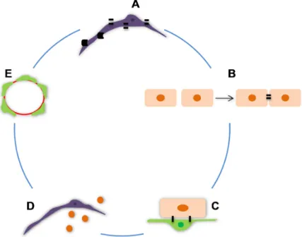
Figure I. 10: The main features of pericytes that determine the blood-brain barrier properties. A,
Expression of tight junctions and barrier-related transporters; B, Role in the formation of tight junctions between endothelial cells (ECs); C, Short distance to endothelial cells and gap, peg-and-socket and adhesion plaques junctions with ECs; D, Production of soluble factors; E, High density of pericytes around brain microvessels.
2.2.2. Participation in vascular development
During vertebrate embryo development, the first functional organ is the vascular system, whose growth must be continuous. The vasculature forms by vasculogenesis (new vessel formation from angioblasts or stem cells) and angiogenesis (sprouting, bridging and intussusceptive growth from existing vessels). Mesenchymal cells differentiate into endothelial tubes that form a primitive blood vessel network from which new blood vessels develop. After formation of the first endothelial tubes they become associated with mural cells that include pericytes and vascular smooth muscle cells (Hellström et al., 2001).
I. INTRODUCTION
16
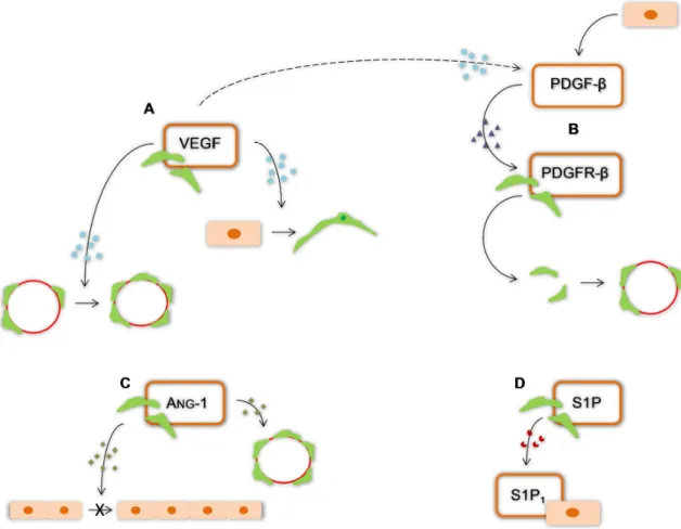
VEGF is very important in this process as it initiates vessel formation and activates a chain of molecular and cellular events that lead to mature vasculature (Jain, 2003). VEGF, which is produced by pericytes under hypoxic (Yamagishi et al., 1999) and hypoglycemia (Hellström et al., 2001) conditions, is crucial to communication with ECs. VEGF induces angiogenic sprouts, positive for the CD31 marker of mature and embryonic ECs, to display α-SMA and desmin, characteristic of pericytic
phenotype. This observation indicates that ECs transform into pericytes or smooth muscle cells and that VEGF plays an important role in this transformation. Furthermore, the number of pericytes covering new capillaries can be increased by VEGF (Hagedorn et al., 2004). Prove of the role of VEGF in angiogenesis is the blockage of its receptor VEGFR2 can temporally normalize a tumor vessel structure (Winkler et al., 2004). Interleukin (IL) -6 is also an important cytokine to pericytes differentiation and may be responsible for recruiting ECs and promoting angiogenesis (Kale et al., 2004).
After formation, the nascent vessels are stabilized by recruiting mural cells, including pericytes, and by generating ECM. This stabilization process is regulated by at least four molecular pathways. The recruitment/differentiation of mural cells, namely pericytes, to sites of angiogenesis or neurovascularization is mediated by the platelet-derived growth factor-β (PDGF-β) produced by ECs (Hellström et al., 2001; Lai and Kuo, 2005; Abramsson et al., 2007), presumably in response to VEGF (Jain, 2003) and by the corresponding receptor PDGFR-β expressed by pericytes (Hellström et al.,
2001; Lai and Kuo 2005; Abramsson et al., 2007). The essential role of this factor was demonstrated by Hellström et al. (2001) through PDGFR-β knockout mice that lacked pericytes along the vessels
and by Abramsson et al. (2007) who showed that the absence of an amino acid motif of PDGF-β
produces defective investment of pericytes in microvascular system. Angs are also important for vascular development and stabilization. Ang-1 produced by pericytes and perivascular cells binds to receptor Tie-2 fostered vascular stabilization and ang-2, expressed by ECs, binds to the same receptor and acts like a destabilizing factor (Jain, 2003; Ballabh et al., 2007; Krueger and Bechmann, 2010) in the absence of VEGF, and restricted to ECs in areas of vascular remodeling (Jain, 2003). Thus, the development of vasculature remains unstable and immature until pericytes or its precursors are recruited (Ballabh et al., 2007; Krueger and Bechmann, 2010). Like mentioned above, in the presence of VEGF, ang-2 facilitates vascular sprouting (Jain, 2003). Therefore, the presence of pericytes regulates negatively ECs proliferation, determining their number, morphology and microvessel architecture (Hellström et al., 2001). Vascular stability is sustained also before pericytes arrive to their position, by binding of sphingosine-1-phosphate (S1P) to its endothelial receptor S1P1 (Krueger and Bechmann, 2010) (Fig. I.11). After binding, small GTPase Rac is activated in ECs (Paik et al., 2004) and N-cadherin, found in peg-socket contacts between ECs and pericytes, and vascular endothelial-cadherins that are normally found in junction complexes between neighboring ECs are expressed (Krueger and Bechmann, 2010). Finally, TGF-β is a multifunctional cytokine that also
promotes vessel maturation through stimulation of ECM production and differentiation of mesenchymal cells to mural cells (Jain, 2003). TGF- β is also important, like ang-1 and its receptor, in
I. INTRODUCTION
17 Al Ahmad et al. (2011) through a novel 3D in vitro model that closely mimics barrier formation and morphology showed the proangiogenic role of pericytes. Pericytes can participate in angiogenesis through NG-2, which is expressed by immature pericytes. Both blocking by antibodies as well as knocking out the gene of NG-2 cancel vascular growth in various models of induced angiogenisis, but it is necessary to have in mind that NG-2 is expressed in more cells of the CNS. Due to their important role in angiogenesis, pericytes may be suggested as a target to pharmacological therapy for tumors (Krueger and Bechmann, 2010)
Figure I. 11: Factors and receptors produced by pericytes that contribute to angiogenesis. A, Vascular endothelial growth factor (VEGF) allows the transformation of endothelial cells into pericytes or smooth muscle cells and increase the number of pericytes covering new capillaries; B, platelet-derived growth factor receptor-β (PDGFR-β) recognizes the platelet-derived growth factor-β (PDGF-β) produced by endothelial cells presumably in response to VEGF. This interaction is responsible for the pericyte recruitment; C, angiopoetin (ang)-1 inhibits endothelial proliferation and is responsible for vascular stabilization; D, sphingosine-1-phosphate (S1P) connects to its endothelial receptor and promotes the continuous stability.
2.2.3. Contractile function
I. INTRODUCTION
18
pericytes express α-SMA (Herman and D’Amore, 1985), with more robust expression in pre-capillaries
compared to mid- and post-capillaries (Nehls and Drenckhahn, 1991; Boado and Pardrige, 1994; Fisher, 2009; Kruger and Bechmann, 2010) (Fig. I.12). Pericytes also possess tropomyposin and myosin that contribute to their contractile capacity (Joyce et al., 1985a, b). However not all pericytes express α-SMA. Bandopadhyay et al. (2001) proved that in CNS pericytes α-SMA is the predominant
contractile protein but was not present in all brain pericytes, which supports the idea that the pericytes constriction ability is not universal.
Figure I. 12:Morphological features of human brain vascular pericytes (HBVP). A, HBVP in primary culture observed by phase contrast microscopy shows the typical morphology. Original magnification: 100x. B, Immunofluorescence analysis of the pericyte marker α-smooth muscle actin (α-SMA). Nuclei were stained with Hoechst 33258 dye. Scale bar: 40µm.
Pericytes also can express receptors for vasoactive substances such as prostacyclin (Fisher, 2009; Kruger and Bechmann, 2010), angiotensin II, endothelin-1, catecholamines, and vasopressin. Ang-2 is a vasoctive peptide that most of the times cause vasoconstriction. Ferrari-Dileo et al. (1996) demonstrated that pericytes have specific ang-2 binding sites. They also found binding sites for vasoactive intestinal polypeptide. Zwieten et al. (1988) discovered the presence of binding sites for vasopressin. The receptors for endothelin-1, other vasoactive peptide, were also located (Yamagishi et al., 1993). Nitric oxide (NO) also affects pericytes, causing their relaxation trough cyclic guanosine monophosphate (Haefliger et el., 1994). With all these evidences, it is considered that pericytes have a contractile function and blood flow regulatory capabilities, especially in pre-capillary arterioles (Balabanov and Dore-Duffy, 1998; Kruger and Bechmann, 2010).
Like already said pericytes are associated to microvessels and have some features of smooth muscle cells, so the absence of these last cells in microvessels turn pericytes their possible contractile substitutes (Bandopadhyay et al., 2001).
Yemisci et al. (2009) showed that pericytes contract during ischemia and even after reopening of the occluded middle cerebral artery. Pericytes also cause segmental narrowing of capillaries, turn erythrocytes trapped in the capillary constrictions and obstruct microvirculation. These authors also proved that peroxynitrite leads to pericytes constriction, wheareas the suppression of oxygen and nitrogen radicals formation can reverse this situation (Fig. I.13).
I. INTRODUCTION
19 Figure I. 13: Pericytes properties that turn them into cells with contractile functions. A, Receptors for vasoactive substances; B, Expression of α-smooth muscle actin.
2.2.4. Immune and phagocytic function
Brain pericytes constitutively express low levels of adhesion molecules (intercellular adhesion molecule-1 and vascular CAM-1), which have potential stimulatory activity in major histocompatiblity complex (MHC) -class II dependent antigen presentation (Veerbek et al., 1995). Thus, pericytes may have the capacity to present antigens to T-lymphocytes. Dore-Duffy and Balabanov (1998) showed the response of primary rat CNS pericytes to interferon у with upregulation of the MHC class II
molecule and present antigen to primed lymphocytes (Balabanov and Dore-Duffy, 1998). Brain pericytes also produce immunoregulatory cytokines like IL-1β and IL-6 (Fabry et al., 1993). TGF-β
produced by pericytes may also function as immunoregulator at the BBB (Dore-Duffy et al., 1996; Balabanov and Dore-Duffy, 1998)
Pericyte lysosomes express acid phosphatase that implies a phagocytic function of pericytes (Fisher, 2009). Pericytes also have components that are recognized by macrophage-selective monoclonal antibodies EBMS/11 and ED2 (Esiri et al., 1986; Balabanov et al., 1996) (Fig. I.14).
I. INTRODUCTION
20
pericytes like immune cells. The expression of acid phosphatase in lysosomes and the possession of components recognized by antibodies for macrophages turn pericytes into phagocytic cells.
2.2.5. Roles on hemostasis
Pericytes appear to be cells with pro- and anti- coagulant activity [60] (Fig. I.15). In fact, Kim et al. [104] showed that pericytes decrease endothelial tissue plasminogen activator (tPA), a serine protease that processes plasminogen into proteolytically active plasmin thus allowing fibrinolysis to occur, and suggested that this effect is mediated by a soluble-derived factor since it was observed in a non-contact ECs-pericytes co-culture model. They also showed that pericytes amplify the LPS-induced enhanced release of plasminogen activator inhibitor-1 (PAI-1), and further showed that pericytes express robust amounts of the antithrombin and antifibronolytic molecule serpin protease nexin-1 (PN-1). Thus, these evidences indicate that pericytes have a complex and important role in brain microvascular hemostasis, with effects that are largely antifibrinolytic. Besides inducing an anti-coagulant response through PN-1, pericytes can differentially regulate expression of pro-anti-coagulant enzyme complexes involved in the extrinsic pathway of blood coagulation. Bouchard et al. [105] have defined functionally active tissue factors on the surface of human brain pericytes that are the primary generator of the coagulation cascade.
Figure I. 15: Pericytes have the capacity to regulate blood clotting through a pro-and anti-coagulant
activity.
2.2.6. Multipotent cells
Besides all these functions, pericytes can be seen like multipotent cells. Pericytes may be a source of multipotent stem cells that differentiate along multiple lineage and may provide trophic support and maintenance in the adult brain (Dore-Duffy et al., 2006). They can differentiate into osteoblasts, chondroblasts, fibroblasts, adipocytes, and smooth muscle cells (Lai and Kuo 2005, Dore-Duffy et al., 2006, Dore-Duffy, P. 2008) (Fig. I.16).
I. INTRODUCTION
21 authors showed that cultured pericytes express mesenchymal stem cells markers, including CD44, CD73, CD90 and CD105.
Recently Dore-Duffy characterizate of CNS capillary pericytes from the transgenic mice harboring a temperature-sensitive mutant of the SV40 virus target T-gene. These Immortopericyte (IMP) are stable and do not differentiate for long periods of time and, at 33°C in the presence of interferon γ. These
authors showed that these cells are α-SMA negative and pluripotent. However they can be induced to differentiate along mesenchymal and neuronal lineage at 37ºC.
Figure I. 16: Pericytes can differentiate into osteoblast, chondroblast, fibroblast, adipocytes and smooth
muscle cells.
3. Neurovascular unit pathology
3.1. Involvement of pericytes in BBB dysfunction
I. INTRODUCTION
22
others. Some of these last are released by ECs and endothelium itself responds to the released agents (Ballabh et al., 2004). On the other hand, the length of brain capillaries is reduced in neurodegenerative diseases, like Alzheimer´s disease, which diminishes the transport of energy substrates and nutrients across the BBB and the elimination of neurotoxins from the brain (Zlokovic 2008).
Pericytes dysfunction or their loss also play an important role in the pathogenesis of some diseases. Reduction of pericytes has already been observed after stroke (Duz et al., 2007), multiple sclerosis (Zlokovic, 2008), brain tumor (Ho, 1985), diabetic retinopathy (Hammes et al., 2002), aging (Frank et al., 1990), and in a variety of angiopathies (Szpak et al., 2007) (Fig. I.14D).
Eberhard et al. (2000) observed different degrees of pericyte recruitment in six different types of malignant human tumors (glioblastomas, renal cell carcinomas, colon carcinomas, mammary carcinomas, lung carcinomas, and prostate carcinomas) indicating differences in the functional state of the tumor vasculature that can reflect variations in maturation degrees of the tumor vascular bed, since pericytes confer stability to the vasculature when present in a large number. These evidences support the possibility of a pericytes targeted therapy against the progression of tumors, especially when they are still immature.
Figure I. 17: Dysfunction of the blood-brain barrier (BBB). A, Normal BBB. The BBB dysfunction includes tight
junctions opening (B), changes in transport systems (C) and reduction of pericytes number (D).
I. INTRODUCTION
23 following ischemia through direct contact with ECs. Al Ahmad et al. (2011) through a 3D BBB model in hypoxia condition showed that EC monolayer exhibited significantly disrupted TJ protein expression, namely claudin-5 and ZO-1, and that the presence of astrocytes and pericytes is essential for optimal localization of these two proteins in cell borders.
3.2. Bilirubin neurotoxicity 3.2.1. Hyperbilirubinemia
3.2.2. Acute bilirubin encephalopathy vs. kernicterus or chronic bilirubin
encephalopathy
Unconjugated bilirubin (UCB) is the end product of heme catabolism (Dennery et al., 2001) and is a tetrapyrrole with propionic side groups. These groups form internal hydrogen bonds with the distal lactam groups that render this molecule nearly insoluble in aqueous medium at physiologic pH. Therefore, UCB circulates in serum bound to albumin. Serum albumin has a binding site for UCB with high affinity and two or more binding sites with lower affinity (Berk and Noyer, 1994; Ostrow et al., 1994, Wennberg, 2000).
During embryonic life bilirubin is produced starting from 12 weeks gestation and is eliminated through maternal circulation. After birth this placental protection is lost, which contributes to UCB accumulation (Brito et al., 2006). Moreover, in neonatal life the short red blood cell lifespan leads to an UCB overproduction, the immaturity of hepatic cells decreases UCB conjugation and the lack of bacterial intestinal flora impairs bilirubin excretion (Porter and Dennis, 2002). These facts, together with the characteristic neonatal hipoalbuminemia (Kaplan and Hammerman, 2005) that causes disturbances of the albumin-bilirubin equilibrium (Ahlfors and Parker, 2010), lead to neonatal hyperbilirubinemia, which occurs in up to 60% of full term newborns and 80% of pertems (Denneryet al., 2001). In addition, other factors may arise and contribute for even higher levels of UCB, including metabolic acidosis, infection, hyperoxia, as well as prematurity and drugs or preservatives (Kaplan and Hammerman, 2005). UCB has the capacity to enter into the brain and in high concentrations can affect and turn yellow specific locations (Ostrow et al., 2004), as the globus pallidus and subthalamic nucleus, as well as the auditory and oculomotor brainstem nuclei (Shapiro, 2010). The cerebellum (Brito et al., 2011) and hippocampus may also be affected (Shapiro, 2010). If untreated, hyperbilirubinemia can develop into UCB encephalopathy or kernicterus depending on neurological damage progress to chronic and permanent or even death (American Academy of Pediatrics, 2004).
3.2.3. Effects of unconjugated bilirubin in the neurovascular unit
I. INTRODUCTION
24
(Palmela et al. 2011). Finally, increases in the neuronal form of NO synthase and nitrites production were shown to occur in the presence of elevated levels of UCB (Brito et al., 2008; Vaz et al., 2010; Brito et al., 2010). Palmela et al. (2011) also showed the upregulation of eNOS expression, increase of nitrites production, decrease of the TEER and increase the permeability in human BMVECs and that UCB crosses the ECs monolayer in a time- and concentration dependent manner (Palmela et al., unpublished).
Figure I. 18: Some effects of unconjugated bilirubin (UCB) in central nervous system cells. EC, Endothelial cell; M, microglia; N, neurons; A, astrocyte; O, oligodendrocytes. The presence of UCB increases the permeability of endothelial cells, leads to death of all cells represented, secretion of cytokines by endothelial cells, microglia, astrocytes and even by neurons, as well as to production of nitrites by endothelial cells, microglia and neurons.
4. Aims
27
II.
MATERIALS AND METHODS
1. Materials
Human brain vascular pericytes (HBVP) and Trypsin/EDTA solution were acquired from ScienCell Research LaboratoriesTM (Carlsbad, California, USA). Dulbecco’s modified Eagle’s medium (DMEM) and fetal bovine serum (FBS), were purchased from Biochrom AG (Berlin, Germany). Antibiotic antimycotic solution (20X), Trypsin/EDTA 1X, human serum albumin (HSA; fraction V, fatty acid free), bovine serum albumin (BSA), Hoechst 33258 dye, poly-L-lysine, poly-D-lysine and organosilanecoated
slides were from Sigma Chemical Co. (St. Louis, MO, USA). Triton X-100 was acquired from Roche Applied Science (Mannheim, Germany). DPX mountant for microscopy was obtained from BDH Prolabo (Poole, UK). Mouse anti-α-SMA was purchased from ABD Serotec (Oxford, UK). Mouse
anti-eNOS Type III antibody was acquired from BD Biosciences (Erembodegem, Belgium). Fluorescein isothiocyanate (FITC)-labeled horse anti-mouse antibody was acquired from Vector laboratories (Burlingame, CA, USA). Antibody Alexa Fluor 488 goat anti-mouse was from Invitrogen CorporationTM (Carlsbad, CA, USA). Primary antibody mouse anti-collagen IV came from Sigma Chemical Co. (St. Louis, MO, USA). DuoSet ELISA kits were from R&D systems® (Minneapolis, MN, USA). TRIzol Plus RNA Purification Kit was from Invitrogen CorporationTM (Carlsbad, CA, USA). Primers for real-time Polymerase chain reaction (RT-PCR) analysis were purchased from Thermo Scientific (Soeborg, Denmark). RevertAid H Minus First Strand cDNA synthesis and Maxima SYBR Green qPCR Master Mix (2x) were obtained from Fermentas (Burlington, Ontario, Canada).
Vector® antigen unmasking solution was from Vector Laboratories, Inc. (Burlingame, CA, U.S.A.) and Dako REAL™ EnVision™ Detection System, Peroxidase/DAB, Rabbit/Mouse was acquired from Dako,Glostrup, Denmark). Mayer’s hematoxilin, H2O2 solution and all other chemicals were of analytical grade and were purchased from Merck (Darmstadt, Germany).
2.
In vitro
studies – pericytes
2.1. Primary cultureII. MATERIALS AND METHODS
28
5000/cm2 in new coated vessels. For immunofluorescence analysis, cells were plated on poly-D -lysine-coated coverslips, previously placed in 24-well culture plates.
2.2. Treatment
UCB was purified according to the method of McDonagh and Assisi (1972). For UCB treatment, a 10 mM stock solution was prepared in 0.1 N NaOH and used immediately after preparation. The pH value was restored to 7.4 by addition of equal amounts of 0.1 N HCl, and all the procedures were performed under light protection to avoid photodegradation. Confluent HBVP were incubated with 50 or 100 µM UCB, or no addition (control), in the presence of 100 µM HSA (UCB/HSA molar ratios of 0.5 and 1.0, respectively), for 1, 4, 24 or 78 h at 37ºC to the cell death, cytokines and nitrites release analysis (Fig. II.1).
Figure II. 1: Schematic representation of the pericytes primary culture and UCB treatment. After being thaw
the cells werecultured in DMEM medium supplemented with 20% fetal bovine serum (FBS) and 1% antibiotic-antimycotic solution until the culture achieved approximately 80% confluence. Then they were detached and incubated with 50 or 100 µM UCB, or no addition (control), in the presence of 100 µM HSA (UCB/HSA molar ratios of 0.5 and 1.0, respectively), for 1, 4, 24 or 78 h at 37ºC to the cell death, cytokines and nitrites release analysis.
2.3. Characterization
Characterization of untreated HBVP was performed by a standard indirect immunocytochemical technique. After 1, 4, 24 and 72 h cells were fixed with 4% paraformaldehyde in phosphate buffered saline (PBS) for 20 minutes at room temperature. Next, permeabilization was performed using 0.2% triton X-100 during 3-5 min and 3% BSA in PBS was used as blocking solution for 1 h at room temperature. Cells were incubated overnight at 4ºC with the mouse anti-α-SMA primary antibody
(1:100) and with the secondary antibody Alexa Fluor 488 goat anti-mouse (1:500) for 1 h at room
100 µM HAS with or without UCB 50 or 100 µM
II. MATERIALS AND METHODS
29 temperature. Both primary and secondary antibodies were diluted in blocking solution. Nuclei were stained with Hoechst 33258 dye in PBS (1:1000) for 2 minutes. DPX was used as mountant medium. In order to have negative controls, some coverslips were incubated only with secondary antibody. This assured that the signal obtained for coverslips incubated with both primary and secondary antibodies was a result of the expression of the target proteins. Fluorescence was visualized using a Leica DFC 490 camera adapted to an AxioScope.A1 microscope (Zeiss). Fifteen red-fluorescence images of random microscopic fields were acquired per sample under 100x magnification. At the end of each time point images were also acquired using a phase contrast microscopy (Olympus, model CK2-TR).
2.4. Assessment of apoptosis
To determine if pericyte death occurs by apoptosis, we evaluated the nuclear morphology after Hoechst staining as previously described (Silva et al., 2001). Fluorescence was visualized using a Leica DFC 490 camera (Leica, Wetzlar, Germany) adapted to an AxioScope.A1 microscope (Zeiss, Göttingen, Germany). Apoptotic nuclei were identified by condensed chromatin, as well as nuclear fragmentation, and were counted in twenty random microscopic fields per sample under 400x magnification.
2.5. Quantification of Cytokine Release
Cytokine release by HBVP was quantified following UCB treatment for 1, 4, 24 or 72 h in 12-well plates. The cytokines IL-6 and VEGF were assessed in duplicate, using the supernatants free from cellular debris by a specific DuoSet ELISA development kits according to the manufacturer’s instructions. Measurements were obtained at a wavelength of 450 nm, with a reference filter of 620 nm, using a microplate reader. The results were expressed as relative secretion vs. control samples.
2.6. Measurement of Cytokine mRNA Expression
IL-6 and VEGF mRNA expression was measured by quantitive RT-PCR using mRNA cells from control (HSA) and UCB treatment. The time points were chosen according to the results of the release of these cytokines. For IL-6 were used the time periods 30, 45 and 60 min, and for VEGF 1, 4 and 8 h. Total RNA was extracted using the TRizol reagent according to the manufacturer’s instructions. Total RNA was quantified using Nanodrop ND-100 Spectrophotometer. One microgram of total RNA was reverse transcribed into cDNA using a RevertAid H Minus First Strand cDNA synthesis kit under the recommended conditions. It was performed quantitive RT-PCR using β-actin as an endogenous
control to normalize the expression levels of cytokines mRNAs. The following sequences were used as primers: IL-6 sense, 5’-GACAGCCACTCACCTCTTCA-3’ and anti-sense,
5’-TTCACCAGGCAAGTCTCCTC-3’ (Wang et al., 2006); VEGF sense,
5’CCCTGATGAGATCGAGTACA-3’ and anti-sense, 5’-CCTCGCCTTGCAACGCGAGT-3’ (Park et al., 2009); β-actin sense, 5’-ACAGAGCCTCGCCTTTGCCG-3’ and anti-sense,
5’-TGGGCCTCGTCGCCCACATA-3’ (NM_001101.3).
II. MATERIALS AND METHODS
30
performed in 8-strip individual tubes with each sample performed in triplicate, and a no-template control was included for each amplificate. The melting temperature of the PCR program was adjusted to 62ºC. Standard curves using a “calibrator” cDNA (chosen among the cDNA samples) were prepared for each target and reference gene. In order to verify the specificity of the amplification, a melt-curve analysis was performed, immediately after the amplification protocol. Non-specific products of PCR were not found in any case. The relative quantification was made using the Pfaffl modification of the ∆∆CT equation (CT, cycle number at which the fluorescence passes the threshold level of detection), taking into account the efficiencies of individual genes. The results were normalized to β
-actin and the initial amount of the template of each sample was determined as relative expression vs. control samples (reference). The relative expression of each sample was calculated by the formula 2 -∆∆CT.
∆CT is a value obtained, for each sample, by the difference between the mean CT value of each cytokine and the mean CT value of β-actin. ∆∆CT of one sample is the difference between its ∆CT value and ∆CT value of the sample chosen as reference.
2.7. Quantification of Nitrite Levels
In order to evaluate if UCB effects on HBVP include the activation of signalling pathways leading to NO production and ultimately to nitrosative stress, the levels of nitrite, a stable end product of NO, were measured in the incubation medium after cell treatment. Supernatants free from cellular debris were mixed with Griess reagent [1% (w/v) sulfanilamide in 5% H3PO4 and 0.1% (w/v) N-(1-naphthyl) ethylenediamine dihydrochloride (v/v) in a 1:1 ratio] in 96-well tissue culture plates for 10 min at room temperature in the dark. The absorbance at 540 nm, with a reference filter of 620 nm, was determined using a microplate reader. Solutions of sodium nitrite (0-200 µM) were used as standards.
2.8. Evaluation of eNOS Expression
The production of nitrites was further confirmed by evaluation of the expression of one of the enzymes responsible for its formation in pericytes, the eNOS (Loesch et al., 2010). Detection of this enzyme was performed by a standard indirect immunocytochemical technique using a mouse anti-eNOS Type III antibody (1:100) as the primary antibody and a horse FITC-labelled anti-mouse antibody (1:227) as the secondary antibody. Fluorescence was visualized using a Leica DFC 490 camera adapted to an AxioScope.A1 microscope. Five to ten green-fluorescence images of random microscopic fields were acquired per sample using a 63X objective.