UNIVERSIDADE DE LISBOA
Faculdade de Medicina Veterinária
USE OF TRI-SOLFEN TO CONTROL PAIN DURING TREATMENT OF
HOOF LESIONS IN DAIRY COWS
ANA MARGARIDA DOS SANTOS FERRADOR
CONSTITUIÇÃO DO JURI ORIENTADOR Doutor Miguel Luís Mendes Saraiva Lima Doutor George Thomas Stilwell Doutora Berta Maria Fernandes Ferreira
São Braz
Doutor George Thomas Stilwell
2018 LISBOA
UNIVERSIDADE DE LISBOA
Faculdade de Medicina Veterinária
USE OF TRI-SOLFEN TO CONTROL PAIN DURING TREATMENT OF
HOOF LESIONS IN DAIRY COWS
ANA MARGARIDA DOS SANTOS FERRADOR
DISSERTAÇÃO DE MESTRADO INTEGRADO EM MEDICINA VETERINÁRIA
CONSTITUIÇÃO DO JURI ORIENTADOR Doutor Miguel Luís Mendes Saraiva Lima Doutor George Thomas Stilwell Doutora Berta Maria Fernandes Ferreira
São Braz
Doutor George Thomas Stilwell
2018 LISBOA
v
Dedicatória
vi ACKNOWLEDGMENTS
Ao Professor George Stilwell, por todos os ensinamentos transmitidos e pelo incansável acompanhamento durante a elaboração deste trabalho. Sem o qual não teria tido a coragem de perseguir a minha grande paixão pela clínica de grandes animais; a sua dedicação e cuidado com o bem-estar animal fez-me redescobrir a, há tanto tempo perdida, vontade de seguir este caminho. E ainda pelo incutido novo bichinho pela observação de aves. Mais que um professor, um mentor.
Ao Engenheiro Nuno Carolino, pela preciosa ajuda na análise estatística do meu trabalho.
À Australian Animal Ethics, por ter permitido o desenvolvimento deste projeto. É de louvar o interesse e preocupação pela melhoria e instituição de inovadoras formas de controlo e maneio da dor animal.
À Eurofins Agroscience, pela prontidão na análise laboratorial das minhas amostras.
Ao CIISA, pelo apoio financeiro disponibilizado para o desenvolvimento deste projeto e futuras divulgações do mesmo.
À Associação Agrícola de São Miguel e toda a sua equipa espetacular, em especial ao Dr. João Vidal, por me terem recebido tão bem. Foi um privilégio aprender a realidade da clínica de campo com tão bons profissionais.
À Typoteam, de onde levei valiosos ensinamentos pessoais e profissionais. Dos maiores desafios surgem as maiores surpresas, e esta equipa foi sem dúvida uma delas. Obrigada por me terem dado a oportunidade de me mostrar a mim mesma que sou capaz.
A todos os amigos que fiz durante este percurso, na Lusófona e FMV, as minhas duas casas igualmente importantes.
A todos os meus amigos, que me acompanham ou foram acompanhando neste percurso.
Às AgroVets e as Vets, as amigas que levo no coração.
Ao melhor grupo de estágio que podia ter pedido, Joaninha e Maria Sara. “Fizemos os 4 uma bela equipa”.
vii
Às minhas melhores amigas de longa data, Teresa, Cláudia e Tânia, que levam uma grande escola a aturar-me e ouvir os meus devaneios. O tempo não afasta, só fortifica.
À minha homónima Margarida, o alto astral personificado. Qualquer que seja o caminho que nos espere, o Xiang vai ter sempre uma mesa para nós. Ensinaste-me a olhar para os problemas de forma descontraída e que há sempre uma razão para dar uma boa gargalhada, mesmo quando tudo parece negro.
À Maria Sara, o maior presente que a FMV me trouxe. Teria sido possível terminar este caminho, mas nunca teria tido a mesma luz. As grandes amizades estão escondidas nos cantinhos menos prováveis e vêm quando menos se espera. Percorrermos este caminho juntas é um privilégio, e assim continuará, onde quer que o Mundo nos leve. Obrigada por seres a minha consciência, por me ouvires e te fazeres ouvir.
À minha família, a minha base. Aos meus avós, aos meus padrinhos, às minhas primas e aos meus tios. Daí surge a verdadeira essência de quem somos.
Aos meus pequeninos, Gui e Guadalupe, as luzes dos meus olhos. Por mais negro que esteja o céu, não há chuva que molhe enquanto estiver convosco.
Ao meu mano, o meu vizinho da porta ao lado. O que vê todos os bolos de areia e formigas que faço desde sempre e continua a acreditar que de lá podem sair obras-primas.
Aos meus pais, as minhas raízes. Se aqui cheguei foi graças a vocês. Em todos os momentos que pensei que não ia ser capaz, todos “não consigo”, vocês incansavelmente ouviram, cuidaram e incentivaram para superar todas as limitações que coloquei a mim mesma. Obrigada por me darem o meu porto seguro, sem cláusulas nem restrições.
viii ABSTRACT
Use of Tri-Solfen to Control Pain During Treatment of Hoof Lesions in Dairy Cows Hoof lesions in dairy cattle have a great impact, either in production as in animal welfare. Trimming may cause severe pain resulting in violent reaction with risk for humans’ safety as well as affecting the animal’s immediate welfare. These interventions are usually carried out by non-veterinarian technicians, without any kind of pain management training. An efficient pain management is not only an ethical obligation, as also allows a better manipulation and meticulous treatment.
The present study had the main purpose to test the efficiency of Tri-Solfen®, with a combination of local anaesthetics – lidocaine and bupivacaine - adrenaline and cetrimide in a topical gel form. The efficiency of this formulation has already been tested in other procedures, such as mulesing, castration, disbudding and tail docking in lambs and calves, significantly reducing pain related behaviours. Being dairy cows a second objective was to assess lidocaine, bupivacaine and metabolites, as well as cetrimide residues in milk to determine the safety of use in milking animals.
The selected cows were in the drying off period and lameness scoring was performed when entering the chute. Before trimming, each animal was randomly distributed to two groups: C – usual trimming with no pain control; T – trimming with local anaesthetics being applied once live corium was exposed. Lesions’ characteristics were registered. Algometry measurements were performed before and after intervention, to assess animal reaction to pressure. During corrective trimming, behaviour observation was done by two persons blind to treatment. Lameness scoring was again performed at the end of the intervention.
Non-parametric tests and analysis of variance were performed. Analysis of data showed that treatment significantly influenced reaction to trimming and lameness score after trimming on the treated group, when compared with the not treated group. Algometry values showed increased pressure threshold after application of Tri-Solfen. Anaesthetics residues are below the LOQ value in all animals after the first milking, except in one sample at the fourth milking. This study suggests that the use of topical local anaesthetics with lidocaine and bupivacaine helps reducing pain inflicted during corrective trimming of severe lesions, enhancing animal welfare and providing trimmer safety due to diminishing pain related behaviours. We also demonstrated that the levels of anaesthetics and/or metabolites residues are very low in all animals in the four milkings after treatment.
ix RESUMO
Uso de Tri-Solfen no Controlo da Dor Durante o Tratamento de Lesões Podais em Bovinos de Leite
As afeções podais em vacas leiteiras têm um enorme impacto quer sobre a produção, quer sobre o bem-estar animal. O desbridamento das lesões pode causar dor intensa, levando a reações do animal que dificultam o maneio e a segurança do mesmo e do operador. Por norma, estas intervenções são realizadas por técnicos não médicos veterinários sem formação no controlo da dor. O maneio eficaz da dor não só é uma obrigação ética, como permite uma mais fácil manipulação e um tratamento mais minucioso. O presente estudo teve como principal objetivo avaliar testar a eficácia de um medicamento, Tri-Solfen®, que tem na sua composição uma associação de anestésicos locais – lidocaína, bupivacaína – adrenalina e cetrimida, na forma de gel tópico. A eficácia desta formulação já foi avaliada noutros procedimentos, como mulesing, castração, descorna e amputação de cauda em borregos e novilhos, tendo reduzido significativamente os comportamentos de dor. Sendo animais leiteiros, um secundo objetivo foi detetar a presença de lidocaína, bupivacaína e cetrimida, em amostras de leite após aplicação do produto. As vacas selecionadas encontravam-se no período de secagem e foram classificadas quanto ao grau de claudicação quando conduzidas ao tronco. Antes do início da aparagem cada vaca foi aleatoriamente alocada a um de dois grupos: C – aparagem sem aplicação do medicamento; TriS – aparagem com aplicação do anestésico tópico sobre a lesão do córion. As características das lesões encontradas foram registadas. Foram efetuados testes de algometria antes e após a intervenção, para avaliar a reação do animal a diferentes graus de pressão. Durante a aparagem curativa, dois observadores cegos ao tratamento avaliaram os comportamentos de dor. O grau de claudicação foi novamente avaliada no fim da intervenção. Na análise estatística dos dados foram aplicados testes não paramétricos e análise de variância. A análise dos dados demonstra existir uma redução da reação à aparagem e do grau de claudicação à saída do tronco, no grupo tratado comparativamente com o grupo não tratado. Os valores de algometria demonstram maior resistência à pressão após aplicação do medicamento. Resíduos anestésicos encontraram-se abaixo do LOQ em todos os animais após a primeira ordenha, exceto numa amostra da quarta ordenha. O estudo parece sugerir que a utilização da combinação de anestésicos locais tópicos reduz a dor durante a aparagem curativa, melhorando o bem-estar animal e aumentando a segurança do operador por redução dos comportamentos associados à dor. Os valores de resíduos anestésicos e/ou metabolitos foram consideravelmente baixos em todos os animais nas quatro ordenhas após aplicação.
x Índex Dedicatória ... v ACKNOWLEDGMENTS ... vi ABSTRACT ... viii RESUMO ... ix Índex ... x LIST OF FIGURES ... xi LIST OF GRAPHICS... xi LIST OF TABLES ... xi
LIST OF ABBREVIATIONS, INITIALS AND ACRONYMS... xi
PART I - DESCRIPTION OF THE TRAINING PERIOD ... 1
PART II – EXPERIMENTAL STUDY: USE OF TRI-SOLFEN TO CONTROL PAIN DURING TRIMMING OF HOOF LESIONS IN DAIRY COWS ... 3
1. LITERATURE REVIEW ... 3
1.1. DIGITAL ANATOMY ... 3
1.2. LAMENESS ... 4
1.3. HOOF PATHOLOGIES ... 7
1.3.1. Horn Tissues/Hoof pathologies ... 7
1.3.2. Digital Skin related pathologies ... 9
1.3.3. Functional and corrective trimming ... 10
1.4. PAIN ... 11
1.4.1. Nociception ... 12
1.4.2. Pathological pain ... 13
1.5. ANIMAL WELFARE IN FEET LESIONS IN DAIRY COWS... 14
1.5.1. Welfare impact ... 14
1.5.2. Pain assessment and management in feet lesions ... 15
1.6. LOCAL ANAESTHETICS ... 18
1.6.1. Tri-Solfen ... 19
1.6.2. Topical anaesthetics residues in animal products ... 20
2. EXPERIMENTAL STUDY ... 24
2.1. OBJECTIVES ... 24
2.2. MATERIAL AND METHODS ... 24
2.2.1. Ethics Statement ... 24
2.2.2. Description of the farms ... 24
2.2.3. Study 1 - Residues’ search in milk after the use of Tri-Solfen ... 24
2.2.3.1. Experimental group ... 25
2.2.3.2. Protocol ... 25
2.2.4. Study 2 - Use of Tri-Solfen for pain management during treatment of hoof lesions in dairy cows ... 26
2.2.4.1. Experimental group ... 26
2.2.4.2. Experimental group extra ... 27
2.2.4.3. Protocol ... 27
2.3. STATISTIC ANALYSIS ... 28
2.4. RESULTS ... 29
2.4.1. Milk residues of Lidocaine, Bupivacaine and their metabolites and Cetrimide ... 29
2.4.2. Lameness scores before and after trimming ... 30
2.4.3. Reaction to trimming ... 31
2.4.4. Falling ... 32
2.4.5. Algometry ... 32
2.5. DISCUSSION ... 33
2.5.1. Milk residues of Lidocaine, Bupivacaine and their metabolites and Cetrimide ... 33
2.5.2. Lameness scores before and after trimming ... 35
2.5.3. Behaviours during trimming ... 35
2.5.4. Algometry ... 36
xi
BIBLIOGRAPHY ... 38
APPENDICES... 43
Appendix I – Poster “Caso clínico atípico de besnoitiose (Besnoitia besnoiti) num touro limousine” ... 43
Appendix II – Poster “Aplicação tópica de anestésicos locais para controlo a dor durante a aparagem curativa de lesões podais de vacas leiteiras – dados preliminares” ... 44
Appendix III – Abstract “Use of topical anaesthesia to control pain during and after trimming hoof lesions in dairy cows” ... 45
Appendix IV - Direcção Geral de Alimentação e Veterinária approval ... 46
Appendix V – Complete residues search analytical methods protocol ... 47
Appendix VI – Observer’s form ... 49
Appendix VII – Operator’s form ... 50
Appendix VIII – Debris classification ... 51
Appendix IX – Falling classification ... 51
Appendix X – Reaction to trimming classification ... 51
Appendix XI - Residues per animal according to milking ……….51
LIST OF FIGURES Figure 1 – Claw zone diagram………...……….3
Figure 2 – Sagittal section of a bovine digit………..4
Figure 3 – Pain system in mammals………12
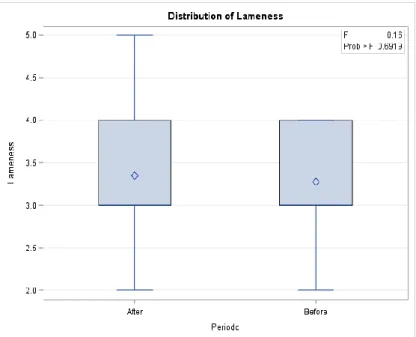
LIST OF GRAPHICS Graphic 1 – Boxplot of ‘Lameness score before trimming’ (Before) and ‘Lameness score after trimming’ (After) in Control group……….……….30
Graphic 2 – Boxplot of ‘Lameness score before trimming’ (Before) and ‘Lameness score after trimming’ (After) in Tri-Solfen group……….31
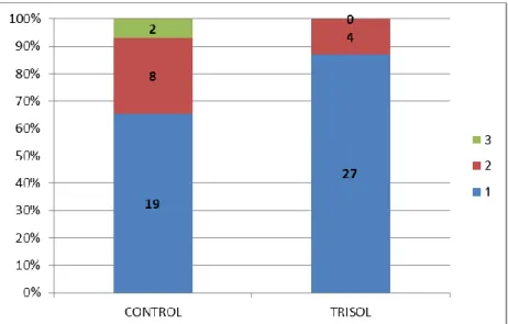
Graphic 3 – Number of animals in each reaction category to trimming in Control (CONTROL) and Tri-Solfen (TRISOL) groups………...31
Graphic 4 - Number of animals falling during the trimming procedure in Control (CONTROL) and Tri-Solfen (TRISOL) groups………...32
LIST OF TABLES Table 1 – Lameness scoring system………..6
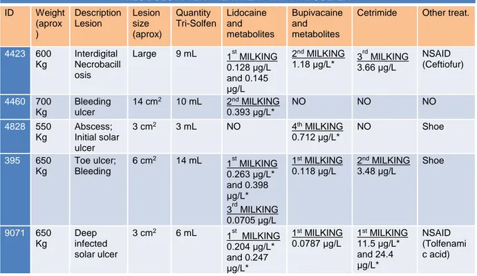
Table 2 – Specification of cases, including weigh, type of lesion and dosage used…………25
Table 3 – Residues’ values per animal, according to pathology and treatment….…………..29
Table 4 – Least squares means from algometry evaluation, on three separate periods and the association of periods, marked bold the differences………...32 LIST OF ABBREVIATIONS, INITIALS AND ACRONYMS
ADI: Acceptable daily intake BW: Body weight
MRL: Maximum residue limits
NSAID: Non-steroidal anti-inflammatories LOD: Limit of detection
LOQ: Limit of quantification
1
PART I - DESCRIPTION OF THE TRAINING PERIOD
The following report has the purpose of presenting the activities which took place during the sixth year of the masters in Veterinary Medicine. The main goals of the internship were to reinforce the previous knowledge acquired during the first five years of academic education, developing clinical reasoning and getting in touch with farm animal’s reality. This period of training was divided in two separate parts, the first one in Lisbon Veterinary Medicine Faculty and the second one in the Associação Agrícola de São Miguel.
The first part took place between September 2017 and February 2018, integrated in Farm Animals Clinic curricular unit, under the supervision of Professor George Stilwell. Multiple sides of clinical practice were approached, including internal medicine, preventive medicine, surgery, obstetrics and reproduction. Clinical field work was developed in farms in the region of Lisbon and Ribatejo, namely:
- One semi-intensive beef farm, where the main focus was on this type of animal production problems, such as obstetrics, parasitic diseases or fattening period diseases (e.g. Bovine Respiratory Disease).
- Three intensive dairy cow farms, being the main clinical cases: obstetric and reproduction related pathologies, lameness or problems concerning milk production.
- The national zootechnic station, concerning a much large spectrum of species, including beef, dairy cows, pigs, sheep and goats.
- One intensive goat farm, for milk production, where contagious ecthyma or orf and caseous lymphadenitis took a substantial role.
In most of the cases, the patients followed were submitted to physical examination, complementary exams, elaboration of a differential diagnostics lists, proceeded by clinical decision and institution of appropriate therapeutics. Several samples were collected for further analysis on Lisbon Veterinary Medicine Faculty laboratory, from faecal samples for parasite search, to encephalon samples with suspect bacterial meningitis. In approximately 90 field service cases, we performed two C-sections, two surgical corrections of abomasum displacement, one resolution of vaginal prolapse, seven necropsies, one calf dehorning, three abscess drains, six obstetric interventions, seven reproductive interventions, one sanitation of a herd, approximately fifty cases of lameness and about sixty seven internal medicine cases. Animal welfare was always taken in consideration, making sure that no animal would be submitted to unnecessary procedures or suffer pain.
Over this first period of training and has additional knowledge, I attended the XIX Jornadas da Associação Portuguesa de Buiatria and to the V Jornadas Técnico-Veterinárias do Campo Branco, getting to know new approaches in ruminants medicine and meeting colleagues working in the field. Also, and arising from the field service performed during these first months, I was co-author of the scientific poster “Caso clínico atípico de
2
besnoitiose (Besnoitia besnoiti) num touro limousine” (Appendix I) presented in the XIX Jornadas da Associação Portuguesa de Buiatria; author of the scientific poster “Aplicação tópica de anestésicos locais para controlo da dor durante a aparagem curativa de lesões podais de vacas leiteiras – dados preliminares“ (Appendix II) presented in the 8º Encontro de Formação da Ordem dos Médicos Veterinários; and co-author of an abstract on “Use of topical anaesthesia to control pain during and after trimming hoof lesions in dairy cows” (Appendix III) accepted for oral communication at the 2018 World Buiatrics Congress.
On the second part of my training, from April to June 2018, I followed the field service team of Associação Agrícola de São Miguel in Azores, supervised by Dr. João Vidal. Along this period I got in touch with several clinical cases in dairy cows, the main animal production in this island. I was involved in the diagnosis and treatment of cases related to internal medicine, surgery, herd health, milk quality services and reproduction management. Getting in touch with field service reality was a major gain to my academic education and a very fulfilling experience.
3
PART II – EXPERIMENTAL STUDY: USE OF TRI-SOLFEN TO CONTROL PAIN DURING TRIMMING OF HOOF LESIONS IN DAIRY COWS
1. LITERATURE REVIEW
1.1. DIGITAL ANATOMY
Bovine digital anatomy is similar in both thoracic and pelvic limbs, only changing the designation of the ventral surface from palmar to plantar respectively. In the extremity of both limbs, phalanges form two functional medial digits (III and IV), separated by the interdigital space, and two vestigial lateral paradigiti or dewclaws (II and V). Only digits III and IV have three phalanges, the proximal (P1), the middle (P2) and distal (P3) phalanges. The functional digits are the ones with surface contact and responsible for weight bearing. Branches of the axillary arteria and vein are responsible for the thoracic limb irrigation and drainage. In the pelvic limb, branches of the external iliac arteria send their flux to the homonym vein, creating a complex venous-arterial flux with huge capillarity.
In the thoracic limb, palmar innervation comes mainly from the medial and ulnar nerves and the dorsal innervation from the superficial branch of the radial and dorsal branch of the ulnar nerves. In the pelvic limb, plantar nerves derives from the tibial nerve, and the dorsal nerves originates from the superficial and deep peroneal nerves (Sisson & Grossman, 1986; Budras & Habel, 2003).
The distal extremity of the digit includes three primarily structures: ungula, corium and bones with their associated structures (vessels, nerves and ligaments).
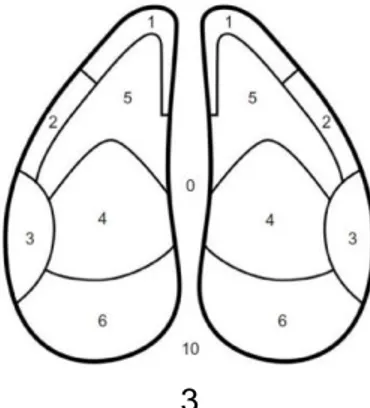
The ungula is the corneal structure giving distal external protection to the digits, covering the skeletal and soft tissue parts. Three distinctly modified layers are present in the hairless skin area in compassing with the haired skin: subcutis, dermis and epidermis. This three layers will have different modifications along the hoof forming five segments: periople, corona, wall, sole and bulb (Budras & Habel, 2003). In the palmar/plantar aspect of the claw it is possible to distinguish the heel, the sole, the white line (area that connects the wall with the sole), and the toe (Figure 1).
Figure 1 – Claw zone diagram: (1) white line at the toe, (2) abaxial white line, (3) abaxial heel - wall junction, (4) sole – heel junction, (5) apex of the sole, and (6) heel (adapt from Risco & Retamal, 2011).
4
Growth is done along the dorsal wall, abaxial and axial from the corona, and ventrally, growing approximately 5 mm per month. The horn tissue will also surround and protect the corium (Stilwell, 2013).
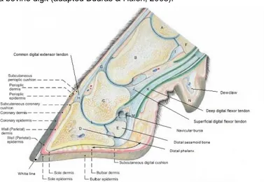
Corium is a live tissue responsible for producing and giving support to the keratinise tissue and is divided in: papillary corium, lamellar corium and digital cushion. It is supplied by nerves and vessels. The lamellar corium is the primary suspensory tissue for the suspensory apparatus of P3 (Figure 2), by virtue of a series of laminar folds anchored on the abaxial, dorsal, and axial surface of P3, and extend outwards to interdigitate with the lamellae of the wall. Beneath P3 there is a support structure composed of loose connective tissue from the solar and perioplic corium, and caudally by the digital cushion. The digital cushion is an important support structure composed of loose connective tissue and varying amounts of adipose tissue (Shearer & van Amstel, 2001).
The internal structures of the digit includes: the distal phalanx (P3), tendons (the common digital extensor tendon and the deep digital flexor tendon), the distal sesamoid bone (navicular) and the navicular bursa (Sisson & Grossman, 1986; Budras & Habel, 2003). The distal phalanx is supported only by connective tissue, ligaments and tendons, creating a 30º to 45º angle with the soil (Stilwell, 2013).
Figure 2 - Sagittal section of a bovine digit (adapted Budras & Halen, 2003).
1.2. LAMENESS
Lameness is considered an abnormality of the normal gait in consequence of lesions, defects, injuries, diseases and/or other factors located somewhere in the limb or the rest of the body, inducing pain or discomfort, serving as a ‘being bothered’-signal given by the animal and as a strategy used by the animal in order to maintain a certain state of comfort or even welfare (Beusker, 2007). Van Nuffel et al. (2015) also describes lameness as a clinical manifestation of painful disorders, mainly related to the locomotor system, resulting in impaired movement or deviation from normal gait or posture, with severity variations. Diseases concerning the hoofs of dairy cattle are presented as one of the main multifactorial
5
disorders that affect these animals (Rabelo et al., 2013), mostly in those maintained in intensive productions (Stilwell, 2013). Pain and discomfort related with foot lesions are predominantly seen as behaviour alterations – lameness, more lying time, less social behaviours etc. (Manske, Hultgren, & Bergsten, 2002a; Capion, Thamsborg, & Enevoldsen, 2009).
Factors such as type of flooring, bedding, calving, management, behaviour or lack of functional trimming can enhance the risk of hoof diseases causing lameness (Holzhauer, Hardenberg, & Bartels, 2008). Due to lameness, severe welfare problems and production losses, like reduced milk yield, weight loss, culling, deaths, replacement cost, infertility, prolonged calving interval, veterinary expenses, drugs and additional stockman’s time will occur (Weaver, St. Jean, & Steiner, 2005). Whay et al. (1997) suggested that parturition and the associated husbandry changes were also critical for the development of lesions in the claws in heifers. Biomechanical causes concerning gait on dairy cows and contamination due to dirtiness leads to hoof lesions are more commonly present on the outer claw of the pelvic limbs, and when in the thoracic limbs, on the inner claw (Stilwell, 2013; Rabelo et al., 2013). In the pelvic limb, the inner claw both the heel bulb and the axial wall are less developed, and the sole is more concave and sloped axially; as for the outer claw, the sole is flatter and create a more stable weight bearing surface. These anatomic differences between the outer and inner claw will result in a less stable weight bearing surface in the inner claw where more weight is naturally displaced to its abaxial wall, especially when cows are housed on hard surfaces. On the other hand, the thoracic limb claws have similar shape and size and there is greater flexibility due to the anatomic arrangement of the shoulder, having a more stable weight distribution (Shearer & van Amstel, 2001).
The diagnosis of lameness is mainly made by observation of the animal standing and walking, since hoof lesions frequently cause primary and secondary chronical pain and a hyperalgesia state (Stilwell, 2013).
Lameness in cows are usually identified by the farmer, hoof trimmer or veterinarian detecting changes in cow gait, posture or behaviour or the presence of hoof lesions during routine trimming (Van Nuffel et al., 2015). Although early stages of lameness are difficult to identify in cows, since cattle tend to show little behavioural response until injuries are advanced due to their stoic nature, several authors tried to create subjective and objective measuring systems to stablish a more accurate and precise scoring and staging of lameness status. Flower & Weary (2009) evaluate gait assessment methods, discussing the reliability and validity of measures used. They considered that subjective methods provide immediate, on-site assessment and do not require technical equipment, however the results can suffer from poor inter and intra-observer reliability. Subjective scores can be consistent within and among observers, especially if the gait assessment scoring system provides detailed classifications of each category and the observers have been trained. For objective methods
6
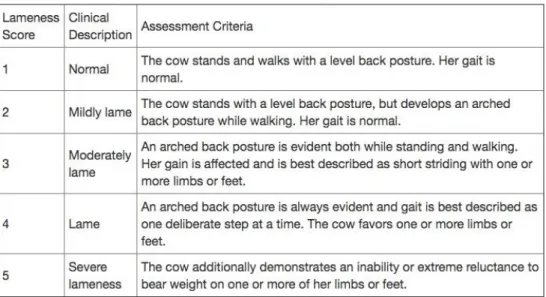
of gait assessment, they concluded that although it helps overcome subjective methods gaps, providing accurate and reliable data, often require sophisticated technology, for kinetic and kinematic measures, limiting their use on farms. Sprecher et al. (1997) developed a subjective 5-point lameness scoring system that assess gait, emphasising back posture as an important parameter of evaluation, trying to determine if the system could predict the reproductive future performance, risk of culling and detect early recognition of lame dairy cattle (Table 1). This system categorized cows into normal or mildly lame (1 and 2) contrasted with moderately to severely lame groups (>2), predicting that cows over group 2 would experience extended intervals from calving to first service, to conception, requiring additional services to become pregnant and be 8.4 times more likely to be culled. Currently, this scoring system is the most used and reliable to assess lameness (Schlageter-Tello et al., 2014), and was the basis in our study.
Table 1- Lameness scoring system (Sprecher et al., 1997).
In a study comparing different methods of lameness detection based on characteristics visually identified, Van Nuffel et al. (2015) described signs of lameness as: changes in gait patterns, considering the speed of walking, stride duration or weight distribution; changes in posture or body movement patterns, like arched-back posture or head movements; changes in weight distribution patterns; and changes in behaviour, such as longer duration spend at the resting, shorter duration at feeding places or grazing due to pain.
Lameness in dairy cattle is a major concern for animal welfare and productivity, needing a serious and intense intervention, despite cause-and-effect relationships have been difficult to determine (Capion et al., 2009). Lameness score of all the population is extremely important to control, determine and properly correct the risk factors (Stilwell, 2013; Schlageter-Tello et al., 2014). In summary, to find and treat properly lame animals it is crucial to periodically assess cattle based on locomotion or behaviour alterations (Van Nuffel et al., 2015).
7 1.3. HOOF PATHOLOGIES
Hoof diseases/lesions are normally divided in two categories: hoof/horn tissue pathologies and digital skin pathologies, frequently coexisting in the same animal and in the same limb (Stilwell, 2013). The most common non-infectious causes of lameness affecting the bovine digit are toe or solar ulcers, white line disease and traumatic lesions of the sole. Some of these illnesses are predisposed by metabolic disorders, like rumen acidosis and laminitis along with other physiological factors that can affect the integrity of the suspensory apparatus of the third phalanx. Mechanical factors, as hard flooring surfaces, overgrowth and altered weight bearing, or traumatic lesions of the sole, exacerbated by abrasive flooring conditions, will contribute to lameness complications (Weaver et al., 2005).
Diseases affecting the ruminant digital skin represent some of the most common and important causes of lameness in cattle, however, unlike lesions that specifically affects the claw, these diseases affect the skin of the interdigital space, heel bulbs and interdigital cleft. Although there are some differences between the way these conditions develop and appear, they are all caused by infectious agents capable of inducing inflammation and lameness (Risco & Retamal, 2011). The type of lesion, anatomic location and hoof structures involved that induce a more painful sensation or a more severe state of lameness still brings questions and disagreement among authors (Rabelo et al., 2013).
Next, a summary description of the most relevant podal pathologies will be presented. 1.3.1. Horn Tissues/Hoof pathologies
Laminitis
By definition, laminitis is a diffuse acute, subacute, subclinical or chronic inflammation of pododerm, usually in several digits. In acute stages, blood and serum exudation are present, followed by later (chronic) grooves on hoof wall, concave profile, widened white line and flat sole. Inherited factors, parturition, feeding stress (subacute ruminal acidosis or SARA) from change of dry cow concentrate diet to high production rations, exacerbation by trauma, as in excessive standing due to reluctance to use cubicles, are some of the predisposing factors considered for this disease (Weaver et al., 2005). Subclinical laminitis can have a serious negative impact since it leads to solar ulcers, shedding sole and white line disease (Stilwell, 2013).
Solar ulcer
This type of lesion is a circumscribed limited reaction of the pododerm often characterised by an erosive defect at the sole-heel junction (Weaver et al., 2005). The affected area will develop haemorrhage and necrotic tissue, reaching the solar surface and exposing the corium with cease growth of the horn tissue (Stilwell, 2013). Damage of the pododerm creating horn defects may appear as a secondary result of laminitis, poor trimming, hormonal factors and heel horn deformity (Weaver et al., 2005; Stilwell, 2013). As a consequence, a
8
laxity of the suspensory apparatus of the third phalanx will lead to a continuous impact of the phalanx on the solar corium (Stilwell, 2013). Shearer & van Amstel (2001) considered that the excessive growth of the toe area can be one cause for the rotation of the third phalanx, creating corium lesions associated with solar ulcers. The caudal border of distal phalanx where the deep digital flexor tendon attaches, is usually in the point of pressure (Weaver et al., 2005). In most cases the lateral posterior claw is the more affected one possibly because of excessive weight-bearing following horn overgrowth (Weaver et al., 2005). Granulation tissue appears where the lesion is in an attempt of healing which can prolapse, increasing the pain, and serve as an entrance to secondary bacterial infections, leading to osteomyelitis, arthritis, septic interphalangeal arthritis and podal abscess (Stilwell, 2013). Hard floor surfaces, hoof overgrowth or loss of the impact protection mechanisms, such as the digital cushion, will contribute to an aggravation and more severe states of these lesions. Therefore, dairy cows are more likely to develop solar ulcers due to housing conditions and type of flooring (Stilwell, 2013).
Toe ulcer
This lesions appear on the anterior extremity of the claw, leading the animal to bear the weight in the heel area. Traditionally was correlated with laminitis, when downward displacement of the apex of the third phalanx caused pressure necrosis of the corium in the toe region with toe ulceration as a consequence. However the development of this lesion is still not fully known (Shearer & Amstel, 2009). Some studies present the relationship between subclinical laminitis with lesion of the toe arteries as a cause, when other authors considered the excessive trimming in association with hard surfaces or floors with too much inclination (Stilwell, 2013). Toe ulcers are a very painful claw lesion with high production cost, as milk production decrease and weight loss is significant (Stilwell, 2013).
White line disease
Being the white line an area with less resistance, predisposing factors such as permanent humidity and laminitis can induce and exacerbate lesions (Stilwell, 2013). White line disease is characterized by an abaxial, or less commonly axial, wall separation from laminae at sole-wall area extending proximally, with cavity impacted with mud and faeces leading to the eventual development of an abscess. The development of the lesions follow the sequence: necrosis of wall laminae caused by pressure, and possibly also of solar laminae, followed by under-running and septic laminitis tracking progressively more proximally after entry of purulent micro-organisms, with absence of natural drainage distally due to impacted material (Weaver et al., 2005). Tissues of the white line giving in will allow foreign bodies to penetrate, with separation of the fibre connections and entrance of micro-organisms (Stilwell, 2013). When an abscess is present the primarily responsible micro-organism is Trueperella pyogenes (Weaver et al., 2005), possibly creating fistulas though the corona area (Stilwell, 2013). This lesion is predisposed by abnormal horn production resulting from laminitic insult;
9
insufficient hoof trimming; or related with previously events of the peripartum (Weaver et al., 2005).
Corkscrew claw
A corkscrew claw is a claw twisted throughout its length in a configuration that displaces the abaxial wall by up to 360°, with one or both lateral posterior claws affected. Although bone molding is present, it is not known whether this is a matter of cause or effect. Periarticular exostoses develop around the distal interphalangeal joint, probably from strain of the distal abaxial collateral ligament. Pressure from the exostosis on the dermis of the wall possibly accounts for the excessive growth of the abaxial wall (Greenough, 2012). Normally this condition only presents itself in animal over three year old and a heritable component is considered (Stilwell, 2013).
Heel erosion
This disorder is an irregular loss of bulbar horn in form of multiple blackish pit or pock-like depressions or later deeper oblique grooves, usually affecting posterior digits more severely than anterior (Weaver et al., 2005). Since dairy cows hoofs are permanently exposed to humidity with low pH levels due to manure, typical of the intensive production system, maceration and destruction of heel tissues takes place easily (Stilwell, 2013). In addition to these chemical and physical actions, Dichelobacter nodosus and Fusobacterium necrophorum can be involved in the erosion process (Weaver et al., 2005).
1.3.2. Digital Skin related pathologies Interdigital necrobacillosis
Interdigital necrobacillosis is an acute inflammation of subcutaneous tissues of interdigital space and adjacent coronary band, spreading to dermis and epidermis, caused by an interdigital microtrauma and post infection with Fusobacterium necrophorum, Bacteroides melaninogenicus and other organisms (Weaver et al., 2005). Advanced cases can develop to digital septic arthritis and eventually release septic thrombi which can trigger endocarditis (Weaver et al., 2005; Stilwell, 2013). The interdigital space is more prone to this infection, providing perfect anaerobic conditions and trauma location.
Digital and interdigital dermatitis
Digital dermatitis is a circumscribed superficial ulceration of skin bordering coronary margin at heels, occasionally more dorsally, being the major lameness problem in some farms. Although not completely clarified, some authors believe there is an involvement of Treponema genus spirochaete, Borrelia burgdorferi, Dichelobacter nodosus and Campylobacter spp. (Weaver et al., 2005). It is a contagious disease, with high humidity and poor hygiene predisposing dairy cattle to develop this condition (Stilwell, 2013). Interdigital dermatitis is an inflammation of interdigital skin without extension to deeper tissues, and variable associated disturbance of horn growth (Weaver et al., 2005). There is no consensus
10
through the scientific community if the difference between digital and interdigital dermatitis is only the localization and extension of the lesions or if the ethology also differs (Stilwell, 2013).
Interdigital hyperplasia
A proliferative reaction of interdigital skin and/or subcutaneous tissues forms a firm mass, developing a skin hyperplasia with secondary ulceration (Weaver et al., 2005). The main causes are chronic infection, such as interdigital necrobacillosis; repeated trauma/irritation of the interdigital space; poor conformation; and is also inherited in some breeds. Most clinical cases are in adults of four to six years and in the posterior limbs (Weaver et al., 2005; Stilwell, 2013).
1.3.3. Functional and corrective trimming
Claw pathologies affect animal welfare and have economic implications, due to costs of treatment, earlier culling, and production losses, being influenced by management and genetics (van der Linde et al., 2010). Regular claw trimming can be beneficial to claw health and animal well-being. Bell et al. (2009) stated that good claw condition is achieved by proper attention to breeding for conformation, proper foot trimming and good foot hygiene prior to first calving has being one of the most important critical control point for the management of lameness in dairy heifers.
Studies on the pathogenesis of sole ulcers and white-line disease clearly show that claw overgrowth leads to disproportionate weight bearing and eventual claw disease. Restoration of appropriate weight bearing within and between claws can be achieved by hoof trimming (Shearer & van Amstel, 2001). Manske et al. (2002b) studied and described the effects of regular claw trimming on the claw health of dairy cows. These authors compared two groups of cows, one of which received autumn trimming, while the other did not. Claw trimming in autumn proved to be associated with a significantly positive effect on the prevalence of lameness, and the risk of claw lesions requiring veterinary treatment between scheduled trimmings was reduced in trimmed relative to non-trimmed cows. These results are in favour of at least two claw trimmings per year, whether due to therapeutic or prophylactic effect. However, the optimal frequency of claw trimming is likely to be determined by factors specific to each farm and each animal. Holzhauer et al. (2008) also proved that dairy cows trimmed preventively at the end of the housing period had a significantly lower risk of getting solar ulcers than cows trimmed at the end of the grazing period.
Proper application of claw trimming methods provides correction of hoof overgrowth that, if left uncorrected, leads to overburdening of the affected claws and eventually to claw disease. When trimming is performed, claws are reduced to their normal shape and proportions, thereby returning the foot normal function and helping avoid lameness caused by improper trimming. Although claw trimming should be performed routinely one or two times per year, in
11
most dairy farms cow’s claws are trimmed only when they become lame or at the dry off period (Shearer & van Amstel, 2001).
To properly apply claw trimming procedures restraining method are extremely important. The best restraint method provides immobilization of the foot for good viewing of the claw and interdigital space and at the same time allows the operator to have free range of movement for trimming procedures, whether done with simple or more complex restraining systems (Shearer & van Amstel, 2001).
Claw trimming can be done by the veterinarian, a claw trimmer or the farmer himself. There are two different trimming technics: functional trimming and corrective trimming. When performing a functional trimming the aims are to restore appropriate weight bearing within each claw; to correct hoof overgrowth that leads to the overburdening of claws and to balance weight bearing between the claws of each foot; and to identify and correct claw lesions at an early stage. If claw lesions are detected, a corrective trimming procedure is required. The proposes of these procedures are to provide rest to the damaged or diseased claw by transferring weight to the healthy claw and to remove loose horn and thin, hard ridges that may cause damage to the underlying corium, always making sure that the corium is not damaged during the procedures. In severe painful cases or if the corrective procedures are unable to create sufficient difference in height between the two claws, application of a block attached to the sound claw is recommended. Cases with severe haemorrhage of the corium or in claw amputation should be addressed with topical treatments under a bandage, taking in account that it has to be removed in order to prevent environmental contamination and further complications (Shearer & van Amstel, 2001).
In digit skin pathologies, foot washes and topical treatments are the regular approaches. Different methods of foot wash in water have been introduced to improve claw hygiene without causing environmental disadvantage and health problems for cows or humans. Fjeldaas et al. (2014) showed that CuSO4 footbaths had a preventive effect on heel horn erosion lesions, as stationary automatic flushing of the hind feet with only water had no beneficial effect on interdigital dermatitis or heel horn erosion.
1.4. PAIN
The International Association for the Study of Pain describes pain as ‘an unpleasant sensory and emotional experience associated with actual or potential tissue damage, or described in terms of such damage’. Thus, pain is not only a physical sensation, but is usually associated with emotional and mental alterations, more or less extended, potentially leading to suffering (Stilwell, 2017).
Absence of pain is of course an essential feature of animal welfare. That is the reason the Farm Animal Welfare Council (FAWC, 1993) included ‘freedom from pain, injury or disease –
12
by prevention or rapid diagnosis and treatment’ as one of the ‘Five Freedoms’ as guidelines for the welfare of all species of livestock.
However, recognition of pain in ruminants is not easy. Wild cattle, from which domesticated animals descend, were preys, submitted to a strong evolutionary pressure to mask signs of pain and its implied weaknesses (Huxley & Whay, 2006 citing Phillips, 2002). Even if not displaying obvious pain behaviours, this does not mean that cattle do not experience it. To recognise signs of pain is a significant challenge for veterinarians, where they are expected to be able to diagnose, grade and treat pain in cattle (Huxley & Whay, 2006; Gleerup et al., 2015).
1.4.1. Nociception
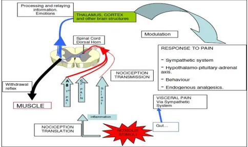
Nociception is the unconscious afferent activity produced in the peripheral and central nervous systems by noxious stimuli, with potential tissue damage, being described by many authors as the first and basic part of the pain mechanism (Figure 3) (Stilwell, 2009). The nociceptor is the primary sensory neuron that is activated by those stimuli, with characteristics thresholds or sensitivities that distinguish them from other sensory nerve fibres (Julius & Basbaum, 2001).
Nociception mechanism rests essentially on two stages – transduction and transmission. In transduction, occurring at the sensory endings of the nociceptors, the noxious stimuli (mechanical, temperature or chemical) is translated into electrical activity. Transmission is the propagation, by membrane depolarization, of the electrical impulses along the sensory nervous fibre to the central nervous system. It is here that occurs the first synapsis between neurons, with glutamate being the predominant excitatory neurotransmitter in all nociceptors. Modulation is also an important component of pain and by which transmission of pain impulses through the spinal cord are inhibited by descending reflexes that originate in the noradrenergic neurons (Stilwell, 2017).
13
When a noxious stimuli is first induced it causes what is called “physiologic pain” or “first pain”, serving as a protective biological function to active a response and repair potential tissue damage. This almost instant transmitted sensation travels through thinly myelinated A fibres, generating the defensive activity. “Chronic pain” happens as a result of C fibres activation, interpreted by the central nervous system as a dull, diffuse, aching or throbbing sensation. “Neuropathic pain” results from injuries to the nerve fibre, being important in animal welfare since it might be the cause of enduring pain after mutilations and for which treatment is very difficult. “Visceral pain” is an unique type of pain for which there are no first and second components, often being poorly localized, deep and dull and usually triggered by other kind of stimuli, namely stretching, compression or ischemia (Stilwell, 2009; Stilwell, 2017).
All nociceptors (except those coming from the head) have their first axons synapse in the dorsal horn of the spinal cord, namely on laminae I, II and V. It is here, by way of interneurons connecting to the ipsilateral ventral horn, that the reflex arch is produced allowing for rapid muscle contraction and body withdrawal from the stimuli source reducing further damage. The signals then travel through a spinothalamic tract or a spinoreticular tract of the spinal cord to several structures of the brain, namely the mesencephalon, thalamus, reticular formation, hypothalamus, limbic system and cortex (Stilwell, 2009). It is in the central nervous system that perception or consciousness of pain occurs, leading to an emotional response and eventually to suffering.
1.4.2. Pathological pain
Pathological pain can be triggered in different types of tissues and classified as inflammatory or neuropathic (Klaumann et al., 2008). Pain resulting from inflammation is very frequent in animals and may cause a great deal of suffering, reducing animal welfare and performance. It is much more difficult to manage than acute pain (Stilwell, 2009).
After tissue damage an inflammatory reaction usually occurs, with vascular components, fibroblastic components and tissue cell components being activated: blood vessels carry circulating precursors that are released into the area of injury and are activated by enzymes; mast cells release histamines and other substances; macrophages activate fibroblasts, which in turn release interleukin and Tumor Necrosis Factor (TNF); cyclooxygenase activates prostaglandin and leukotrienes and more. Pain may be exacerbated and triggering thresholds are reduced, when nociceptor terminals are exposed to these products of tissue damage and inflammation, called “inflammatory soup”. The acidic pH level of the inflammatory soup is also important in nerve sensitization. This will trigger the terminals to sensitize or excite the nociceptor by interacting with cell-surface receptors expressed by these neurons (Julius & Basbaum, 2001; Klaumann et al., 2008).
14
Another type of nociceptors, the silent nociceptors, become hyper-excitable when sensitized by the inflammatory soup. This leads to a “primary sensitization” or “primary hyperalgesia”, in which almost any stimulus is felt as pain, generating a constant state of pain. The secondary peripheral hyperalgesia occurs when local vasodilatation, plasma extravasation and extension of the inflammatory soup results in an additional amplification of the inflammatory response by reducing other nerve endings threshold to stimuli, producing pain even without tissue damage (Stilwell, 2009). Peripheral nociceptors’ activation also results in a use dependent neuronal plasticity in the spinal cord that modifies the subsequent performance of the nociceptive pathway by hyperalgesia (an increased or prolonged response to noxious inputs) or allodynia (pain caused by a stimulus that does not normally inflict pain) (Stilwell et al., 2009; Tranquilli et al., 2013).
Prolonged noxious stimuli produces greater sensitivity to subsequent stimuli. This hypersensitivity status is probably what occurs in cows with chronic lameness with pain continuing for a long time even after treatment of the primary hoof lesion (Whay et al., 1998; Stilwell, 2009).
1.5. ANIMAL WELFARE IN FEET LESIONS IN DAIRY COWS
1.5.1. Welfare impact
When under stressful production conditions dairy cows can more easily acquire production diseases, leading to pain or death, raising both ethical and economical concerns (Hultgren, Manske, & Bergsten, 2004). Hoof diseases and lesions causing lameness are one of the most important.
The Farm Animal Welfare Council (FAWC, 1997) stated that “lameness is an extremely painful condition and steps must be taken, as a matter of urgency, to reduce the incidence”. Gait alterations are usually a manifestation of discomfort or pain, caused mainly by claw lesion in dairy cows (Van Nuffel et al., 2015). Lameness, for reasons of prevalence and individual suffering, is considered to be the most severe welfare problem for dairy cows (Stilwell, 2013). Lesions of the hoof horn tissue are the source of most cases of painful lameness. Interdigital or digital skin lesions have a short duration period if properly treated, in contrast with claw lesions that can be long lasting even when treated (Hultgren et al., 2004). We know little of how much cattle suffer during a lameness episode. Pain is a subjective experience drawing on both physiological and emotional components (Whay et al., 1997). The absence of obvious signs of pain or lower locomotion scores in certain hoof lesions do not necessarily means the absence of pain sensation, indicating either that these lesions are causing different severities of lameness, or that the case definitions used is not sensitive enough to detect all lesions (and possibly discomfort) (Tadich, Flor, & Green, 2010).
Whay et al. (1997) tried to associate locomotion, claw lesions and nociceptive threshold in dairy heifers during the peri-partum period, describing the development of claw lameness in
15
heifers at the time of their first parturition and the relationship between claw pathology, gait score and hyperalgesia, as indicated by nociceptive thresholds. They determined that as lameness increases the nociceptive threshold significantly decreases, demonstrating sensitization to the stimulus. The increased sensitivity to a mechanical stimulus indicates that the limb is in a hypersensitive or hyperalgesic state. Even if not possible to distinguish between peripheral sensitization or spinal sensitization, this study confirms that the locomotion changes were likely to be due in part to the animal's hyperalgesic state and not only as a result of biomechanical restriction of movements.
In a study performed by Bruijnis et al. (2012) assessing the welfare impact of a hoof lesion, a specific impact of foot disorders on dairy cow welfare was shown, mirroring differences in foot disorder painfulness, duration and incidence. Pain induced by this type of lesion causes negative effects in all three domains, causing impaired health and functioning, suffering and affects the ability to perform natural behaviour, as well as compromising the longevity of cows, as the associated lameness and poor performance are important reasons to cull cows prematurely.
To reduce lameness cases, farmers need to be aware of the number of lame cows and the severity of lameness in their herd (Van Nuffel et al., 2015). The usually accepted methodologies to classify lameness stand on detect changes in gait, posture or behaviour of the cows, done using subjective methods such as visual observations for locomotion, which is easy to apply and inexpensive, making the implementation of regular and systematic assessment of gait an ethical obligation. Improvement in education and training, either in recognition of subtle signs of pain exhibited by dairy cows as well as in the acknowledgment of the benefits of analgesia, can minimise lameness negative consequences on animal welfare and health (Becker et al., 2013).
1.5.2. Pain assessment and management in feet lesions
Pain causes harmful effects in interconnected areas: animal welfare, arising ethical challenges; animal physiology, with biological effects; and productivity, having negative economic impact (Stilwell, 2017).
Cows change their way of walking to relieve pain, so an abnormal locomotion is considered an indicator of an underlying problem that induces pain (Flower & Weary, 2009). Due to dairy cattle stoic nature, many authors have been trying to properly assess pain in these animals by detection of several changes in their behaviour as signs of pain. A single sign or measurement cannot be used as an accurate information of how the animal is felling in a particular moment, since animals react differently to stress and pain (Stilwell et al., 2009; Stilwell, 2017).
A study performed by Gleerup et al. (2015) tried to assess bovine pain in general using the most significant signs of pain by creating a Cow Pain Scale. Attention towards the
16
surroundings, head position, ears position, facial expressions, response to approach and back position were the six signs considered as the more reliable and accurate. The resort to this scale showed substantial inter-observer agreement between the two observers and an easier evaluation of the progression of each clinical case.
Shearer et al. (2013) summarized the assessment of pain in lame cattle relaying on seven primary key-points:
- Locomotion or Lameness Scoring Systems
An arched back, already defined as the key behavioural change evaluated in the Sprecher lameness scoring system, is frequently associated with lameness, as is “bobbing” of the head during locomotion. Shortening or lengthening of the stride and the degree of abduction or adduction of the limbs also can be an indicator of problems related with the limbs. Others are: changes in claw placement, the alignment of the pin bones when walking, reluctance in the animal’s willingness to move (being frequently associated with lameness affecting multiple claws) and changes in the stance phase, resulting in the animal maintaining its weight on the sound limb for as long as possible to minimize weight-bearing time on the lame limb.
- Pressure Mats
This type of devices help to determine the contact pressure, contact area, and stance phase duration in the affected claws, and therefore to detect lameness when present.
- Weighing Platform
The use weighing platform was described to measure the redistribution of weight of cattle limbs that occurs in response to pain associated with lameness. Cattle redistribute weight to avoid uncomfortable surfaces and distribute weight away from a limb with discomfort primarily toward the contralateral limb.
- Nociceptive Threshold Tests
The hyperalgesia caused by lameness, can create a more sensitive and exaggerated reaction to stimuli compared with sound animals. When a mechanical pneumatic blunt pin is pressed on the dorsal aspect of the digit with gradually increasing pressure, the pressure at which the reaction occurs is recorded as the nociceptive threshold. This method allows to quantify regional sensitivity and, potential pain objectively. A pressure device, an algometer, can be used in these cases, showing that the animal retracts the limb when the pressure reaches the pain threshold.
- Heart Rate
Heart rate means tends to be less in lower lameness scoring, in comparison to higher lameness scorings.
17
Measurement of cortisol levels is used to quantify response magnitude and duration to acutely painful states and procedures and these seem to correspond to the predicted noxiousness (McCarthy et al., 2016).
- Accelerometers
Continuously measuring gravitational force in multiple axes, accelerometers’ values can be processed to determine activity and postural behaviours occurring on lameness situations.
To effectively manage pain in cattle, reducing primary and acute pain and preventing secondary (central or peripheral) hypersensitivity should be combined (Nolan, 2000 cited in Stilwell, 2009). The main concern of practitioners and farmers is usually the first or acute pain, often neglecting the control of pathologic or chronic pain, since it is less obvious, the control is more expensive and it does not pose safety problems for the operator (Stilwell , 2009).
Pain management in lame cattle can be approached according to the following principles (Shearer et al., 2013):
- Corrective Trimming
Pain relieve and a faster recovery period from claw lesions can be accomplished by reducing the possibility of further complication associated with abscess formation, post-procedural pain minimization and adjust weight bearing on diseased or damaged claws.
- Anaesthesia of the Lower Limb and Foot
Anaesthesia is indicated whenever it is necessary to perform procedures that may be very painful to the animal. There are at least two methods to achieve this: intravenous regional anaesthesia under a tourniquet or ring block, both easy to perform under field conditions. These methods not only alleviates discomfort, but also lessens movement of the foot associated with corrective trimming adjacent to sensitive tissues of the corium, facilitating trimming procedures and reducing the potential to accidental damage of healthy tissues.
- Analgesia
A multimodal approach using analgesics, such as local anaesthetics, NSAIDs, and sedative-analgesics, may be beneficial when treating lame cattle. The use of local anaesthetics, such as lidocaine, reduces gait scores and effects distribution of weight acutely. Non-steroidal anti-inflammatories (NSAID) (e.g. flunixin meglumine) have demonstrated substantial analgesia in induced lameness models illustrated through modifications of gait and improved pressures placed on the affected foot and claw. However, in field trials, NSAID use have yielded variable results with only mild improvement in locomotion score and in nociceptive thresholds (Chapinal, de Passillé, Rushen, & Wagner, 2010; Shearer et al., 2013). Animals treated under sedative-analgesics, such as xylazine, demonstrated lower levels of cortisol and stood longer and had reduced gait scores in the first hour (Shearer et al., 2013).
18
The two most important reason for not using proper analgesia in cattle are the ability to identify signs of pain, sometimes suppressed by these animals, or because the economic consequences of not using analgesia are negligible (Stilwell , 2009). On a survey performed by Becker et al. (2013), concerning attitudes taken against pain and painful procedures in dairy cattle, it was showed that reducing pain to the lowest possible level when treating lameness cases was much more important to farmers than to bovine practitioners. Also common painful procedures, such as corrective trimming of solar ulcers, were frequently performed without any kind of anaesthesia by practitioners, farmers and claw-trimmers, even if the cost was not a limitation to most of them. The level of pain caused by lameness treatments and the sensitivity of dairy cows to pain was not unanimous, showing lake of education and training, even as the awareness on the benefits of proper pain management. The reasons pointed out by practitioners for not properly manage severe pain in some pathologic conditions are reduced cost-effectiveness, low practicability (few long acting drugs available), long withdrawal periods and lack of legal license (Stilwell , 2009). However, it is the veterinarian ethical obligation to sensitize and encourage towards the implementation and importance of pain recognition and control.
1.6. LOCAL ANAESTHETICS
In animal production, local or regional anaesthesia are preferred methods for pain management due to practicability and economic reasons.
Local anaesthetics reversibly bind to sodium channels and block impulse conduction along nerve fibres, by inhibiting the generation and conduction of ionic fluxes responsible for membrane depolarization (Lomax, Sheil, & Windsor, 2013; Tranquilli et al., 2013). The interruption of neural transmission in sensory afferent nerves or tracts by a local anaesthetic drug after local tissue infiltration, regional nerve blocks, or epidural injection effectively prevents or reduces pain or nociceptive input. Analgesia in the desensitized area also removes the immediate secondary (central) sensitization to pain and reduces the central facilitation of the nociceptive pathway, preventing or reducing pain escalation response (Lomax, Sheil, & Windsor, 2008). Absorption of local anaesthetics may also be accomplished through mucous membranes or damaged skin to reach the nerve fibres (Tranquilli et al., 2013). Systemic absorption is generally slow, keeping the active substances concentrated at the site and slowing its metabolism, prolonging intensity and duration (Lomax et al., 2013). The duration of effect of local anaesthetics at the site is inversely related to the rate of systemic absorption. Vascular absorption rate varies directly with the vascularity of the injection site and the physicochemical and pharmacological properties and dose of the local anaesthetic. Lidocaine (being a good vasodilator) removal from the site of injection is faster, making lidocaine a shorter-acting anaesthetic (60 to 120 minutes). On other side, bupivacaine is slowly “washed out” from isolated nerves in vitro, and it is not readily removed
19
by the bloodstream from nerve membranes, making its duration of action long (180 to 480 minutes) (Tranquilli et al., 2013).
In cattle the anaesthesia of the foot may be accomplished by: ring block, consisting in a local anaesthetic infiltration of the tissues around the limb; intravenous regional anaesthesia, with local anaesthetic injection into an accessible superficial vein in an extremity isolated from circulation by placing a tourniquet on the animal’s leg; regional analgesia, desensitizing specific nerves; or by general anaesthesia especially in aggressive animals or for procedures requiring complete immobilization for asepsis and safety during operation (Tranquilli et al., 2013).
Two local anaesthetics are included in a topical gel, called Tri-Solfen. This product has been tried in several different medical procedures in different species, with the aim of providing local anaesthesia in an easy non-invasive way. Tri-Solfen is a spray-on topical anaesthetic, haemostatic and antiseptic gel agent that contains lidocaine, bupivacaine, adrenaline and cetrimide (Lomax et al., 2008, 2013). Application of this gel in open wounds leads to a rapid and prolonged anaesthesia of the area, as well as prevented subsequent pain escalation response. Also, the presence of a gel base can act as a barrier, attenuating pain by coating damaged nerve endings and providing a barrier against ongoing environmental exposure and tactile stimulation (Lomax et al., 2013). The efficiency of this anaesthetic combination was the subject of our study and more detailed explanation will be given in the next topic.
1.6.1. Tri-Solfen
Tri-Solfen (Bayer Animal Health, Australia) is a commercially available only in Australia as a topical anaesthetic, haemostatic and antiseptic formulation for the alleviation of pain in farm animals. Its composition includes lidocaine hydrochloride (40.6 g/L), bupivacaine hydrochloride (4.5 g/L), adrenaline acid tartrate (24.8 mg/L), and cetrimide (5.0 g/L) in a gel base (Australian Pesticides and Veterinary Medicines Authority [APVMA], 2005). The association of the two local anaesthetics was selected to deliver rapid onset of wound anaesthesia action of lidocaine with the prolonged duration effect of bupivacaine. Adrenaline is included as a vasoconstrictor, increasing local anaesthetic compounds at the site of administration and reducing absorption of the compounds away from the traumatised nerve tissue and also reducing the risk of systemic toxicity, and increasing local activity. The vasoconstrictor properties of adrenaline also reduces bleeding, and slows the blood flow to the wound, thereby suppressing the inflammatory cascade and resulting in a reduction on associated pain caused by accumulation of inflammatory mediators. Cetrimide, a topical antiseptic compound, provides antisepsis conditions for procedures performed in non-sterile farm environments. Tri-Solfen is a non-sterile liquid solution, designed for on-farm use, supplied in multi-dose backpack containers with a metered dose spray applicator. The