* Extracted from the thesis: “Tecnologia para o autoexame ocular: estudo comparativo sobre uso da cartilha impressa versus virtual”, Programa de Pós-Graduação em Enfermagem, Universidade Federal do Ceará, 2014.
1 Centro Universitário Estácio do
Ceará, Fortaleza, CE, Brazil.
2 Universidade Federal do Rio Grande
do Norte, Natal, RN, Brazil.
3 Universidade Federal do Ceará,
Programa de Pós-Graduação em Enfermagem, Fortaleza, Ceará, Brazil.
Received: 06/12/2017 Approved: 12/19/2017 Corresponding author:
Nelson Miguel Galindo Neto BR 232, Km 208, s/n
CEP 55200-000 – Pesqueira, PE, Brazil [email protected]
ORIGINAL ARTICLE DOI: http://dx.doi.org/10.1590/S1980-220X2017024703326
Technology for performing ocular self-examination: comparison
between printed and virtual booklets*
Tecnologia para realização do autoexame ocular: comparação entre cartilha impressa e virtual Tecnología para la realización del autoexamen ocular: comparación entre cartilla impresa y virtual
Jennara Candido Nascimento1, Maria Alzete Lima2, Lívia Moreira Barros3, Nelson Miguel Galindo Neto3, Lorita Marlena Freitag Pagliuca3, Joselany Áfio Caetano3
How to cite this article:
Nascimento JC, Lima MA, Barros LM, Galindo Neto NM, Pagliuca LMF, Caetano JA. Technology for performing ocular self-examination: comparison between printed and virtual booklets. Rev Esc Enferm USP. 2018;52:e03326. DOI: http://dx.doi.org/10.1590/S1980-220X2017024703326
ABSTRACT
Objective: Comparing the results of the ocular self-examination performed with the aid of printed and virtual versions of an educational booklet. Method: A quasi-experimental study carried out in a state (public) school of a capital in northeast Brazil, with 100 students equally divided into control and intervention groups according to age, gender, schooling and economic status. Pearson’s Chi-square test and Fisher’s exact test were applied with a significance level of 5%. Results: The results of the self-examination obtained by the virtual and printed booklets were statistically similar, except for the item ‘Alterations of the pupillary reflex’, in which the virtual booklet was more effective for its identification (p=0.049). Conclusion: The printed and virtual versions of the ocular educational booklet have similar efficacy for performing ocular self-examination.
DESCRIPTORS
INTRODUCTION
The right to good eyesight must be recognized as an expressive component of public health, above all because it enables the full development of human intellectual and laboral potential. Knowing the complaints and eye problems of a geographical region enables planning public resources and developing strategies aimed at reducing and controlling visual impairment and blindness.
A relevant factor that makes it difficult to cope with eye conditions is non-identification by the population of the need to seek ophthalmologic care. In turn, not seeking the appropriate care results in increased assistance at emergency services, and sometimes with diseases that constitute outpa-tient care in which the condition tends to be more severe(1-2). In this context, we can highlight the need to contribute to recognizing vision-related changes so that the awareness of the need for specialized care becomes clear. In order to achieve this recognition, an effective strategy that should be taught to the population is ocular self-examination. This study focuses on the self-analysis performed by the individ-ual in order to identify visindivid-ual changes, impairment of visindivid-ual acuity and/or abnormalities in ocular structures.
It is therefore pertinent to contribute to extending care beyond clinics and hospital units with educational actions that use technologies such as folders, booklets and manuals for self-care of the eyes(3). This fact points to the need to develop materials on the subject that favor screening for reducing cases of blindness due to preventable causes(4). Thus, virtual and printed versions of the booklet were created(3,5) regarding ocular self-examination.
In addition to creating (the material) and in line with Evidence-Based Practice (EBP), it is important to compare the types of technologies so that the choice for the tech-nology version to be used is based on scientific evidence. Therefore, the purpose of this study is to compare the results of an ocular self-examination carried out with the aid of the printed and virtual versions of the educational booklet.
METHOD
This was a quasi-experimental study developed in the secondary and high school levels at a public school in Fortaleza, CE, selected due to the fact that the students did not undergo ocular evaluation prior to the data collection, and also due to the absence of a health promotion program on this subject in the institution.
In this study two educational technologies focused on ocu-lar self-examination were evaluated: a booklet designed and val-idated by Caetano and Pagliuca(6),which consists of a 14-page illustrated and colorful booklet printed on coated paper, com-prising a cover, presentation, necessary material and description of the ocular self-examination technique; and a virtual booklet developed and validated by Lima et al.(3),following the analysis steps and planning, modeling, implementation, evaluation and maintenance by Falkembach for its construction.
The theme, the learning objective and the content for the virtual version were adapted from the printed version of the booklet to be used online, considering the theoretical
premises on distance education(3). Both booklets present sim-ple information that enables the reader to carry out an ocular self-examination based on verifying their visual acuity (far and near), the visual field (peripheral vision and central vision), ocular movement, and external ocular structures, allowing for early identification of alterations present in the eyes.
The population was composed of students enrolled in the secondary and high school education, and the sample was selected by convenience. Inclusion criteria included having physical conditions for performing an ocular self-examination and basic ability in computer use, which included turning the equipment on and off, accessing icons on the desktop, and han-dling the basic tools offered for navigation. The exclusion cri-terion was presenting ophthalmological evidence of blindness. The formula used for sample calculation to compare groups was: n=(Zα+Zβ)2x2xP(1-P)/d2; considering Zα (con-fidence level) of 95%, Zβ (power) of 80%, and after conduct-ing a pilot study, P (proportion of occurrence of the outcome) of 44% and d (clinically important difference) of 30%. Thus, 43 participants were obtained for each study group; however, a total of 50 participants was obtained for each group after a 4% increase due to possible losses (experimental and control), totaling 100 people to integrate the sample. The sampling was done by convenience, and participants’ distribution into the groups was performed by the main researcher and occurred with pairing considering age, gender, schooling and economic situation. Participant blinding could not be carried out, since the purpose of the study was presented to the volunteers due to ethical reasons, and they became aware to which group they had been allocated to when they were assigned the printed or virtual version of the booklet.
Data collection took place from December 2012 to December 2013. It should be noted that this time interval was required since the research took place in a school envi-ronment which has breaks, vacations and evaluations/exams when it is impossible to collect data.
An instrument was used to record participant characteri-zation containing variables on the results obtained during the ocular self-examination. These variables included: far and near visual acuity for each eye, evaluation of the external ocular structures (eyelids, conjunctiva and sclera, roundness, flatness, color and uniformity of the iris, isochoria and pupillary pho-torreaction), the presence of alterations in peripheral vision (blurring and visual dimming, diplopia and anopsia), and changes in central vision (blank areas, wavy vision, blind spots and flashing lights for monocular vision). The instrument also provided a space for written complaints. A blank space was left available for the variables “far and near visual acuity” for recording the row for each scale in which it was possible for the reader to see during the examination. The available answers for the other variables were “yes” or “no”, if the reader noticed any changes during their ocular self-examination.
The Jaeger card was used to evaluate near visual acuity, known as a portable visual tracker, which differs from the first scale by presenting fewer lines (six in this case, J1 to J6), and by the graduation between them which ranges from 0.37 m to 1.25 m. The student holds the card 33 centimeters away from their eyes and test one eye at a time, identifying up to which line they are able to read well. In this case, vision was considered altered when the individual only visualized the larger lines with spacing greater than J2. It is noteworthy that the students were instructed to wear their glasses and/or corrective lenses during the test in both assessments.
For evaluating external ocular structures, the presence of secretion, edema, redness, nodules, lesions, trichiasis, entropion and ectropion were considered as altered results. Other symptoms such as tearing, burning and pain upon movement have also been evaluated as they are indicative of inflammatory processes such as scleritis and episcleritis. The students evaluated both eyes one at a time with the aid of a mirror and cotton swabs. We emphasize that both versions of the booklet present illustrations corresponding to each alteration to help identify abnormal findings.
Regarding the pupil and iris, isochoric, light-reactive and accommodative pupils were considered normal. Alterations of the pupillary reflex to light, presence of nystagmus, rhyth-mic, involuntary and bilateral movements of the eyeball, in addition to double vision represented the main alterations that should be recorded in the instrument.
The Amsler grid was used to detect changes in the cen-tral visual field. For this investigation, the main abnormality recorded by the students was distortion in the lines that make up the screen associated with central blind spots. The expected normal response consists of undistorted visualiza-tion of the lines that compose the grid.
In conducting this study, this instrument was validated by three doctorate students with experience in the ocular health theme, in a previous study regarding the validation phase of the printed booklet(7). Before the definitive data collection began, the instrument was pre-tested with 19 students duly enrolled in a public school. The test duration ranged from 17 to 30 minutes, in which students could read the guidelines included in the printed booklet, perform the self-examination based on the information presented and fill out the data collection instrument. No need for instrument modification was observed after completion of the pre-test. The students’ invitation to participate was done by the principal researcher in the classrooms and during class hours, according to previously determined schedules with the school coordinator and after the teachers had given con-sent to the schedule. All the groups were informed about the research that would be conducted and were instructed that those interested in participating should attend the reserved room.
The evaluations were performed in the school in a quiet private room with good lighting. The materials required for the examinations were made available over the table: Reduced Snellen Card, Jaeger scale, Amsler Grid, five meters of string, tape, cotton swabs, mirror, alcohol gel, newspaper, the instrument for recording the results and pens.
The printed version of the booklet was used by the morn-ing period students, while the virtual booklet was used by the afternoon students. Printed copies of the booklet were made available for the control group as the participants individually entered the room provided for the examination, and they were welcomed by a member of the research team who instructed them to read the material and then to start performing the steps contained in the booklet for carrying out the ocular self-examination. All the evaluations that comprise the ocular self-examination were listed in this area. To perform the eval-uations, the students simply had to move the cursor to the icon marked with the name of the exam to be performed and click. Then, a new window opened containing the necessary instruc-tions for the evaluation. This procedure was repeated for the other tests. Each student was asked to perform the exams according to their understanding of the reading. Questions were clarified at the end of each session after completion of the self-examination and after the material had been returned. For the intervention group, a computer with the elec-tronic version of the booklet (available in the desktop area) was made available. Participants in this group were instructed to click on the icon titled “Ocular self-examination” to access the booklet, in which all steps of the self-examina-tion were available for access. In addiself-examina-tion, the electronic version provided a video tutorial that could be accessed by the participants.
Thus, after using one of the versions of the booklet (printed for the control group and virtual for the intervention group), the participants then performed the self-examination once. Two trained nursing students provided support during the collection period and accompanied the students during the self-examina-tion stages, but without interfering in their conducts.
In order to analyze the data, measures of central ten-dency and dispersion (means and standard deviation) were used, and Pearson’s Chi-square and Fisher’s exact tests were performed for comparison between proportions. The level of statistical significance for the tests was set at 5%.
The study complied with national and international standards of ethics for research with human beings, in accordance with Resolution 196/96 of the National Health Council, and was approved in October 2012 by the Ethics Committee of the Universidade Federal do Ceará (opinion number 118.180).
RESULTS
There was female prevalence for both groups in the total of 100 students who performed the ocular self-examination. Regarding age, both groups had a predominating age group of between 15 to 18 years (76.7%).
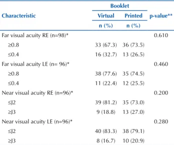
Table 1 – Synthesis of far and near visual acuity evaluation results using the ocular self-examination among students – Fortaleza, CE, Brazil, 2014.
Characteristic
Booklet
p-value**
Virtual Printed
n (%) n (%)
Far visual acuity RE (n=98)* 0.610
≥0.8 33 (67.3) 36 (73.5)
≤0.4 16 (32.7) 13 (26.5) Far visual acuity LE (n= 96)* 0.460
≥0.8 38 (77.6) 35 (74.5)
≤0.4 11 (22.4) 12 (25.5) Near visual acuity RE (n=96)* 0.200
≤J2 39 (81.2) 35 (73.0)
≥J3 9 (18.8) 13 (27.0) Near visual acuity LE (n=96)* 0.280
≤J2 40 (83.3) 38 (79.1)
≥J3 8 (16.7) 10 (20.9)
*The numbers differ for each variable due to not recorded responses. RE − right eye; LE – left eye; **Chi-square test of linear trend.
Note: (n=100).
According to Table 2 (evaluation of external ocular structures), the students obtained a higher proportion of results considered normal during the investigation of the eyelids (96%), conjunctiva and sclera (92%), iris (90%) and pupil (69%). However, the evaluation of the pupil showed a statistically significant difference (p=0.049), which indicates an association between the use of the virtual booklet and recognition of alterations in the pupillary reflex.
Table 2 – Synthesis of the results regarding external ocular struc-ture evaluation using the virtual and printed version of the book-lets among students – Fortaleza, CE, Brazil, 2014.
Booklet
p-value**
Virtual Printed
n (%) n (%)
Eyelids: warning signs during the
examination? (n=98)* 0.117 Yes 0 (0.0) 4 (8.2)
No 49 (100.0) 45 (91.8) Conjunctiva and sclera: warning
signs during the examination?
(n=98)* 0.059
Yes 1 (2.0) 7 (14.3) No 48 (98.0) 42 (85.7) Regular iris, round and flat and of
uniform color? (n=94)* 0.303 Yes 46 (93.9) 39 (86.7) No 3 (6.1) 6 (13.3) Pupil: similar size in both eyes?
Does it increase and decrease
according to light? (n=96)* 0.049 Yes 29 (59.2) 37 (78.7) No 20 (40.8) 10 (21.3)
* The numbers differ for each variable due to not recorded responses; ** Fisher’s Exact Test.
Note: (n=100).
Regarding visual field evaluation, the majority of stu-dents reported having no alterations in peripheral vision, such as visual blurriness (53%), dimming of vision (87%), absence of areas (94%) or double vision (81%). However, the proportion of outcomes classified as altered during the central vision assessment was 28%.
In general, the alterations perceived during the central vision assessment include distortions of blank areas in obser-vations carried out using both eyes, and wavy vision, blind spots and flashing lights for monocular vision. No statistical significance was found regarding the association between altered results and the booklet version used (Table 3).
During the tests, the groups were similar in relation to registering signs and symptoms of visual disturbances, and the most frequent complaints were headache, lacrimation, double vision and sensitivity to light.
Table 3 – Synthesis of visual field and central vision evaluation results using virtual and printed versions of the booklets among students – Fortaleza, CE, Brazil, 2014.
Booklet
p-value**
Virtual Printed
n (%) n (%)
Has any area become blurred?
(n=98)* 0.840
Yes 22 (44.9) 24 (49.0) No 27 (55.1) 25 (51.0) Has any area become dark? (n=97)* 0.147
Yes 9 (18.8) 4 (8.2) No 39 (81.2) 45 (91.8) Has any area gone missing? (n=97)* 0.678
Yes 2 (4.2) 4 (8.2) No 46 (95.8) 45 (91.8) Are all Amsler grid lines straight and
all squares visible? (n=98)* 0.263 Yes 32 (65.3) 38 (77.6) No 17 (34.7) 11 (22.4) Did you have double vision?
(n=97)* 0.307
Yes 11 (22.9) 7 (14.3) No 37 (77.1) 42 (85.7)
* The numbers differ for each variable due to not recorded responses; **Fisher’s Exact Test.
Note: (n=100).
DISCUSSION
The external validity of the findings is anchored in the fact that there is homogeneity in the profile of public school students; thus, the results obtained from this study’s sample may be convergent with what would be found in other public schools, with samples selected in a similar way and exposed to the same interventions.
Establishing screening behavior is complex and there are several reasons for its underutilization, which may include cultural customs, susceptibility to unrecognized illness, limited education, and age. Approximately 20% of school children present some type of ocular disorder related to bio-logical, social and environmental aspects(8).
In view of the above, educational interventions medi-ated by light technologies such as the booklet for ocular self-examination can promote positive screening behav-iors and is a viable strategy that must be incorporated into nursing care in any care setting. Thus, screenings using Information and Communication Technologies (ICT) rep-resent a strategy that tends to radically change the panorama of late detection of visual impairments(9).
Therefore, it is essential that materials address guidelines on eye health at accessible reading levels, both in the printed and the onlineversions(4). Printed materials allow individuals to coordinate their learning process according to their own reading speed, representing a valuable contribution to patient education, since it allows constant reinforcement of informa-tion(10). On the other hand, virtual materials complement the written text by enabling increased patient understanding of care through multimedia tools such as images and videos(11). A study conducted in Saudi Arabia to identify patients’ preferences regarding the resources to be used for eye health education evidenced that online materials, videos and smart-phone applications were preferred(5). The use of understandable language and visual effects are frequent, making online materials more attractive, contributing to understanding the content and to the interaction between patient and health professional(10,12). Either the printed or virtual booklets for ocular self-examination synthesize a physical examination of the visual system in easy steps to be performed at home or at health institutions, representing a simple and easily acquired material to detect eye health conditions. The techniques described are used to assess visual acuity (far and near), external ocular structures, the visual field (peripheral vision and central vision) and ocular movement.
The most common causes of reduced visual acuity in schoolchildren of all ages, genders, and ethnicities are refractive errors such as farsightedness, astigmatism, myo-pia, strabismus and amblyopia(13). The delay in identifying these changes as well as its correct treatment is one of the main causes of school dropout and repetition among chil-dren in Brazil(14).
Studies indicate a prevalence of reduced visual acuity ranging from 6% to 22% depending on the implemented sample(15-18). The prevalence of reduced far and near visual acuity found in this study was higher for the right eye (29.6% and 22.9%, respectively).
With regard to ocular diseases, the literature refers to a higher prevalence of diseases affecting the cornea and
conjunctiva, followed by those that affect the eyelids and tear duct apparatus(19). In this context, evaluating the pupil becomes important for detecting changes in response to light. The proportion of 31.3% of alterations in this aspect was observed among the students who performed the ocular self-examination, in which most of the records were related to the use of the virtual booklet.
The Amsler grid is very relevant to detect alterations in the central visual field, since it is possible to detect small central scotomas with this method. The technique consists of looking at a small point in the center of a 10 cm grid and determining whether they see wavy lines, or if there are absent lines or any deformation(19). According to the stu-dents’ self-perception after using the grid, the most frequent alteration was presence of visual turbidity (46.9%).
The role of nurses in preventing visual problems is of the utmost importance for detecting change and offering imme-diate assistance by the competent health services. Moreover, it is up to them to guide parents, teachers and family mem-bers about the common signs and symptoms of eye diseases and on the importance of diagnosis and immediate inter-vention for preinter-vention of more serious optical problems(20). The consequences of untreated visual impairment not only affect school performance but also social behavior, which can lead to occupational accidents and generate a high socioeconomic burden for the country(17). Most of the signs and symptoms of such (visual) deficiency are related to clinically important diseases such as myopia (approaching the object to see it better, frowning, tightening the eyes), hyperopia (blurred vision and headache) and astigmatism (blurred image, tearing)(18). Therefore, the investigation of these elements is of fundamental importance in order to avoid more severe conditions occurring, such as low vision and blindness.
The individual perception of health and disease, the sub-jectivity that permeates the criteria of choice in the context of health and individual preferences are determinants of the individual’s choice regarding their health(21). The fact that students did not know about the ocular self-examination and its purpose highlights the gaps in schools, but also among the general population regarding educational-preventive actions in the field of ocular health. This reality massively contributes to underreporting of vision changes and to the increase in ocular diseases such as refractive errors.
The earlier the detection of visual impairment problems in the school-age child is performed, the better the results obtained by the implemented treatment will be(19). Therefore, it is essential to clarify the population about the health-disease process in the ocular context, focusing on the possibility of early intervention and control of health problems, which could avoid or minimize restorative and rehabilitative treatments, mainly because they are not able to fully restore ocular health.
CONCLUSION
Both the virtual and printed booklet versions showed sim-ilar results regarding the ocular self-examination for identify-ing visual changes, impairment in visual acuity and abnormal-ities in ocular structures. The only item in which there was a difference was Alterations of the pupillary reflex, in which the virtual booklet was more effective in identifying it.
Scientific support regarding the similar effectiveness of the booklet versions can contribute to decision-making by health and education professionals involved in the screen-ing of eye diseases at school for the choice for the most
appropriate educational material to be used in their reality. Thus, human and material resources can be invested to make the printed or virtual versions of the booklet available when pertinent to the context, and highlighting the possibility of sub-identification of alterations in the pupillary reflex in case the printed booklet option is used.
Further studies are required using both booklet versions in different contexts and with different subjects, in addition to comparing these with other types of technology on the theme to widen the state of art, and thus supporting the best choice of technology based on scientific evidence.
REFERENCES
1. Barreto MS, Arruda GO, Marcon SS. How adult men use and evaluate health services. Rev Eletr Enf [Internet]. 2015 [cited 2016 Set 07];17(3):1-8. Available from: https://revistas.ufg.br/fen/article/view/29622/20773
2. Cassettari SSR, Mello ALSF. Demand and type of care provided in emergency services in the city of Florianópolis, Brazil. Texto Contexto Enferm [Internet], 2017 [cited 2017 June 02]; 26(1):e3400015. Available from: http://www.scielo.br/pdf/tce/v26n1/1980-265X-tce-26-01-e3400015.pdf
3. Lima MA, Pagliuca LMF, Nascimento JC, Caetano JA. Virtual guide on ocular self-examination to support the self-care practice for people with HIV/aids. Rev Esc Enferm USP [Internet]. 2014 [cited 2016 Set 07];48(2):281-7. Available from: http://www.scielo.br/pdf/reeusp/ v48n2/0080-6234-reeusp-48-02-285.pdf
4. Williams AM, Muir KW, Rosdahl JA. Readability of patient education materials in ophthalmology: a single-institution study and systematic review. BMC Ophthalmol [online]. 2016 [cited 2017 Jan 13];16:133. Available from https://www.ncbi.nlm.nih.gov/pmc/articles/PMC4973096/ 5. Nascimento JC, Souza ELV, Almeida PC, Pagliuca LMF, Caetano JA. Percepções de clientes com HIV/AIDS sobre a cartilha para o autoexame
ocular. Rev Enferm UERJ [Internet]. 2014 [citado 2017 jan. 13];22(6):748-52. Disponível em: http://www.facenf.uerj.br/v22n6/v22n6a04.pdf 6. Caetano JA, Pagliuca LM. Cartilha sobre auto-exame ocular para portadores do HIV/AIDS como tecnologia emancipatória: relato de
experiência. Rev Eletr Enf [Internet]. 2006 [citado 2017 nov. 6];8(2):241-9. Disponível em: www.revistas.ufg.br/index.php/fen/article/ view/7039
7. Nascimento JC, Lima MA, Almeida PC, Pagliuca LMF, Caetano JA. Assessment of the virtual guide on eye self-examination in the context of HIV/AIDS. Acta Paul Enferm [Internet]. 2012 [cited 2017 Nov 10];25(spe1):87-93. Available from: http://www.scielo.br/pdf/ape/ v25nspe1/14.pdf
8. Couto Junior A, Oliveira LAG. The main causes of blindness and low vision in school for blind. Rev Bras Oftalmol [Internet]. 2016 [cited 2016 Set 07];75(1):26-29. Available from: http://www.scielo.br/pdf/rbof/v75n1/en_0034-7280-rbof-75-01-0026.pdf
9. Brady CJ, Eghrari AO, Labrique AB. Smartphone-based visual acuity measurement for screening and clinical assessment. JAMA. 2015;314(24):2682-3. DOI: 10.1001/jama.2015
10. Huang G, Fang CH, Agarwal N, Bhagat N, Eloy JA, Langer PD. Assessment of online patient education materials from major ophthalmologic associations. JAMA Ophthalmol. 2015;133(4):449-54.
RESUMO
Objetivo: Comparar os resultados do autoexame ocular realizado com auxílio das versões impressa e virtual de cartilha educativa.
Método: Estudo quase-experimental realizado em escola estadual de capital do nordeste brasileiro, com 100 estudantes divididos igualmente em grupo controle e intervenção, mediante pareamento quanto à idade, ao sexo, à escolaridade e à situação econômica. Foram aplicados Testes Qui-quadrado de Pearson e Exato de Fisher, com nível significância de 5%. Resultados: Os resultados do autoexame obtidos pelas cartilhas virtual e impressa foram estatisticamente semelhantes, exceto no item Alterações do reflexo pupilar, no qual a cartilha virtual foi mais eficaz para sua identificação (p=0,049). Conclusão: As versões impressa e virtual da cartilha educativa ocular possuem eficácia semelhante para a realização do autoexame ocular.
DESCRITORES
Saúde Ocular; Autoexame; Educação em Saúde; Tecnologia Educacional; Saúde Escolar.
RESUMEN
Objetivo: Comparar los resultados del autoexamen ocular realizado con auxilio de las versiones impresa y virtual de cartilla educativa.
Método: Estudio cuasi-experimental llevado a cabo en escuela estadual de capital del nordeste brasileño, con 100 estudiantes divididos igualmente en grupo control e intervención, mediante pareamiento en cuanto a la edad, el sexo, la sexualidad y situación económica. Fueron aplicadas Pruebas de Chi cuadrado de Pearson y Exacta de Fisher, con nivel de significación del 5%. Resultados: Los resultados del autoexamen obtenidos por las cartillas virtual e impresa fueron estadísticamente semejantes, excepto por el punto Modificaciones del reflejo pupilar, en el que la cartilla virtual fue más eficaz para su identificación (p=0,049). Conclusión: Las versiones impresa y virtual de cartilla educativa ocular tienen efectividad semejante para la realización del autoexamen ocular.
DESCRIPTORES
11. Hansberry DR, Agarwal N, Shah R, Schmitt PJ, Baredes S, Setzen M, et al. Analysis of the readability of patient education materials from surgical subspecialties. Laryngoscope. 2014;124(2):405-12.
12. John AM, John ES, Hansberry DR, Thomas PJ, Guo S. Analysis of online patient education materials in pediatric ophthalmology. J AAPOS. 2015;19(5):430-4.
13. Aldebasi YH. Prevalence of correctable visual impairment in primary school children in Qassim Province, Saudi Arabia. J Optom [Internet]. 2014[cited 2017 Jan 13];7(3):168-76. Available from: https://www.ncbi.nlm.nih.gov/pmc/articles/PMC4087181/
14. Cavalcanti Júnior J, Rebouças CB, Dantas RA, Pagliuca LM. Conhecimento de professores sobre sinais/sintomas indicativos de baixa acuidade visual em escolares.. J Nurs UFPE On line [Internet]. 2015 [citado 2016 dez. 10];9(4):7289-94. Disponível em: https://periodicos. ufpe.br/revistas/revistaenfermagem/article/view/13586/16396
15. Avó HS, Marcomini LAG. Relationship between self-reported vision and vision function measured in the first ophthalmologic evaluation. Rev Bras Oftalmol [Internet]. 2016 [cited 2016 Aug 08];75(1):45-9. Available from: http://www.scielo.br/pdf/rbof/v75n1/en_0034-7280-rbof-75-01-0045.pdf
16. Ribeiro GB, Coelho ALD, Chaves PHP, Macedo RL, Silva TAB. Ophthalmologic screening of children of public schools in Belo Horizonte/ MG: an overview about the visual impairment in children. Rev Bras Oftalmol [Internet]. 2015 [cited 2016 Aug 08];74(5):288-91. Available from: http://www.scielo.br/pdf/rbof/v74n5/en_0034-7280-rbof-74-05-0288.pdf
17. Moreira Neto CA, Moreira ATR, Moreira LB. Visual acuity evaluation in children of the elementary school of Curitiba. Rev Bras Oftalmol [Internet]. 2014 [cited 2016 Aug 08];73(4):216-19. Available from: http://www.scielo.br/pdf/rbof/v73n4/en_0034-7280-rbof-73-04-0216.pdf 18. Oliveira RS, Parizotto AV, Caleffi MF, Beal C, Yeh WSS, Vicensi MC. Avaliação da acuidade visual em escolares no município de Herval
d’Oeste, Santa Catarina, Brasil. Rev Bras Med Fam Comunidade [Internet]. 2013 [citado 2016 ago. 08];8(28):180-6. Disponível em: https://rbmfc.org.br/rbmfc/article/view/rbmfc8(28)544
19. Rocha MNAM, Ávila MP, Isaac DLC, Mendonça LSM, Nakanishi L, Auad LJ. Prevalence of eye diseases and refractive errors in children seen at a referral center for ophthalmology in the central-west region, Brazil. Rev Bras Oftalmol [online]. 2014 [cited 2016 Aug 08];73(4):225-9. Available from: http://www.scielo.br/pdf/rbof/v73n4/en_0034-7280-rbof-73-04-0225.pdf
20. Champfleur NM, Champfleur SM, Galanaud D, Leboucq N, Bonafé A. Imaging of the optic chiasm and retrochiasmal visual pathways. Diagn Interv Imaging. 2013;94(10):957-71.
21. Sanchez RM, Ciconelli RM. Conceitos de acesso à saúde. Rev Panam Salud Publica. 2012;31(3):260-8.