194
C
ASER
EPORT1University Hospital, Faculty of Medicine, São Paulo University (USP), São Paulo/SP, Brazil.
2Strabismus Unit, University Hospital, Faculty of Medicine, São Paulo University (USP), São Paulo/SP, Brazil.
Study conducted at the Strabismus Unit of the University Hospital of São Paulo University (USP), São Paulo, SP, Brazil.
Strabismus surgery in a patient
with Saethre-Chotzen syndrome
Correção de estrabismo em paciente com
síndrome de Saethre-Chotzen
Thiago Gonçalves dos Santos Martins
1, Veridiana Valence Melo Meuleman
2, Fábio Richieri Hanania
2, Mariza Polati
2The authors declare no conflicts of interest
Received for publication: 14/11/2011 - Accepted for publication: 26/9/2012
A
BSTRACTSaethre-Chotzen syndrome is a very rare congenital syndrome characterized by craniosynostosis. The incidence of it is around 1: 50,000 live births. Intelligence is usually normal, but a few affected individuals may have mild to moderate mental retardation. Children with Saethre-Chotzen syndrome should be evaluated by members of an experienced interdisciplinary team as treatment usually involves many different specialities. The strabismus surgery in these patients is difficult, because they usually have anomalous insertion and misdirection of the extraocular muscles. Imaging techniques are recommended in order to investigate the anatomical aspects of the extraocular muscles and their insertions.
Keywords: Strabismus/diagnosis; Saethre-Chotzen syndrome; Case reports
R
ESUMOA síndrome de Saethre-Chotzen é uma doença rara, que pode causar alterações craniofaciais e estrabismo. A incidência é de 1 para 50.000 nascidos vivos. A inteligência costuma ser normal, mas alguns casos podem ter retardo mental. Crianças com essa síndrome devem ser acompanhadas por uma equipe multidisciplinar. A correção do estrabismo nesses pacientes pode ser mais difícil, devido à ocorrência frequente de inserções anômalas dos músculos extraoculares. Recomendam-se técnicas de imagem para avaliar even-tuais alterações das inserções e trajeto dos músculos extraoculares.
Descritores: Estrabismo/diagnóstico; Síndrome de Saethre-Chotzen; Relato de casos
195
I
NTRODUCTIONT
he Saethre-Chotzen syndrome is an autosomal dominantcraniosynostosis with an incidence of 1:50,000 live births(1).
The anomaly is due to the premature fusion of cranial sutures. Plagiocephaly results from the premature closure of only one side of the coronal suture, leading to facial asymmetry with elevation of the ipsilateral orbit and eyebrow. The intelligence of patients is usually not affected, and the condition may be associated with syndactyly and strabismus.
C
ASE REPORTT.A.L., a 13-year-old female patient from Mauá/SP, Brazil, was referred to the Strabismus Unit of the University Hospital of São Paulo University (HCFMUSP) with a history of abnormal head position since birth. She was followed-up from the age of 8 years in the Medical Genetics and Neurosurgery units, where she received the diagnosis of Saethre-Chotzen Syndrome. In July 1998 she underwent surgery to correct craniosynostosis, after being diagnosed with plagiocephaly.
Strabismus surgery in a patient with Saethre-Chotzen syndrome
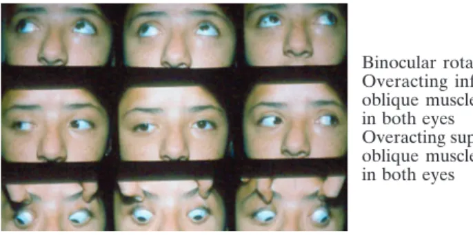
Figure 1A. First examination of extrinsic ocular muscles (October 5th, 2001)
Binocular rotation: Overacting inferior oblique muscles, +2 in both eyes Overacting superior oblique muscles, +3 in both eyes
Upper boxes show measures with right eye fixation and lower boxes show measures with left eye fixation. Measures for near fixation were done in the straight ahead and down gaze positions.
Medical History
Birth by caesarean delivery at term without complications, weighting 3750 grams. Her neuropsychomotor development was normal.
Ophthalmic Examination
Corrected visual acuity of 0.2 (+4.00 SD, -1.00 CD X 90) in the right eye and 0.6 (+1.00 SD) in the left eye. Biomicroscopy and fundus examination were normal.
The first examination of extrinsic ocular muscles, performed
on October 5th, 2001, showed:
Monocular central fixation, stable in both eyes.
Binocular fixation = without correction and with far and near optical correction, right exotropia (XT) and hypotropia, deviation of the right eye.
Right excyclotropia 5° in the double Maddox rod test. Alternate prism cover test: With correction (Figure 1A) and versions (Figure 1B).
During the examination it was noted that the patient tilted her head towards the right shoulder. Also, in the evaluation of
Figure 2A. Examination of extrinsic ocular muscles (August 21st, 2002)
Upper boxes show measures with right eye fixation and lower boxes show measures with left eye fixation. Measures for near fixation were done in the straight ahead and down gaze positions
Figure 2B. Examination of extrinsic ocular muscles (August 21st, 2002)
Figure 1B. Examination of extrinsic ocular muscles (October 5th, 2001)
Figure 3B. Examination of extrinsic ocular muscles (November 12th, 2008)
Torticollis was absent.
Binocular rotation: Underacting right inferior oblique muscle, -1
Overacting superior oblique muscles, +3 in both eyes Slight lowering of both eyes in adduction
Figure 3A. Examination of extrinsic ocular muscles (November 12th, 2008)
Upper boxes show measures with right eye fixation and lower boxes show measures with left eye fixation. Measures for near fixation were done in the straight ahead and down gaze positions
Binocular rotation: Underacting right lateral rectus, -1 Underacting left la-teral rectus, -1 Overacting superior oblique muscles, +3 in both eyes
Far
Near
Far
Near Far
196
Corresponding author:
Thiago Gonçalves dos Santos Martins
Prédio dos Ambulatórios do Hospital das Clínicas da Faculdade de Medicina da Universidade de São Paulo - Serviço de Oftalmologia
Av. Dr. Enéas de Carvalho Aguiar, prédio dos ambulatórios, 6º andar, sala 8
CEP: 05403-000 - São Paulo (SP), Brazil. E-mail: [email protected]
Figure 4. CT scan performed four years after the first procedure
Rev Bras Oftalmol. 2013; 72 (3): 194-6
Martins TGS, Meuleman VVM, Hanania FR, Polati M
eye versions, she appeared to have anisotropy in “X” (the deviation increased when he looked up and down), although this was not confirmed in the measurements taken during the alternate prism cover test. She was first operated in the right
eye on April 18th, 2002, as this eye had the worst visual acuity.
There was no restriction of movement in both eyes in the forced duction test. A 6.0 mm recession of the lateral rectus and a 6.0 mm resection of the medial rectus were performed; these muscles were moved upward, with correction of their oblique muscle
insertions.The lateral rectus muscle followed an oblique path,
from the bottom up to its insertion. Furthermore, the scleral insertion of both muscles had an oblique orientation, with their superior end being 1.0 mm closer to the limbus than the inferior end.
Examination of extrinsic ocular muscles four months after the procedure showed:
Alternate prism cover test: With optical correction (Figure 2A) and versions (Figure 2B).
The patient continued to tilt her head towards the right shoulder and presented anisotropy in X, but the overaction of oblique muscles had decreased.
A second operation on the left eye was planned to occur two years after operating the right eye. There was no restriction of ocular movement in the forced duction test.
During the procedure, it was noted that the lateral rectus muscle insertion was 8.0 mm from the limbus, and the medial rectus was 6.0 mm from the limbus (insertions were not oblique). The lateral rectus insertion was more inferior; its superior end corresponded to the 3-9h corneal meridian.
A 6.0 mm recession of the lateral rectus and a 6.0 mm resection of the medial rectus were performed. Recession of the lateral rectus was done following the anomalous direction of the insertion.
A year and a half after the second procedure, examination of the extrinsic ocular muscles showed: Binocular fixation = without correction and with far and near optical correction, exotropia, deviation of the right eye (XT RE); alternate prism cover test: with optical correction (Figure 3A) and versions (Fi-gure 3B).
The patient no longer had torticollis, but still had slightly overacting oblique muscles.
A CT scan performed four years after the first procedure showed no anatomical abnormalities of the extraocular muscles (Figure 4).
D
ISCUSSIONApproximately 56% of patients with craniosynostosis have
strabismus2. In these patients, the extraocular muscles may be
absent or may present changes in thickness, fibrosis, and
anomalous paths and insertions. Malformation of the orbit,
amblyopia and intracranial hypertension syndrome may occur.3
In the case presented here, treatment and surgical planning were difficult due to the anomalous insertion of extraocular muscles, as well as the facial asymmetry.
Imaging tests such as MRI, CT and ocular ultrasonography can be used to support surgical planning in
ca-ses of strabismus associated with craniossinostosis.2 With these
tests, surgical planning can done with previous knowledge of the anatomic changes of extraocular muscles, thus avoiding unnecessary incisions and inadvertent muscle injury and reducing the number of procedures needed to correct deviations and improve results. In the case presented here, imaging tests did not show changes in muscle paths, as the imaging technique was not specifically intended to study the problem, but those changes were then found during surgery. In such cases, the physician should state their diagnostic hypothesis when requesting the imaging test, so that the paths of affected muscles can be studied more accurately.
The orbital asymmetry, with the right orbit lower than the left, and the transoperative findings of anomalous muscle insertions and paths can perhaps explain some clinical findings which were initially considered as paradoxical, such as torticollis and anisotropy not confirmed by objective data measurement with prisms.
C
ONCLUSIONThis case report illustrates the surgical difficulties found in such cases: The anomalous muscle paths were only detected during the operation, even though imaging tests had been performed preoperatively, failing to find the anomaly. Requests for imaging tests should include proper instructions to the radiologist as to what to investigate, so that the results can actually support surgical planning.
R
EFERÊNCIAS1. Behrman RE, Jenson HB, Kliegman RM, editors. Nelson tratado de pediatria. Tradução da 17a ed. Rio de Janeiro: Elsevier; 2005. p. 2113-4. 2. Somani S, Mackeen LD, Morad Y, Buncic JR, Armstrong DC, Phillips JH, et al. Assessment of extraocular muscles position and anatomy by 3-dimensional ultrasonography: a trial in craniosynostosis patients. J AAPOS. 2003;7(1):54-9.