Cláudia Filipa Cunha Antunes
Setembro de 2013
Relevance of tissue macrophages
in lung development
UMinho|20
13
Cláudia F
ilipa Cunha Antunes
R
ele
vance of tissue macrophages in lung de
velopment
Universidade do Minho
Escola de Ciências da Saúde
This work was performed under supervision of
Sandra Maria Araújo Costa, PhD
Cláudia Filipa Cunha Antunes
Setembro de 2013
Master Thesis
Master in Health Sciences
Relevance of tissue macrophages
in lung development
Universidade do Minho
Escola de Ciências da Saúde
v
Acknowledgments
Não construimos uma tese sozinhos, embora muito do que apresentamos no final seja não mais do que o somatório de muitos momentos de luta connosco próprios.
Ainda assim, há pessoas cujo empenho e companheirismo salutares merecem um especial agradecmento.
Á minha orientadora Sandra Costa, deixo a minha sincera gratidão pela orientação, dedicação, pela extrema fibra que foi demonstrando sempre que surgiam problemas e não foram poucos. Porque começamos a construír tudo do zero com tudo o que isso implica.
À comissão organizadora do mestrado em Ciências da Saúde, pela oportunidade que me deu de o poder integrar.
A todo o ICVS – Instituto de Investigação de Ciências da Vida e da Saúde, em particular ao Domínio das Ciências Cirúrgicas.
Ao laboratório de Oncologia onde fiquei grande parte da tese e que muitas vezes auxiliou nas minhas pequenas/grandes dúvidas (Vera,Filipa, Susana,Céline, Ricardo A. e Ricardo C.).
Ao laboratório de Desenvolvimento, onde encontrei a Sheeba, Raquel, AnaLuce, Hugo, Duda.
Á Marina S. pelos PCR’s, pelas imunos, pela tua ajuda Voluntária!
À Cristina e Marina A., minhas companheiras das intermináveis horas de estereologia! Aos serviços de Histologia e Biologia Molecular, pelo apoio técnico, Miguel, Luís e Goreti muito obrigada!
À minha família e amigos. Aos meus pais por financiarem o meu trabalho durante este tempo e por muito mais.. À minha irmã. Às minhas amigas de todo o sempre: Susana e Francisca. Aos meus amigos de muitos anos: Zé, Sílvia, Guida, Marta e Carla.
ix
Abstract
Macrophages are found in almost every tissue, from embryonic to adulthood, with trophic roles during development. In organs such as brain, kidney, mammary gland and pancreas, macrophages reside in considerable number and provide important regulatory functions that shape their development and maturation. Some studies have demonstrated the regulation of developmental morphogenesis by macrophages, more specifically in vascular system formation and branching morphogenesis. Lung is a branching organ and its morphogenesis is dependent on interactions between epithelial and endothelial compartments. Indeed, in fetal lung, macrophages represent around 2-5% of the total lung cells. However, their precise contribution to lung development remains unknown. In this way, the main goal of this work was to unfold the functional role of tissue macrophages on lung development. Therefore, we aimed to understand whether and how macrophage ablation impaired fetal and neonatal lung development, using a macrophage-deficient mouse model (Csf1r-/- mice). The first aim was to characterize lung
morphology of Csf1r-/- versus wild type and Csf1r+/- mice. We performed lung stereological analysis
(calculation of mesenchymal, epithelial and air space volume density (VD)) at pseudoglandular (embryonic day (E) 15.5), fetal saccular (E18.5) and at post-natal saccular (after birth, P0) developmental stages. The lung mesenchymal VD in Csf1r+/- and Csf1r-/- mice was increased
when compared with wild type mice in all stages analysed. At E18.5, beyond an increase in lung mesenchymal, a decrease of epithelial and airspace VD was observed in Csf1r-/- mice. Finally, at
P0 stage, Csf1r-/- derived lungs showed a decrease in bronchiolar and air space VD as well as an
increase in the mesenchymal VD compared with control group. These results suggested a delay in lung morphogenesis associated with macrophage tissue ablation. Keeping in mind that macrophages support angiogenesis during organ development and remodelling, we assessed vessel density on lungs, by immunohistochemical analysis of CD31 endothelial cell marker. At P0, lungs from Csf1r-/- mice presented a significant decrease in vessel density when compared
with wild type mice (p value=0.020). This finding demonstrates a deficiency in lung vascular network associated with macrophage ablation. Furthermore, we quantified RNA expression levels of key angiogenic factors (VEGF-A, Flk-1, Ang-1, Ang-2, Hif-1a and Fgf2) in lung from the different mice groups at E15.5, E18.5 and after birth, P0. At E15.5 stage, an up regulation of VEGF-A, Flk-1, Ang-2 and Fgf2 transcripts was found. At E18.5 a significant reduction of VEGF-A and Ang-2
x
RNA levels was demonstrated in Csf1r+/- and Csf1r-/- groups when compared with wild type lungs.
However, the opposite trend was observed in P0 knockouts lungs, with an increase in VEGF-A, Flk-1, Ang-1 and Ang-2 transcripts expression levels. This could be explained by compensation mechanisms trying to reverse the deficient lung vasculature (less vessels) observed at P0. In conclusion, all these evidences demonstrate that macrophage ablation is critical to lung development, impairing airway epithelial and vascular compartments.
xi
Resumo
Os macrófagos estão presentes em quase todos os tecidos desde a fase embrionária até à fase adulta, associados a funções tróficas durante o desenvolvimento. Em órgãos como cérebro, rim, glândula mamária e pâncreas, os macrófagos estão presentes em largo número, regulando o desenvolvimento da sua forma e maturação. A literatura tem demonstrado que processos da morfogénese do desenvolvimento como a ramificação e a formação do sistema vascular são altamente regulados pelos macrófagos. E o pulmão é também um órgão de ramificação, dependente da inter-ligação entre a componente epitelial e endotelial. Durante o desenvolvimento fetal deste órgão, os macrófagos representam cerca de 2 a 5 % do número total de células, permanecendo por identicar qual a sua relevância. Neste sentido, o objectivo deste trabalho foi perceber se e como a deleção dos macrófagos influência o desenvolvimento fetal e neo-natal do pulmão, usando um modelo de ratinho com a ablação dos macrófagos (Csf1r
-/-ratinho). O nosso primeiro alvo de estudo foi caracterizar a morfologia dos pulmões que provêm de animais Csf1r-/- por comparação com os Csf1+/- e a estirpe selvagem. Desse modo, recorremos
a análises esterológicas para calcular a densidade de volume (DV) de uma série de parâmetros como mesênquima, epitélio, espaço aéreo, em pontos específicos do desenvolvimento; no estadio pseudoglandular (desenvolvimento embriónico (E) 15.5), fetal sacular (E18.5) e pós-natal sacular (depois do nascimento, P0). A tendência observada sugere que os pulmões dos ratinhos Csf1r-/- e Csf1r+/- apresentam um aumento da densidade de volume de mesênquima
quando comparado com o grupo controlo em todos os estadios. Especificamente a E 18.5, para além da densidade de volume de mesênquima estar aumentada, um decréscimo do epitélio e espaço aéreo foi observado para os Csf1r-/- animais. Por último, logo após o nacimento (estadio
P0), os animais Csf1r-/- apresentaram uma diminuição da DV do epitélio bronquiolar e do espaço
aéreo comparando com a estirpe selvagem. Estes resultados sugerem um atraso na morfogénse pulmonar associado à deleção dos macrófagos tecidulares. Por outro lado, a regulação dos macrófagos no processo angiogénico durante o desenvolvimento e no processo de remodelação dos vasos, suportou o seguinte foco de estudo. Nós analisamos a densidade dos vasos pulmonares, utilizando o marcador endotelial CD31, por imunohistoquímica. A P0, os pulmões dos animais com o genótipo Csf1r-/- apresentaram um decréscimo significativo comparado com o
xii
associado com a ablação dos macrófagos. Mais, avaliamos os níveis de expressão de RNA de factores angiogénicos chave como VEGF-A, Flk-1, Ang1, Ang2, Hif-1a e Fgf2, nos pulmões dos diferentes grupos de animais, a E15.5, E 18.5 e P0. A E 15.5 um aumento de VEGF-A, Flk-1, Ang-2 e Fgf2 foram encontrados. A E18.5, um decréscimo nos niveis de expressão de VEGF-A e Ang-2 para os animais Csf1r+/- and Csf1r-/- foi demonstrado. Mas quando avaliamos estes factores
no estadio P0, um aumento significativo de VEGF-A, Flk-1, Ang-1 e Ang-2 foram encontrados comparando com o grupo controlo. Este resultado pode dever-se há existência de mecanismos compensatórios que tentam reverter a dificiente vasculaura (menor número de vasos) a P0. Em conclusão, estas evidências demonstram que a deleção dos macrófagos é critica para o desenvolvimento do pulmão, resultando no dano do epitélio das vias aéreas e da vasculatura.
xiii
xv
T
ABLE OFC
ONTENTSAcknowledgments ... v
Abstract ... ix
Resumo ... xi
Abbreviation List ... xix
Figures and Tables ... xxv
1. INTRODUCTION ... 29
1.1.Lung development ... 29
1.1.2 Mouse and Human lung developmental stages ... 29
1.1.3. Cellular Differentiation of the Lung ... 31
1.1.4. Development of the Pulmonary Vasculature ... 32
1.2. Macrophages ... 33
1.2.1.Macrophage origin and differentiation ... 33
1.2.2 CSF1/CSF1R Pathway ... 35
1.2.3.Macrophages subsets ... 36
1.2.4. Relevance of Tissue Macrophages ... 38
1.2.4.1.Bone morphogenesis ... 39
1.2.4.2.Mammary gland development ... 40
1.2.4.3.Neural networking/Reproductive system ... 41
1.2.4.4.Brain ... 42
1.2.4.5.Angiogenesis ... 42
1.2.4.6.Pancreas ... 44
1.2.4.7.Kidney ... 44
1.2.5.Lung macrophages and inflammatory mediators in lung development ... 45
1.2.5.1. Lung macrophages ... 45
1.2.5.2. Cytokines as mediators in lung development ... 46
1.10 Rationale and Aims ... 49
2. MATERIALS AND METHODS ... 53
2.1. Animals ... 53
2.2. Study design ... 53
2.3. PCR analyses for Genotyping ... 53
2.4. Lung-to-body weight ratios ... 54
2.6. Stereological Analyze ... 54
xvi
2.8. Vessel Density quantification ... 56
2.9. RNA extraction and cDNA synthesis ... 56
2.10. Quantitative Real-Time PCR (q-PCR) ... 57
2.11. Weigert’s staining ... 57
2.12. Arterial thickness evaluation ... 58
2.13. Statistical Analysis ... 58
3. RESULTS ... 61
3.1. Impact of macrophages deletion in pseudoglandular stage of lung development (E15.5) ... 61
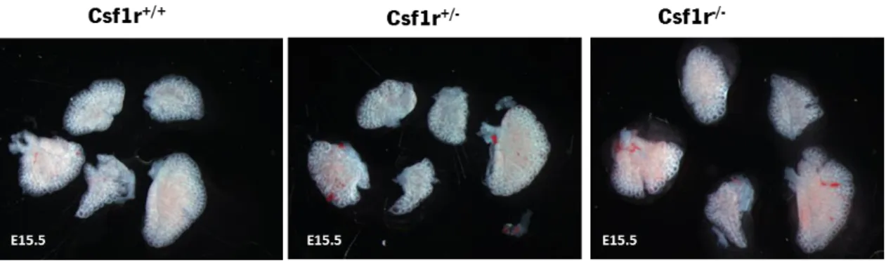
3.1.1. Lung gross morphology ... 61
3.1.2 Lung bud number ... 61
3.1.3. Lung morphology ... 63
1.1.4.Lung vasculature ... 64
3.1.5. Gene expression of angiogenic factors ... 66
3.2. Impact of macrophages deletion in fetal saccular stage of lung development (E18.5) ... 67
3.2.1. Lung gross morphology ... 67
3.2.2. Lung morphology ... 68
3.2.3. Lung vasculature ... 69
3.2.4. Gene expression of angiogenic factors ... 71
3.2.5. Arterial thickness ... 72
3.3. Impact of macrophages deletion in post-natal saccular stage of lung development (P0) ... 73
3.3.1. Lung gross morphology ... 73
3.3.2. Lung-to-body weight ratios ... 73
3.3. Neonatal blood gasometry ... 74
3.3.4. Lung morphology ... 75
3.3.5. Lung vasculature ... 76
3.3.6. Gene expression of angiogenic factors ... 77
3.3.7. Arterial thickness ... 78
4. DISCUSSION ... 83
5. FUTURE PERSPECTIVES ... 93
xix
Abbreviation List
Ang: Angiopoietin Arg1: Arginase 1
BDP: Bronchopulmonary Dysplasia Ccl17: Chemokine C-C motif ligand 17 cDNA: complementary Deoxyribonucleic Acid Csf1: Colony stimulated factor
Csf1r: Colony stimulated factor receptor CCSP: Clara Cell Secretory Protein DAB: 3,3’-diamino-benzidine ECS:Endothelial Cells E: Embryonic Day
ECM: Extracellular Matrix
EDTA: Ethylenediamine Tetraacetic Acid
FGF: Fibroblast Growth Factor Flk-1: Fetal Liver Kinase 1
GAPDH: Glyceraldehyde 3-Phosphate Dehydrogenase GM-CFU: Granulocyte/Macrophage Colony-Forming Unit GnRH: Gonadotropin-Releasing Hormone
H2O2: Hydrogen Peroxide
HIF-1a: Hypoxia-inducible Factor IL: Interleukin
IPA: Isopropanol
xx
JAK/STAT: Janus Kinase/Signal Transducer and Activator of Transcription LH: Luteinizing Hormone
LIF: Leukaemia Inhibitory Factor LPS: Lipopolysaccharides
MAPK: Mitogen-Activated Protein Kinases MDV: Microvessel Density
MIN: Minute
mmHg: Millimeter of Mercury
MPS- Mononuclear Phagocyte System Mrc1: Mannose receptor 1
M1: Classically activated macrophages M2: Alternative activated macrophages
NF-KB: Nuclear Factor kappa-light-chain-enhancer of activated B cells P: Partial pressure
PBS: Phosphate Buffered Saline PCR: Polymerase Chain Reaction
PECAM: Platelet Endothelial Cell Adhesion Molecule PFA: Paraformaldehyde
PH: Pulmonary Hypertension PPSCs: Bone marrow stem cells PI3K: Phosphatidylinositol 3-kinase
qRT-PCR: Quantitative Reverse Transcription Polymerase Chain Reaction RANKL: Receptor Activator of Nuclear factor-kB Ligand
RT: Room Temperature
RTKs: Protein-Tyrosine Kinase Receptors Sat: Saturation
xxi Shh: Sonic hedgehog
SEC: Second
STAT: Signal Transducers and Activators of Transcription SP-C: Surfactant Protein C
SZ: Subventricular Zone TEBs: Terminal End Buds
TGF-β- Transforming Growth Factor-β TNF-α: Tumor Necrosis Factor VD: Volume Density
VEGF: Vascular Endothelial Growth Factor
VEGFR: Vascular Endothelial Growth Factor Receptor Wnt: Derived from Drosophila Wingless and the mouse Int
xxv
Figures and Tables
Figure 1. Principal stages of lung development in mouse and human. ... 30 Figure 2. Cell types of the respiratory system... 31 Figure 3. Macrophage origin and differentiation. ... 35 Figure 4. CSF1/CSF1R Pathway ... 36 Figure 5. Macrophage activation and polarization. ... 37 Figure 6. Distribution of body tissue macrophages.. ... 38 Figure 7. The trophic role of macrophages in bone morphogenesis.. ... 39 Figure 8. The trophic role of macrophages in ductal branching... 40 Figure 9. Macrophages interference in angiogenic and vascular eye development.. ... 43 Figure 10. Gross morphology evaluation of lung embryos at E15.5 stage. ... 61 Figure 11. Representative pictures of left lung lobe at E15.5. ... 62 Figure 12. Number of buds in left lung lobe at E15.5 stage.. ... 62 Figure 13. Lung embryos histological sections at E15.5 stage. ... 63 Figure 14. Lung stereological analyses at E15.5 stage. ... 64 Figure 15. CD31 immunohistochemical staining in lung embryos at E15.5. ... 65 Figure 16. Vessel density in lung embryos at E 15.5 stage. ... 65 Figure 17. Angiogenic factors gene expression profile by qPCR of lungs at E 15.5. ... 67 Figure 18. Gross morphology evaluation of lung embryos at E18.5 stage. ... 68 Figure 19. Lung embryos histology at E18.5 stage. ... 68 Figure 20. Lung embryos histology at E18.5 stage ... 69 Figure 21. CD31 immunohistochemical staining in lung embryos at E18.5. . ... 70 Figure 22. Vessel Density quantification at E 18.5... 70 Figure 23. Angiogenic factors gene expression profile by qPCR of lung at E 18.5 ... 71 Figure 24. Arterial thickness at E18.5 stage.. ... 72 Figure 25. Arterial thickness evaluation at E18.5 stage. ... 72 Figure 26. Gross morphology evaluation of lung newborns at P0 stage.. ... 73 Figure 27. Left, right and total lung-to-body weight ratio at P0 stage. ... 74 Figure 28. Lung histology at P0 stage. ... 75 Figure 29. Sterelogical analyses at P0 developmental stage. ... 76 Figure 30. CD31 immunohistochemical staining in lung embryos at P0 stage. ... 77 Figure 31. Angiogenic factors gene expression profile by qPCR of lung newborns at P0 .... 78 Figure 32. Arterial thickness at P0 stage. ... 79 Figure 33. Arterial thickness evaluation at P0 stage... 79 Table 1. Neonatal blood gasometric evaluation after birth ... 75
29
1. INTRODUCTION
1.1. Lung development
Lung development is a highly regulated process. Its development is coordinated from the primary bud stage until the generation of millions of alveolar gas exchange units [1]. This process arises from embryogenesis and continues after birth which involves structural organization and functional maturity [2]. Gas exchange allows the oxygenation of red blood cells in the circulatory system and the simultaneous removal of carbon dioxide, being one of the most important characteristics of respiratory system [3]. Until the lung is fully capable to play this process a complex development program should take place. Mammalian lung development starts with the differentiation of ventral foregut endoderm into various epithelial cell types that contour the inner surface of the trachea and two primary buds, the primordial lung[4]. The endoderm gives rise to multiple resident epithelial cells and the mesoderm to vascular structures, airway smooth muscle and others [5]. After the primary lung buds formation, the two main bronchi of the primordial lung extend into the surrounding mesenchyme and begin the process of branching morphogenesis [1, 6]. The two main bronchi of the primordial lung are subdivided into sequentially finer buds, the bronchioles, which are surrounded by parabronchial smooth muscle, originating the common respiratory tree. This process is essential to elaborate the orchestration of the bronchiolesand their endings, alveoli, highly specialized structures that allow gas exchange process. The branching program of the lung epithelium is accompanied at the same time by the development of the pulmonary vasculature, constituted by a blood vessels network, which covers the alveoli [1, 4]. It is the tight connection between the airways and the blood vessels the key for dynamic and complex respiratory process, which allows an efficient gas exchange.
1.1.2 Mouse and Human lung developmental stages
In Humans, the initiation of pulmonary development arises as a bifurcation at the posterior end of the laryngotracheal groove, occuring at 3-4 weeks of gestation. In rodents, the two buds that will originate right and left lung arise from the foregut endoderm, appearing on embryonic day (E) 9.5 of embryo development [2, 7]. Each lung bud develops into left and right lungs [2]. In both, humans and mice, lung constitution is asymmetrical. In humans, left lung
30
comprises two lobes while the right lung consists of three lobes. On the other hand, the left mice lung presents a single lobe and the right lung is composed of four lobes [6].
In both species, the embryological development of respiratory system can be divided into five principal stages (figure 1). In mouse, the first stage corresponds to lung primordium appearance (E8-9); followed by pseudoglandular stage, during which the primary buds generate a complex tree-like structure (E10-16); canalicular stage, where the terminal buds become narrower (E16-18); saccular stage, which involves the developing of numerous small sacs that are the precursors of the alveoli (E18.5 to postnatal day (P) 5); and lastly, alveolar stage (starts at birth and extends almost 4 weeks into postnatal life) associated with the increase of terminal saccules, alveolar ducts, and alveoli. In human embryos, during the embryonic period a pulmonary protuberance containing two buttons develops, with the formation of the trachea and bronchus (3-4 weeks); pseudoglandular period, during which organogenesis occurs allowing the complete formation of bronchial tree and the airways (5-17 weeks); canalicular period, when respiratory bronchiole appears and vascular network increases (16-26 weeks); saccular period, with expansion of air spaces (24-38 weeks) and alveolar period, when alveolar development occurs by the formation of secondary and tertiary septa (36 weeks-2years). For human the first four stages take place in utero and the last ends after birth. In mice, the fourth stage ends after birth and the last occurs exclusively after birth [1, 2, 8].
Figure 1. Principal stages of lung development in mouse and human: diagrammatic representations of the timeline and developmental organization (adapted from[9]).
31 1.1.3. Cellular Differentiation of the Lung
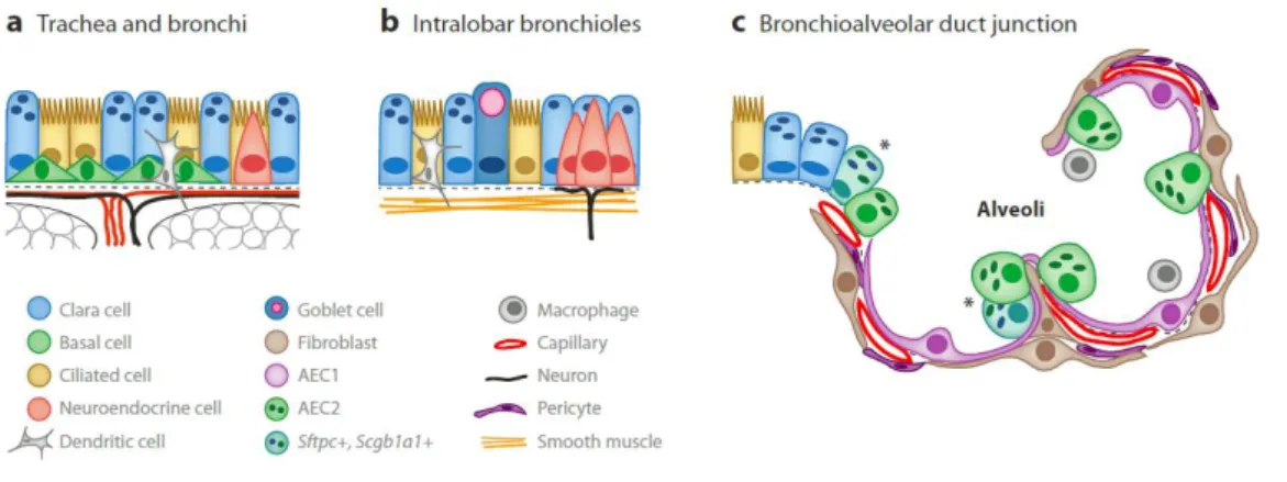
In mice, the current model of epithelial development describes that airway branching morphogenesis occurs during the pseudoglandular stage. The terminal buds contain a pool of highly proliferative multipotent progenitor cells, which give rise to several types of cells [1]. The differentiated airway tree is composed by a complex and large number of cells, which change significantly along the proximal-distal regions (figure 2). In adult mice, the proximal pseudostratified epithelium, composed by large airways (tracheobronchial region) shows three major cell types: ciliated, basal and clara secretory cells. Ciliated cells are first detected in the trachea and mainstem bronchi at approximately E14, being typically marked by expression of FoxJ1 transcription factor. Basal cells are observed in the trachea at E10.5 associated with expression of the transcription factor Trp63. Clara cells are typically marked by synthesis of the secretoglobin family member Scgb1a1, (also known as CC10 or CCSP), which appears around E17 [1, 10, 11]. Concerning distal columnar epithelium, composed by small airways (intralobar bronchioles), the cell population is similar to the large airways, with no basal cells and more number of ciliated cells, neuroendocrine cells and Clara cells. Neuroendocrine cells are among the first cells to differentiate within the respiratory epithelium, with the starts of Dll1 expression at E13.5[1] [10]. During the canalicular and saccular stages, the terminal or acinar tubes narrow and give rise to small saccules and the walls of these sacs, the primary septae.
Figure 2. Cell types of the respiratory system. (a) Large airways (tracheobronchial region) are covered by a pseudostratified epithelium composed of basal cells, ciliated and secretory cells. (b) Small airways (intralobar bronchioles) a columnar epithelium containing ciliated, neuroendocrine and Clara cells. (c) The alveoli are composed by epithelial type 1 cells (AEC1) and cuboidal AEC2 cells and few cells secreting secretoglobulina Scgb1a1 (asterisks). The alveolar epithelial cells are closely associated with mesenchymal cells such as fibroblasts, endothelial cells and pericytes[10].
32
The endoderm begins to differentiate into the two main specialized cell types: type 1 and type 2 alveolar epithelial cells (AEC1 and AEC2) [10]. The type 1 cells constitute about 95% of the alveolar epithelium and lining the alveolar saccules, characteristically express aquaporin 5 and T1alpha. The type 2 cells comprise 5-10% of the alveolar surface and are characterized by production of surfactant-associated protein C (Sftpc or SPC) [5, 7]. Alveolar epithelial cells are closely associated with mesenchymal cells including fibroblasts, vascular smooth muscle cells, endothelial cells, vascular pericytes and macrophages [1].
1.1.4. Development of the Pulmonary Vasculature
The vascular development takes place throughout lung development with alterations in the structure of the capillary network. During mouse development, the vascular network of the lung is established from E≈10.5 and remains to develop postnatally. This system is composed of endothelial cells surrounded by vascular smooth muscle, pericytes, and other mesenchymal cells. All these cellular components are specifically organized to ensure the precise function of pulmonary vascular system [12]. Particularly, inhibition of lung endothelial cell development is accompanied by inhibition of airway epithelial cell differentiation and maturation [13]. Moreover, although significant airway remodeling occurs postnatally, the primary vascular network is established early in development, being essential for viability at birth [12]. Therefore, the crosstalk between the developing vasculature and the developing airways is a key event during lung development.
The angiogenesis and vasculogenesis are the crucial processes, which allow the lung vascular development since early stages [13]. Vasculogenesis is characterized to take place at early stages of embryo development, with a formation of new blood vessels de novo. On the other hand, angiogenesis promotes blood vessel formation from extension of pre-existing ones [14]. Concerning molecular players of pulmonary vasculature development, the pathways VEGF/VEGFR, Angiopoietin/TIE, Ephrins/Eph receptors, and Notch/Jagged appear to be essential in the regulation of pulmonary vascularization [15]. For instance, Ephrin B2 expressed by the microvasculature is critical for lung alveolization and secondary septae formation [16] [17]. Little is known about angiopoetins (Ang) expression during development, but for instance, overexpression of Ang-1 causes severe pulmonary hypertension and is increased in lungs from human patients with pulmonary hypertension [18]. Regarding Notch/Jagged pathway, for instance Notch-1 and Jagged-1 gene expression increased progressively from early to later lung
33
development, on vascular and nonvascular cells, suggesting their contribution during lung development. [19]. VEGF-A/VEGFR-2 signaling is a crucial pathway, which regulates physiological
and pathological lung formation. This pathway has central action on vascular system, as a mitogen, survival, and differentiation factor for endothelial cells [20]. However, their relevance has been also demonstrated in pulmonary epithelial morphogenesis. For instance, in neo-natal mice, the neutralization of VEGF-A or VEGFR-2 (Flk-1) leads to a lung immature with less complex alveolar [21].
Furthermore, using mouse embryonic lung cultures treated with an antisense oligodeoxynucleotide to the VEGFR-2 was observed the loss of VEGF function which leads to a decreased in epithelial branching, epithelial and mesenchymal proliferation, and a downregulation of BMP-4 expression, an important regulator of lung development [22]. On the other hand, VEGF-A stimulation increases epithelial branching morphogenesis on lung embryonic cultures with an increases of Sp-c as well as Bmp-4 mRNA levels [22]. VEGF-A hyper expression in respiratory epithelium mice leads to lung vasculature alteration associated with disruption of the lung acinar structure, branching morphogenesis and inhibition of Type I cell differentiation [23]. These observations indicate that lung morphogenesis is critically dependent on lung neovascularization and VEGF/ VEGFR-2 pathway plays a key role in lung patterning. Together all these evidences demonstrated the close link between airway and vascular development, since the disruption of one system has critical consequences on the development of the other, and the other way around.
1.2. Macrophages
1.2.1. Macrophage origin and differentiation
Macrophages belong to the mononuclear phagocyte system (MPS). It includes bone-marrow-derived precursor cells, monocytes present in the peripheral blood and mature macrophages existent in the tissues [24]. Macrophages are normally identified in murine tissues using the markers, CD68, F4/80 and/or CD11b [25]. CD68, whose expression is restricted to mononuclear phagocytes, is a lysosomal antigen expressed (at high levels) in monocyte/macrophages [26]. F4/80 is a well-characterized membrane protein and the best known marker for mouse macrophages and blood monocytes [27]. CD11b is a leukocyte-specific receptor and is regarded as a marker for monocyte/macrophages, granulocytes, and natural
34
killer cells[28] In mice, the first population of macrophages is found in the late head-fold stage at E7.5 and has maternal origin [29]. However, the first embryonic macrophages, which derive from the primitive endoderm of the yolk sac, are found at E8 and sprout to the anterior structures of the embryo and don’t express mature macrophage markers, such as F4/80. These macrophages don’t differentiate from monocyte cells but directly from mesenchymal progenitor cells [30]. The first site of definite haematopoiesis is the yolk sac, followed at E10 by the aorta– gonads–mesonephros region of the embryo, where the second pool of hematopoietic progenitor cells and F4/80 positive cells are found [31-33]. By E10.5 to E11 the primitive liver becomes the principal site of haematopoiesis and from this moment, macrophages that have differentiated from monocytic progenitor cells colonize the entire embryo [32, 33]. After birth, the bone structures are formed and the bone marrow becomes the main site of haematopoiesis and the MPS is established [34]. Macrophages derive from pluripotent hematopoietic stem cells under the influence of numerous factors. The bone marrow stem cells (BMSCs) differentiate in multiple macrophages progenitor stages: from granulocyte/macrophage colony-forming unit (GM-CFU) to macrophage CFU (M-CFU) to monoblast to pro-monocyte and for last macrophage [24]. Transcription factors as PU.1 are important to macrophage differentiation. However, this is required only for macrophages differentiation from monocyte precursor, once PU.1-deficient mice is normal until the macrophages differentiation through the monocyte precursor starts [35] [32]. However, others factors are necessary for the differentiation of each macrophages cells precursors. The production of macrophages by the liver, and then by the bone marrow, is controlled in large part by the growth factor macrophage colony-stimulating factor (CSF-1). This growth factor allows the differentiation of macrophages from monocytes as well as their proliferation and viability in vitro [24]. Circulating CSF-1 is produced by endothelial cells in blood vessels and, together with locally produced CSF-1, regulates the survival, proliferation and differentiation of mononuclear phagocytes and osteoclasts (figure 3) [36].
35 Figure 3. Macrophage origin and differentiation. Progenitors cells of MPS system arise from bone marrow, where from pluripotent stem cells (PPSCs) others progenitor are formed. The multipotent progenitors are granulocyte/macrophage colony-forming unit (GM-CFU), which differentiate in monoblast and this last in pro-monocyte. In blood vessels, endothelial cells produce CSF-1, which allows the differentiation of pro-monocyte coming from the bone marrow in monocytes. In the tissues, local produced CSF-1 allows the differentiation of monocytes in tissue macrophages. (Adapted from [24, 36])
1.2.2. CSF1/CSF1R Pathway
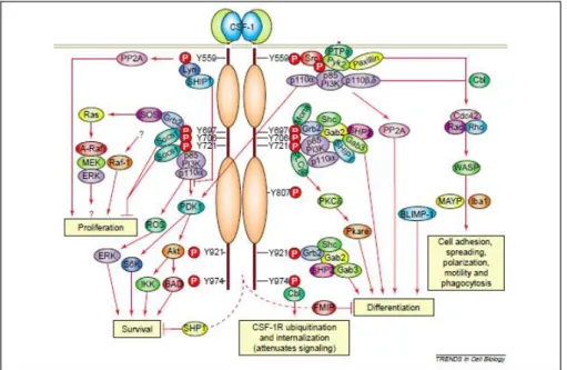
Csf-1 acts on its target cells by binding to colony stimulating factor 1 receptor (Csf1r), a cell-surface tyrosine kinase receptor expressed in macrophages and all trophoblast cell lineages [31]. The activation of this pathway is allowed by the link of Csf1 to the receptor Csf1r, which triggers their autophosphorylation and successive phosphorylation of downstream molecules. Phosphorylation of most of tyrosine residues creates docking sites for a variety of signaling molecules such as the p85 regulatory subunit of phosphatidylinositol 3-kinase (PI3K), Casitas B lineage (Cbl) and Grb2-associated binder-3 (Gab3), to mediate macrophage survival, differentiation, morphology and motility (Figure 4) [37-39]. Beyond Csf1, macrophages can also be grown and differentiated from monocytic progenitors in the presence of GM-CSF and to a lesser extent under the action of IL-3. The double action of these two factors allows the generation of mature macrophages [40, 41]. Mice with null mutation in the Csf1 gene or with null mutation in Csf1r are both deeply depleted of most macrophage populations. However, these mice are not totally depleted of macrophages, as is the case of spleen resident macrophages, thymus and lymph nodes derived-macrophages [36, 42].
Nevertheless, Csf1 and Csf1r phenotypes mutant animals, together with PU.1 are the best models to study the trophic roles played by tissue macrophages [24]. The phenotype of the PU.1
36
knockout is characterized by the depletion of B cells and granulocytes and also a significant decrease in the size of many macrophage populations [24]. The phenotypes found in Csf1 and Csf1r mutant mice are similar, which means that Csf1r is the only receptor for Csf1. However, the severity of some phenotypes is more evident in Csf1r-/- mice, which rarely live beyond a few
weeks of age. The phenotypes include complete loss of epidermal Langerhans cells and microglial cells. Conversely, these populations of cells are relatively normal in the Csf-1op/op mice.
This could be explained by the existence of other ligands that could link to the Csf1r, as IL-34, which supports myeloid-cell development in vitro [43].
Figure 4. CSF1/CSF1R Pathway. Colony-stimulating factor-1 receptor (Csf-1r) activation leads to regulation of different signaling pathways in myeloid cells. CSF1 binding to CSF-1R kinase triggered its tyrosine phosphorylation and generate the activation of signaling molecules associated with the receptor through their phosphotyrosine-binding domains [36].
1.2.3. Macrophages subsets
Macrophages display functional plasticity, which allows their polarization in a diversity of subsets (figure 5). The microbial and cytokine environment drives macrophages to express specialized and polarized functional properties allowing their wide functional heterogeneity [44]. Several macrophages subsets with distinct functions have been described.
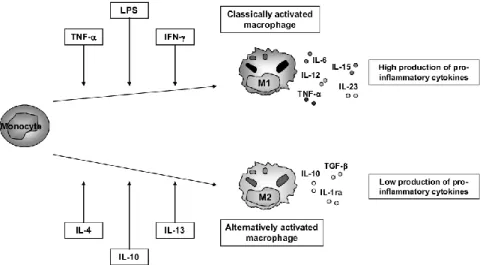
Macrophage activation can be either pro-inflammatory or anti-inflammatory leading to tissue damage or renewal and wound healing [45]. Classically activated macrophages, designated as M1, mediate defense of the host from intracellular pathogens, being activated by lipopolysaccharide and interferon- γ. These macrophages are commonly characterized by interleukin (IL)–12high, IL-23high and IL-10low phenotype. They express pro-inflammatory cytokines
37
such as IL-12, IL-23, IL-1beta, IL-15, IL-18 and TNFalpha, which are involved in the T helper 1 (TH1)-cell-mediated immune resolution of infection. They also produce chemokines and abundant amounts of reactive oxygen and nitrogen intermediates [44, 45]. Functionally, M1 cells enhance endocytic functions and improve the capacity to kill intracellular parasites [46, 47]. Alternatively activated macrophages, defined as M2, have anti-inflammatory function, enhance debris scavenging, angiogenesis, wound healing, tissue remodeling and repair [24]. They are involved in processes such as extracellular matrix (ECM) production and release of trophic factors. M2 cells have been described in a number of variants, depending upon the stimuli used to generate them. These cells generally share an IL-12low, IL-23low, IL-10high phenotype; have high levels of scavenger,
mannose and galactose-type receptors. They ensure the production of ornithine and polyamines through the arginase pathway and are IL-1 receptor antagonist (IL-1ra) high, IL-1decoyR high, IL-1blow
and caspase1low [48]. They can be subdivided in M2a, stimulated by type II cytokines IL-4 or IL-13;
M2b, obtained by triggering of Fcgamma receptors in the presence of a Toll receptor stimulus; and M2c which includes deactivation programs elicited by GC, IL-10 or TGFbeta [45]. The different types of macrophages are specified by the microenvironment, although there is considerable plasticity between distinct types. These two distinct profiles show that the macrophage activation can be either pro-inflammatory or anti-inflammatory, contributing to tissue destruction or regeneration. The diverse macrophages functionality has contributed to demonstrate their tissue trophic role namely through their implication in pathology [49].
Figure 5. Macrophage activation and polarization. Within the tissue, monocytes can polarize in M1-type, classically activated macrophages, when it’s exposed to pro-inflammatory cytokines such as TNF-α and IFN- γ or LPS. And these macrophage populations are characterized by high production of pro-inflammatory cytokines such as TNF-, IL-6, IL-12, IL-15 and IL-23. On the other hand, when monocytes are stimulated by IL-4, IL-13 or IL-10, they generate alternative activated macrophages (M2-type), which produce low levels of pro-inflammatory markers and high levels of anti-inflammatory mediators such as IL-10, IL1ra and TGF- β[50].
38
1.2.4. Relevance of Tissue Macrophages
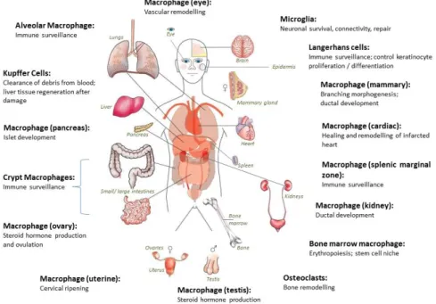
In mammals, macrophages are found throughout the body tissues (figure 6). They are strategically located to answer inflammatory conditions, being able to ingest and process external materials, dead cells and debris and call for additional macrophages [24].
Figure 6. Distribution of body tissue macrophages. Macrophage precursors are released into the circulation as monocytes and then migrate into all body tissues, differentiating into mature macrophages. They are strategically located throughout the body where they have specialized functions [51].
Based on their anatomical localization and functional phenotype, macrophages are divided into subpopulations of mature tissue macrophages [24, 49]. Tissue-resident macrophages comprise osteoclasts (bone), alveolar macrophages (lung), histiocytes (interstitial connective tissue), Kupffer cells (liver), among others[49]. The levels of macrophages in these tissues are variable. For instance in microglial cells in the brain and Kupffer cells in the liver they constitute 10–20% of all cells , but in other tissues their levels are low, such as at the musculo–skeletal junctions [52, 53]. Besides the recognized function of macrophages in disorders induced by pathogens, it is being increasingly accepted that they also have unique non-pathogen induced functions in tissue homeostasis, remodeling and reorganization. Higher levels of macrophages are present in almost all developing organs, with a spike correlated with key periods of organogenesis [54]. Among other functions, macrophages are crucial in processes such as branching morphogenesis, neural patterning, angiogenesis, bone morphogenesis and the generation of
39
adipose tissue [24]. They are fundamental in the normal development of several organs, as the mammary gland, pancreas and kidney, and the development of these organs is mainly characterized by branching morphogenesis, which is decisive during lung organogenesis [31, 55, 56]. In all these cases, the macrophage depletion leads to tissue impairment formation, responsible by their inadequate function.
1.2.4.1. Bone morphogenesis
Analysis of spontaneous mouse mutants or mice with experimental gene disruptions has highlighted key steps in the differentiation and activation of osteoclasts. Specifically, the use of CSF-1op/op mice determined that the ablation of CSF1 results in a defective development of a
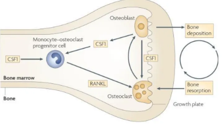
common monocyte-osteoclast bone marrow progenitor cell. This progenitor cell under the stimulus of the receptor activator of nuclear factor-kB ligand (RANKL) becomes a multi-nucleated functional osteoclast (figure 7)[57].
Figure 7. The trophic role of macrophages in bone morphogenesis. The formation of monocyte- osteoclast progenitor is dependent of Colony stimulating factor 1 (CSF1) in the bone marrow that then proliferates and differentiates in the presence of receptor activator of nuclear factor-κB ligand (RANKL) give arise to multinuclear osteoclasts[24].
The most striking phenotype of CSF-1op/op mice is osteopetrosis, with skeletal abnormalities and
tooth eruption failure due to osteoclasts depletion. This phenotype is also common in Csf1-/- rats
(known as toothless rats; tl rats) and PU.1-deficient mice that reveal early myeloid defects and an impaired development of both macrophages and osteoclasts [42, 58, 59]. CSF1 is also responsible to enhance mature osteoclastic bone reabsortion [60]. Remodeling of bone occurs throughout adult life and is controlled by the interaction of bone marrow stromal cells/osteoblasts and osteoclasts. Without functional osteoclasts, the normal re-sculpted of bone doesn’t happen, leading to osteopetrosis. Osteopetrosis causes the fail in the marrow cavity
40
orchestration, which leads to ablation of the most part of haematopoiesis that normally occurs in the bone marrow, although still occurs extramedullary hematopoiesis in the liver and spleen [61]. Csf1op/op mice also show the absence of teeth because the tooth buds, although formed, cannot
erupt through the jaw [42, 59]. This osteopetrotic phenotype is one of the best examples of the irreplaceable function of specialized macrophages and their tissue-trophic role.
1.2.4.2. Mammary gland development
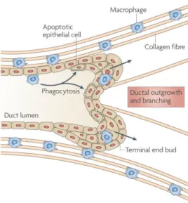
Macrophages are an important component of the mammary gland stroma and the ablation of this cell population results in defective ductal elongation, decreases fibroblast growth factor (FGF)-induced mammary epithelial proliferation, suggesting their direct or indirect actuation in mammary epithelial proliferation [62, 63]. This fact could be explained because several cell types, including epithelial, fibroblasts, adipocytes and endothelial cells, composing the mammary gland, and macrophages are known to interact [55]. In mice, the initial mammary ducts give place to multilaminate bulbous termini called terminal end buds (TEBs) [24]. At the same time that TEBs begin to grow during puberty, macrophages are recruited and align along the neck of the TEB structures and their presence is critical for proper ductal elongation (figure 8).
Figure 8. The trophic role of macrophages in ductal branching. Macrophages are associated with epithelial structures including terminal end buds (TEBs) and are often associated with collagen fibres, which they help to form. Loss of macrophages results in defective ductal elongation and disrupts collagen fibrogenesis [24].
Studies in CSF-1op/op mice demonstrated the correlation between macrophages action and
mammary development. These mice showed a decrease in ductal elongation associated with the fact that macrophages were not recruited to the TEB structures and the rate of outgrowth of these structures and their branching was decreased [64]. On the other hand, the cross of these
41
mice to transgenic mice overexpressing CSF-1 under the control of the mouse mammary tumor virus (MMTV) promoter overcame the atrophic mammary gland, meaning that this effect is due to local production of CSF-1 rather than a systemic effect [65]. Although the mechanism of macrophage activity remains not fully understood, the formation of collagen fibrils but not collagen I synthesis is inhibited in CSF-1op/op mice. The restoration of macrophage activity results in
the formation of normal fibrils and normal ductal outgrowth and branching, being evident the relevance of macrophage in matrix remodeling of mammary gland [66]. It is accepted that different populations of macrophages may exist in mammary gland and these populations may reside at different levels of M1/M2 with actions dictated by external stimuli.
1.2.4.3. Neural networking/Reproductive system
Macrophages are found throughout reproductive tissues [67]. In this way, one of the approaches to determine their role, using CSF-1op/op mice, shows severe defects in the
reproductive system. One of the implications is associated gonadal tissue. In male mice, macrophages are closely associated with testosterone- producing Leydig cells in the testis, and their ablation using a liposome encapsulated clodronate suppress testosterone synthesis. Female mice shows poor ovulation rates, since that macrophages are recruited to the interstitium of the developing ovarian follicle, and the maximum number of these cells is present before ovulation. Nevertheless, analyses of CSF-1 nullizygous mice suggest that the primary reproductive defect occurs in the regulation of the hypothalamic-pituitary-gonadal axis [24]. In male mice, the concentration of circulating luteinizing hormone (LH), which is the pituitary hormone responsible for testosterone biosynthesis, is reduced by 90% compared to control mice [68]. To determine whether this was due to a primary defect in the pituitary, CSF-1op/op mice were treated with the
GnRH agonist, histrelin, and the levels of circulating LH were rescued [68]. These data suggest that CSF-1op/op male mice have a primary defect in their hypothalamic axis. In female, there is also
decreased negative feedback response to oestrogen removal and a full absence of the positive feedback to oestrogen that causes the surge in LH, resulting in impairment of the ovulation [24]. These evidences are consistent with the relevance of macrophages during the development and maintenance of reproductive system and neural networking.
42
1.2.4.4. Brain
In the brain the number of macrophages is large, known as microglial cells, which represent around 15 % of total value of brain cells. These cells seem to be the only ones that express CSF1R in the brain during development and in adult-hood [53, 69]. The brain of CSF-1op/op mice is
grossly normal, with unremarkable cytoarchitecture, and microglia appear to be normal in the adult, but these animals shows a small development delay in their acquisition [70, 71]. On the other hand, in CSF1r-/- mice there is a complete absence of microglia population, resulting in
strong defects in brain such as swollen ventricles, thinned cortex and decrease in olfactory bulbs[72]. This different phenotype could be explained by the IL-34 action, acting as an alternative ligand for CSF1R. In vitro studies of rat embryonic brains, the CSF1 treatment induced outgrowth and neuronal survival in mixed cultures but not in pure neuronal cultures [73]. Although the precise molecular base of microglial cells in brain remains unknown, it is instructive the relevance during wound repair orchestration after brain injury [74]. For instance, in paraplegic rats the macrophage implantation is followed by a regeneration of spinal cord, and in ischemia model the selective ablation of microglial cells exacerbates ischemic injury in the brain [75]. Microglial cells have been recognized to produce growth factors, such as insulin-like growth factor 1 (IGF1), neurotrophic and protective factors [24]. These data shows the relevance of macrophages in the establishment of neuronal circuitry during development.
1.2.4.5. Angiogenesis
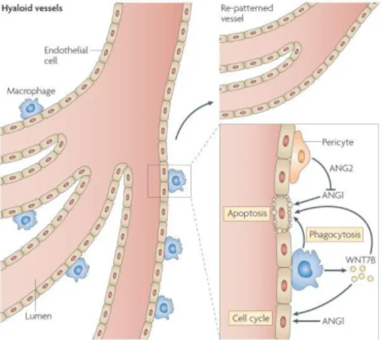
Macrophages have been recognized as important players of angiogenesis during wound healing, producing a variety of factors that can regulate vascular potential [24]. However, only more recently their relevance in developmental angiogenesis has been explored. During eye development, the role of macrophage in vascular remodeling was showed. Macrophage ablation, either using a suicide gene approach or through their loss in PU.1-deficient mice, results in a failure of remodeling and persistence of the eye vascular deficient structure post-natally [76]. The mechanism of macrophage action is through their production of WNT7B, which stimulates neighboring vascular endothelial cells to enter the S phase of the cell cycle. More, the action of Ang-2 produced by pericytes leads to endothelial cells undergoes apoptosis to be phagocytosed
43
by the macrophages (figure 9) [77, 78]. Therefore, macrophages allow that the vascular remodeling occurs, and ensures the phagocytosis of apoptotic endothelial cells.
Figure 9. Macrophages interference in angiogenic and vascular eye development. The hyaloid vessel system remodeling is dependent of the vessels synthesize WNT7B, which leads to vascular endothelial cells to enter the DNA synthesis phase of the cell cycle. And the angiopoietin 2 (Ang-2), produced by pericytes blocks the survival signal from Ang-1, which allows that the endothelial cells suffering apoptosis are phagocytosed by the macrophages [24].
In brain development was shown that these cells promote vascular networking and this effect appears as being due to an interaction with endothelial tip cells. This association was observed during all phases of vascular networking formation. Also it was showed that yolk sac–derived macrophages colonized the embryonic mouse brain independently of vessels, as previously reported for the chick macrophages accumulated in the subventricular zone (SZ) between E 10.0 and E 11.5. From this moment, the vessels began to sprout laterally and fused with neighboring vessel branches to form the subventricular vascular plexus. Furthermore, the number of macrophages in the SZ peaked at E 11.5, the major phase of vascular networking. The ablation of macrophages using PU.1-null mice results in hindbrains with significant reduction in the number of vessel intersections and thereby decreased subventricular vascular plexus (SVP) complexity. Fewer connections also formed between neighboring radial vessels compared with wild-type hindbrains at the time when vascular networking was initiated in deeper brain layers. CSF1- deficient hindbrains had an SVP with reduced complexity, similar to PU.1-null mice [79]. These findings suggest that early tissue macrophages play an essential role in brain angiogenesis.
44
1.2.4.6. Pancreas
In the primary postnatal pancreas of different mouse strains, mature macrophages have been detected from birth on. The number of F4/80 positive cells significantly decreases from birth to weaning. This fact could be explained by the exocrine expansion associated with the postnatal development of the pancreas, but can also mean the earliest F4/80 fetal macrophages disappearance. Despite the critical influence of macrophages in the development and pathogenesis of diabetogenesis, as a result of their infiltration in intra-islet, they have not been clearly described in noninflamatory conditions. Under physiological conditions, the characterization of macrophage population was performed, and macrophages were first observed in the pancreas at E14.5 and remain present in the fetal pancreas at E17.5. It was also observed the presence of macrophage precursors at E12.5 in the fetal pancreas, pancreatic buds along with stomach and duodenal loops [56]. The F4/80+ cells, observed at E14.5, do not likely enter the pancreas via blood circulation, since that functional blood vessel are not observed until E15.5 in the murine pancreas [80].The association between macrophages and tissue organogenesis was described. In vivo studies demonstrated that macrophage ablation results in abnormal postnatal islet morphogenesis and impaired pancreatic cell proliferation [81]. Pancreatic explants were excised and cultured with or without M-CSF. F4/80+ cells were observed in the explants, for both cases, after 5 days of culture. However, the supplementation with M-CSF induced a significant increase in the number of F4/80+ cells in the explant area. Also, it was examined that the addition of M-CSF, and the consequent increase in macrophage number, increase more than fourfold the number of positive insulin cells (insulin- and glucagon-expressing cells) in the cultured explants. Possibly, this effect is mediated via differentiation and activation of the macrophage precursors that were present in the explant [56]. These results allowed concluding an important role in the development of fetal pancreas macrophages.
1.2.4.7. Kidney
Fetal macrophages are among the first hematopoietic cells found in the kidney, appearing in the renal interstitium between E11.5 and 12. It was demonstrated the co-localization of F4/80 with Csf1r-positive cells in the interstitium surrounding the ureteric bud, using a Csf1r-EGFP mice. Their localization appears to be contiguous to the vascular development bed between the metanephric mesenchyme and the metanephric ducts, suggesting that possibly macrophages
45
enter the renal mesenchyme from or with the circulation. The supplementation with CSF-1 to E11.5-12 kidney explants culture resulted in a significant increase in the number of branch tips and nephrons [31]. Associated with this effect was showed the increase in EGFP-positive cells as a result of an increase in the macrophage population. However, there are several ways to the CSF1 actuation in the kidney development, such as macrophage response with the production of critical factors, as growth signal mediating surface tethered, or transmembrane proteins able to signal to adjacent renal cells. Other effect could be an increase in the clearance of apoptotic cells due to macrophage activation. However, apoptotic markers in the explants are not significant different with or without CSF-1 stimulation. Other possibility is the CSF-1 effect in the number of resident macrophages, which increases the proliferative environment. Finally, CSF-1 could directly signalize to another cell population within the kidney. Rather than an immunological role assigned to macrophages, these findings suggest a trophic role during embryonic kidney development [31]. During post-natal development, a similar approach was used to identify the relevance of macrophages in postnatal organ growth and kidney repair. CSF-1 administration to newborn mice increased overall body weight and kidney weight and volume. Moreover, using Csf1r-reporter mouse it was showed that this effect was accompanied by an increase in macrophage numbers in the kidneys.
1.2.5. Lung macrophages and inflammatory mediators in lung development
1.2.5.1. Lung macrophages
The adult lung macrophage population could be subdivided in two principal types: alveolar and interstitial macrophages, localized in separate anatomical compartments in the lung, including the air spaces and lung connective tissue, respectively [82, 83]. Alveolar macrophages are localized within the alveolus, strategically located to answer as primary defense of the lung against inhaled particles [84, 85]. Interstitial macrophages are closely associated with matrix and connective-tissue of lung, representing around 40% of the total macrophages in lung tissue [86]. Interstitial macrophage exhibits a power greater than alveolar to proliferative and this capacity is possible related with the maintenance of lung macrophage pool [40].
During mice lung development, macrophages are found since the begining of development around E10, and remains during throughout development [87, 88].
46
Immunofluorescence labeling to F4/80 macrophage marker in E12.5 embryonic lungs, showed that macrophages are located extensively accumulated within developing lungs, particularly within branch points. At E15.5 the relative abundance of macrophages in lung is around 5% of total cells. More, their characterization shows enrichment in a lot of genes common with tumor associated macrophages (TAM), suggesting similarities between fetal and tumor environments [31]. On the other hand, more recent findings showed that macrophages are closely associated with maturation of lung. During the saccular stage at post-natal day (P) 1 and P 5, around 8% of total lung cells are macrophages. The increase of these cells is greater during postnatal development, particularly, during alveolarisation, from P 14-21 approaching the 16%, and at 3 months a significant resident macrophage population is maintained. More, during alveolarization it was showed the upregulation of Arginase 1 (Arg1), mannose receptor 1 (Mrc1) and chemokine C-C motif ligand 17 (Ccl17), a phenotype of an M2 or tissue remodeling macrophages [88]. These facts shows that macrophages are present during all lung development but their levels are variable. Despite the deep characterization of macrophage population during post-natal periods and to lesser extent during fetal development, the real contribution of macrophages to lung development remains unknown.
1.2.5.2. Cytokines as mediators in lung development
Some cytokines commonly secreted or regulated by macrophages have been demonstrated to modulate fetal lung branching and maturation [89, 90]. Besides their crucial role in inflammation and the immune response, some cytokines are important players in hematopoiesis, liver and neuronal regeneration, embryonic development and fertility. Their action is mediated by the activation of target genes involved in crucial processes of development such as differentiation, survival, apoptosis and proliferation [89]. During lung development, the relevance of some cytokines has been described. For instance, IL-6 is constitutively expressed in rat pulmonary primitive epithelium, displaying highest levels during earliest stages, which decrease during the following stages of development. Also LIF is important during lung development. Although Leukemia inhibitory factor (LIF) knockout mice have normal lung formation, it is known that LIF, together with insulin-like growth factor I (IGF-I), regulates lung maturation. LIF in addition to IGF-I null mutant mice aggravates pulmonary immaturity, namely LIF/IGF-I double deficient embryos show lung hypoplasia and defective differentiation of the
47
alveolar epithelium and vasculogenesis. Likewise, TGFβ is expressed at high levels during normal lung development, being crucial for branching morphogenesis and epithelial cell differentiation with maturation of surfactant synthesis [91]. Other cytokine associated with lung development is TNF-a receptor found throughout the embryonic lung and the supplementation with TNF-a in vitro of embryonic lung primordial have a stimulatory effect on branching morphogenesis and surfactant-associated protein (SP-A) expression [92].
Concerning lung maturation, the overexpression of pro-inflammatory mediators, for
instance TNF-α, transforming growth factor α (TGF-α), IL-11, or IL-6, in mice lungs results in pathologic situations leading to impairment in alveolarization. The overexpression of the enumerated factors results in fewer and larger alveoli and fibrosis. [93]. The previous data suggests that lung development is highly regulated by cytokine environmental. Being macrophages relevant players in cytokines expression, their possible relevance during lung development is overwhelming.
49 1.10 Rationale and Aims
Besides their function during the development in other organs, macrophage function during lung development remains unknown. Therefore, this master thesis aims to understand the relevance of tissue macrophage ablation during lung development. Tacking in account that disruption of Csf1r gene results in large depletions of macrophages in most tissues, in order to accomplish this aim, we perform a characterization of lung development using a macrophage-deficient mouse model (Csf1r-/-). Three specifics points of lung development were evaluated: the
pseudoglandular (at E15.5), the fetal saccular stage (E18.5) and the post-natal saccular stage (P0). For all stages tree groups/genotypes Csf1+/+- Csf1r-/+ and Csf1r-/- were compared:
- At pseudoglandular stage it was performed the evaluation of gross morphology, bud number quantification and lung morphology characterization, in which stereological analyses was used. For the last evaluation it was used stereological analysis. Specifically, it was measured epithelial, mesenchymal and air space volume density (VD). Additionally, it was performed evaluation of lung vasculature: vessel density quantification using the endothelial cell marker CD31 (PECAM-1), by immunohistochemistry. Beyond this, assessment of transcripts expression patterns of angiogenic factors and associated molecules (such as VEGF-A, VEGFR2, Ang-1, Ang-2, Hif1α and Fgf2) was performed by q-RT-PCR.
- At the fetal saccular stage (E18.5) it was also evaluated lung morphology by the same parameters described. Concerning lung vasculature, beyond the parameters evaluated at E15.5 stage, it was evaluated the artery thickness, using Weigert’s staining.
- At post-natal saccular stage (P0), beyond the previous lung morphology evaluation at E15.5 and E18.5, lung-to-body weight ratios were accessed. Regarding, stereological analysis, beyond the mesenchymal, epithelial and air space volume, it was also evaluated the large and small airway epithelium VD. Blood gasometrical evaluation was performed as readout of lung functionality. Regarding lung vasculature all parameters evaluated at E18.5 stage were also evaluated.
53 2. MATERIALS AND METHODS
2.1. Animals
All procedures and protocols were carried out in accordance with European Union Directive 86/609/EEC and NIH guidelines on animal care and experimentation. Mice of mixed genetic background (C3H/C57BL/6) and deficient in Csf1r (Csf1r–/-) were kindly provided by Prof.
E. Richard Stanley, Albert Einstein College of Medicine University Yeshiva, Bronx, USA. Animals were fed ad libitum and exposed to day-night cycles alternatively every 12h.
2.2. Study design
The Csf1r+/- animals were crossed and the vaginal plug checked 12 hours after and
defined as embryonic gestational day 0.5 (E0.5). At E15.5 and E18.5 stages, dam were sacrificed by cervical dislocation, embryos were removed by caesarean section. Embryos and newborns (post-natal day (P) 0) were sacrificed by decapitation and lung excised under stereomicroscope (Olympus SZX7). Depending on the analysis required, different lung tissue processing was performed. To evaluate mice genotype, a small piece of tissue of each animal was collected. The tree different embryos/newborns genotypes were assessed using PCR analyses. Then, the animals were subdivided into 3 groups: Csf1r+/+ (wild type or control); Csf1r+/-
and Csf1r-/-.
2.3. PCR analyses for Genotyping
Pellet was diluted in ultrapure H2O and let to dissolve overnight. Detection of CSF1R
transgene was done using a primer specific to a fragment Csf1r. Genomic DNA was extracted from tissue pieces and incubated overnight at 55° in lysis buffer (100 mM TRIS (pH8), 0.2 % sodium dodecyl sulfate (SDS), 5mM EDTA) and proteinase K (85μg/ml). After centrifuged 10 min at 14000g, isopropanol was added to the supernatants collected, centrifuged 5 min at 14000g, the new supernatant discarded and pellet dried by incubation at RT. Pellet was diluted in ultrapure H2O and let to dissolve overnight. Detection of Csf1r transgene was done using a set of
specific primers to a fragment Csf1r.The final concentrations in the PCR reactions were: 50ng of extracted DNA, 200 μM of each dNTP, 1.5mM MgCl2, 1.25 U Taq DNA Polymerase (Promega) and 0.4 μM of each primers ( Intron 2 Forward: 5’AGACTCATTCGAACCAGAGAGC-3’;
Csf1r-54
InEx 3 Reverse: 5’-GAATTTGGAGTCCTCACCTTTG -3’; Csf1R-Neo-Forward: 5’ CCGGTAGAATTCCTCGAGTCTA-3’). Amplification (MycyclerTmthermalcycler, BIO-RAD) was
performed as follows: 92° for 20 sec (initial strand separation); 35 cycles of 94º (20 sec), 58º (45 sec) and 72º (1 min); and 72° for 7 min (final elongation step). The PCR fragment size expected to presence of Csf1r gene (wild type animals) was 385 bp and to absence (Csf1r-/-) was
308 bp. These were visualized by electrophoresis in 4% agarose gels.
2.4. Lung-to-body weight ratios
After birth (P0), newborn were killed by decapitation and lung excised under a stereomicroscope. The right and left lungs and the entire animal were weighed independently and lung-to-body weight ratios were assessed. The evaluation was performed to the tree genotypes (Csf1r+/+: n=8; Csf1r-/+: n=11 and Csf1r-/-: n=4).
2.5. Neonatal blood gasometric evaluation
The newborn were killed by decapitation and the blood collected (neck bleeding) to perform gasometric evaluation. The parameters calculated were: pH, PCO2 (Partial Pressure)
(mmHg), PO2 (mmHg), SatO2 (Saturation) (%) and Lactate (mmol/L) using a gasometric analyser
(i- Stat1 analyser; Abbott, Chicago, IL, USA). The evaluation was performed for the tree genotypes at P0 stage (Csf1r+/+: n=4; Csf1r-/+: n=5 and Csf1r-/-: n=2).
2.6. Stereological Analyze
To stereological analyses, the trachea was cannulated and the lungs were insufflated with fixation solution (glutaraldehyde 2,5% in PBS) under a constant pressure of 1 rpm during 1 min using a peristaltic pump (Gilson, Model Minipul S3). The lungs were dissected and kept in fixation solution on agitation, until its processing. Briefly, the lungs were dehydrated in increasing concentrations of ethanol and xylol, embedded in methacrylate, consecutively cut in slides with 15 µm and stained with hematoxylin-amoniacal. Stereological analysis was carried out by one person, blinded to the genotype of the animal, on a Zeiss microscope Axioplan 2 (Tokyo, Japan) using the Stereo Investigator version 5.2 software (MicroBrightField). A fixed range of 30 to 30
55
slides per animal was used; the 15µm-thick hematoxylin stained slides from each animal were analyzed using a grid of test points. The total number of points and intersections in each lung were >1,000. Morphometric parameters, such as total, mesenchymal, epithelial, air space, alveolar and bronchiolar lung tissue volume were calculated according to the point and intersection counting. Briefly, all points (intersections) which laid over a point count were counted and were represented as a proportion of total points. The volumes were calculate by the Cavalieri principle VL = h·∑P(sl)·d2(h- thickness, sI-point number, d-distance between grid points) [94]. The
percentage of volume density relative to all lung tissue was calculated for Csf1r+/+, Csf1r-/+ and
Csf1r-/- animals (n=4, n=5 and n=4 lungs were analyzed at E15.5; n=4, n=4 and n=4 at E18.5;
and n=4 n=5 and n=4 at P0, respectively).
2.7. Immunohistochemistry
To immunohistochemical studies the trachea was cannulated and the lungs were insufflated with fixation solution (paraformaldehyde (PAF) 4% in PBS) without pressure control. Then, lungs were dissecated and fixated overnight at RT with agitation. Afterwards lungs were dehydrated in increasing concentrations of ethanol and xylol, embedded in paraffin and 3 µm slides were used. Briefly, paraffin slides were deparaffinized and rehydrated. Antigen retrieval trials were performed in: 10mM of citrate buffer pH=6.0 in microwave or water-bath at 100°C, by 15 and 20 min, respectively. To inactivate endogenous peroxidases, slides were incubated in 3% hydrogen peroxide in MeOH for 10 min. Then, slides were incubated with Avidin/ Biotin for 15 min each and after with normal horse serum (HNS) (VECTASTAIN Elite ABC Kit) for 30 min, followed by an overnight incubation in diluted primary antibody PECAM-1/CD31 (Santa Cruz) in a 1:200 dilution. After, slides were incubated with biotinylated horse anti-goat secondary antibody (Vector Labs) in a 1:500 dilution in HNS for 1h, and next, peroxidase-labeled reagent for 5 min (VECTASTAIN® ABC reagent, Vector Labs). Slides were incubated with 3,3’-diamino-benzidine (DAB + Substrate System, DakoCytomation). Finally, slides were counterstained with hematoxylin and permanently mounted. All dilutions were made in PBS, pH 7.5, and all incubations were performed in humid chambers at room temperature. Between each step in the staining procedure (except before incubation with primary antibody), the slides were rinsed twice in PBS. In all reactions a negative control was included, where the staining was performed with the same protocol above described without primary antibody.
![Figure 1. Principal stages of lung development in mouse and human: diagrammatic representations of the timeline and developmental organization (adapted from[9])](https://thumb-eu.123doks.com/thumbv2/123dok_br/17577161.818480/30.892.183.707.722.968/figure-principal-development-diagrammatic-representations-timeline-developmental-organization.webp)