AR
TIGO ORIGINAL / ORIGINAL AR
TICLE
INTRODUCTION
The hepatopulmonary syndrome (HPS) is an al-teration in the vasculature of the lung secondary to a chronic liver disease, usually cirrhosis, characterized by the triad of liver disease, abnormalities in gas exchange (increase of alveolar-arterial gradient of oxygen, with or without hypoxemia) and evidence of intrapulmonary vasodilations (IVD)(12, 21, 29, 34).
Liver cirrhosis causes a series of systemic mani-festations, including changes in the pulmonary vasculature as idiopathic pulmonary hypertension, anastomoses between the portal circulation and pul-monary and the HPS.
The moderate dyspnea is a common inding in patients with liver cirrhosis, may result from com-pression of lung parenchyma by ascites and/or stroke pleural(1), concomitant pathologies that affect the lungs and liver. However, severe hypoxemia (PaO2 <60 mmHg) is less common and, in the absence of associated cardiopulmonary disease, strongly suggests the presence of HPS(5, 8).
Hypoxemia [PaO2 < 70 mmHg or increase of
alveo-ARE THE SPIDER ANGIOMAS
SKIN MARKERS OF
HEPATOPULMONARY SYNDROME?
Américo de Oliveira
SILVÉRIO
1,2, Dayanne Cintra
GUIMARÃES
1,
Larissa Fernanda Queiroz
ELIAS
1, Érika Oliveira
MILANEZ
1and Silvano
NAVES
2ABSTRACT – Context – Hepatopathies can signiicantly inluence both veins and arteries, these changes may cause some cutaneous stigmas, such as spider angioma (SA) and some systemic vascular changes, such as those observed in hepatopulmonary syndrome (HPS). Based on this common pathophysiological root we can assume that the SA can be skin markers of HPS. Objective – The objective of this study is to assess whether there is a relationship between the presence of SA and HPS. Methods– Records of 40 patients with liver cirrhosis who underwent contrast echocardiography were evaluated, in which we researched the description of SA, physical examination, and other clinical and laboratory data. For diagnosis of HPS we use these signs of the disease: presence of liver disease (cirrhosis in the case), abnormalities in gas exchange by arterial blood gases, and evidence of pulmonary vasodilations by the contrast echocardiography. Results – The SA were found in 21/40 (52.5%) patients and hepatopulmonary syndrome in 9/40 (22.5%). The HPS was observed in 8/21 (38.1%) of patients with SA and 1/19 (5.3%) patients were without this sign (P<0.01). We found no statistically signiicant difference between the SA and the presence of HPS with sex or age. Patients with SA had a higher hypoxemia [PaO2 84.8 ± 11.5 mmHg and 19.8 ± 14.7 mmHg alveolar-arterial gradient of oxygen (AAG)] than those without SA (PaO2 90.8 ± 10.7 mmHg and 10.9 ± 11.7 AAG mmHg) (P<0.05). Conclusion - Our indings show a correlation between the presence of SA and HPS, suggesting that the SA may be cutaneous markers of HPS.
HEADINGS – Liver diseases, complications. Skin diseases vascular. Hepatopulmonary syndrome.
Declared conflict of interest of all authors: none
1 Departamento de Medicina, Pontifícia Universidade Católica de Goiás (PUC-Goiás); 2 Hospital Geral de Goiânia (HGG).
Correspondence: Américo de Oliveira Silvério. Rua Bárbara, quadra 41, lote 9 – Condomínio Portal do Sol 2 – 7884-651 – Goiânia, GO, Brasil. E-mail: americosilverio@ hotmail.com
lar-arterial gradient of oxygen (AAG) 15 mmHg] is multifactorial, being caused either by intrapulmonary vascular dilations, and by changes in alveolar-capillary diffusion of oxygen and/or the imbalance in the venti-lation/alveolar perfusion(12, 14, 16, 23, 29).
Still little is known about the pathogenesis of in-trapulmonary vascular dilation, it is believed that it is due to a number of factors such as the imbalance between vasoconstrictors and vasodilators substances, and/or increased sensitivity of pulmonary endothe-lium to a vasodilator and/or hyporesponsiveness to vasoconstrictors substances(12, 14, 16, 23, 29).
This imbalance between vasodilator and vasocon-strictor substances, is also involved in the pathophys-iology of others vascular abnormalities observed in cirrhosis, such as the clubbing, palmar erythema and SA(21, 22).
asso-ciated with chronic liver diseases, its prevalence is variable, ranging from 4 to 47% depending on diagnostic criteria and methods used(23).
Assuming that both the HPS and the SA have a common pathophysiological root, that is an imbalance between vaso-constrictor and vasodilator substances, and the suggestion of some authors, that SA are most commonly seen in patients with the HPS, we can assume that SA could be used as skin markers of HPS. This would allow an earlier and less costly diagnosis of the syndrome, and thus a more effective therapeutic approach. This study aims to evaluate if exists a relationship between SA and HPS.
METHODS
We prospectively evaluated 45 patients with liver cirrhosis diagnosis seen at the Gastroenterology and Hepatology Service of the General Hospital of Goiânia. The diagnosis of cirrhosis was based on clinical and laboratory criteria (history, physical examination, ultrasound indings and biochemical data and/ or CT scan of the abdomen) and/or histology.
The study included patients of both sexes suffering from chronic liver disease, aged above 18 years old and who vol-untarily agreed to participate. These patients underwent a clinical interview and physical examination, focusing on the presence of SA. Blood samples were collected to perform liver function tests, complete blood count. Arterial blood gases were taken with patients sitting and breathing room air. The AAG was calculated using the alveolar gas equation. Subsequently, a simple echocardiography and a contrast echocardiography (CEE) have been made.
The presence of SA was based on physical examination data. We consider as carriers of HPS patients who met the following criteria: presence of chronic liver disease, abnor-malities in arterial oxygenation (AAG 15 mmHg) and evidence of intrapulmonary vasodilations (IVD) the CEE(29).
For the research of IVD we used CEE performed by an experienced researcher using the method of microbubbles. The technique is based on the rapid infusion of 10 mL saline solution (NaCl 0.9%) in the right median cubital vein(33), after
its agitation and production of microbubbles. These should be viewed in the right heart chambers, with the help of a transducer positioned in the left parasternal region, since they are echogenic. Under normal conditions, the microbubbles are retained in the pulmonary capillary bed, as in patients with IVD they are displayed in the left atrium in 3 to 6 car-diac cycles after being observed in the right chambers(33). Its appearance before the third cycle suggests the presence of intracardiac communication(33).
Our study was approved by the Ethics Committee of human and animal research of General Hospital of Goiânia.
Statistical calculations were performed using the program Epi Info 3.5.2 (Centers for Disease Control Epidemiology Program Ofice, Atlanta, Georgia). To evaluate the associ-ation between qualitative categorical variables it was used the chi-square test of Pearson, with the Yates test correction when necessary. We used the Kruskal-Wallis test to test the correlation of quantitative independent variables. The results were expressed as mean ± standard deviation (SD) and sig-niicance level in all tests was set at 5%.
RESULTS
Of the 45 patients initially evaluated ive were excluded (2 because they had atrio-ventricular communication, 2 for not having adequate acoustic window to perform the CEE and one for not having inished the protocol). The 40 (88.9%) remaining patients composed our study group. Of these, 23 (57.5%) were men and 17 (42.5%) women. Our population was composed mostly by whites (65% - 26 patients) and the average age was 48.6 ± 11.7 years old (ranging from 19 to 71 years old).
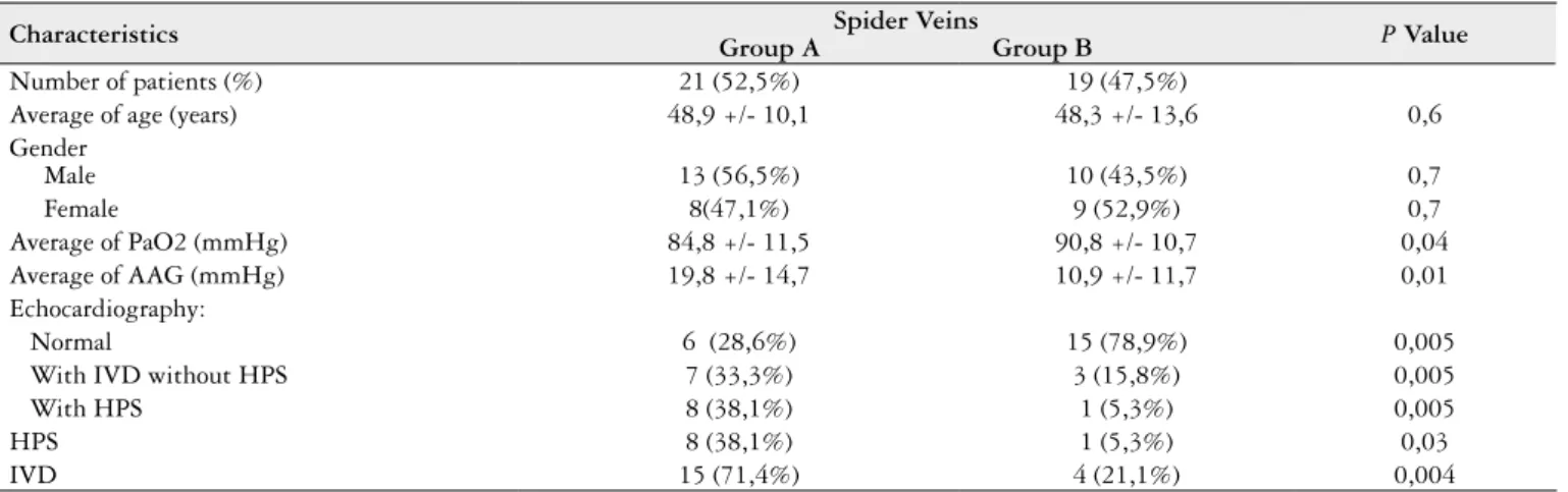
Patients were divided into two groups according to the presence or absence of SA. Group A: composed of 21 patients (52.5%) with SA, the average age was 48.9 ± 10.1 years old, 13/21 (56.5%) were male. Group B: composed of 19 patients (47.5%) without SA, the average age was 48.3 ± 13.6 years old and 10/19 (43.5%) were male. There was no statistically signi-icant difference between these variables. Table 1 summarizes the demographics data of the researched population.
TABLE 1. Characteristics of the 40 patients divided according to the presence of spider veins
Characteristics Group A Group BSpider Veins P Value
Number of patients (%) 21 (52,5%) 19 (47,5%)
Average of age (years) 48,9 +/- 10,1 48,3 +/- 13,6 0,6
Gender
Male 13 (56,5%) 10 (43,5%) 0,7
Female 8(47,1%) 9 (52,9%) 0,7
Average of PaO2 (mmHg) 84,8 +/- 11,5 90,8 +/- 10,7 0,04
Average of AAG (mmHg) 19,8 +/- 14,7 10,9 +/- 11,7 0,01
Echocardiography:
Normal 6 (28,6%) 15 (78,9%) 0,005
With IVD without HPS 7 (33,3%) 3 (15,8%) 0,005
With HPS 8 (38,1%) 1 (5,3%) 0,005
HPS 8 (38,1%) 1 (5,3%) 0,03
IVD 15 (71,4%) 4 (21,1%) 0,004
The IVD were present in 19/40 (47.5%), and in 9/40 (22.5%) also met the other criteria for HPS.
When comparing the two groups we observed that the IVD were present in 15/21 (71.4%) patients of group A and in 4/19 (21.1%) patients of group B (P<0.005), with relative risk 3.4 (1.3 to 8.4). About the HPS, it was present in 8/21 (38.1%) and in 1/19 (5.3%) respectively (P<0.05), with a relative risk of 5.2 (0,8 to 33.9). The mean PaO2 and AAG were respectively 84.8 ± 11.5 mmHg and 19.8 ± 14.7 mmHg in group A and 90.8 ± 10.7 mmHg and 10.9 ± 11.7 in group B (P<0.05).
Most patients 36/40 (90%) had A Child-Pugh index, which does not allow to evaluate relations of SA with the severity of liver disease.
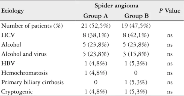
The hepatitis C virus isolated or associated with alco-hol consumption were the most common etiologic agents of cirrhosis, present in 23/40 (57.5%) and 18/40 (45.0%) respectively. There was no statistically signiicant difference between the presence of SA and the etiology of cirrhosis (P = 0.68) (Table 2).
TABLE 2. Correlation between the presence of spider veins and cirrhosis etiology
Etiology Spider angioma P Value Group A Group B
Number of patients (%) 21 (52,5%) 19 (47,5%)
HCV 8 (38,1%) 8 (42,1%) ns
Alcohol 5 (23,8%) 5 (23,8%) ns
Alcohol and virus 5 (23,8%) 3 (15,8%) ns
HBV 1 (4,8%) 1 (5,3%) ns
Hemochromatosis 1 (4,8%) 0 ns
Primary biliary cirrhosis 0 1 (5,3%) ns
Cryptogenic 1 (4,8%) 1 (5,3%) ns
HCV = hepatitis C virus; HBV = hepatitis B virus
DISCUSSION
Galen’s view that the liver controls blood in the veins lost prestige as the true nature of the vascular system was becoming evident(17). However, information obtained from the study of patients with liver disease have revealed that these diseases can signiicantly inluence both the arteries and veins(11, 17, 30).
In chronic liver disease we can observe the inluence of portal hypertension in the vascular system. There is dilation of the veins of the thorax and abdomen(17) and formation of collateral circulation between portal and systemic cir-culation(10, 20, 35), because of the opening and dilation and hypertrophy of preexisting vascular channels(6, 30).
Ectopic varicose veins may occur in the digestive tub(6). We can observe the presence on skin of SA, most commonly on the face and upper chest, the palms of the hands may become hyperemic (palmar erythema) and we can ind cya-nosis, edema in the nail bed and it subsequently can rise to the digital clubbing(4, 17).
The SA are known to be a cutaneous stigma of chronic liver disease(18, 26), occurring in approximately 15% of patients with Laennec’s cirrhosis(26). In patients with this sign, esopha-geal varicose veins and collateral circulation in the abdomen appear to be two times more frequent(7, 14).
In the pulmonary vasculature we can also observe changes resulting from chronic liver disease. Idiopathic pulmonary hypertension can arise, anastomoses between the portal and the pulmonary circulation and HPS(19, 31).
The pathogenesis of intrapulmonary vascular dilatation is still debated, several mechanisms have been proposed, including: imbalance between vasoconstrictors and vasodi-lators substances; the increase of pulmonary endothelium sensitivity to a vasodilator not produced or not metabolized by the diseased liver, or even hyporesponsiveness to vasocon-strictor substances(12, 14, 16, 23, 31).
Among the vasodilator substances postulated to explain the pathogenesis of HPS, the nitric oxide (NO) wins a role. The NO is a potent endothelial vasodilator and could be responsible for intrapulmonary vasodilatation, since in cirrhotic patients the serum levels of this substance are ele-vated, possibly due to endotoxemy observed in this group of patients, which would induce an increased production of NO (9, 12, 14, 15, 23).
Therefore, both the HPS and the SA seem to have a com-mon pathophysiological root, which is the imbalance between vasoconstrictors and vasodilators substances.
Some authors have suggested that the SA is most com-monly seen in patients with the HPS(1, 3, 18, 27, 28), although other authors have not found the same relationship(13, 24). In the classic story of Rydell and Hoffbauer(18) the authors have called attention to the fact that the vast majority of cirrhotic patients with cyanosis and digital clubbing associated exhibit one or more vascular stigmas of cirrhosis, especially SA(18). Andrivet et al.(3) found the signal in seven of nine cirrhotic patients with hypoxemia. Rodriguez-Roisin et al.(28) demon-strated that patients with SA have higher gas exchange ab-normalities, reduced pulmonary vasoconstrictor response to hypoxia, reduced pulmonary vascular resistance and systemic and major changes in the relation ventilation/perfusion than patients without the sign(28). In contrast, Lima et al.(24) eval-uating the clinical features of cirrhotic patients candidates for liver transplantation, observed the SA in 8/9 (88.8%) of patients with HPS and in 35/47 (74.5%) of cirrhotics without the syndrome (P = 0.6). Fallon et al.(13) also evaluating can-didates for liver transplantation, also found no difference in the prevalence of SA in patients with or without HPS (37% and 39% respectively, P = 0.78). In our study, the SA were found in 8/9 (88.9%) patients with HPS, in 7/10 (70.0%) of patients with IVD without hypoxemia and in 6/21 (28.6%) of patients without IVD (P<0.005). Suggesting that the SA could not only be related to HPS but as well as to the degree of IVD, and may also be a predictive marker of HPS.
such abnormalities (P<0.01). Mohammad Alizadeh AH et al.(25) found the presence of IVD in 17/54 (31.4%) of re-searched patients, of these, 10/54 (18.3%) met criteria for the HPS and 7/54 (13%) had only IVD, deined as subclinical cases of the syndrome. In this study, HPS was found in 9/40 (22.5%) patients. Despite that, 10/40 (25.0%) patients had IVD, however, without fulilling the diagnostic criteria of the syndrome, which may represent a frustrated form of it, as the subclinical cases found in the study of Mohammad Alizadeh AH et al.(25).
The average of PaO2 was lower in patients with SA (84.8 ± 11.5 mm Hg) compared with patients without the sign (90.8 ± 10.7 mm Hg) with P<0.05; and the average AAG in the irst group was higher (19.8 ± 14.7), compared with 10.9 ± 11.7 in the second (P<0.05). These indings suggest that patients with SA are more hypoxemic than patients without SA.
Based on our indings we suggest that the SA are skin markers of the presence of HPS. However, it is necessary to perform further studies to correlate the presence of HPS with the number and size of the SA.
Silvério AO, Guimarães DC, Elias LFQ, Milanez EO, Naves S. São as aranhas vasculares marcadores cutâneos da síndrome hepatopulmonar?. Arq Gastroenterol. 2013,50(3):175-9.
RESUMO – Contexto - As hepatopatias podem inluenciar de forma considerável tanto as veias quanto as artérias, dessas alterações podem surgir alguns estigmas cutâneos, como as aranhas vasculares (AV) e algumas alterações vasculares sistêmicas, como as observadas na Síndrome Hepatopulmonar (SHP). Baseados nessa possível raiz isiopatogênica comum, podemos supor que as AV sejam marcadores cutâneos da SHP. Objetivo - Avaliar se há relação entre a presença das AV e a SHP. Métodos - Foram avaliados os prontuários de 40 pacientes com cirrose hepática submetidos a ecocardiograia com contraste (ECC), nos quais pesquisamos a descrição de AV, no exame físico, e outros dados clínicos e laboratoriais. Para diagnóstico da SHP utilizamos os seguintes critério: presença de hepatopatia (no caso cirrose), de anormalidades nas trocas gasosas pela gasometria arterial, e evidências de vasodilatações pulmonares pela ecocardiograia com contraste. Resultados - As AV foram encontradas em 21/40 (52,5%) pacientes e a síndrome hepatopulmonar em 9/40 (22,5%). A SHP foi observada em 8/21 (38,1%) dos pacientes com AV e em 1/19 (5,3%) dos pacientes sem esse sinal (P<0,01). Não encontramos diferença estatisticamente signiicativa entre a presença das AV e da SHP com sexo ou faixa etária. Pacientes com AV apresentaram maior hipoxemia (PaO2 84,8 ± 11,5 mmHg e GAA 19,8 ± 14,7 mmHg) que os sem AV (PaO2 90,8 ± 10,7 mmHg e GAA 10,9 ± 11,7 mmHg) (P<0,05).
Conclusão - Nossos achados mostram correlação entre a presença das AV e a SHP, sugerindo que as AV possam ser marcadores cutâneos da SHP.
REFERENCES
1. Akiyama M, Inamoto N. Arteriovenous haemangioma in chronic liver disease: clinical and histopathological features of four cases. Br J Dermatol. 2001;144:604-9. 2. Alonso-Martínez JL, Zozaya-Urmeneta JM, García-Sanchotena JL, Olaz-Pre-ciado F, Estébanez-Estébanez C, Berjón-Reyero J. Hepatopulmonary syndrome: relationship with liver dysfunction and systemic hemodynamic disorder Med Clin (Barc) 2004;123:721-5.
3. Andrivet P, Cadranel J, Housset B, Herigault R, Harf A, Adnot S. Mechanisms of impaired arterial oxygenation in patients with liver cirrhosis and severe respiratory insuficiency. Effects of indomethacin. Chest. 1993;103:500-7.
4. Bashour FA. Clubbing of the digits: physiologic considerations. J. Lab. Clin. Med. 1961;58:613-21.
5. Berthelot P, Walker JG, Sherlock S, Reid L. Arterial changes in the lungs in cirrhosis of the liver-lung spider nevi. N Eng J Med. 1966;274:291-8.
6. Bosch J, Pizcueta P, Feu F, Fernández M, García-Pagán JC. Pathophysiology of portal hypertension. Gastroenterol Clin North Am. 1992;21:1-14.
7. Brick IB, Palmer ED. Esophageal varices and vascular spiders (nevi araneosi) in cirrhosis of the liver. J Am Med Assoc. 1954;155:8-10.
8. Brett SJ, Evans TW. Endogenous nitric oxide in exhaled human breath. A new means of monitoring airway disease activity or another NO-NO? Chest. 1996;110:873–4.
9. Cahill PA, Wu Y, Sitzmann JV. Altered adenylyl cyclase activities and G-protein abnormalities in portal hypertensive rabbits. J Clin Invest. 1994;93:2691-700. 10. Calabresi P, Abelmann WH. Porto-caval and porto-pulmonary anastomoses in
Laennec’s cirrhosis and in heart failure. J Clin Invest. 1957;36:1257-65. 11. Chang SW, Ohara N. Pulmonary circulatory dysfunction in rats with biliary
cirrhosis. Am Rev Respir Dis. 1992;145:798-805.
12. Colle I, Van Steenkiste C, Geerts A, Van Vlierberghe H. Hepatopulmonary syndrome and portopulmonary hypertension: what’s new? Acta Gastroenterol Belg. 2007;70:203-9.
13. Fallon MB, Krowka MJ, Brown RS, Trotter JF, Zacks S, Roberts KE, Shah VH, Kaplowitz N, Forman L, Wille K, Kawut SM; Pulmonary Vascular Complications of Liver Disease Study Group. Impact of hepatopulmonary syndrome on quality of life and survival in liver transplant candidates. Gastroenterology. 2008;135:1168-75. 14. Ferreira PP, Zollinger CC, Bittencourt PL. Síndrome Hepatopulmonar.
Gastro-enterol Endosc Dig. 2008;27:5-13.
15. Finn SM, Rowland M, Lawlor F, Kinsella W, Chan L, Byrne O, O’Mahony O, Bourke B. The signiicance of cutaneous spider naevi in children. Arch Dis Child. 2006;91:604-5.
16. Freire R, Mangualde J, Vieira AM, Lobato C, Alves AL, Cremers MI, Augusto F, Caetano F, Oliveira AP. Síndrome Hepatopulmonar em doentes com cirrose hepática: importância da sua pesquisa sistemática e impacto no prognóstico. J Port Gastrenterol 2007;14:176-83.
17. Fritts HW Jr. Systemic circulatory adjustments in hepatic disease. Med Clin North Am. 1963;47:563-78.
18. Hoffbauer FW, Rydell R. Multiple pulmonary arteriovenous istulas in juvenile cirrhosis. Am J Med. 1956;21:450-60.
19. Jiva TM. Unexplained hypoxemia in liver disease: The Hepato-pulmonary syn-drome. Journal of Critical Illness. 1994;9:934-47.
20. Khaliq SU, Kay JM, Heath D. Porta-pulmonary venous anastomoses in experi-mental cirrhosis of the liver in rats. J. Pathol. 1972;107:167-74.
21. Krowka, M.J., Cortese, D.A.; Hepatopulmonary syndrome. Current concepts in diagnostic and therapeutic considerations. Chest. 1994;105:1528-37.
22. Lange PA, Stoller JK. The hepatopulmonary syndrome. Ann Intern Med. 1995;122:521-9.
23. Li CP, Lee FY, Hwang SJ, Lu RH, Lee WP, Chao Y, Wang SS, Chang FY, Whang-Peng J, Lee SD. Spider angiomas in patients with liver cirrhosis: role of vascular endothelial growth factor and basic ibroblast growth factor. World J Gastroenterol. 2003;9:2832-5.
24. Lima BL, França AV, Pazin-Filho A, Araújo WM, Martinez JA, Maciel BC, Simões MV, Terra-Filho J, Martinelli AL. Frequency, clinical characteristics, and respiratory parameters of hepatopulmonary syndrome. Mayo Clin Proc. 2004;79:42-8.
25. Mohammad Alizadeh AH, Fatemi SR, Mirzaee V, Khoshbaten M, Talebipour B, Shariian A, Khoram Z, Haj-sheikh-oleslami F, Gholamreza-shirazi M, Zali MR. Clinical features of hepatopulmonary syndrome in cirrhotic patients. World J Gastroenterol. 2006;12:1954-6.
26. Ratnoff OD, Patek AJ Jr. The natural history of Laennec’s cirrhosis of the liver. Na analysis of 386 cases. Medicine. 1942;21:207-68.
27. Robin ED, Horn B, Goris ML, Theodore J, Kessel AV, Mazoub J, Tilkian A. Detection, quantitation and pathophysiology of lung spiders. Trans Assoc Am Physicians. 1975;88:202-16.
28. Rodriguez-Roisin R, Roca J, Agusti AG, Mastai R, Wagner PD, Bosch J. Gas exchange and pulmonary vascular reactivity in patients with liver dcirrhosis. Am Rev Respir Dis. 1987;135:1085-92.
29. Rodríguez-Roisin R, Krowka MJ, Hervé P, Fallon MB; ERS Task Force Pulmo-nary-Hepatic Vascular Disorders (PHD) Scientiic Committee. Pulmonary–He-patic vascular Disorders (PHD). Eur Respir J. 2004;24:861-80.
30. Schraufnagel DE, Patel KR. Sphincters in pulmonary veins: An anatomic study in rats. Am. Rev. Respir. Dis. 1990;141:721-26.
31. Schraufnagel DE, Kay JM. Structural and pathologic changes in the lung vascu-lature in chronic liver disease. Clin Chest Med. 1996;17:1-15.
32. Sharma V, Aggarwal S. Spider nevi and chronic liver disease. Indian J Dermatol Venereol Leprol. 2009;75:403.
33. Shub C, Tajik AJ, Seward JB, Dines DE. Detecting intrapulmonary right-to-left shunt with contrast echocardiography. Observations in a patient with diffuse pulmonary arteriovenous istulas. Mayo Clin. Proc. 1976;51:81-4.
34. Silvério AO, Garcia E, Rosa H. Síndrome Hepatopulmonar. In: Mattos AA, Dantas W. Compêndio de Hepatologia.2 ed. São Paulo: Fundo Editorial Byk, 2001.cap 44:685-96.
35. Stein H, Stein S. Digital clubbing in cirrhosis of the liver. Lancet. 1961;2:999-1000. 36. Vedamurthy M, Vedamurthy A. Spider nevi: a presenting feature of chronic liver
disease. Indian J Dermatol Venereol Leprol 2008;74:397-8.