Dissertação de Mestrado
Mestrado em Bioquímica Aplicada – Biomedicina
Trabalho efectuado sob a orientação do
Prof.ª Doutora Cristina Pereira-Wilson
Doutor Cristóvão Lima
Janeiro de 2015
Cátia Daniela Oliveira Lima Ferreira Machado
Ursolic and Oleanolic acid effects on
cellular cholesterol and impact on
signaling pathways of cell death and
proliferation
Nome: Cátia Daniela Oliveira Lima Ferreira Machado Endereço eletrónico: [email protected]
Número do Cartão de Cidadão: 13478368 Telefone: 926962130
Título da dissertação:
Ursolic and Oleanolic acid effects on cellular cholesterol and impact on signaling pathways of cell death and proliferation
Orientadores:
Professora Doutora Cristina Pereira-Wilson Doutor Cristóvão Lima
Ano de conclusão: 2015
Mestrado em Bioquímica Aplicada, ramo de Biomedicina
DE ACORDO COM A LEGISLAÇÃO EM VIGOR, NÃO É PERMITIDA A REPRODUÇÃO DE QUALQUER PARTE DESTA TESE/TRABALHO
Universidade do Minho, 30 de Janeiro de 2015
iii
AGRADECIMENTOS
“No meio da dificuldade encontra-se a oportunidade”
Albert Einstein
Em primeiro lugar gostaria de agradecer à Profª. Doutora Cristina Pereira-Wilson, orientadora deste trabalho, pela oportunidade de trabalhar neste projeto e integrar a sua equipa de investigação, pela orientação, pelo apoio e tempo disponibilizado e acima de tudo por partilhar o seu conhecimento científico comigo.
Gostaria de agradecer também ao Dr. Cristóvão Lima, meu coorientador, pelo apoio e incentivo, e pelas sugestões construtivas ao longo deste trabalho.
Um especial agradecimento á Dalila, Carla, Cristina e Ana Rita por todo o tempo e paciência que tiveram comigo, pelos ensinamentos, e principalmente pelas conversas, simpatia, amizade, e momentos de descontração.
Aos restantes colegas de laboratório, pelo bom ambiente de trabalho e boa disposição, pelo suporte e pelo espirito de equipa demonstrado.
Aos meus pais e irmã um agradecimento muito especial pelo apoio incondicional, por me darem a oportunidade de estar aqui e por nunca duvidarem das minhas capacidades. Graças a eles e ao excelente exemplo de luta e garra que me mostraram ao longo da minha vida sou a pessoa que sou hoje. Foi com eles que aprendi que a vida não é fácil, que as coisas não nos aparecem de mão beijada e que se lutarmos conseguimos ultrapassar todas as nossas dificuldades! Obrigada!
Por fim mas não menos importante, um sincero obrigado ao meu marido, Hernani, pelo apoio incondicional que me deste durante este longo percurso. Por nunca me teres deixado desistir nos momentos que mais me fui abaixo e por teres sempre um sorriso nos lábios e um abraço apertado quando mais precisei. Obrigado por teres depositado toda a confiança em mim e por seres o meu porto seguro! Obrigado por tudo! Amo-te!
v
Ursolic and Oleanolic acid effects on cellular cholesterol and impact on signaling pathways of cell death and proliferation
ABSTRACT
Cancer is a disease characterized by the uncontrolled growth of abnormal cells that beyond the limits of a cell division in normal conditions. Colorectal cancer (CRC) is the third most common tumors and is a major cause of cancer related death worldwide. Cholesterol metabolism has been established as possible source of therapeutic targets in cancer progression, because the cholesterol synthesis pathway provides farnesyl pyrophosphates groups essential toprenylation of proteins involved in cell proliferation and cancer growth. For this reason, targeting cholesterol synthesis with statins (HMG CoA reductase inhibitors) has been explored but with limited results in the clinic due to the toxicity of high dose statin treatment.Additionally, cholesterol is involved in proliferative cell signalling through receptors activation upstream of RAS and PI3K because it is a major constituent of lipid rafts. Thus, the aim of this project is to test if natural compounds with structure similar to cholesterol, such ursolic acid (UA) and oleanolic acid (OA) can alter the lipid composition and influence signalling pathways in order to inhibit the proliferative activity of carcinoma colorectal cells (HCT116). For this we evaluated the effect of the triterpenoids in the amount of cellular cholesterol by cholesterol quantification assay and found that the UA reduces and OA increase cholesterol cellular levels.These effects may be affect AKT signaling pathway induced by insulin because our results indicate that UA, but not OA, causes a decrease in p-AKT.Furthermore, by the method of nuclear condensation we evaluated apoptosis, and both the UA and OA increased tumor necrosis factor alpha (TNF-α) induced apoptosis. These results help to take another step in understanding the mechanisms of action of these natural compounds, helping control the progression of cancer.
vii
Efeito do ácido ursólico e oleanólico no colesterol celular e o seu impacto nas vias de sinalização de morte celular e proliferação
RESUMO
O cancro é uma patologia caracterizada pelo crescimento descontrolado de células anormais, que ultrapassam os limites de uma divisão celular em condições normais. O cancro colorrectal representa mundialmente o terceiro cancro mais comum e a maior causa de morte relatada por cancro. O metabolismo do colesterol tem sido proposto como possível alvo terapêutico na progressão desta neoplasia, porque a via do mevalonato responsável pela síntese deste composto proporciona grupos farnesil pirofosfatos essenciais à prenilação de proteínas que ativam vias relacionadas com a proliferação e o crescimento celular. Por esta razão, o combate da síntese de colesterol com estatinas (inibidores da HMG CoA Reductase) tem sido explorado mas com resultados limitados na prática clínica devido a toxicidade causada pelo tratamento com elevadas doses de estatinas. Adicionalmente verifica-se que o colesterol está também envolvido na proliferação através da ação de recetores de membrana que ativam a via da RAS e do PI3K, por ser o maior constituinte dos lipid
rafts. Assim este projeto visa testar se compostos naturais com estrutura semelhante à
do colesterol, como o ácido ursólico (AU) e ácido oleanólico (AO), conseguem alterar a composição lipídica e influenciar vias de sinalização de maneira a inibir a atividade proliferativa em células do carcinoma colorrectal (HCT116). Para tal avaliamos o efeito dos triterpenoides na quantidade de colesterol celular através do método de quantificação de colesterol, e verificamos que o AU diminui os níveis celulares e o AO provoca o seu aumento. Estes efeitos podem afetar a via de sinalização AKT induzida pela insulina, porque os nossos resultados indicam que o AU, mas não o AO, provoca uma diminuição da proteína p-AKT. Por outro lado, pelo método de condensação nuclear avaliamos a apoptose, e tanto o AU como AO aumentam a apoptose induzida pelo Fator tumoral de necrose alfa (TNF-α). Estes resultados ajudam a dar mais um passo na compreensão dos mecanismos de ação destes compostos naturais, ajudando no controlo da progressão do cancro.
ix CONTENTS Agradecimentos ... iii Abstract ... v Resumo ... vii Abbreviations list ... xi List of figures ... xv 1. Introduction ... 1 1.1-Cancer ... 3 1.2-Colorectal cancer ... 4 1.3-Lipid Rafts ... 7
1.3.1- Apoptosis and lipid rafts dependent membrane receptors ... 9
1.3.1- Cell proliferation and lipid rafts dependent membrane receptors ... 13
1.4-Cholesterol biosynthesis ... 17
1.4.1- Mevalonate pathway... 17
1.4.2- Sterol regulatory element binding protein ... 18
1.4.3- Statins ... 21
1.5-Natural compounds and chemoprevention ... 22
1.5.1- Triterpenoids ... 23
1.5.1.1- Ursolic Acid ... 25
1.5.1.2- Oleanolic Acid ... 26
2. Research objectives ... 27
3. Material and Methods ... 31
3.1 - Cell Culture, conditions and reagents ... 33
3.2 - Cell Viability by MTT reduction assay ... 34
3.3 - Protein extraction and Western Blot analysis ... 35
3.4 - Nuclear Condensation ... 36
3.5 - Cholesterol Assay ... 37
3.6 - Statistical analysis ... 39
4. Results ... 41
x
4.3 – Effects of UA and OA on TNF-α induction in cell death ... 46
4.4 – Effects of UA and OA on induction of proliferative pathways by insulin... 49
5. Discussion and Conclusions ... 53
5.1 – Discussion ... 55
5.2 – Conclusions ... 62
5.3 – Future Perspectives ... 64
6. References ... 65
xi
ABBREVIATIONS LIST
AKT – Protein kinase B
APC – Adenomatous polyposis coli BSA – Bovine serum albumin Cav-1 – Caveolin-1
Chol – Cholesterol
COPII – Coatomer protein complex II CRC – Colorectal cancer
DISC – Death-inducing signaling complex DMAPP – Dimethylallyl pyrophosphate DMSO – Dimethyl sulfoxide
DNA – Deoxyribonucleic acid
EDTA – Ethylenediamine tetraacetic acid EGF – Epidermal growth factor
EGFR – Epidermal growth factor receptor ER – Endoplasmic reticulum
ERK – Extracellular-signal-regulated kinase
FADD – Fas-associated protein with death domain FAP – Familial adenomatous polyposis
FAS – Fatty acid synthase FBS – Fetal bovine serum GDP – Guanosine diphosphate GTP – Guanosine triphosphate
HMG CoA – 3-hydroxy-3-methylglutaryl CoA
xii
Insig – Insulin-induced gene product IPP – Isopentanyl pyrophosphate IR – Insulin receptor
LDL – Low density lipoproteins
MAPK – Mitogen-activated protein kinase MMR – Mismatch repair
MTT – 3-(4, 5-dimethylthiazol-2-yl)-2,5-diphenyltetrazolium bromide MβCD – Methyl-β-cyclodextrin
Na3VO4 – Sodium orthovanadate
NaF – Sodium fluoride
NF-kB – Nuclear factor kappa-light-chain-enhancer of activated B cells OA – Oleanolic acid
PBS – Phosphate-buffered saline PDGF – Platelet derived growth factor PFA – Paraformaldehyde
PI3K – Phosphatidylinositol 3 kinase PMSF – Phenylmethylsulfonyl fluoride PVDF – Polyvinylidene difluoride Rcel – Ras converting enzyme
Rip – Regulated intramembranous proteolysis ROS – Reactive oxygen species
RPMI – Roswell park memorial institute médium RTK – Receptor tyrosine kinase
S1P – Site-1-protease S2P – Site-2-protease
xiii
SCAP – SREBP cleavage-activating protein Simv – Simvastatin
SRE – Sterol regulatory element
SREBP – Sterol regulatory element-binding protein SSD – Sterol sensing domain
TCC – Total cellular cholesterol
TNFR – Tumor necrosis factor receptor TNF-α – Tumor necrosis factor alpha
TRADD – Tumor necrosis factor receptor 1-associated death domain protein TRAIL – TNF-related apoptosis-inducing ligand
xv
LIST OF FIGURES
Figure 1.1 – The hallmarks of cancer. Adapted from (Hanahan and Weinberg, 2011) ... 4 Figure 1.2 – Schematic representation of some key genetic alterations during colorectal cancer (CRC) carcinogenesis ... 5 Figure 1.3 – Structure and composition of membrane lipid rafts. From (Chichlowski and Hale, 2008) ... 7 Figure 1.4 – Influence of disruption of membrane rafts domains in Fas signaling pathway. Reproduced from (Hryniewicz-Jankowska et al., 2014). ... 11 Figure 1.5 – Example how depletion of cholesterol in lipid rafts can modify early events in TNFR family signaling. Reproduced from (Muppidi et al., 2004)... 12 Figure 1.6 – Schematic representation of Insulin receptor pathways: PI3K mediating metabolic responses and Ras mediating mitogenic signaling. Adapted from (Mlinar et al., 2006) ... 14 Figure 1.7 – Activation of protein RAS after hydrolysis of GTP to GDP and their signaling pathways. Adapted from (Van der Weyden and Adams, 2007) ... 16 Figure 1.8 – Schematic representation of the Mevalonate pathway. Adapted from (Thurnher et al., 2013) ... 17 Figure 1.9 – Mechanism of action of SREBPs. Reproduced from (Xiao and Song, 2013). ... 19 Figure 1.10 – Regulation of SREBP by insulin signaling. Reproduced from (Shao and Espenshade, 2012) . ... 20 Figure 1.11 – Structure of Cholesterol (A), Ursolic acid (B) and Oleanolic acid (C). ... 24 Figure 3.1 – Evaluation of nuclear condensation by Hoechst staining in HCT116 cells exposed to OA treatment after 24h of incubation. Amplification 40x. White arrows indicate cells with nuclear condensation. ... 37 Figure 3.2 – Representative scheme of general steps involved in standard cholesterol assay. ... 39 Figure 4.1 – Effects of triterpenoids (UA and OA) and Simvastatin on cell viability in HCT116 cells by MTT assay. ... 44 Figura 4.2 – Effect on quantity of cholesterol cellular after incubation of 24h (A) and 72h (B) with the different compounds. ... 46
xvi
Figure 4.4 – Effect of 24h (A,B) and 72h (C,D) of incubation with triterpenoids, simvastatin and MβCD on p-AKT and p-ERK protein expression, in HCT116 cells. The protein levels were evaluated by Western Blot. ... 51 Figure 5.1 – Effect of insulin in AKT signaling pathway. Adapted from (Krycer et al., 2010) .. 62 Figure A – Effect of 16h of incubation with triterpenoids, simvastatin and MβCD on SREBP1, SREBP2 and FAS protein expression, in HCT116 cells. The protein levels were evaluated by Western Blot.. ... 83
3
1.1-Cancer
Cancer is a disease that affects millions of people worldwide. According to the World Health Organization, in 2012 there were 8.2 million deaths caused by cancer, while there were 32.6 million people living with cancer. Also 14.1 million new cases of cancer were estimated worldwide, 7.4 million of these in men and 6.7 million in women. This number is expected to increase to 24 million by 2035 (Worldwide cancer statistics|WCRF, 2010),(GLOBOCAN 2012).
Cancer is a disease characterized by the uncontrolled growth of abnormal cells, which break the rules of cell division. Normal cells are subject to a set of signals that indicate if they have to divide, differentiate or die. Cancer cells acquire autonomy with respect to these signals, resulting in uncontrolled growth and proliferation. The disease involves modifications, and/or mutations in the genome of the cell, which yields abnormal proteins responsible for cell imbalance, resulting in a slow progression and accumulation of abnormal cell masses, the tumors (Hejmadi, 2010).
Generally, multiple mutations in key genes are required to convert a normal cell into a cancer cell. This is the principle of the so-called “Multi-Hit Model” of Cancer. In this model, cell suffers a mutation that would give it a tenuous growth advantage. The progeny cells could then suffer a second mutation that could lead uncontrollably growth and form a small benign tumor. If a third mutation occurs in these cells could allow it to outgrow the others and overcome constraints imposed by the tumor microenvironment, its descendants may form a mass of cells, each one with three mutations. Finally, an additional mutation in one of these cells could permit its progeny to invade the blood system and create progeny colonies at other sites, forming metastasis (Lodish, 2013).
The specific characteristics that distinguish cancer cells from normal cells are so called “hallmarks of cancer”. Originally six: self-sufficiency in growth signals; insensitivity to antigrowth signals; capacity to evading apoptosis; limitless replicative potential; sustained angiogenesis; tissue invasion and metastasis (Hanahan and Weinberg, 2000). In 2011,
Hanahan and Weinberg added two additional hallmarks: the capacity to modify cellular
metabolism to support neoplastic proliferation and capacity to evade immunological destruction. The authors added also two new characteristics: genome instability and
4
mutation; and promotion of tumor inflammation (figure 1.1) (Hanahan and Weinberg, 2011).
The origin of cancer cells is associated with defects in cell cycle. Changes in the functioning of cell cycle regulatory genes are responsible for the development of cancer. Two classes of genes, oncogenes and tumor suppressor genes are related to cell cycle regulation. The oncogenes are responsible for the production of proteins that act in the stimulation of the cell cycle, while tumor suppressor genes are responsible for the production of proteins that act by inhibiting the cell cycle (Osborne et al., 2004).
1.2-Colorectal cancer
Colorectal cancer (CRC) is one of the most common cancers and is a major cause of cancer death related worldwide. According Globocan 2012, CRC was the third most common cancer with nearly 1.4 million (9.7%) new cases in 2012. This type of cancer is the third most common in men (746,000 cases, 10.0% of the total) and the second in women (614,000 cases, 9.2% of the total) worldwide. Most cases of CRC (55%) occur in more developed
5
regions of the world. In less developed regions the incidence is lower with more deaths, this reflect a low survival in these regions (GLOBOCAN 2012). After 5 years of the diagnosis, survival rates are 90% for localized disease, 65% for regional disease and 10% for metastatic CRC (Deschoolmeester et al., 2010). Its mortality rate can be reduced if the diagnosis is performed in the early stages of the disease, so it is necessary that prevention screenings are made, including simple tests to the stool or colonoscopy, recommended from 50 years in healthy individuals.
The development of CRC result from the progressive transformation of normal epithelium to adenocarcinoma and may have two origins: genetic or environmental (Kheirelseid et al., 2013). At genetic level it is known that individuals who suffer from Crohn's disease or chronic colitis have a higher probability for developing CRC as well as individuals who suffer from inflammation of the intestines, which corresponds to 1-2% of CRC cases (Stolfi et al., 2012). It is also known that 15% of CRC are originated from familial. The hereditary CRC can be divided depending on whether there are or not colonic polyps: familial adenomatous polyposis (FAP) and hereditary nonpolyposis colorectal cancer (HNPCC). The FAP is characterized by the development of many adenomas in the colon and rectum during the second decade of life and is caused by a germline mutation in the adenomatous polyposis coli (APC) gene (figure 1.2) (Fearon, 2011; Half et al., 2009).
APC is a tumor suppressor gene situated on the extensive arm of chromosome 5 in band q21.The region that encodes is divided into 15 exons and codifies a large protein with 309kD. The APC protein has many domains that mediate oligomerization and the binding to
Figure 1.2 - Schematic representation of some key genetic alterations during colorectal cancer (CRC) carcinogenesis. Mutations in APC gene occur in the initiation process of CRC carcinogenesis, while mutations in the
6
a diversity of intracellular proteins, which have an significant role in initiation of transduction, transcriptional activation and cell adhesion (Half et al., 2009).
The HNPCC, also known as Lynch syndrome, is the most common form of hereditary CRC that is initiated by germline mutations in mismatch repair (MMR) system associated with DNA-methylation changes and somatic inactivation of the wild-type parental allele. The loss of functional MMR system in HNPCC patients occurs by mutations or silencing of MMR genes that leads to genomic instability and thus to the development of CRC (Markowitz and Bertagnolli, 2009; Kheirelseid et al., 2013)
However the majority of cases occur spontaneously in individuals with no family history or genetic predisposition. In these cases there is an influence of environmental factors. Age is a considerable risk factor, because even though the cancer occurs at all ages it is more common (60% of cases) among elderly people (over 65), which occurs due to hormonal and immune changes (Hejmadi, 2010); the absence of exercise associated with obesity rate, is also considered a potential carcinogen. Other factors to consider are the consumption of alcohol and tobacco because when consumed in excess and combined can cause irreparable damage in DNA. Worldwide, 3.6% of all cancers derive from chronic alcohol consumption. The alcohol acts as a solvent allowing other substances, notably from smoke tobacco, to penetrate cell membranes. In addition, the alcohol leads to an increase in blood acetaldehyde produced by alcohol dehydrogenase and cytochrome P4502E1 (Seitz and Stickel, 2010), leading to a slower metabolization by the enzyme acetaldehyde dehydrogenase (Fedirko et al., 2011). These compound (acetaldehyde) is highly toxic and carcinogenic when in circulation because it interferes with the synthesis and repair of DNA causing mutations. It is also believed that activation of cytochrome P4502E1 leads to formation of reactive oxygen species (ROS), and activation of various pro carcinogens present in the diet and in tobacco smoke, which facilitates the carcinogenic process (Seitz and Stickel, 2010).
A contributing factor to the development of the CRC is the diet. Individuals who have a fat diet rich in animal protein and high consumption of meat, especially red meat, have a
7
higher risk of developing the disease. The fatty acid composition of meat, heterocyclic amines formed during food preparation, the nitrosamines present in processed products and iron from the meat have been suggested as potential factors that may explain the increased risk of CRC (Järvinen and Knekt, 2001). Moreover diets rich in animal fat leads to increased levels of triglycerides and cholesterol which may also be a risk factor. Several epidemiological studies have shown that a diet rich in fruits, vegetables and whole grains with high amounts of bioactive phytochemicals are associated with the decrease risk of development chronic diseases and the carcinogenesis process. Therefore, these foods have strong antioxidant and anti-proliferative effects helping in the body healthy maintenance (Liu, 2004; Pan et al., 2008; Lima et al., 2006).
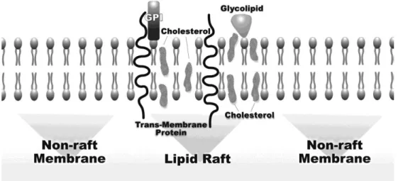
1.3-Lipid Rafts
The lipid rafts are defined as low density microdomains and resistant to detergents within the lipid bilayer of the plasma membrane with a relatively small size (50 nm). They are constituted by transmembrane proteins, GPI-anchored proteins and lipids, namely cholesterol and sphingolipids (figure1.3) (Staubach and Hanisch, 2011). This composition has impact on their characteristics, namely fluidity and stability, and allows building platforms rich in diverse signaling molecules such as EGFR, Src, AKT, heat shock protein 90 (Hsp90), and Fas (CD95) (Lee et al., 2014; Sharma et al., 2002). Lipid rafts have been associated with many biological functions of sorting and signaling to regulate cell survival, proliferation, migration, adhesion and in apoptotic pathway (Onodera et al., 2013; Lee et al., 2014). The heterogenic and dynamics composition of these lipid rafts contributes to the
8
large number of signals capable of being transmitted from the external cell membrane to cytoplasmic organelles or even to the nucleus (Mollinedo and Gajate, 2006).
The lipid rafts can be divided into two subclasses: the planar lipid rafts, which do not correspond to invaginate microdomains and which possess morphological flaws; and the caveolar rafts, a subclass specialized in tubular invagination of the plasma membrane with specific proteins, caveolins, including caveolin-1 (Cav-1). This is a classical protein hairpin that plays a key role in processes of endocytosis and transport. Cav-1 also controls signal transduction events and serve as a scaffolding protein in the organization of signaling molecules. Cav-1 serves as an adapter that initiates the signaling pathway-specific RAS-ERK and can block other routes involved in the apoptotic process (Staubach and Hanisch, 2011). Moreover, act as stabilizer of rafts on the cell surface by recruiting receptors or sequestrating cholesterol to these regions of the membrane (Lajoie and Nabi, 2007).
The increase of total cholesterol levels in circulation automatically leads to an increase of its concentration in lipid rafts (Mollinedo and Gajate, 2014). Thus, a progressive increase in cholesterol content in the membrane contributes to the expansion of lipid rafts and oncogenic pathways progression (Patra, 2008). Cholesterol is the most abundant lipid component of these raft domains and its concentration levels are essential for the formation, stabilization and organization of these structures. This sterol is a molecule that has more affinity for sphingolipids than unsaturated phospholipid. Therefore, the cholesterol acts as a barrier of hydrocarbon chains of sphingolipids allowing to maintain the raft attached and functioning as “dynamics glue”. Without cholesterol the membranes become fluid and permeable to some molecules (Simons and Ehehalt, 2002).
Thus cholesterol is crucial to maintain cellular homeostasis and provide platforms capable of activating pro and anti - apoptotic or proliferative pathways of signaling (George and Wu, 2012; Li et al., 2006). Recent studies show that changes in cholesterol levels of these rafts can directly induce the activation of multiple pathways, activation or deactivation of kinases, activation of apoptotic membrane receptors and increased levels of intracellular calcium. Some of these membrane proteins can be activated in these microdomains in the absence of their ligands. This type of activation of them occurs by aggregation of receptor molecules in lipid rafts. This occurs due to changes in lipid and
9
protein contents of these domains, which consequently results in the formation of signaling platforms (George and Wu, 2012).
The observation that many receptors after ligand stimulation are able to move inside or outside lipid rafts suggests that the association of these compartments is not continuous and is subject to regulation. The association of receptors with rafts is an equilibrium process that can be moved to one side or other based on exterior conditions (Pike, 2005).
1.3.1- Apoptosis and lipid rafts dependent membrane receptors
Lower rate of death cells than of proliferation is one of the essential factors for development of cancer, and consequently the capacity for the cells resist to apoptotic stimuli is a hallmark of cancer. Lipid rafts have been recently involved in the triggering of death receptor-mediated apoptosis.
Apoptosis or programmed cell death is a cellular mechanism that allows the cell to defend and protect from neoplastic development by eliminating genetically damaged cells or excess cells. Apoptosis is therefore responsible for maintaining cellular homeostasis and it is characterized by marked changes in cellular morphology, including plasma membrane blebbing, DNA fragmentation, chromatin condensation, nuclear breakdown and the presence of membrane associated apoptotic bodies (Patra, 2008).
This programmed cell death in mammalian cells is regulated by two major signaling pathways: the intrinsic pathway which is activated by intracellular signals from the mitochondria and the extrinsic pathway which is activated by the binding of apoptotic ligands to death receptors on the cell surface (Gajate et al., 2009; Fouqué et al., 2014). The intrinsic pathway is activated when there is accumulation of DNA damage, viral infections or by dysregulation of mitochondrial function. These stimuli lead to release of cytochrome c into the cytoplasm by mitochondria, where cytochrome c binds the adapter Apaf-1 and pro-caspase 9 to form the apoptosome. This complex leads to the activation of pro-caspase 9 and hence the pathway of caspases (caspase -3, -6, and -7) that allows cell death. On the other hand the extrinsic pathway of apoptosis occurs when there is activation of death receptors of the membrane. Membrane death receptors belong to the tumor necrosis factor (TNF) superfamily, which includes the Fas/CD95, the Tumor necrosis factor receptor (TNFR), and
10
the TNF-related apoptosis-inducing ligand (TRAIL) receptors (Wu, 2009). Death receptors consists of more than 20 proteins with a broad variety of biological functions that have two regions of high homology: extracellular cysteine-rich domains and a cytoplasmic domain called “death domain” with 80 amino acids, which have an important role in transmitting the death signal from the cell surface to intracellular signaling pathways (Mollinedo and Gajate, 2006). When activated, these receptors recruit adaptor proteins such as Fas-associating protein with a death domain (FADD) or tumor necrosis factor receptor 1-associated death domain protein (TRADD) for the membrane microdomains and pro-caspase 9 to form the death-inducing signaling complex (DISC). This complex activates caspase 8, resulting in the subsequent activation of caspases -3, -6 and -7, or activates the intrinsic pathway through caspase-8-mediated Bid cleavage, which activates the release of cytochrome c from mitochondria via interaction with Bax and Bak and indirectly activating caspases-3, -6, -7 and inducing the apoptosis (Wu, 2009; Gajate et al., 2009).
The redistribution of tumor necrosis factor family receptors into and out of lipid rafts may dynamically regulate the competence and outcomes of signaling by these receptors. The integrity of membrane rafts is required for the initiation of apoptosis, because these domains provide a signaling platform for the activation of pro-apoptotic membrane receptor molecules, and this occurs via receptor oligomerization by agents that promote the integrity of the membrane rafts in the absence of receptor ligand molecules (Hryniewicz-Jankowska et al., 2014). An example of this phenomenon is Fas (CD95) receptor, a member of the family of TNF receptors, which converts apoptotic signals in the presence of its ligand FasL and plays a crucial role in apoptosis. However, this receptor can be also activated in the absence of its ligand (Mollinedo and Gajate, 2006). FAS-mediated apoptosis involves the translocation of Fas into the lipid rafts. This recruitment of Fas and its downstream effectors into membrane rafts may provide a mechanism for amplifying Fas signaling by reorganizing membrane microdomains and bringing molecules together for this domains reducing the space, and facilitating protein-protein interactions among different signaling molecules and pathways. It is well documented that Fas oligomerization in lipid raft microdomains results in successive recruitment of FADD and then procaspase-8, allowing formation of DISC. The activation of this receptor also results in recruitment of additional pro-apoptotic proteins into the lipid raft and/or activation of the JNK apoptotic pathway (George and Wu, 2012).
11
The disruption of lipid rafts by agents such as metil-β-cyclodextrin (MβCD) may, therefore, negatively interfere with death receptors mediated apoptosis (figure 1.4).
A study conducted by Xu et al. using the triterpene Avicin D, showed that this compound enhances Fas translocation to lipid rafts, where it associates with FADD, inducing apoptotic signals. On the other hand the use of MβCD, which is a cholesterol-depleting compound that extracts cholesterol from the plasma membrane and perturbs lipid raft organization, blocks the clustering of this receptor and its DISC complex to rafts domains thereby inhibiting apoptosis (figure1.4) (Xu et al., 2009). This example demonstrates the importance of the stability of lipid rafts for the clustering of the death receptor Fas which is crucial to trigger apoptotic signal transduction. Fas-mediated apoptosis can be induced not only by FasL but also by TNF-α (Du et al., 2014). TNF-α is a major pro-inflammatory cytokine, synthesized as a 26 kDa transmembrane type II protein of 233 amino acids, involved in inflammation and initiation of the acute phase reaction by promoting survival/inflammatory signaling or inducing cell death (Fouqué et al. 2014). TNF binds to their membrane receptor TNFR, another death-inducing receptor that belongs to the tumor necrosis factor superfamily, which mediates biological fates from inflammation to apoptosis and other forms of programmed cell death. The binding of TNF-α to its receptors initiates a complex array of signaling events, mainly through the activation of the MAPK stress signaling cascade
Figure 1.4 – Influence of disruption of membrane rafts domains in Fas signaling pathway. Reproduced from
(Hryniewicz-Jankowska et al., 2014).
12
and of the Nuclear factor kappa-light-chain-enhancer of activated B cells (NF-κB) transcription factor.
After binding of TNF-α to TNFR, this is moved to lipid rafts where there is the formation of Complex I, constituted by RIP1, TRAF2, TRADD and clAP1 proteins, which translates signals that lead to activation of NF-κB responsible for inflammation. When the activity of NF-κB is not necessary for the cell the RIP1, TRADD and TRAF2 dissociates from TNFR to recruit FADD and the caspase-8 to form a new complex (Complex II) and initiate cell death. NF-κB inhibits cell death through increasing the expression of anti-apoptotic genes that directly inhibit caspase-8 activation in Complex II (Legler et al., 2003; Muppidi et al., 2004).
Cholesterol depletion (by MβCD for example), disrupted the structure of lipid rafts and the formation of the complex I occurs outside of lipid rafts and can´t signal efficiently to NF-κB. So in conditions produced by cholesterol depletion, occurs a change of the Figure 1.5 – Example how depletion of cholesterol in lipid rafts can modify early events in TNFR family signaling. Reproduced from (Muppidi et al., 2004)
13
inflammation signaling for apoptosis (figure 1.5) because lipid rafts disruption blocks the ubiquitination of the adaptor proteins RIP1 and TRADD in the TNFR signaling complex and prevents NF-κB induction by complex I. Can also inhibit the synthesis of growth factors resulting in increased sensitivity of the cells to apoptosis mediated by TNF-α (Muppidi et al., 2004).
The both Fas and TNF receptors are examples that disruption of lipid rafts integrity can lead to anti (Fas) or pro-apoptotic (TNFR) effects, depending on the stimulation or apoptotic factors involved. For example, for TNF receptor the MβCD, mevastatin and cholesterol oxidase have all demonstrated pro-apoptotic effects after disruption of lipid rafts in keratinocytes. For Fas receptor the MβCD is able to inhibit Avicin D-induced apoptosis in Jurkat cells (George and Wu, 2012).
1.3.1- Cell proliferation and lipid rafts dependent membrane receptors
In many types of cancer, proliferation and cell growth are the result of an abnormal signaling by growth factor receptors. Lipid rafts also act as signaling platforms for proliferative growth factor mediated signaling. Many receptor tyrosine kinases including the EGF receptor (EGFR), the PDGF receptor and Insulin receptor (IR), have been shown to be localized in these microdomains, and in all cases the alteration on cholesterol content of rafts controlled the signaling by these receptors (Hryniewicz-Jankowska et al., 2014; Pike, 2005).
EGFR is a member of the family of receptor type tyrosine kinases (RTK's), consisting of three domains: an extracellular domain of 620 amino acids that recognizes and binds to the EGF; a single transmembrane lipophilic domain of 24 amino acid penetrating through the membrane alpha helix shaped; and a cytoplasmic domain that include an intrinsic tyrosine kinase domain. The stimulation of the EGFR receptors present in lipid rafts induces a conformational change in the extracellular domain of the receptor, which consequently promotes homo or heterodimerization with other RTK's. This dimerization activates the intrinsic activity of the receptor, resulting in auto phosphorylation of tyrosine residues at the C-terminal (Brand and Wheeler, 2012; Pike, 2005).
The lowering of cholesterol levels in lipid rafts or their disruption by using MβCD stimulates ligand activation and EGFR phosphorylation (Wang and Paller, 2006; Tekpli et al.,
14
2013). Moreover several groups have shown that cholesterol depletion causes an increase in the amount of bound EGF, and on the other hand increased cholesterol levels leads to a reduction in binding of EGF to EGFR. This seems to indicate that the receptor location in lipid rafts impairs the ability of EGF binding, because restriction of spatial character. The depletion of cholesterol is still associated with an increase in receptor dimerization induced by EGF, suggesting that cholesterol changes in lipid rafts affect not only receptor binding but also the signaling (Lambert et al., 2009; Pike, 2005; Ringerike et al., 2002). But this effects is still controversial in the literature because a study performed by Zhuang et al. showed that caveolin-negative prostate cancer cell line LNCaP treated with an agent that disrupts the rafts suppressed ligand-dependent EGFR phosphorylation in the rafts fraction of the membrane and re-formation of this microdomains with cholesterol restored EGFR/AKT pathway (Zhuang et al., 2002).
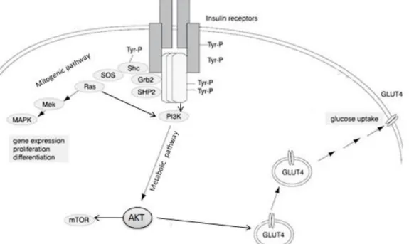
The insulin signaling cascade is also proposed to function via lipid rafts. Insulin is a major anabolic hormone controlling critical steps such as cellular glucose uptake and lipid metabolism. Insulin plays an important role in vesicle trafficking, stimulation of protein kinases and phosphatases, promotion of cellular growth and differentiation and activation or repression of transcription. Insulin exerts its effects via binding to the IR (Saltiel and Pessin, 2002). The IR is a heterotetrameric bifunctional complex, consisting of 2 extracellular α subunits that bind insulin and 2 transmembrane β subunits which contain tyrosine kinase
Figure 1.6 – Schematic representation of Insulin receptor pathways: PI3K mediating metabolic responses and Ras mediating mitogenic signaling. Adapted from (Mlinar et al., 2006)
15
activity. After insulin binding to the α subunit, the phosphorylation of one β domain is induced as a result of the increase catalytic activity of the receptor kinase activity (Chang et al. 2005) that phosphorylates and recruits different substrate adaptors of IRS family causing activation of several signaling pathways. Insulin uses the PI3K/AKT signaling pathway to exert most of their metabolic activities such uptake of glucose. Insulin leads to an increased phosphorylation of AKT but also induces mitogenic signaling transmitted via RAS/MAPK pathway (figure 1.6) (Sánchez-Wandelmer et al., 2009).
In cells that have caveolae the IR are constitutively sequestered in caveolae, but in case of caveolae-negative cells the receptors are recruited into rafts only by the addiction of insulin (Pike, 2003). IR directly interacts with caveolin-1 and stimulates their tyrosine phosphorylation (Saltiel and Pessin, 2003). Cholesterol depletion and loss of lipid rafts structures in plasma membrane did not reduce the quantity of IR or their affinity for insulin. A study by Parpal et al. reported that the interaction with the immediate downstream mediator molecule IRS-1 and metabolic control (such uptake of glucose) is, however, inhibited. As a consequence of cholesterol depletion, the further downstream propagation of the signal to AKT phosphorylation was being blocked, while insulin signaling via the MAPK pathway remains intact (Parpal et al., 2001; Karlsson et al., 2004). When cholesterol levels were then reestablished in these cells, protein phosphorylation mediated by insulin returned to normal, and lipid rafts were again morphologically recognizable (Cohen et al., 2003).
The activation of these RTK´s leads to the activation of the signaling pathway of the RAS protein. This protein is one the most studied oncoproteins, because is one component of the cascades RAS-RAF-MAPK and RAS-PI3K-AKT-mTOR. RAS plays an important role in the initiation of cell proliferation. The RAS oncogene is mutated in approximately 25% of human cancers (Hejmadi, 2010), and are noted in 50% of CRC cases (Bos, 1989). This mutation changes the RAS protein into an oncoprotein, characterized by constitutive activation, which in turn triggers an uncontrolled growth, making the cells fail to turn off their proliferative state (Hejmadi 2010). Under normal conditions, the RAS protein is activated by growth factors (Bos, 1989), through binding one molecule of guanosine triphosphate (GTP), thereby increasing affinity for subsequent effector molecules in the cascade. This is later disabled when the hydrolysis of GTP to guanosine diphosphate (GDP) occurs (Brand & Wheeler
16
2012). When there is a mutation in RAS the hydrolysis of GTP does not occur and the molecule is constantly active and with sufficient autonomy to induce proliferation independently of receptors signaling and extracellular ligands (Swanson and Hohl, 2006).
RAS activation causes a conformational modification in the RAF kinase, which leads to activation of MAPK nuclear transporter that is responsible for cellular proliferation. RAS also recruits PI3K to the membrane leading to activation of AKT protein, and consequently mTOR, which induces protein synthesis and other metabolic effects, and inhibition of several pro-apoptotic signals (figure 1.7) (Brand and Wheeler, 2012).
Post-translational modifications of RAS, namely prenylation, are important for RAS signaling. RAS protein begins as a hydrophilic globular protein exhibiting a C-terminal region with the sequence - CAAX (C a cysteine, A an aliphatic acid - valine, leucine, isoleucine - and X any amino acid), which corresponds to a mark for post-translational modifications, including farnesylation, proteolysis and methylation (Fehrenbacher et al., 2009; Hancock and Parton, 2005). RAS is synthesized in the cytoplasm, where its CAAX sequence is recognized by farnesyltransferase enzyme that adds a farnesyl group to the terminal
Figure 1.7 – Activation of protein RAS after hydrolysis of GTP to GDP and their signaling pathways. Adapted
17
cysteine. Then farnesylated proteins are sent to the ER where they find Ras converting enzyme (Rce1) that remove the AAX amino acids and the isoprenylcysteine carboxyl methyltransferase (Icmt) that subsequently add a methyl group to the carboxyl cysteine (Fehrenbacher et al., 2009). The RAS is ultimately sent to the plasma membrane, especial for rafts domains and may also be regulated by local accumulation of anionic phospholipids (Rocks et al., 2006). The post-translational modification of RAS protein, required for its membrane insertion and subsequent activation depends on the synthesis of lipid molecules by the mevalonate pathway. Therefore RAS signaling depends on cholesterol homeostasis because it depends on prenylation for activating and on lipid rafts composition for assembling its signaling targets.
1.4-Cholesterol biosynthesis
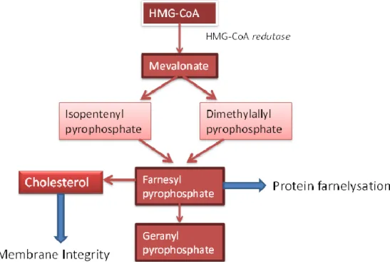
1.4.1-Mevalonate pathway
This pathway, which takes place in the cytoplasm, is a highly conserved metabolic cascade responsible for the synthesis of various products. In the first step of the pathway 3-hydroxy-3-methylglutaryl CoA (HMG CoA) reductase converts HMG CoA to mevalonate (Thurnher et al., 2013). HMG CoA reductase is the enzyme that catalyzes the limiting step of
18
the mevalonate pathway, and it is essential for the synthesis of isoprenoid compounds and cholesterol, dolichol and ubiquinone (figure 1.8).
The isoprenoids (farnesyl pyrophosphate and geranyl pyrophosphate) are essential for the post-translational modifications of many proteins, including RAS (Winiarska et al., 2008), and are synthesized by condensation of isopentenyl pyrophosphate (IPP), a compound of five carbon atoms, and its isomer dimethylallyl pyrophosphate (DMAPP) (Thurnher et al., 2013).
The regulation of mevalonate pathway and cholesterol homeostasis is regulated by a family of transcription factors called sterol regulatory element-binding protein 2 (SREBP2) (Yellaturu et al., 2009).
1.4.2 - Sterol regulatory element binding protein
SREBP-2 function as transcription factor that activate specific genes involved in cholesterol biosynthesis and endocytosis of low density lipoproteins (LDL) (Edwards et al., 2000).
In humans there are two SREBP genes: SREBP-1 and SREBP-2 that encode three different proteins. The SREBP-1 gene encodes two proteins, the SREBP1a and SREBP1c derived from distinct promoters. The gene SREBP-2 encodes the SREBP2 protein. In humans, the SREBP1c primarily regulates FA metabolism, such as fatty acid synthase (FASN) gene and SREBP2 is responsible for the activation of genes of cholesterol biosynthesis: genes such as HMG CoA reductase and LDL receptor (Krycer et al., 2010). Human SREBP2 contains 1141 amino acids and is 47% identical to SREBP1a with its 1147 amino acids (Xiao and Song, 2013); (Sato, 2010).
The SREBPs are synthesized as 125kDa membrane proteins of the ER in the form helix-loop-helix-leucine zipper structure: an N-terminal with 480 amino acids; an intermediate hydrophobic region of 80 amino acids and a regulatory domain in the C-terminal with 590 amino acids (Daemen et al., 2013; Xiao and Song, 2013). The N-terminus of SREBP2 region is rich in glutamine, proline, serine, and glycine (Shimano, 2001).
19
After synthesis of the pre-SREBP, this protein binds to the SREBP cleavage-activating protein (SCAP) in ER and then this complex is displaced from the ER to the Golgi apparatus. The sequence of amino acids MELADL, found in cytosolic loop 6 of SCAP, is required for coatomer protein complex II (COPII) binds to SREBP-SCAP and shall transport (Xiao and Song, 2013). The protein SCAP has two different domains in each molecule: the N-terminal has eight transmembrane helices and contains the sterol sensing domain (SSD) and that that recognize the SREBPs; the C-terminal on the other hand contains five WD repeats, with 40 amino acids each one, which allows the interactions protein-protein. This is the region of SCAP that forms a complex with C-terminal domain of SREBP (Sato, 2010). When cellular cholesterol levels are low, pre SREBP binds to SCAP, and after entering the Golgi complex is cleaved by site-1-protease (S1P) that causes a conformational change in the sterol and allows this binding to site-2-protease (S2P). S2P cleavage occurs within the transmembrane region, a process called Regulated intramembranous proteolysis (Rip). This cleavage results in the liberation of terminal domain from membrane (Edwards et al., 2000). Thus the N-terminus of SREBP enters the nucleus by binding to ATCACCCCAC sequence of cis-element called sterol regulatory element (SRE) promoting gene expression (figure 1.9).
20
When cholesterol levels are elevated the SCAP directly binds to cholesterol promoting a conformational change in protein. This conformation allows the binding of SCAP to Insig (insulin-induced gene product), a protein residing in the ER, preventing movement of the SCAP-SREBP complex to the Golgi apparatus and thus blocking activation of SREBP’s target genes. The binding of Insig prevents the COPII from recognizing the MELADL sequence (Shimano, 2001; Daemen et al., 2013). In this case Insig can still bind to the N-terminus of HMG CoA reductase in order to recruit E3 ubiquitin ligase and allow access to the proteasome, extracting the enzyme from the membrane and allowing its degradation (Xiao and Song, 2013).
There are two genes, INSIG1 and INSIG2 that codify two proteins: the Insig-1 and Insig-2, respectively, which are 59% identical. These genes control tissue and signal specific regulation of SREBP. Insig-1 is the dominant isoform and the only that are subject to proteasome dependent degradation; high levels of cholesterol inhibit ER to Golgi transport of SREBP by stabilization of the Insig-1. On the other hand Insig-2 is more stable, is expressed at low levels and its suppressed by insulin (Shao and Espenshade, 2012; Bengoechea-Alonso and Ericsson, 2007). Some studies indicate that SREBP1/SCAP complex is usually associated with Insig-2 and the SREBP2/SCAP complex interacts with Insig-1 (Sato, 2010).
21
Recent studies demonstrated that formation of mature SREBPs is controlled in various levels, namely in response to changes in the levels of insulin, oxysterols and polyunsatured fatty acids (figure 1.10) (Edwards et al., 2000).
Insig-1 is one of the most readily inducible genes by insulin but gene expression of Insig-2 is suppressed. This because insulin stimulates proteolytic cleavage of mature SREBP1c increasing the affinity these for the SCAP through COPII vesicle enhancing the activation of SREBP targets genes and reduces the amount of Insig-2 protein by quickly degradation of the cognate mRNA (Yellaturu et al., 2009).
SREBP only not involving in sterol regulated events but are also targets of intracellular signaling pathways such MAPK kinase and PI3K/AKT. This insulin stimulated SREBP1c activation is PI3K/AKT dependent, because the induction of this pathway potentiates the ER to Golgi transport and the proteolytic activation (Rochira et al., 2013; Shao and Espenshade, 2012).
1.4.3-Statins
HMG CoA reductase inhibitors, known as statins, were originally identified in 1970 by the Japanese Biochemical Akira Endo and received commercial approval for the treatment of hypercholesterolemia in 1980 because these were capable of reducing endogenous cholesterol synthesis, leading to a decrease of LDL in circulation. Furthermore, statins appear in various processes involved with pleiotropic effects including inhibition of cell proliferation, induction of apoptosis, modulation of inflammation and angiogenesis (Lochhead and Chan, 2013).
In the context of cancer therapy, inhibitors of the enzyme HMG CoA reductase have been proposed as anti-tumor agents due to evidence that farnesylation by farnesyl group resulting from the use of this pathway is the crucial step for anchoring the protein RAS to membrane and also because the cells of colorectal cancer show high levels of expression and activity of HMG CoA reductase compared to normal tissue (Calleros et al., 2009). Some antitumor effects of statins could also result from the capacity of these drugs to interrupt the formation of lipid rafts (Winiarska et al., 2008).
22
The first statins were of natural origin, deriving from fungal fermentation - lovastatin, simvastatin, pravastatin, mevastatin – but there are now many synthetic statins – fluvastatin, atorvastatin, rosuvastatin, pitavastatin. All have a lactone ring: when in the inactive form this ring is closed, when in the active form the ring it’s open. Just the form with the open ring has the ability to block the catalytic activity of HMG CoA reductase, because it mimics an intermediate of the mevalonate pathway and binds to its active site with more affinity than the natural substrate (Wong et al., 2002). Studies conclude that the use of statins in the long term actually reduce the risk of pancreatic cancer in 59%, prostate cancer in 50% and 49% the risk of CRC compared to patients not undergoing treatment using to statins (Takahashi et al., 2006).
Despite their beneficial effects, many studies have also reported cytotoxic effects exerted by statins, however although direct toxicity to normal muscle cells and endothelium have been described, other normal cell types are unaffected by statins. In contrast, strong toxicity is induced in many tumor cells, which may consequently result in programmed cell death (Corcos and Le Jossic-Corcos, 2013). In other studies, it is observed that use of simvastatin, the most widely used because it is of natural origin and with high stability, significantly decreased the concentration of cholesterol in the lipid rafts, thereby inhibiting AKT cascade and inhibits proliferation (Lochhead and Chan, 2013; Miraglia et al., 2012). However, the dose and time required is still to be determined, because unfortunately the anti-proliferative effects in vitro are observed at concentrations too high for the clinical reality (Osmak, 2012)
1.5-Natural compounds and chemoprevention
Epidemiological studies show that cancer correlates with the diets poor in fruits and vegetables. Therefore, during the last decade several studies have been performed to understand the cellular and molecular mechanisms underlying the process of carcinogenesis and leading to the development of methodologies for dietary prevention of cancer, called chemoprevention. The objective of chemoprevention is the use of noncytotoxic natural (or synthetic) agents to delay, inhibit or reverse the growth and the development of malignant
23
cells through modulation of metabolic mechanisms and signaling pathways (Yadav et al., 2010), (Pan et al., 2008),(Newman et al., 2000).
The interest and the use of natural compounds extracts derived from fruits, vegetables and whole grains, increased in recent years because they offer a great range of advantages: low toxicity; easier to obtain; and favorable physiological functions (Yi et al., 2010; Pan et al., 2008). For this reason 70% of the drugs used in modern medicine as anti-carcer have their roots in products derivate from nature (Prasad et al., 2011). The National Cancer Institute of the United States has identified around 40 plant foods that exert preventive effects against cancer, including turmeric, soybean, green tea, ginger, cabbage, red wine and cauliflower.
Phytochemicals are defined as bioactive nonnutrient plant compounds, in other words, are plant chemicals (Liu, 2004). Phytochemicals can be grouped in different classes such as: phenolics, alkaloids, carotenoids, organosulfur compounds, and terpenoids (or isoprenoids) (Pan et al., 2008).
Terpenoids are lipophilic phytochemicals which have a multicyclic structure (Thoppil and Bishayee, 2011; Pan et al., 2008). These compounds are derived from isoprene units [CH2=C(CH3)CH=CH2] which have oxygen atoms in multiple functional groups. According to
the number of carbon units, terpenoids can be classified as: monoterpenes (e.g., carvone, geraniol), diterpenes (e.g. retinol and trans-retinoic acid), triterpenes [e.g., betulinic acid (BA), lupeol, oleanic acid (OA), and ursolic acid (UA)], and tetraterpenes (Thoppil and Bishayee, 2011). More than 40,000 different terpenoids are known to occur in nature and according to various studies on development of drugs for cancer, many of these compounds have been shown to inhibit cell proliferation and metastasis by different mechanisms of action (Bishayee et al., 2011).
1.5.1-Triterpenoids
In particular the triterpenoids are an important class of bioactive phytochemicals in plants synthesized by cyclization of squalene, a triterpene hydrocarbon and precursor of all steroids (Bishayee et al., 2011). They are organic metabolites of isopentenyl pyrophosphate
24
oligomers characterized by the basic structure of 30-carbon isoprenoid composed from six isoprene units (C30H48), arranged in five rings with several oxygen atoms attached (Yadav et al., 2010; Yan et al., 2013).Triterpenoids are distributed throughout the plant kingdom in the form of free triterpenoids, triterpenic glycosides (saponins), phytosterols and/or their precursors (Patlolla and Rao, 2012).
These compounds have a high variety of benefits for human health, especially antioxidant, antiviral, antimicrobial and hypoallergenic. Recent studies have not only confirmed these proprieties, but also showed that triterpenoids are cytotoxic to cancerous cells without showing any toxicity in normal cells (Bishayee et al., 2011). Studies indicate that these compounds may act at different stages of tumor development: inhibiting initiation and promotion, inducing cell differentiation and apoptosis, regulating various signaling pathways, down regulating of transcription factors (e.g., nuclear factor-kappaB [NF-κB]), modifying expression of anti-apoptotic proteins (e.g., bcl-2, bcl-xL) or promoters of cell proliferation (e.g., cyclooxygenase-2 [COX-2], cyclin D1, c-myc), as well as suppressing invasion and metastasis (Thoppil and Bishayee, 2011; Yadav et al., 2010).
Ursolic acid Oleanolic acid
Cholesterol
A
B
C
25
There are about 20,000 triterpenoids from several fruits and medicinal plants, that can be subclassified into several groups, including: lupanes, oleananes, ursanes, holostanes, sqalenes, limonoids, dammaranes, (Shanmugam et al., 2013; Bishayee et al., 2011). In the present work we used two pentacyclic triterpenoid isomers – ursolic acid (UA) and oleanolic acid (OA) because they have structure similar to cholesterol (figure 1.11).
1.5.1.1- Ursolic Acid
Ursolic acid (3β-hydroxy-urs-12-en-28-oic acid) is a pentacyclic triterpenic acid (figure 1.11-B) identified in various plants, herbs and fruits namely in Salvia species, rosemary, olive, apples, peares and berries. This natural compound may appear in the form of free acid or as aglycone precursor for saponins (Shanmugam et al., 2013; Prasad et al., 2011).
The first study with the UA on anti-mammary cancer effects was conducted by
Es-Saandy and coworkers, and according to this study the UA acts as an inhibitor of
proliferation by altering the cell cycle at the G1 phase (Es-Saady et al., 1996). Other studies have shown that UA exhibits a large spectrum of properties, such as anti-inflammatory, antioxidant, hepatoprotective, anti-tumor and anti-metastatic with low toxicity. This effect against proliferation correlates with suppression of EGFR/MAPK pathway by inhibition of EGFR phosphorylation and AKT regulation; low Bcl-2 and Bcl-x and inducing apoptosis (Shan et al., 2009). A study conducted by Xavier et al. 2009, shows that UA not affect the expression of p-ERK but inhibits p-AKT in CO115 cells, without affecting the expression of KRAS protein and has PI3K as one of its molecular targets. It also showed that this compound inhibits proliferation and induces apoptosis in HCT15 cells (Xavier et al., 2009). The same author demonstrated that UA could be involved in modulation of autophagy through activation of JNK signaling in HCT15 cells and by inducing the accumulation of p63 levels (Xavier et al., 2013).
26
1.5.1.2- Oleanolic Acid
Oleanolic acid (3b-hydroxyolean-12-en-28-oic acid) is a member of oleanane triterpene family and an isomer of UA (figure 1.11-C). OA and UA have a similar chemical structure varying only in the position of a methyl group, in E ring. When the methyl group in position C19 of UA is moved to the position C20 is formed the OA (Pollier and Goossens, 2012).
This pentacyclic triterpenic acid is isolated from more than 1620 plant species, including many food and medicinal plants, and similarity to the UA, OA have pharmacological activities, such as hepatoprotective effects, and anti-inflammatory, antioxidant, or anticancer activities (Juan et al., 2008; Pollier and Goossens, 2012). OA induces apoptosis in HL-60 and HCT15 cells through the release of cytochrome c into the cytosol and thus leading activation of the caspases pathways. This effect is also verified by PARP cleavage in HL-60 cells and suppression of activation of NF-kB and Bcl-2 proteins. Several studies have also shown that the antitumor activity of the OA occurs through cell cycle arrest and inhibition of DNA replication (Shyu et al., 2010; Shanmugam et al., 2012; Juan et al., 2008).
29
Hypercholesterolemia has been implicated in cancer development and progression. Cholesterol is an amphipathic molecule present in the cell membranes which plays an important role in the regulation of cellular homeostasis, in particular the integrity, fluidity and membrane permeability. Cholesterol is therefore an important constituent of membrane microdomains called lipid rafts. These specialized microdomains are also rich in proteins involved in anti and pro apoptotic signaling and are determinants of survival and proliferation.
Cholesterol lowering drugs such as statins have been proposed to play a crucial role in the progression of cancer, because the mevalonate pathway responsible for the synthesis of this compound provides farnesyl pyrophosphate groups essential for the prenylation of the RAS oncogene in membrane, which activates related proliferation pathways and cell growth.
The triterpenoids OA and UA are natural compounds which have chemical structures similar to cholesterol. Thus the aim of our study is to verify:
1. If these compounds possess anti-cancer activity by altering cellular cholesterol levels;
2. Death and proliferative signaling pathways of colorectal cancer cells (HCT116) 2.1. Effects on TNF-α signaling.
33
3.1 - Cell Culture, conditions and reagents
HCT116 cell line (derived from a human colorectal carcinoma) was kindly provided by Dr. M. Rohde from The Danish Cancer Society and grown in RPMI-1640 medium (Sigma-Aldrich, St. Louis, MO, USA) supplemented with 6% fetal bovine serum (FBS) (Biochrom KG, Berlin, Germany), antibiotic-antimitotic 1% solution (Sigma-Aldrich, St. Louis, MO, USA), sodium pyruvate 0.1mM, sodium bicarbonate 2g/L and HEPES 10mM (Sigma-Aldrich, St. Louis, MO, USA). Cells were maintained in 25cm2, not toxic and transparent tissue culture flasks (TPP), at 37°C in a humidified atmosphere with 5% CO2 and 95% air. The medium was changed twice a week. Cellular subcultures
were performed when confluence reached values close to 80-90%. For subcultures and plating, the adherent cells were detached with trypsin solution 1% (Sigma-Aldrich, St. Louis, MO, USA) and trypsin was neutralized by adding fresh medium. The cell density of the suspension was determined using a haemocytometer and was calculated by the formula: Mean of cells in the four quadrants x dilution factor x 104 = number of cells per mL.
Simvastatin, UA and OA were purchased from Sigma-Aldrich (St. Louis, MO, USA) and were dissolved in dimethyl sulfoxide (DMSO) to stock solutions, aliquots were kept at -20°C. The methyl-β-cyclodextrin (MβCD), cholesterol and insulin were also obtained from Sigma-Aldrich (St. Louis, MO, USA). The MβCD was dissolved in RPMI medium without serum, and aliquots were stored at -20°C at a concentration of 5mM and for the experiments we used 1.5mM. Furthermore, insulin was dissolved in water and stored at 4°C in concentration of 100µM, for the experiments we used 2µL of stock for 2mL of medium. The stock solution of TNF-α was also stored at -20⁰C in concentration of 10µg/mL and for the condensation assay we used 2µL of stock for 2mL of medium. The DMSO concentration that is placed in the culture medium in all conditions did not exceed 0.5% (v/v) and the controls were treated only with the vehicle (DMSO).
34
3.2 - Cell Viability by MTT reduction assay
The MTT assay allows the evaluation of cell viability by dehydrogenases associated metabolic reduction of 3-(4, 5-dimethylthiazol-2-yl)-2,5-diphenyltetrazolium bromide (MTT), a yellow compound, to a purple colored formazan. Only metabolically active cells are capable to reducing this compound and therefore the number of viable cells is directly proportional to the amount of formazan crystals produced (Chang et al. 2013).
HCT116 cells were plated in 24-well plates two days before incubation with compounds to a cellular density of 0.6x10⁵cell/mL and left to adhere for 48h. Following, fresh medium was added to cells containing the test compounds: UA at final concentration of 2.5, 5, 10 and 15µM; OA at 5, 10, 20, 30 and 40µM; Simvastatin at 10 and 30µM for 48h incubation time.
One hour before the end of incubation time, 50µL MTT (5mg/mL, diluted in phosphate-buffered saline (PBS) solution and maintained in the dark and at -20°C) (Sigma-Aldrich, St. Louis, MO, USA) were added to each well and cells were incubated at 37°C, with 5% CO2 during 1h. After this time the supernatant was removed,
formazan crystals were dissolved in 1mL of ethanol / DMSO solution (proportion 1:1) and 200µL of each condition was transferred for a 96-well plate. The reading of absorbance was done at 570 and 690nm using a micro plate reader (Molecular Devices Spectra Max Plus 384). As a blank, 200μL of DMSO/ethanol solution were used. The results were expressed as percentage relative to the control (untreated cells). The IC50
value of compounds was calculated as the concentration at which 50% reduction in cell viability was detected relative to untreated controls.
35
3.3 - Protein extraction and Western Blot analysis
HCT116 cells were plated in 6-multiwell culture at a density of 0.6x10⁵cell/mL. After treatment with compounds at different incubation times or concentrations, the cells were washed with PBS solution, trypsinized and medium was added for neutralizing the trypsin and were collected into a respective falcon. The samples were centrifuged at 500g for 5 min at 4°C. After this the supernatant was discarded up to 500µl, samples were resuspended and transferred to individual eppendorf tubes. The samples were centrifuged at 1500g for 3 min at 4°C, and the pellet was resuspended in lysis buffer (1% Triton in 150mM NaCl, 50mM Tris-base (pH 7.5), 1mM EDTA, 0.1% SDS, 1% Sodium deoxycholate) supplemented with 20mM of sodium fluoride (NaF), 1mM phenylmethylsulfonyl fluoride (PMSF), 20mM of Sodium orthovanadate (Na3VO4), and
protease inhibitor cocktail (Roche, Mannheim, Germany). After 10 min on ice the samples were centrifuged at 10 000g for 10 min, obtaining in supernatant the total protein.
Protein quantification of each sample was performed using the kit Bio-Rad DC protein assay (Bio-Rad Laboratories, Hercules, CA, USA) and a bovine serum albumin (BSA) used as protein standard. For Western Blot, 20μg of protein were loaded and separated on 8% SDS-polyacrilamide gel and then transferred to Hybond-P polyvinylidene difluoride (PVDF) membranes (Westran Clear Signal, USA). The membranes were blocked in TPBS (PBS with 0.05% Tween-20) containing 5% (w/v) nonfat dry milk under shaking for at least 2h, washed three times in TPBS solution and incubated with primary antibody (table 3.1) overnight. After being washed again with TPBS, the membranes were incubated with secondary antibody (containing IgG horseradish peroxidase) for 1h at room temperature. The immunoreactive bands were detected using the Clarity Western ECL Substrate (Bio-Rad Laboratories, Hercules, CA, USA) under a chemiluminescence detection system, the ChemiDoc XRS (Bio-Rad Laboratories, Hercules, CA, USA). β-actin protein was used as the control. The area intensity of the bands was quantify using Quantity One software (Bio-Rad) and standardized relative to control.