Universidade de Lisboa Faculdade de Medicina Dentária
MICROINFILTRAÇÃO COM CONTAMINAÇÃO SALIVAR EM RESTAURAÇÕES CLASSE V (ESTUDO IN VITRO)
Cláudia Galrinho
Mestrado integrado em Medicina Dentária
Universidade de Lisboa Faculdade de Medicina Dentária
MICROINFILTRAÇÃO COM CONTAMINAÇÃO SALIVAR EM RESTAURAÇÕES CLASSE V (ESTUDO IN VITRO)
Dissertação orientada pela Professora Doutora Sofia Arantes e Oliveira
Cláudia Galrinho
Mestrado integrado em Medicina Dentária 2012/2013
i
Ao meu irmão
ii
ÍNDICE
AGRADECIMENTOS ... iii ABSTRACT ... ix 1. INTRODUCTION ... 1 2. OBJECTIVES ... 53. MATERIALS AND METHODS ... 7
4. RESULTS ... 16
5. DISCUSSION ... 18
6. REFERENCES ... 23
Appendix I ... 28
iii
AGRADECIMENTOS
Quero prestar o meu profundo agradecimento às pessoas envolvidas, de uma forma ou de outra, neste trabalho.
À minha orientadora, a Professora Doutora Sofia Arantes-Oliveira, pela
sabedoria que soube transmitir, pela paciência e pelo sentido de humor. Mas sobretudo por me ensinar ,de forma graciosa, a saber lidar com o erro.
Ao Professor Doutor Jaime Portugal pelos ensinamentos e observções pertinentes.
Aos meus pais e ao meu irmão. Eles conhecem o limite do meu amor, da minha força, da minha fraqueza e das minhas dúvidas. Porém, fico na incerteza que conheçam a dimensão da minha gratidão por eles. Não tem fim.
À Sofia Martins, Magno Dinis, João Bernardo e Lénia Caracóis, amigos
incondicionais que fiz durante o meu percurso académico. Devo-lhes carinho e o apoio. À Lama Beseisso, minha colega e amiga para a vida. Por me ensinar a trabalhar em equipa, e por me fazer sorrir em dias menos felizes. Agradeço a lealdade e o seu eterno companheirismo.
iv
RESUMO
:
O desenvolvimento da medicina tem promovido um aumento da esperança média de vida nos últimos tempos (Mackenbach., 2013). O aumento significativo no número dos idosos é acompanhado por um conjunto de problemas muito específicos no que respeita aos cuidados de saúde oral, já que os estes estão a viver mais e a reter e os seus dentes naturais durante mais tempo (Chalmers., 2006). Os idosos são mais vulneráveis à cárie devido a várias condições médicas, tais como o Síndrome de Sjogren e radiação da cabeça e pescoço que reduzem a produção de saliva (Turner et al., 2007). Além disso, estes pacientes são frequentemente medicados com drogas que também reduzem o fluxo de saliva e causam xerostomia (Gareri et al., 1998; Baldoni et al., 2010). Um dos problemas específicos da população idosa relaciona-se com a maior prevalência de cárie radicular em comparação com os adultos jovens .
A localização da cárie radicular está associada à idade e à recessão gengival, que expõe a superfície da raiz (Watanabe., 2003; Meneghim et al., 2002). Um dos principais problemas destas lesões é a maior susceptibilidade das superfícies radiculares à cárie, devido ao elevado pH crítico da dentina (de 6,8 a 6,0) em comparação com o pH crítico do esmalte ( 5,9 a 5,2.).Outro problema deste tipo de lesão está associado ao isolamento em relação à contaminação salivar, devido à proximidade com tecidos gengivais e dificuldades de acesso interproximal; à grande quantidade de água e menor quantidade de matéria inorgânica na dentina. São factores críticos porque afetam a resistência de união à dentina (Hoppenbrouwers et al., 1986).
A microinfiltração corresponde à passagem indetectável de bactérias, fluidos, moléculas ou iões entre as paredes da cavidade e do material de restauração (Kidd., 1976 apud Alani and Toh., 1997). A integridade marginal depende de numerosos factores, incluindo o tipo de material restaurador utilizado, do tipo de cavidade e as condições de isolamento (Murray et al., 2000). Portanto, a selecção de um material em detrimentos de outro é de importância crítica para a diferença entre o sucesso e falha de uma restauração a longo prazo (Maryniuk et al., 1986).
O ionómero de vidro modificado por resina, o ionómero de vidro convencional, os compómeros, resinas compostas e amálgama são os materiais restauradores
v
frequentemente usados para restaurar lesões profundas de cárie (Nicholson., 2006; Mickenautsch et al., 2011; Sakrana et al., 200).
Objectivo
O principal objetivo do presente estudo é avaliar a influência da contaminação com saliva na microinfiltração de restaurações classe V restauradas com de ionómero de vidro e resina composta com dois diferentes sistemas adesivos (etch-and-rinse e self-etch)
Materiais e métodos
30 dentes foram seccionados ao meio, longitudinalmente com uma lâmina de diamante (Diamond Wafering Blade - Buehler, Série 15HC Diamante N º 11-4244, Deutschland);em cada metade foi realizada uma cavidade de classe V (3 × 2 × 2 milímetros) na superfície da raiz com uma de broca cilindríca de diamante, sob refrigeração a água. As amostras foram divididas aleatoriamente em três grupos de acordo com o material a estudar, (Grupo 1 – Ionofill plus, VOCO; Grupo 2 - Solobond M, VOCO e Grandioso, VOCO; Grupo 3 - FuturabondM, VOCO e Grandioso, VOCO). Em cada grupo de 10, os espécimes foram restaurados de acordo com as instruções do fabricante e os outros 10 foram contaminados com saliva antes do material restaurador. Contaminação Saliva foi realizado com saliva fresca e aplicada durante 5 segundos com uma microescova antes da inserção do material restaurador e após a fotopolimerização do adesivo o adesivo nos grupos em que se utilizou adesivo (groups 2 e 3).
- Nos grupos 2 e 3 após a contaminação salivar, a resina composta foi inserida em incrementos de 2 milímetros e fotopolimerizados por 20seg. (600mW/cm2- Fotopolimerizador de halogénio XL3000 de série n º 105944, 3M ESPE Dental Products, St Paul, MN, EUA) . Todos os espécimes foram sujeitoa a termociclagem
- As amostras foram isoladas com cera na câmara pulpar e ápex com e cera e verniz deixando uma margem de 1 milímetro à restauração
- As amostras foram, em seguida, imersas em solução de azul de metileno a 2%, durante 4 h e cuidadosamente lavadas com água corrente destilada.
vi
- Cada amostra foi seccionada longitudinalmente em 2 segmentos com uma lâmina de diamante 0,3 (Diamond wafering Blade -Buehler, Série 15HC Diamante N º 11-4244, Alemanha), com um dispositivo de corte (Isomet 1000 - Buehler, Illinois, EUA).
- As superfícies expostas foram polidas e examinadas em um microscópio estereoscópico (Meiji Techno EMZ-8TR n serial. º 411.479-Meiji Techno Co., Saitama, Japão), com aquisição da imagem digital, a fim de quantificar o grau de infiltração.
- Cada superfície foi classificada de acordo com a classificação ISO 14765 (2003). Os dados foram analisados com testes estatísticos de Kruskal-Wallis, Mann-Whitney com correcção Bonfferroni. O teste Wilcoxon foi realizado para permitir comparações de 2 variaveis dependentes (infiltração no esmalte e infiltração na dentina).
Resultados
Nas margens de esmalte a percentagem de grau de microinfiltração variou de 18% no grau 2, para 46% a de grau 0. Nas margens de dentina, a percentagem variou de 5% no grau de 1 a 75% no grau 3.
De acordo com o Teste de Kruskall-Wallis, a microinfiltração no esmalte e na dentina revelaram diferenças entre os grupos (p <0,05). Para os 15 comparações realizadas com os testes de Mann-Whitney posthoc, o teste de Bonferroni corrigiu a significância estatística para p <0,003.
No esmalte o grau de microinfiltração foi inferior para o sistema de restauração self-etch/composito tanto com contaminação como sem contaminação salivar (p <0,003). Estes valores foram seguidos pelo etch-rinse/composite e ionómero de vidro sem contaminação com saliva e, finalmente, por etch-rinse/composite e de ionómero de vidro com contaminação salivar.
Em dentina, os grupos etch-rinse/composito e ionómero de vidro sem contaminação salivar obtiveram menor infiltração do que os outros grupos (p <0,003).
O self-etch/composito foi o único sistema de restauração onde a contaminação salivar não influenciou a microinfiltração no esmalte e na dentina (p <0,003).
vii
O grau de microinfiltração foi mais baixa no esmalte do que em dentina (p <0,05) para os grupos do self-etch/composito (3a e 3b), com e sem contaminação salivar. Para os demais grupos testados não houve diferenças no grau de microinfiltração entre esmalte e dentina.
Discussão
No presente estudo os materiais utilizados demonstraram ter comportamentos diferentes. O maior grau de microinfiltração, em dentina e esmalte, foi registado nas restaurações de etch-and-rinse/composito e ionómero de vidro. Os resultados estão em concordância com o esperado no que respeita ao etch-and-rinse, mas são controversos no caso do ionómero, uma vez que é tido como um material mais tolerante à contaminação salivar (McLean et al. 1985) e por aderir melhor a estruturas humedecidas (Burges and Gallo, 2002) . Este facto sugere por um lado que mais estudos têm que ser realizados com diferentes marcas de materiais, além de que o tecido dentário é uma variável que é difícil de controlar e pode interferir com os resultados.
O sistema de restauração self-etch/compósito não foi influenciado pela contaminação em ambas as margens, apesar de ter sido menos eficaz nas margens em dentina. Este resultado não seria de esperar uma vez que o pH do self-etch não suficientemente baixo para acondicionar o esmalte tanto quanto a dentina, uma vez que o grau de mineralização é diferente (Moszner et al., 2005). Apesar disso este resultado é semelhante a estudos anteriores nos quais os self-etch também teve piores resultados em dentina (Brackett et al, 2003; Fabianelli, 2003). Nesse sentido os materiais utilizados tiveram influência nas diferenças registadas entre o esmalte e a dentina, apesar deste facto só se verificar no grupo do self-etch.
Dada a sobrevalorização da microinfiltração pelo azul de metileno, pode não ser correto selecionar um material em detrimento de outro só com base num estudo, além de existirem variáveis não desejáveis que podem contribuir para alterar os resultados como a marca do material a sensibilidade do operador e a variabilidade da estrutura dentária.
viii
Conclusões
A partir da análise dos resultados obtidos no presente estudo in vitro, concluiu-se que:
- O grau de microinfiltração nas margens em esmalte e dentina foi semelhante nas restaurações em ionómero de vidro e etch-and-rinse/composito, diferindo apenas no grupo do self-etch/composito.
- A microinfiltração foi diferente em dentina e esmalte apenas nas restaurações em self-etch/composito.
- O mesmo estudo deveria de ser realizado com outras marcas comerciais dos mesmos materiais: de ionómero de vidro, self-etch e ecth-and-rinse.
- Outro tipo de estudo, in vivo ou in vitro (para além do estudo da microinfiltração), deveria ser realizado de modo a atingir o mesmo objectivo deste estudo.
ix
ABSTRACT
The main aim of the present study was to evaluate the influence of saliva contamination in the microleakage of a glass ionomer material and a resin composite bonded with two different adhesive systems.
In this study 30 teeth are half cut, longitudinally with a diamond blade and in each half a class V cavity was performed. Specimens were randomly divided by three groups according to the material to study; (Group 1- Ionofill molar, VOCO; Group 2- Solobond M, VOCO and Grandioso, VOCO; Group 3- FuturabondM, VOCO and Grandioso, VOCO). In each group 10 specimens were restored according to the manufacturer´s instructions and 10 were contaminated with fresh saliva prior to the restorative material and after curing the adhesive in the groups where adhesive was used (groups2 and 3). Composite resin were inserted in groups 2 and 3 with 2 increments of 2mm and cured for 40sec each. All specimens were thermal cycled by immersion in two interchanging baths of 5ºC and 55ºC, for 500 cycles. Specimens were isolated with wax and nail polished and immersed in a 2% methylene blue solution for 4h. Each specimen was sectioned across the restoration in three segments of 1mm each with a diamond saw at a cutting device and microleakage degree evaluated under a stereomicroscope. According to Kruskall-Wallis, both enamel and dentin microleakage revealed differences between groups (p<0,05). Enamel microleakage was lowest for the self-etch/composite restoration system either with saliva or without saliva contamination (p<0,003). In dentin, etch-rinse/composite and glass ionomer without saliva contamination groups yielded significantly lower microleakage than the other groups (p<0,003). The self-etch/composite was the only restoration system where saliva contamination did not influenced enamel and dentine microleakage (p<0,003). The degree of microleakage for the self-etch/composite groups with and without saliva contamination, was found to be lower in enamel than in dentin (p<0,05). These results suggest that microleakage at the enamel and dentine margins of a restoration performed with saliva contamination is affected by the restorative system used.
x
PALAVRAS-CHAVE:
Restaurações classe V; Microinfiltração; Contaminação salivar; Ionómero de Vidro; Sistemas AdesivosKEYWORDS:
Class V restorations; Microleakage; Saliva contamination; Glass Ionomer; Adhesive systems1
1. INTRODUCTION
The development of medicine has promoted an increase in life expectancy in recent times (Mackenbach., 2013). This significant increase in the number of the elderly is followed by a set of very specific problems with regard to oral health care.
Many factors such as eating habits and presence of plaque are responsible for caries in the elderly (Selwitzet et al., 2007) that have become an important dental problem since patients are retaining their natural teeth longer (Chalmers., 2006).
The elderly are more vulnerable to caries also due to several medical conditions such as Sjogren’s syndrome and head and neck radiation that lead to reduce salivary output (Turner et al., 2007). It is known that the salivary buffering and sugar clearance effects of saliva that prevent demineralization play a major role as an anti-cariogenic factor (Cassolato et al., 2003).
Furthermore, these patients are frequently medicated with drugs that also reduce saliva flow and cause dry mouth (Gareri et al., 1998; Baldoni et al., 2010), such as diuretics, beta blockers, tricycle antidepressants, antihistamines, anticonvulsants and antipsychotic (Olver., 2006).
One of the specific problems of this population relates to the higher prevalence of root caries in the elderly compared to younger adults (Ritter et al., 2010)
Root caries lesions have been described in the literature without a general consensus regarding involvement of the cemento-enamel junction (Bignozzi et al., 2013).
Most authors agree that root caries are located on the root surface of a tooth, usually close to or below the gingival margin (Lynch et al., 1994). It and its location have been positively associated with age and gingival recession (Watanabe., 2003; Meneghim et al., 2002) that expose the root surface.
According to a study performed by Hoppenbrouwers et al, the higher susceptibility of root surfaces to caries is due not only to the much higher critical pH of the root hard tissue (from 6,8 to 6,0) compared to the enamel critical pH (5,9 to 5,2) but
2
also to the much greater demineralization rate of root than enamel at a buffer solution (pH5) undersaturation with respect to hydroxyapatite. The authors attribute this higher solubility of the root mineral with respect to that of the surface enamel mineral to the much higher carbonate and magnesium contents of the root mineral (Hoppenbrouwers et al., 1986)
Challenges encountered in the restoration of these lesions differ from those posed by coronal lesions (Amer et al., 2012). Saliva contamination more probably occurs in regions near or at the gingival margin and many carious lesions are found in these areas where isolation is difficult (Yoo et al., 2006; Sattabanasuk et al., 2006). One of the consequences of saliva contamination is the reduction of bond strength of adhesive systems (Barghi et al., 1991; Hitmi and Attal., 1999; Powers et al., 1995).
High bond strength values lead to the maintenance of marginal sealing that is crucial to prevent secondary caries, as well as pulpal infection and ultimately loss of pulp vitality due to microleakage (Guéders and Geetrs, 2011; Murray et al., 2002).
Microleakage may be defined as undetectable passage of bacteria, fluids, molecules or ions between the cavity walls and the material of restoration (Kidd., 1976 apud Alani and Toh., 1997). The marginal integrity depends on numerous factors, including the type of restorative material used, the type of cavity and the isolation conditions (Murray et al., 2002; Costa P., 2006). Therefore, the selection of one material over another is of critical importance to the difference between success and failure of a long-term restoration (Maryniuk et al., 1986). The relationship between marginal leakage and type of restorative materials used has been extensively studied both in clinical and laboratory experiments (Amer and Kolker, 2012) and microleakage of a restoration may vary over time. Resin-based composites in association with dental adhesives are believed to loose sealing ability over time, enabling microleakage (Lundin and Noren, 1991).
Resin-modified glass ionomer, glass ionomer, compomer, composite resin, and amalgam restorative materials are frequently used to restore carious root lesions (Nicholson, 2006; Mickenautsch et al., 2011; Sakrana et al., 2004). Amalgam has been extensively used in the past due to its many advantages such as low technical sensitivity, self-sealing margins (as a consequence of corrosion) and good wear resistance, however, it´s use is rapidly declining due to alleged adverse health effects
3
related to the release of mercury (Bates, 2006; Al-Saleha and Al-Sedairib, 2011). This has been a controversial subject since some authors agree that there is no reason to discontinue use of amalgam as the standard of care for caries in posterior teeth (Bellinger et al., 2006). Nevertheless, amalgam also has aesthetic shortcomings (Manhart et al., 2001) and plastic alternatives are being increasingly used (Dijken, 2000; Collins et al., 1998; Raskin et al., 1999)
Composite resins are the most commonly used material nowadays, since they combine aesthetic, physical and mechanical properties: modulus of elasticity and ability to establish adhesion to tooth structure, allowing more conservative restorations (Martins el al., 2007). However they are highly expensive, time-consuming and need a technique-sensitive adhesive procedure by coupling with dentin bonding agents.
Simplification in the adhesive technique has become a major requirement in the current dental practice. Three-step bonding systems are now considered too complicated and time consuming (Coelho et al., 2012). They have been replaced by “one-bottle” and “self-etching” adhesives. These bonding systems contain acidic hydrophilic monomers, which perform etching and monomer penetration simultaneously; therefore separated etching, rinsing and drying phases do not exist when using these materials (Peumans et al., 2005; Moszner et al., 2005). Reduced number of working phases has diminished the technical sensitivity and the possibility of errors while using. However, significant improvement in bond strengths with these materials is yet to be proved. Several recent studies suggested that self-etch adhesives might be more resistant to salivary contamination because hydrophilic adhesive solutions, specifically products with acetone or ethanol based, may perform better in saliva contamination (Kermanshah et al., 2010).
Other weakness of the composite material is the lack of dimensional stability due not only to the polymerization shrinkage, but also to the thermal expansion coefficient, which is higher than the one from dental hard tissues (Bullard et al., 1988, Rossomando and Wendt, 1995). The thermal expansion coefficient is the expansion and the contraction of the restorative material when subjected to changes in temperature (Anusavice and Phillips, 2003), and is one of the most important factors that influence the microleakage (Bullard et al., 1988; Tan and Santini, 2005).
4
Both higher thermal expansion coefficient and polymerization shrinkage, compete with the bond strength of the adhesive system and challenges marginal integrity and sealing ability especially in the dentin segment (Manhart et al., 2001), where high bond strengths are more difficult to achieve (Hashimoto et al., 2003; Spencer et al., 2010).
Admittedly, a glass ionomer have thermal expansion coefficient similar to dentin’s (Kaplan et al., 1992) and will undergo polymerization shrinkage to a smaller degree than a resin composite restorative material (Bowen et al., 1982), and although their chemical adhesion to tooth structure lead to lower bond strength values than resin adhesive materials the less stress due to lower shrinkage could imply lower microleakage. Matis et al, have reported an 80% 10-year retention rate with Ketac- Fil, a conventional glass ionomer (Matis et al.,1996).
Glass ionomers adhere best to moist tooth structures, and drying reduces bond strength and increases leakage significantly, which could be a reason for the materials to behave adequately in saliva contamination conditions (Burges and Gallo, 2002). On the other hand, poor mechanical properties, such as low fracture strength, toughness and wear, limit their extensive use in dentistry as a filling material in stress-bearing area (Burges and Gallo, 2002). On the other hand, glass ionomers have good biocompatibility, and by incorporating fluorine, they exhibit an anticariogenic potential. Two clinical trials have shown a 30% reduction in recurrent caries around glass ionomer restorations in high caries risk patients (Erickson et al., 2001; Haveman et al, 1999). Clearly, these materials are the materials of choice in high caries—risk patients (Burges and Gallo, 2002). Furthermore, a study by Arcoria, showed that when a glass ionomer liner was used in both dental amalgam and glass ionomer restorations is successful in what concerns the reduction of microleakage (Arcoria, 1999).
5
2. OBJECTIVES
The main aim of the present study was to evaluate the influence of saliva contamination, in enamel and dentin microleakage, of a glass ionomer material and a resin composite bonded with two different adhesive systems (etch and rinse one step and self-etching adhesive).
Regarding the main objective, the following hypotheses were tested:
Hypothesis a)
H0: The materials studied yielded the same degree of enamel microleakage
H1: The materials studied yielded different degrees of enamel microleakage
Hypothesis b)
H0: The materials studied yielded the same degree of dentin microleakage
H1: The materials studied yielded different degrees of dentin microleakage
Hypothesis c)
H0: Saliva contamination had no influence on the enamel microleakage of the materials studied.
H1: Saliva contamination influenced the enamel microleakage of the materials studied.
Hypothesis d)
H0: Saliva contamination had no influence on the dentin microleakage of the materials studied.
H1: Saliva contamination influenced the dentin microleakage of the materials studied.
6 Hypothesis e)
H0: The material tested did not influence the differences between enamel and dentin microleakage degrees.
H1: The material tested influenced the differences between enamel and dentin microleakage degrees.
7
3. MATERIALS AND METHODS
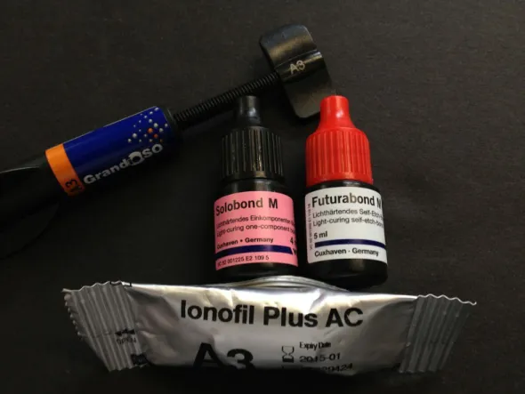
The materials used in the study were a glass ionomer, Ionofil Plus AC plus (color A3; VAL: 2/2015, lot no.: 135342; Voco GmbH, Cuxhaven, German); two adhesive systems: Solobond M (VAL: 8/2015, lot no.:1307330; Voco GmbH, Cuxhaven, German) and Futurabond M (VAL: 1/2015, LOT no.: 1305252; Voco GmbH, Cuxhaven, German) and a resin composite: Grandioso SO (Color A3; VAL 7/2015, lot no.:1304305; Voco GmbH, Cuxhaven, German). Materials composition is presented in Table X below (figure 3.1 and table 3.1).
Name Application Procedure Composition
Ionofil Plus AC
Mixing (10sec);
Application directly into the cavity within 30 s after completion of mixing.
Glass polyalkenoate cement
Solobond M Etch with 37% phosphoric acid, for 15 sec Application of Solobond M homogeneously with a Micro Tim and allow to act for 30 s. Dispersion of Solobond M with a faint air jet
Curing with LED/halogen light for 20 s
Acetone 50%-100%;
2- hidroxyethyl methacrylate 10- 25%; BisGMA 10-25%;
Acidic adhesive Monomer 5-10%; Hidroxypropylmethacrylate <2.5%; Catalyst <2.5%
Futurabond M Rinse cavity thoroughly with water. Remove excess moisture with a faint air jet. Application of Futurabond M onto a mixing palette. Apply with a suitable applicator and allow it to act for 20 s. Dry the adhesive layer with an air jet for at least 5 s.
Polymerization with blue light (halogen or LED light) for 10 s.
Urethanedimethacrylate 25-50%; Ethanol 10-25%;
Acidic adhesive monomer 5-10%; 2-hydroxyelthyl methacrylate 2,5-5%; Catalyst <2,5%
Grandioso SO Application of GrandioSO in the prepared cavity, adapt with a suitable instrument. Insert in 2 increments of 2mm and cure for 20sec each with a halogen curing light with the intensity of 600mW/cm2, measured with a curing radiometer
Resin: BisGMA; BisEMA; tegdma; Camphorquinone; Butylated Hydroxytoluene
Filler: Glass Ceramic (1μm); Silicon Dioxide nano-particles (20-40nm) Pigments ( Iron oxide; titatium dioxide) Table 3.1: Composition and application procedures of the materials used
8
Figure 3.1 Materials used
3.1 SPECIMENS PREPARATION
Thirty human permanent molars were used, after being stored in 0,5% chloramine (Chloramine T Trihydrate – Merck KGaA, Darmstadt, Germany) at 4ºC, for a maximum of a month (ISO, 2003). Before being used teeth were clean (from dental calculus, soft tissues and debris) and stored in distilled water (4ºC) (ISO, 2003). All the teeth were half cut, longitudinally with a with a 0,3mm thick diamond blade (Diamond Wafering Blade – Buehler, Series 15HC Diamond Nº11-4244, Germany) in a Precision Saw machine (IsoMet® 1000 Precision Saw from Buehler, Illinois, USA), obtaining 60 specimens (figures 3.2 and 3.3)
9
Figure 3.2. Precision Saw machine (IsoMet® 1000 Precision Saw from Buehler, Illinois, USA).
Figure 3.3. Diamond blade (Diamond Wafering Blade – Buehler, Series 15HC Diamond Nº11-4244, Germany).
In each half a class V cavity (figure 3.4) was performed (3×2×2mm) at the root surface (mesial and distal) with a cylinder diamond bur (Bush, Pfings & Company, NJ, USA), in turbine under water refrigeration. All the cavities were performed by the same operator. The cavities had 3 mm length, 2 mm wide, 2 mm deep. The cervical wall was 1mm apical to the cementum enamel junction. Cavity margins were in enamel and dentin.
10
Specimens were randomly divided in three groups (table 3.2) according to the study material; (Group 1- Ionofill Plus AC, VOCO; Group 2- Solobond M, VOCO and Grandioso, VOCO; Group 3- FuturabondM, VOCO and Grandioso, VOCO).
In each group 10 specimens were restored according to the manufacturer´s instructions and 10 were contaminated with saliva prior to the restorative material application.
All the cavities were washed with a water syringe and dried for 5 sec, followed by the restorative procedure in group 1b, 2b and 3b. Saliva contamination was performed in groups 1a, 2a, an 3a with fresh saliva collected from a researcher, applied onto the surface of the cavity with a syringe (10μL) and spread for 5 sec with a micro brush. It was performed after washing and drying the cavity in group 1a and after adhesive curing in groups 2a and 3a.
Composite resin was inserted in 2 increments of 2mm and cured for 20sec each with a halogen curing light (Curing Light XL3000 serial nº 105944, 3M ESPE Dental Products, St Paul, MN, USA) with the intensity of 600mW/cm2, measured with a curing radiometer (Model 100, Demetron Research Corp., Danbury, USA). Glass ionomer was inserted in bulk and allowed to cure for 6 min prior to storage (figure 3.5).
11
Before contamination Saliva contamination Restorative material system
Group 1 (a) Water rinsed 5 sec Ionofill plus AC
Group 1 (b) Water rinsed none Ionofill Plus AC
Group 2 (a) Acid etched+ rinse+ etch and rinse+ light curing
5 sec Grandioso
Group 2 (b) Acid etched+ rinse+ etch and rinse+ light curing
none Grandioso
Group 3 (a) Self-etch+ light cured 5 sec Grandioso Group 3 (b) Self-etch+light cured none Grandioso
Table 3.2- Schematics of the groups
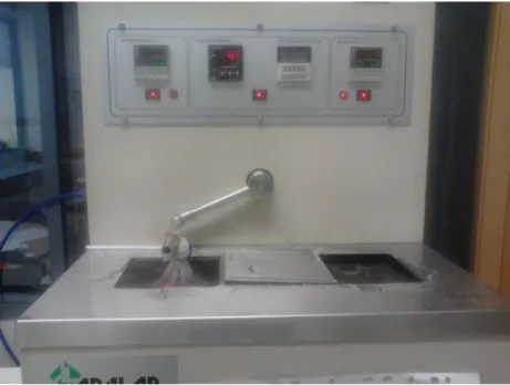
After the restorative procedures, the specimens were stored for 24 hours in an incubator at 37ºC and 100% relative humidity (ISO, 2003). Specimens were then thermal cycled (figure 3.6) by 20sec immersion in two interchanging baths of 5ºC and 55ºC, with 5sec dwell time, for 500 cycles (ISO, 2003), and stored for 24 hrs more in the same conditions as described above.
12 3.2 MICROLEAKAGE TEST
Apex and pulp chamber were isolated with sticky wax and nail polish (Fariha et al., 2012) was applied over the surface leaving a 1mm frame from the restoration margin (figures 3.7 and 3.8).
Figure 3.7 Specimen isolation with Sticky wax
Figure 3.8 Specimen isolation with nail polish
13
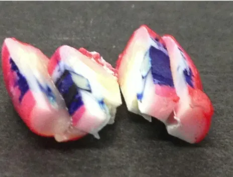
Specimens were then immersed in a 2% methylene blue solution for 4h and thorou ghly rinsed under running water. Each specimen was then sectioned across the restoration in two segments of 1mm each (Figure 3.9) with a cutting device (Isomet 1000 - Buehler, Illinois, USA).
Figure 3.9 Specimen segments
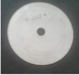
Exposed surfaces were polished with polishing disks (silicon carbide grinding
paper, 800-grit, Buehler II, Germany) and examined at a stereomicroscope (Meiji
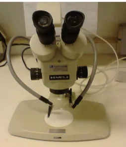
Techno EMZ-8TR serial n.º 411479-Meiji Techno Co., Saitama, Japan), in order to quantify the degree of infiltration (Figures 3.10 and 3.11).
14
Figure 3.10. 800-grit polishing disk
(Buehler II, Germany)
Figure 3.11 Stereomicroscope (Meiji Techno EMZ-8TR)
Each surface was classified using the ISO 14765 (2003) classification method (table 3.3).
Enamel Classification
0 No infiltration
1 Infiltration in the enamel wall
2 Infiltration in the dentin wall without the pulpal wall 3 Infiltration in the dentin wall up to the pulpal wall Dentin Classification
0 No infiltration
1 Infiltration in the dentin wall (less than 50%)
2 Infiltration in the dentin wall (50% or more) without the pulpal wall
3 Infiltration in the dentin wall up to the pulpal wall Table 3.3: Classification of microleakage adopted and altered from ISO 14765, 2003
For each margin (enamel and dentin) microleakage data was analysed with nonparametric statistical tests (Kruskal-Wallis, Mann-Whitney with Bonfferroni
15
correction). A Wilcoxon test was performed in order to compare 2 related variables (enamel and dentin microleakage). Statistical significance was set at 5%.
16
4. RESULTS
Percentages of microleakage degrees for enamel margins are presented in figure 4.1, and for dentin margins are presented in figure 4.2.
The microleakage frequencies found are presented in table 1 and 2 in appendix I. In enamel margins percentage of microleakage degree varied from 18% in degree 2, to 46% in degree 0. In dentin margins, percentage of microleakage degree varied from 5% in degree 1 to 75% in degree 3.
Table 4.3 Percentage of microleakage degrees found for each restorative material system, with and without saliva contamination, in enamel. Different letters represent statistical differences found with Mann-Whitney test (p<0,003).
Statistical tests performed are presented in annex II.
According to Kruskall-Wallis, both enamel and dentin microleakage revealed differences between groups (p<0,05). For the 15 comparisons performed with Mann-Whitney posthoc tests, Bonferroni test corrected the statistical significance to p<0,003.
Enamel microleakage was lowest for the self-etch/composite restoration system either with saliva or without saliva contamination (p<0,003). These values were followed by the etch-rinse/composite and glass ionomer without saliva contamination and finally by etch-rinse/composite and glass ionomer with saliva contamination.
A A B
17
Table 4.4 Percentage of microleakage degrees found for each restorative material system, with and without saliva contamination, in dentin. Different letters represent statistical differences found with Mann-Whitney test (p<0,003).
In dentin, etch-rinse/composite and glass ionomer without saliva contamination groups yielded significantly lower microleakage than the other groups (p<0,003). No other differences were found.
The self-etch/composite was the only restoration system where saliva contamination did not influenced enamel and dentine microleakage (p<0,003).
The degree of microleakage was found to be lower in enamel than in dentin (p<0,05) for the self-etch/composite groups (3a and 3b) with and without saliva contamination. For the other groups tested there were no differences in microleakage degree between enamel and dentin margins.
B B A
18
5. DISCUSSION
Salivary contamination is a frequent problem in restorative procedures, especially when the isolation with rubber dam is difficult to obtain. This represents a considerable issue since the quality of the adhesion between the restorative material and the tooth will be challenged and this bond is considered a key factor in determining the longevity of a restoration (Kermanshah et al., 2010; Chang et al., 2010; Koppolu et al., 2012).
Laboratory methods are frequently used to predict in vivo performance of restorative materials (Fabianelli, 2004), and they are considered a good complement to the more costly and time-consuming clinical study approach (Lucena et al., 2011).
Microleakage analysis is a laboratory method used to examine marginal seal between the restorative material and the tooth surface (Lucena et al., 2011). There are in vitro methods to evaluate the adhesion to tooth structure such as shear and micro-shear bond strength (Placido et al., 2007), as well as microtensile bond strength (Heintze et al., 2011). However, the different properties of materials used in this study (composite material and glass ionomer) such as polymerization contraction and bond strength could be misleading in a bond strength study (Alnazzawi and Watts, 2012; Kaplan et al., 1992). In fact, high bond strengths are required for a resin composite due to the high stress at the bonded interface generated by the polymerization contraction and high coefficient of thermal expansion (Alnazzawi and Watts, 2012). Conversely, glass ionomers require less bond strengths values to maintain a good marginal seal, since they do not suffer from polymerization contraction and have a coefficient of thermal expansion closer to the tooth (Kaplan et al., 1992).
Coefficient of thermal expansion can be manipulated by thermal cycling (Alnazzawi and Watts, 2012). This treatment subjects specimens to extreme temperature changes, similarly to what occurs naturally in the oral cavity (Cenci et al., 2008). However, there are disagreeing opinions about the influence of thermal cycling on microleakage. Some authors reported the absence of any influence, recommending higher number of cycles (Rossomando and Wendt, 1995; Bijella and da Silva, 2000; Pazinatto et al., 2003; Veronezi et al., 2002), while others show an increase of microleakage at the cementum-dentin-restoration interface after thermal stressing
19
(Hakimeh et al., 2000; Cenci et al., 2008). Because there is no consensus between authors, this study followed recommendations established internationally (ISO, 2003), to enable a comparison of results between different studies.
Whole healthy human saliva has been established as an acceptable contamination medium by several authors, and was used in the present work (Oonsombat et al., 2002; Zeppieri et al., 2003). One of the main issues of saliva contamination was the timing. The ideal time for contamination would be after etching, since according to Jordan (1993, apud Lopes et al., 2007) the surface free energy doubles after etching, which is main reason for a good wettability by the adhesive system and the action of saliva would reduce this energy, placing adhesion at risk.
Saliva contamination of the surface after etching would not be feasible in the current study because one of the adhesive system used, a self-etching material, combines in same solution the acidified primer and the adhesive (Peumans et al., 2005; Moszner et al., 2005), not using a separated etching. A study by Kermanshah et al, showed that saliva reduced to the same degree the bond strength of an etch-and-rinse adhesive system when the contamination was performed after the etching step or after the polymerization of the adhesive. Therefore, in the present study the contamination was performed after the adhesive polymerization both in the etch-and–rinse and in the self-etch group (Kermanshah et al., 2010).
When saliva influences adhesion to tooth structure, a gap at the margin of the restoration is formed and can be exposed and easily quantified by means of color dye penetration (Fabianelli, 2004). For this procedure specimens need to be isolated first with sticky wax and nail polish. This preparation is recommended to study marginal leakage without the confounding influence of dentine permeability (Gale e Darvell, 1999). Colour dye penetration studies are the most commonly employed techniques (Lucena et all 2011), due to facility of storage, application and visualizing the penetration of dyes (Youngson et al., 1998). In this study, methylene blue was employed as a tracer to evaluate the degree of infiltration, with a concentration of 2% and an immersion time of 4 hours, a protocol followed in other studies (Camargo et al.,
2006; Sattabanasuk et al., 2006; Alani and Toh, 1997).
Microleakage can be analysed quantitatively or qualitatively and although there are reports of no differences between the methods (Camargo et al., 2006; Veronezi, et
20
al., 2002), in the present work microleakage was evaluated by a quantitative method recommended by ISO (ISO, 2003), since as reported by Nunes et al., the qualitative method used to measure the degree of microleakage is visual and adopts an empirical scale, thus depending on visual acuity and judgment of examiners (Nunes et al., 2005).
Still, results from the quantitative analysis should be considered carefully, since each specimen is sectioned across the restoration in two segments and the exposed surfaces are then analyse, therefore this method only determines the penetration depth along the plane of one tooth section and depends on how and where the tooth is sectioned (Alani and Toh, 1997; Sun et al., 2009).
A low shrinkage during setting and a coefficient of thermal expansion similar to dentin (Kaplan et al., 1992), could determine lower microleakage to the glass ionomer material, although this result was not verified in the present study. In fact, glass ionomer restoration without saliva contamination yielded the same dentin and enamel microleakage as etch-and-rinse/composite restoration and higher enamel microleakage than the self-etch treated composite restorations. These results lead to the rejection of the null hypothesis a) and b).
Although there are no other studies, to the author’s knowledge, that compare the materials used in the present study, Kaplan (1992) compared microleakage in cervical restorations of etch-and-rinse/composite with glass ionomer showing better results when glass ionomer was used, and this would be our expectation for this study, even though the materials used were not the same, and further studies should be developed using other brands of glass ionomer, etch-and-rinse and composite materials.
In the present study and in the presence of saliva contamination, materials behaved differently, thus null hypothesis c) and d) had to be rejected. Saliva contamination led to higher enamel and dentin microleakage of etch-and-rinse/composite restoration and of the glass ionomer restorations. These results were expected for the etch-and-rinse/composite restoration, but can be considered controversial for the glass ionomer since it has been referred to as insensible to saliva contamination (McLean et al. 1985) and to adhere best to moist tooth structures (Burges and Gallo, 2002), reinforcing the idea that further studies must be carried with different brands of materials. Another aspect that influence the results is the fact that human tooth was used, which is so uncontrolled as a variable, as well as the operator variability.
Self-21
etch/composite restorative system was not influenced by saliva contamination, in both margins, as in a study by Kermanshah (Kermanshah et al 2010).
Conversely, the self-etch/composite restorative system was less effective in restraining microleakage along dentin margins than along the enamel margins. This was not expected since it was reported that the self-etching pH is not low enough to condition the enamel surface as well as it does for the dentin surface, due to mineral differences in the dental tissue (Moszner et al., 2005). Despite that contradiction, these results are in accordance with other studies (Brackett et al, 2003; Fabianelli, 2003), in which self-etch materials also had worst results in dentin margins.
These results lead to the rejection of null hypothesis e), the material tested influenced the differences between dentin and enamel microleakage. Even though this null hypothesis had to be rejected, this effect was only registered for the self-etch/composite material, since enamel and dentin microleakage were the same in the glass ionomer and in the etch-and-rinse/composite group.
By testing the marginal sealing ability of restorative materials with dye penetration method alone, one cannot conclude superiority of one material over the other, especially because of the overestimation of infiltration that can be attributed to methylene blue (Fabianelli, 2004). Further studies are needed to determine the best material to be used in challenging situations such as contaminated class V restorations.
Clinical Significance
Due to the discrepant data attained in the present study it is recommended that conclusion of superiority of one material over the other are not drawn from microleakage analysis alone, although it seems that the self-etch material studied was less influenced by the presence of saliva contamination.
22
6. CONCLUSIONS
:
Within the limitations of an in vitro study the results obtained led to the following conclusions:
- Enamel and dentin microleakage degrees were similar for the etch-and-rinse/composite and the glass ionomer restorations, differing only in with the self-etch/composite restorations
- Enamel and dentin microleakage yielded by the self-etch/composite restorations were less influenced by saliva contamination than the other groups.
- The present study should be repeated with other commercial brands of glass ionomer, self-etch, etch-and-rinse and composite materials.
- In vivo and other in vitro studies, besides microleakage should be carried out to fulfil the same objective as in the present study.
23
7. REFERENCES
1- Alani AH, Toh CG (1997). Detection of microleakage around dental restorations: a review. Oper Dent 22(4): 173-185.
2- Alnazzawi A, Watts, D C (2012). Simultaneous determination of polymerization shrinkage, exotherm and thermal expansion coefficient for dental resin-compositesDent Mater 28(12):1240-9
3- Al-Saleh I, Al-Sedairi, Al Anoud (2011). Mercury (Hg) burden in children: the impact of dental amalgam. The Science of the total environment 409 (16):3003-3015.
4- Amer RS, Kolker JL (2013). Restoration of root surface caries in vulnerable elderly patients: a review of the literature. Spec Care Dentist 33(3):141-9. 5- Anusavice KJ, Phillips RWSodm (2003). Phillips' science of dental materials.
11th ed. ed. St. Louis, Mo. [Great Britain], Saunders.
6- Arcoria CJ, Vitasek B, DeWald J P, Wagner M J (1990). Microleakage in restorations with glass ionomer liners after thermocycling. Journal of dentistry 18(2): 107-12.
7- Barghi N, Knight GT, Berry TG (1991). Comparing two methods of moisture control in bonding to enamel: a clinical study. Oper Dent 16(4):130-5.
8- Bates M (2006).Mercury amalgam dental fillings: an epidemiologic assessment. International Journal of Hygiene and Environmental Health 209(4): 309-316. 9- Bellinger C , Trachtenberg F, Barregard L, Tavares M, Cernichiari E, Daniel D,
McKinlay S (2006). Neuropsychological and Renal Effects of Dental Amalgam in Children. JAMA 295 (15):1775-1783.
10- Bignozzi I, Crea A, Capri D, Littarru C, Lajolo C, Tatakis DN (2013).Root caries: a periodontal perspective. J. Periodont. Res.
11- Bijella MF, da Silva SM (2000).In vitro quantitative evaluation of marginal microleakage in Class II restorations confected with a glass ionomer cement and two composite resins. Pesqui Odontol Bras 15(4): 277-282.
12- Bowen RL, Rapson JE, Dickson G (1982).Hardening shrinkage and hygroscopic expansion of composite resins . J Dent Res 61(5):654-8.
13- Brackett WW, Haisch LD, Pearce MG, Brackett MG (2004). Microleakage of Class V resin composite restorations placed with self-etching adhesives. J Prosthet Dent 91(1): 42-45.
14- Bullard RH, Leinfelder KF, Russell CM (1988). Effect of coefficient of thermal expansion on microleakage. J Am Dent Assoc 116(7): 871-874.
15- Burgess.J, DDS, MS, John R. Gallo, DDS (2002). Treating root-surface caries. Dent Clin N Am 46(2):385-404
16- Camargo DA, Sinhoreti MA, Correr-Sobrinho L, de Sousa Neto MD, Consani S (2006). Influence of the methodology and evaluation criteria on determining microleakage in dentin-restorative interfaces. Clin Oral Investig 10(4): 317-323.
24
17- Cassolato SF (2003).Turnbull RS. Xerostomia: clinical aspects and treatment. Gerodontology 20(2):64-77.
18- Cenci MS, Pereira-Cenci T, Donassollo TA, Sommer L, Strapasson A, Demarco FF (2008). Influence of thermal stress on marginal integrity of restorative materials. J Appl Oral Sci 16(2): 106-110.
19- Chalmers, Jane M (2006). Minimal Intervention Dentistry: Part 2. Strategies for Addressing Restorative Challenges in Older Patients. J Can Dent Assoc 72(5):435–40.
20- Chang BH Cho, Lim RY , Kyung SH, Park DS, Oh TS, Yoo HM (2010). Effects of Blood Contamination on Microtensile Bond Strength to Dentin of Three Self-etch Adhesives. Operative dentistry 35(3): 330-336.
21- Coelho, Ana; Canta, João P.; Martins, Jorge N.R.; Oliveira, Sofia A.; Marques, Paula(2012).Perspetiva histórica e conceitos atuais dos sistemas adesivos amelodentinários – revisão da literatura Publicado na Rev Port Estomatol Med Dent Cir Maxilofac 53 :39-46.
22- Collins R, Bryant W, Hodgel V.A (1998). Clinical evaluation of posterior composite resin restorations: 8-year findings Journal of Dentistry 26(4):311-317. 23- Costa Pfeifer CS, Braga RR, Cardoso PE. (2006). Influence of cavity
dimensions, insertion technique and adhesive system on microleakage of class v restorations. Jada 137(2):197-202.
24- Dijken, J.W.V (2000). Direct resin composite inlays/onlays: an 11 year follow-up. J Dent. 28(5):299-306.
25- Erickson RL, McComb D, Wood RE, Maximiw WG (2001). Clinical inhibitation of secondary caries for xerostomic patients. J Dent Res27(5):430-7. 26- Fabianelli, A (2004). A study into the significance of tracing microleakage by
color die infiltration (PhD Thesis). School of Dental Medicine, University of Siena;1-233.
27- Fabianelli A, Kugel G, Ferrari M (2003). Efficacy of self-etching primer on sealing margins of Class II restorations. Am J Dent 16 (1):37-41
28- Fariha N, Shahid M, Bena N (2012). In vivo evaluatin of microleakage of nano-filled resin composite using teo different restorative techniques. Pakistan Oral & Dental Journal 32(2):311-14
29- Gale MS, Darvell BW (1999). Thermal cycling procedures for laboratory testing of dental restorations. J Dent27(2):89-99.
30- Gareri G, Bevacqua I, Mattace R, Ferreri G and Giovambattista De Sarro (1998). Antidepressant Drugs in the Elderly. Gen. Pharmac 30 (4): 465–475. 31- Geerts S, Bolette A, Seidel L, Guéders A (2012). An Evaluation of Leakage of
Two Etch and Rinse and Two Self-Etch Adhesives after Thermocycling. International journal of dentistry. Int J Dent 2012:852841.
32- Giachetti L , Russo D, Bertini F, Pierleoni F (2007). Effect of operator skill in relation to microleakage of total-etch and self-etch bonding systems. Journal of dentistry 35 (3): 289–29.
25
33- Guéders A, Geerts S (2011). Relationship between Operator Skill and In Vitro Microleakage of Different Adhesive Systems in Class V Restorations. ISRN Dent 2011: 285624.
34- Hakimeh S,Vaidyanathan J, Houpt M L, Vaidyanathan T K, Von Hagen S (2000). Microleakage of compomer class V restorations: effect of load cycling, thermal cycling, and cavity shape differences. The Journal of prosthetic dentistry. 83(2):194-203.
35- Hashimoto M, Hiroki O, Hidehiko S, Masayuki K, Haruhisa O (2003). In vitro degradation of resin–dentin bonds analyzed by microtensile bond test,scanning and transmission electron microscopy. Biomaterials 24(21):3795–3803.
36- Haveman C, Burgess J, Summitt JB (1999). A clinical comparison of restorative materials for caries in xerostomic patients. J Dent Res 78:286.
37- Heintze SD, Thunpithayakul C, Armstrong SR, Rousson V Correlation between microtensile bond strength data and clinical outcome of Class V restorations. Dental materials (2011). Dent Mater.27(2):114-25.
38- Hitmi L, Attal JP, Degrange M (1999). Influence of the time-point of salivary contamination on dentin shear bond strength of 3 dentin adhesive systems. J Adhes Dent. 1(3):219-32.
39- Hoppenbrouwers, Driessens F.C.M, Borggreven J.M.P.M (1986). The Vulnerability of Unexposed Human Dental Roots toDemineralization. J Dent Res 65(7):955-958.
40- ISO (2003). Dental materials – Testing of adhesion to tooth structure. Geneva, Switzerland, International Organization for Standardization. ISO/TS 11405. 41- Kaplan I, Mincer HH, Harris EF, Cloyd JS (1992). Microleakage of composite
resin and glass ionomer cement restorations in retentive and nonretentive cervical cavity preparations. J Prosthet Dent 68(6)16-23.
42- Kermanshah H, Ghabraei Sh, Bitaraf T (2010). Effect of salivary contamination during different bonding stages on shear dentin bond strength of one-step self-etch and total self-etch adhesive. Journal of Dentistry, Tehran University of Medical Sciences, Tehran, Iran7(3):132-8.
43- Koppolu M, Gogala D, Thangala V, Deepthi M, Sasidhar N (2012). Effect of saliva and blood contamination on the bond strength of self-etching adhesive system- An in vitro study. J Conserv Dent. 15(3):270-3.
44- Lopes GC, Thys DG, Klaus P, Oliveira GM, Widmer N (2007). Enamel acid etching: a review. Compend Contin Educ Dent. 28 (1):18-24.
45- Lucena C, Lopez JM, Abalos C, Robles V, Pulgar R (2011). Statistical errors in microleakage studies in operative dentistry: A survey of the literature 2001– 2009. Eur J Oral Sci 119(6):504-10.
46- Lundin SA, Norén JG (1991). Marginal leakage in occlusally loaded, etched, class-II composite resin restorations Acta Odontol Scand49(4):247-54.
47- Lynch E, Beighton D (1994). A comparison of primary root caries lesions classified according to colour. Caries Res 28 (4):233-9.
48- Mackenbach J, Yannan Hu, Caspar W.N. Looman (2013). Democratization and life expectancy in Europe, 1960-2008. Social Science & Medicine xxx 1e10.
26
49- Manhart J, Chen HY, Mehl A, Weber K, Hickel R (2001). Marginal quality and microleakage of adhesive class V restorations.J Dent 29(2):123-130.
50- Martins JL, Dotto SR, Travassos RMC, Cardoso TF (2007). Avaliação da microinfiltração marginal em esmalte com a utilização de dois sistemas adesivos. Revista Dentística on line (16): 114-19.
51- Maryniuk GA, Kaplan SH (1986). Longevity of restorations: survey results of dentists' estimates and attitudes. J Am Dent Assoc 112(1):39-45.
52- Matis BA, Cochran M, Carlson T (1996). Longevity of glass-ionomer restorative materials: results of a 10-year evaluation. Quint Int. 27:373–82.
53- McLean J W, Prosser H J, Wilson A. D (1985). The use of glass ionomer cements in bonding composite resins to dentin. Br. Dent J 158:410-414.
54- Meneghim M, Pereira A, Silva F (2002). Prevalence of root caries and periodontal conditions in an elderly institutionalized population from Piracicaba – SP. Pesqui Odontol Bras 16(1):50-56.
55- Mickenautsch S, Mount G, Yengopal V(2011).Therapeutic effect of glass ionomers: an overview of evidence. Australian Dental Journal. 56(1):10-5
56- Moszner N, Ulrich S, Zimmermann J (2005). Chemical aspects of self-etching enamel–dentin adhesives: A systematic review. Dental Materials 21(10):895-910.
57- Munck J, Van Landuyt K, Peumans M, Poitevin A, Lambrechts P, Braem M (2005). A critical review of the durability of adhesion to tooth tissue: methods and results. J Dent Res 84(2): 118-132.
58- Murray PE, Hafez AA, Smith AJ, Cox CF (2002). Bacterial microleakage and pulp inflammation associated with various restorative materials. Dent Mater 18(6): 470-478.
59- Nicholson J W (2007). Polyacid-modified composite resins (“compomers”) and their use in clinical dentistry. Dental materials 23( 5): 615–622.
60- Nunes MCP, Franco EB, Pereira JC (2005). Marginal microleakage: critical analysis of methodology. Salusvita24(3): 487-502.
61- Olver, Ian N (2006). Xerostomia: a common adverse effect of drugs and radiation. Australian Prescriber 29:97-8.
62- Oonsombat C, Bishara SE, Ajlouni R (2003). The effect of blood contamination on the shear bond strength of orthodontic brackets with the use of a new self-etch primer. Am J Or- thod Dentofacial Orthop 123:547-550.
63- Pazinatto FB, Campos BB, Costa LC, Atta MT (2003). Effect of the number of thermocycles on microleakage of resin composite restorations.Pesquisa Odontológica Brasileira 17(4):337-41.
64- Peumans M, Kanumilli P, De Munck J, Van Landuyt K, Lambrechts P, Van MeerbeekB(2005).Clinical effectiveness of contemporary adhesives: A systematic review of current clinical trials. Dent Mater 21(9):864-81.
65- Placido E, Meira JB, Lima RG, Muench A, de Souza RM, Ballester RY (2007). Shear versus micro-shear bond strength test: A finite element stress analysis. Dental materials 23(9):1086-92.
27
66- Powers JM, Finger WJ, Xie J (1995). Bonding of composite to contaminated human dentin and enamel. J Prosthet Dent4(1):28-32.
67- Raskin A, D'Hoore W, Gonthier S, Degrange M, Dejou J (2001).Reliability of in vitro microleakage tests: a literature review.J Adhes Dent 3(4): 295-308.
68- Rossomando KJ, Wendt SL (1995).Thermocycling and dwell times in microleakage evaluation for bonded restorations.Dent Mater 11(1): 47-51. 69- Ritter AV, Shugars DA, Bader JD (2010). Root caries risk indicators: a
systematic review of risk models. Community Dent Oral Epidemiol 38(5):383-97.
70- Sakrana AA, Tanoue N, Kawasaki K, Matsumura H (2004). One-year clinical evaluation of two composite materials used for anterior class V restorations. Journal of Oral Rehabilitation 31(10):985-90.
71- Sattabanasuk V, Shimada Y, Tagami J (2006). Effects of saliva contamination on dentin bond strength using all-in- one adhesives. Journal of Adhesive Dentistry 8(5) 311-318.
72- Spencer P, Ye Q, Park J, Topp E, Marangos O. Wang Y, Bohaty B, Singh V (2010).Adhesive/Dentin interface: the weak link in the composite restoration. Annals of biomedical engineering 38 (6): 1989-2003.
73- Selwitz, R.,Ismail, A., Pitts, Nigel B(2007).Dental caries. Lancet. Vol 369 January 6.
74- Sun J, Fang R, Lin N, Eidelman N, Lin-Gibson S (2009). Nondestructive quantification of leakage at the tooth-composite interface and its correlation with material performance parameters.Biomaterials 30(27): 4457-4462.
75- Tan CL, Santini A (2005). Marginal microleakage around class V cavities restored with glass ceramic inserts of different coefficients of thermal expansion.J Clin Dent.16(1): 26-31.
76- Turner M, Jonathan A Dry Mouth and Its Effects on the Oral Health of Elderly People. The Journal of the American Dental Association. 2007;138, 15S-20S. 77- Veronezi MC, Ishikiriama A, Bastos MTAA, Franco EB (2002). Influência da
ciclagem térmica e do método de avaliaçäo na determinaçäo da microinfiltraçäo em restaurações de resina composta/Influence of the thermocycles and the evaluation methods in the assessing the marginal microleakage of composite resin restorations.Rev. Fac. Odontol. Lins 14(1): 9-18.
78- Watanabe M (2003).Root Caries Prevalence in a Group of Brazilian Adult Dental Patients. Braz Dent J 14(3): 153-156.
79- Youngson CC, Jones JC, Manogue M, Smith IS (1998). In vitro dentinal penetration by tracers used in microleakage studies. Int Endod J 31(2): 90-99. 80- Yoo HM, Oh TS, Pereira PNR (2006).Effect of Saliva Contamination on the
Microshear Bond Strength of One-step Self-etching Adhesive Systems to Dentin Operative Dentistry 31(1):127-134.
81- Zeppieri I L, Chung CH, Mante F K (2003). Effect of saliva on shear bond strength of an orthodontic adhesive used with moisture-insensitive and self-etching primers. American Journal of Orthodontics and Dentofacial Orthopedics.124(4):414-9.
28
Appendix I
29
Frequency tables
Table-1 Frequency of microleakage in Enamel. The most frequent value of microleakage in Enamel was 0, and the less frequent was 2.
Table-2 Frequency of microleakage in Dentine. The most frequent value of microleakage in Dentine was 3, and the less frequent was 1.
Microleakage Frequency Percent Valid Percent Cumulative Percent 0 46 38,3 38,3 38,3 1 24 20,0 20,0 58,3 2 18 15,0 15,0 73,3 3 32 26,7 26,7 100,0 Total 120 100,0 100,0
Microleakage Frequency Percent Valid Percent Cumulative Percent 0 31 25,8 25,8 25,8 1 5 4,2 4,2 30,0 2 9 7,5 7,5 37,5 3 75 62,5 62,5 100,0 Total 120 100,0 100,0
30
Figure 1 Microscopy image of a specimen with Self-etch without contamination (1.5X magnification)
Figure 2 Microscopy image of a specimen with Self-etch without contamination (3.5 X magnification)
31