Rev Odontol UNESP. 2016 Jan-Feb; 45(1): 33-40 © 2016 - ISSN 1807-2577 ORIGINAL ARTICLE
Doi: http://dx.doi.org/10.1590/1807-2577.08815
he hardness and chemical changes in demineralized primary
dentin treated by luoride and glass ionomer cement
As mudanças químicas e de dureza na dentina decidua desmineralizada tratada com luoreto e
cimento de ionômero de vidro
Gisele Fernandes DIAS
a*, Ana Cláudia Rodrigues CHIBINSKI
a, Fábio André dos SANTOS
a,
Viviane HASS
a, Fabiana Bucholdz Teixeira ALVES
a, Denise Stadler WAMBIER
aaUEPG – Universidade Estadual de Ponta Grossa, Ponta Grossa, PR, Brasil
Resumo
Introdução: O fluoreto desempenha importante papel no controle da cárie dental. Objetivo: Avaliar as trocas químicas entre cimentos de ionômero de vidro de alta viscosidade (CIV) e dentina decídua com aplicação de fluoreto de sódio (NaF) a 2% em alterações de dureza dentinária a partir da incorporação de cálcio, fosfato e fluoreto. Material e método: Cavidades Classe I foram preparadas em 40 molares hígidos divididos em 2 grupos (n=20), de acordo com a condição dentinária: hígida (1) e desmineralizada (2). Subgrupos (n=10) foram formados para avaliar a ação isolada do CIV ou associado com NaF (F). Este estudo in vitro avaliou as trocas químicas sob duas condições: dentina hígida e desmineralizada (ciclagem de pH) para simular a perda mineral que ocorre em lesões de cárie. Grupo G1 e G2 receberam restaurações de CIV; Grupos G1F e G2F receberam NaF antes do CIV. Os espécimes foram preparados para microdureza Knoop e Micro-Raman. Para análise estatística foi utilizada Anova 2 fatores (α = 0.05). Os dados do Micro-Raman foram descritos qualitativamente. Resultado: O aumento de dureza foi observado em todos os sítios de contato direto com CIV, em ambas dentinas em todos os grupos (p<0.001); não foi observado diferença em microdureza após aplicação do NaF (p>0.05). Na avaliação do Micro-Raman, o contato direto do CIV/dentina tanto hígida quanto desmineralizada resultou em um aumento do pico do fosfato dentinário. Conclusão: As trocas químicas entre o CIV e dentina desmineralizada podem induzir mudanças das propriedades mecânicas do substrato e a captação de íons minerais (fosfato) ocorre sem a influência do NaF.
Descritores: Cimentos de ionômero de vidro; dentina; tratamento restaurador atraumático; cárie dentária; fluoreto.
Abstract
Background: Fluoride plays an important role in the control of dental caries. Aim: To evaluate the chemical exchange between restoration of glass ionomer cement of high viscosity (GIC) and primary dentin with application of sodium fluoride (NaF) 2% through changes in hardness from uptake of calcium, phosphate and fluoride. Material and method: Class I cavities were prepared in 40 sound primary molars, and the sample was divided into two groups (n=20) according to dentin condition: sound (1) and demineralized (2). Sub-groups (n=10) were formed to investigate the isolated action of the GIC or the association with NaF (F). This in vitro study examined the chemical exchange under two conditions, sound and demineralized dentin (pH cycling), to simulate the occurrence of mineral loss for the caries lesion. G1 and G2 received GIC restoration only; groups G1F and G2F received NaF before GIC restoration. The specimens were prepared for Knoop hardness test and micro-Raman spectroscopy. A two-way ANOVA test (α = 0.05) was used for statistical analysis. Micro-Raman data were qualitatively described. Result: Increased hardness was observed in all the sites of direct contact with GIC in sound and demineralized dentin for all groups (p<0.001); no difference was observed in microhardness after application of NaF (p>0.05). In the evaluation of micro-Raman, direct contact between GIC and dentin for sound and demineralized dentin resulted in increased peaks of phosphate.
Conclusion. The exchange between GIC and demineralized dentin may induce changes of mechanical properties of the substrate, and uptake of mineral ions (phosphate) occurs without the influence of NaF.
INTRODUCTION
he contemporary approach to restorative procedures in pediatric dentistry is based in minimally invasive dentistry. It favors partial caries removal and the maintenance of carious dentin, which still has the potential for reorganization. In primary teeth, clinical researches have already proved this potential ater restoration using glass ionomer cements1-3 (GIC) or other restorative materials4-6. Nevertheless,
there is a trend in research toward study of the interaction between demineralized dentin and bioactive materials such as glass ionomer cements. When using GIC, there is some ion exchange between ions that are released during the setting reaction of the material and the dentin minerals. his reaction allows not only the mineral gain of dentin7 but also a more efective adhesion that afects the longevity
of the restoration.
he main ions involved in this process are calcium, strontium, aluminum and luoride7. In particular, luoride has always been a
subject of research because of its participation in DES-RE mechanism8
and because GIC’s property of luoride release and reload makes this material a reservoir of luoride in the mouth.
he topical application of luoride in the surface of enamel/dentin produces a calcium luoride (CaF2) layer with a high concentration of ionic luoride, which controls dental solubility in situations of high cariogenic challenge9. Notwithstanding, there is little information
available about the action of luoride in demineralized10 and sealed
dentin as considered in this paper.
We hypothesized that the chemical exchange between luoride solution and demineralized dentin, along with restoration of the glass ionomer cement, would improve the mechanical properties of the dentin. As a result, the adhesion and longevity of the restoration could be positively inluenced.
herefore, this paper’s objective was to evaluate the chemical exchange between high-viscosity glass ionomer cement restoration and primary demineralized dentin with the application of sodium luoride (NaF) 2% through changes in dentin hardness from uptake of calcium, phosphate and luoride.
MATERIAL AND METHOD
his research was approved by the Ethics in Research Committee, protocol #134.145.
Sample
he sample consisted of 40 primary molars without any crown defects or carious lesions. he remaining root portions were removed using a high-speed diamond bur (2121 KG Sorensen, Cotia, SP, Brazil), and the teeth were stored in distilled water at room temperature to avoid dehydration until cavity preparation.
Study design
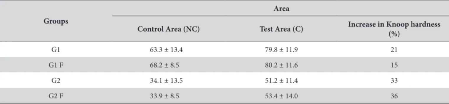
Knoop hardness and micro-Raman tests were used to evaluate the following dentin conditions: sound/demineralized, treated or not treated with NaF solution and with/without contact with GIC. For this purpose, class I cavities were prepared in the 40 selected primary molars. he teeth were divided into 2 groups (n=20) according to the dentin condition: sound (G1) and demineralized (G2). Subgroups (n=10) were formed to analyze the isolated action of the GIC and the action associated with sodium luoride (F).
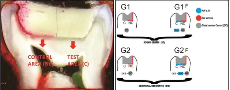
he cavities were divided into halves, one of which was isolated with nail varnish, thus ensuring a control area in each group (Figure 1). All cavities were restored with a high-viscosity glass ionomer cement, but only G1F and G2F received a neutral solution of NaF 2% before restoration (Figure 1).
Preparation of the Class I Cavities
A drilling apparatus (El Quip , São Carlos, SP, Brazil) was used in the preparation of the cavities, which were standardized (depth=2 mm, length=6 mm and wide=4 mm). he size of the cavities was checked with a caliper (Digimess, São Paulo, SP, Brazil).
he preparation of the cavities was started in the center of the occlusal surface of each primary molar. Tungsten carbide
burs #8 with an initial depth of 1.5 mm were used to reach the inal size set at 2mm with a diamond high rotation #3131, under continuous cooling. Ater 8 cavities, drills were replaced by new ones. To complete this step, the teeth were taken to the ultrasonic tank (Cristófoli Biosafety, Campo Mourão, PR, Brazil) for 280 s to remove debris from the cavity preparation. Visual examination of specimens with a stereoscopic magnifying glass (10×) allowed veriication of whether the pulp chamber was exposed by cavity preparation. If so, the tooth was discarded.
Procedures for pH Cycling
he teeth of G2 (n=20) were subjected to dentin demineralization to simulate carious lesions using the method of pH cycling according to the protocol already used11. To protect the external area of dental
crowns, two layers of nail varnish (Colorama, Rio de Janeiro, RJ, Brazil) were applied, avoiding contact with the walls of the cavity.
To establish the DES-RE cycle, each specimen was immersed for 8 h in demineralizing solution and for 16 h in remineralizing solution. he volume of the demineralizing and remineralizing solution was 10 ml for each tooth in every cycle. his cycle was repeated for 14 consecutive days at room temperature without stirring. he solutions used in pH cycling were manipulated in the Pharmaceutical Science Lab at UEPG. he formulations12 were as
follows: demineralizing solution (pH 4.8) with 2.2 mM calcium chloride (CaCl2); 2.2 mM phosphate Sodium (Na2PO4) and 50 mM acetic acid; and remineralizing solution (pH 7.0): 1.5 mM calcium chloride (CaCl2), 0.9 mM phosphate Sodium (Na2PO4) and 0.15 mM potassium chloride (KCl).
he restorative procedures were carried out right ater the DES-RE cycle was inished for all the specimens.
Restorative Procedures
All cavities were illed with high-viscosity GIC (Ketac Molar Easymix, 3M ESPE, St. Paul, MN, USA).
he protocol used is described above. he cavities were treated with Ketac liquid (3M ESPE, St. Paul, MN, USA) for 10 s, washed with air/water spray for 20 s, dried with a gentle stream of dry compressed air and immediately illed with the GIC. he GIC was dosed at a ratio of 2:2 (powder and liquid), and manipulated on the block by mixing with a plastic spatula (Dulex, Rio de Janeiro, RJ, Brazil). he mixture was inserted with an applicator syringe (Precision Maquira , Maringa, PR, Brazil) until the cavity was illed, followed by the “press inger” technique (30 s) with polyester tape (K-Dent Quimidrol , Joinville, SC, Brazil). he glass ionomer was allowed to set for 3 min, then protected with a layer of petroleum jelly (Rioquímica, São José do Rio Preto, SP, Brasil) in accordance with the instructions of the manufacturer. Following restoration, the teeth were stored in a humidiier for 24 h at 37 °C.
Topical Application of Neutral Solution of NaF
Topical application of neutral solution of NaF was carried out only for G1F and G2F (Figure 1). For these groups, ater the application of the Ketac liquid and before the restoration, a neutral solution of NaF at 2% was applied on the dentin for 1 min with the aid of a
cotton ball, and the volume to each cavity was standardized with a cotton ball. he excess solution was removed with ilter paper discs.
Before the restorative procedures, all the cavities received a layer of nail varnish on the mesial side of the cavity. he research design stipulated the division of the cavity into two sites: control area (NC) and test area (C). his was done to guarantee that the GIC would not be in contact with the dentin, as well as to make sure that the NaF solution would be the only chemical element in contact with the dentin.
Preparation of Specimens for Tests
he teeth were ixed in a cutting machine (Isomet 1000 Precision Saw Buehler, Lake Bluf, IL, USA) and were sectioned vertically with a diamond disk (1.3 mm Precision Saw, Lake Bluf IL, USA) at 300 rpm to obtain dental slices (n=3) with approximately 1.1 mm thickness.
Two dental slices were mounted in the center of PVC (Tiger, Joinville, SC, Brazil) cylinders (12×20 mm), which were attached with double-sided tape (3M, SUMARE, SP, Brazil) on a glass plate. he cylinders were illed with colorless acrylic resin (JET, Clear Field, SP, Brazil) made by the powder and liquid technique
he embedded slices were taken to the rotary polishing machine (Arotec, Cotia, SP, Brazil) to perform the polishing of specimens. A sequence of silicon carbide sandpapers (3M Brazil, Sumaré, SP, Brazil) was used under intense water irrigation. Final polishing was performed with diamond paste (Arotec, Cotia, SP, Brasil) of grain 1/¼ µm. For removal of waste, the specimens were washed for 12 min in an ultrasonic tank. Finally, they were stored at 37 °C for 24 h in a 100% humidity environment.
Microhardness Test
he microhardness analysis was performed on a microhardness apparatus (Shimadzu, Kyoto, Japan) with a Knoop indenter. For 30 s, the applied loads were 25 g for sound dentin and 10g for demineralized dentin12.
he loads were applied on the specimens 50 µm below the cavity loor. hree diferent measures were made at 100 µm distance from each other at the same depth. he mean between these three measures was considered the microhardness value of the specimen.
Micro-Raman Spectroscopy
he same specimens used for the microhardness test were subjected to analysis of mineral composition through micro-Raman spectroscopy (Bruker Optik GmbH, Ettlingen, Baden-Württemberg, Germany). he apparatus was calibrated irst to zero and then for the values of coeicients using a sample of silicone.
he test included the following parameters: a neon laser with 532 nm wavelength and 20 mW, a spatial resolution of ≈ 3 mM, spectral resolution of ≈ 5 cm–1, 20 s accumulation time and
just below the tooth-restoration interface in a random place with a three-point mapping analysis: 0, 0.45 and 90 µm in depth.
he micro-Raman spectroscopy detected the chemical content of the dentin through the vibrational molecular characteristics of energy12. he representative spectra of calcium phosphate
(corresponding to 960 cm –1) were identiied, and peaks of diferent
vibrational modes of the phosphate group (591 cm–1, 430 cm–1),
carbonate (1070 cm -1), collagen (1270-1453 cm–1) and peak CaF 2
were represented by the interval of 322 cm-1 present in the samples.
Statistical Analysis
he distribution of normality was veriied with D’Agostino & Pearson and Shapiro Wilks tests; homogeneity of variances was tested with Levene’s test. he hardness data were analyzed using ANOVA 2 criteria considering the factors dentin (sound and demineralized), treatment (with and without luoride) and treating dentin interaction. he level of signiicance used was 5% (α = 0.05). he data micro-Raman spectra were analyzed qualitatively by groups. he statistical program GraphPad Prism version 5 for Windows (GraphPad Sotware, San Diego California, USA) was used for data analysis of hardness.
RESULT
he means and standard deviations from Knoop microhardness are displayed on Table 1. Here, we can note that there is an increase in Knoop hardness (KHN) in diferent groups. here was a great percentage increase of Knoop hardness for Group 2F, which revealed great uptake of luoride by the demineralized dentin. Enhanced hardness values were observed in the test area in sound and demineralized dentin when compared to the control area (p<0.0001). here was a signiicant diference between the control area (NC) and the test area (C) in all groups assessed (Table 1).
A signiicant diference in Knoop hardness was found for dentin (sound and demineralized) in the control area (p<0.0001) and the test area with the restorative material (p<0.0001). Changes in hardness occurred because of direct contact of the restorative material with the dentin surface in both conditions. Treatment with NaF did not result in a signiicant diference for the dentin (sound and demineralized) in either the control area (p=0.358) or the test area (p=0.642). he interaction dentin x NaF did not efect signiicant change in the hardness of dentin, and there was
no signiicant diference in either the control area (p=0.309) or the test area (p=0.751). Pre-treatment with NaF was not efective in improving the condition of the dentin (Figure 2).
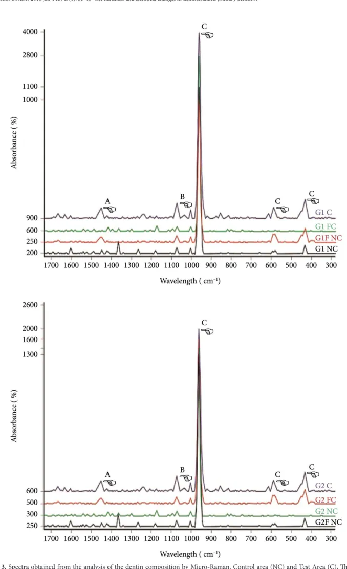
he data obtained by micro-Raman spectroscopy are shown in Figure 3. he range of the spectral region involved (300-1700 cm–1)
was identiied in all groups through the largest intensity peak indicated: 960 cm–1 (PO
4
–3- phosphate), 591 cm–1 and 430 cm–1
(peak vibrational modes of the phosphate group), which represent the calcium hydroxyapatite. he peak 1070 cm–1 (carbonate)
represents the product of collagen ibers within the GIC12. he peak
of 1270-1453 cm–1 characterizes collagen in the range, and 322 cm–1
is CaF2. he identiication of the peak of CaF2 was sought in G2B and G1B, as the product formed ater topical application of luoride, whose reaction with luoride apatite is the product of precipitation of luoride ions13.
Table 1. Means, standard deviations and percentage increase in Knoop hardness (KHN) in diferent groups
Groups
Area
Control Area (NC) Test Area (C) Increase in Knoop hardness (%)
G1 63.3 ± 13.4 79.8 ± 11.9 21
G1 F 68.2 ± 8.5 80.2 ± 11.6 15
G2 34.1 ± 13.5 51.2 ± 11.4 33
G2 F 33.9 ± 8.5 53.4 ± 14.0 36
DISCUSSION
We based our research in an in vitro model previously presented in the literature10 to study ionic exchanges between demineralized
dentin and GIC. As such, the present paper evaluated changes in the mineralization of dentin from uptake of calcium, phosphate and luoride in demineralized primary dentin ater GIC restorations, with or without application of a neutral solution of NaF 2%. In this paper, we observed that direct contact between dentin/GIC resulted in increased hardness and phosphate concentration in dentin substrate.
he increased hardness of demineralized dentin suggests an improvement4,10,13 that is compatible with the changes in mineral
composition. It is important to point out that these alterations weren’t dependent of the luorides in the GIC or the solution that was applied before tooth restoration, since we did not detect an increase in the luoride levels in sound or demineralized dentin. his contradicts our hyphothesis, since we believed that when conditioning the dentin with a sot acid like the Ketac liquid, it would be enough to potentialize the action of a neutral luoride solution. he luoride in neutral solution probably wasn’t reactive enough to form a stable mineral layer or to penetrate the dentin substrate. On the other hand, the luoride released by the GIC wasn’t incorporated on the dentin either. herefore, we can say, based on our indings, that the improved mechanical property seen in the dentin substrate was dependent on the phosphate ions only. herefore, this result may be due to the ionic exchange in the tooth-restoration interface13,14. Ionic elements like sodium, calcium,
aluminum and phosphorus migrate to demineralized dentin13,15,16.
his mineral uptake promotes changes in the hardness of dentin, as we demonstrate in our paper. he relationship between hardness and mineral content of dental hard tissues is well documented in the literature17,18.
he method used in our study produces artiicial caries lesions based on a pH cycle model11 that simulates the DES-RE mechanism
and the pH variation associated with the carious process. he mineral loss resulting from this method translated into a signiicant reduction in Knoop hardness for the demineralized dentin, which reached a maximum of half of the values from sound dentin.
hese modiications were evident in the micro-Raman analysis. We could identify the representative peaks from the mineral phase of the dentin and from apatite hidroxicarbonate, which is the product of the reaction between GIC and dentin19,20, as well as the
phosphate peaks in the sites that have direct contact with GIC. he research design stipulated the division of the cavity into two sites: control area (NC) and test area (C). his was done in an attempt to minimize bias when comparing the dentin substrate with treatment or not. As such, we compared samples of dentin from
the same tooth and at the same depth, and inherent variations in mineral content between diferent teeth did not inluence the results.
Fluoride application before GIC restoration did not increase the luoride uptake in the dentin. One possible explanation is that a lower pH environment should be achieved in the dentin/GIC interface in order for an additional luoride solution to make some diference in the ion exchange proccess21.
here are reports that show that the pattern of luoride and strontium penetration in dentin ater GIC restoration is consistent with the mineralization process22. Even so there is no
evidence that ultrastructural remineralization occurs in the intra or interibrillar demineralized collagen matrix23. Hence, the term
“remineralization” isn’t being used correctly. As a matter of fact, GIC promotes a “mineral gain” in the demineralized dentin, with enhanced calcium and phosphorus contents4,16, but it cannot be
considered remineralization23.
Chemical analysis using micro-Raman spectroscopy complemented the characterization of the dentin underlying the GIC restoration. It provided microscopic data for a wide swath of specimens through vibrational molelular alterations24,25. We were able to detect the
peak of 960 cm–1, which represents phosphate, in all tested groups.
his is the most intense micro-Raman peak in dental hard tissues which indicates mineral accretion in dentin.
In this study, the methodology allowed us to assess the isolated action of the GIC, thus the microhardness test was efective. Gains in hardness and changes in mineral composition may induce immediate improvement in the condition of dentin4,10,13, and no
changes were observed with application of a neutral solution of NaF 2%. herefore, the results reinforce the importance of GIC as a “therapeutic” and irstchoice material for minimally invasive restorations, especially in the ART strategy.
CONCLUSION
A neutral solution of NaF 2% did not modify the mineral and mechanical characteristics of sound and demineralized dentin. he increase in dentin hardness and changes in mineral content were due to ion exchange from GIC to dentin.
ACKNOWLEDGEMENTS
his study was funded by CAPES. We would like to thank Professors Dr. Paulo Vitor Farago and Dr. Yasmine Mendes Puppo for their assistance with the solutions used in pH cycling manipulated in the Pharmaceutical Science Lab at UEPG and the graduate student Paola Crystine Machado for all assistance at the lab.
REFERENCES
1. Massara MLA, Alves JB, Brandão PRG. Atraumatic restorative treatment: clinical, ultrastructural and chemical analysis. Caries Res. 2002 Nov-Dec;36(6):430-6. http://dx.doi.org/10.1159/000066534. PMid:12459616.
3. Chibinski AC, Reis A, Kreich EM, Tanaka JL, Wambier DS. Evaluation of primary carious dentin after cavity sealing in deep lesions: a 10 to 13-month follow-up. Pediatr Dent. 2013 May-Jun;35(3):E107-12. PMid:23756304.
4. Marchi JJ, Froner AM, Alves HL, Bergmann CP, Araújo FB. Analysis of primary tooth dentin after indirect pulp capping. J Dent Child (Chic). 2008 Sep-Dec;75(3):295-300. PMid:19040817.
5. Casagrande L, Falster CA, Di Hipolito V, De Góes MF, Straffon LH, Nör JE, et al. Effect of adhesive restorations over incomplete dentin caries removal: 5-year follow-up study in primary teeth. J Dent Child (Chic). 2009 May-Aug;76(2):117-22. PMid:19619424.
6. Lula EC, Almeida LJ Jr, Alves CM, Monteiro-Neto V, Ribeiro CC. Partial caries removal in primary teeth: association of clinical parameters with microbiological status. Caries Res. 2011;45(3):275-80. http://dx.doi.org/10.1159/000325854. PMid:21576960.
7. Watson TF, Atmeh AR, Sajini S, Cook RJ, Festy F. Present and future of glass-ionomers and calcium-silicate cements as bioactive materials in dentistry: biophotonics-based interfacial analyses in health and disease. Dent Mater. 2014 Jan;30(1):50-61. http://dx.doi.org/10.1016/j. dental.2013.08.202. PMid:24113131.
8. Lippert F, Hara AT, Martinez-Mier EA, Zero DT. In vitro caries lesion rehardening and enamel fluoride uptake from fluoride varnishes as a function of application mode. Am J Dent. 2013 Apr;26(2):81-5. PMid:24073530.
9. Lynch RJ, Navada R, Walia R. Low-levels of fluoride in plaque and saliva and their effects on the demineralisation and remineralisation of enamel; role of fluoride toothpastes. Int Dent J. 2004;54(5 Suppl 1):304-9. http://dx.doi.org/10.1111/j.1875-595X.2004.tb00003.x. PMid:15509081.
10. Ngo HC, Mount G, McIntyre J, Do L. An in vitro model for the study of chemical exchange between glass ionomer restorations and partially demineralized dentin using a minimally invasive restorative technique. J Dent. 2011 Dec;39(Suppl 2):S20-6. http://dx.doi. org/10.1016/j.jdent.2011.10.016. PMid:22101125.
11. ten Cate JM, Duijsters PP. Alternating demineralization and remineralization of artificial enamel lesions. Caries Res. 1982;16(3):201-10. http://dx.doi.org/10.1159/000260599. PMid:6953998.
12. Marquezan M, Corrêa FN, Sanabe ME, Rodrigues Filho LE, Hebling J, Guedes-Pinto AC, et al. Artificial methods of dentine caries induction: a hardness and morphological comparative study. Arch Oral Biol. 2009 Dec;54(12):1111-7. http://dx.doi.org/10.1016/j. archoralbio.2009.09.007. PMid:19878926.
13. Nicholson JW. Glass ionomer dental cements: update. Materials Techonology. 2010 Mar;25(1):8-13. http://dx.doi.org/10.1179/1753555 09X12614966220506.
14. Sidhu SK. Glass-ionomer cement restorative materials: a sticky subject? Aust Dent J. 2011 Jun;56(Suppl 1):23-30. http://dx.doi.org/10.1111/ j.1834-7819.2010.01293.x. PMid:21564113.
15. Baliga MS, Bhat SS. Effect of fluorides from various restorative materials on remineralization of adjacent tooth: an in vitro study. J Indian Soc Pedod Prev Dent. 2010 Apr-Jun;28(2):84-90. http://dx.doi.org/10.4103/0970-4388.66742. PMid:20660973.
16. Bezerra AC, Novaes RC, Faber J, Frencken JE, Leal SC. Ion concentration adjacent to glass-ionomer restorations in primary molars. Dent Mater. 2012 Nov;28(11):e259-63. http://dx.doi.org/10.1016/j.dental.2012.08.014. PMid:22999372.
17. Arnaud TM, de Barros Neto B, Diniz FB. Chitosan effect on dental enamel de-remineralization: an in vitro evaluation. J Dent. 2010 Nov;38(11):848-52. http://dx.doi.org/10.1016/j.jdent.2010.06.004. PMid:20600551.
18. Angker L, Nockolds C, Swain MV, Kilpatrick N. Correlating the mechanical properties to the mineral content of carious dentine--a comparative study using an ultra-micro indentation system (UMIS) and SEM-BSE signals. Arch Oral Biol. 2004 May;49(5):369-78. http:// dx.doi.org/10.1016/j.archoralbio.2003.12.005. PMid:15041484.
19. Banerjee A, Paolinelis G, Socker M, McDonald F, Watson TF. An in vitro investigation of the effectiveness of bioactive glass air-abrasion in the ‘selective’ removal of orthodontic resin adhesive. Eur J Oral Sci. 2008 Oct;116(5):488-92. http://dx.doi.org/10.1111/j.1600-0722.2008.00561.x. PMid:18821993.
20. Sauro S, Thompson I, Watson TF. Effects of common dental materials used in preventive or operative dentistry on dentin permeability andremineralization. Oper Dent. 2011 Mar-Apr;36(2):222-30. http://dx.doi.org/10.2341/10-225-L. PMid:21777102.
21. Ngo HC, Mount G, Mc Intyre J, Tuisuva J, Von Doussa RJ. Chemical exchange between glass-ionomer restorations and residual carious dentine in permanent molars: an in vivo study. J Dent. 2006 Sep;34(8):608-13. http://dx.doi.org/10.1016/j.jdent.2005.12.012. PMid:16540227.
22. Kim YK, Yiu CK, Kim JR, Gu L, Kim SK, Weller RN, et al. Failure of a glass ionomer to remineralize apatite-depleted dentin. J Dent Res. 2010 Mar;89(3):230-5. http://dx.doi.org/10.1177/0022034509357172. PMid:20110510.
23. Chowdary MV, Kumar KK, Thakur K, Anand A, Kurien J, Krishna CM, et al. Discrimination of normal and malignant mucosal tissues of the colon by Raman spectroscopy. Photomed Laser Surg. 2007 Aug;25(4):269-74. http://dx.doi.org/10.1089/pho.2006.2066. PMid:17803383.
24. Almahdy A, Downey FC, Sauro S, Cook RJ, Sherriff M, Richards D, et al. Microbiochemical analysis of carious dentine using Raman and fluorescence spectroscopy. Caries Res. 2012;46(5):432-40. http://dx.doi.org/10.1159/000339487. PMid:22739587.
CONFLICTS OF INTERESTS
he authors declare no conlicts of interest.
*CORRESPONDING AUTHOR
Gisele Fernandes Dias, Departamento de Odontologia, UEPG – Universidade Estadual de Ponta Grossa, Rua Coronel Francisco Ribas, 396, apto 4, Centro, Ponta Grossa - PR, Brasil, e-mail: [email protected]