Anaplasma phagocytophilum and
human granulocytic anaplasmosis in Portugal
Ana Sofia Pereira dos Santos
Dissertação apresentada ao Instituto de Higiene e Medicina Tropical,
Universidade Nova de Lisboa, para obtenção do Grau de Doutor no Ramo de
Ciências Biomédicas, Especialidade de Microbiologia
LISBOA
2007
Neutrophil with Anaplasma phagocytophilum morulae. Pheripheral blood smear stained with Diff-Quik. Photograph kindly provided by Professor J Stephen Dumler, The Johns Hopkins University School of Medicine, Baltimore.
Neutrófilo com uma mórula de Anaplasma phagocytophilum. Esfregaço de sangue periférico corado com Diff-Quik. Fotografia gentilmente cedida pelo Professor J Stephen Dumler, The Johns Hopkins University School of Medicine, Baltimore.
ORIENTAÇÃO:
Doutora Fátima Bacellar
Investigadora Principal Aposentada
Centro de Estudos de Vectores e Doenças Infecciosas Instituto Nacional de Saúde Dr. Ricardo Jorge, Lisboa Professor John Stephen Dumler
Professor Associado
Division of Medical Microbiology, Department of Pathology The Johns Hopkins University School of Medicine, Baltimore
COMISSÃO TUTORIAL:
Doutora Margarida Collares Pereira Investigadora Principal com Habilitação Instituto de Higiene e Medicina Tropical Universidade Nova de Lisboa, Lisboa Professor Doutor Paulo Almeida Professor Associado
Instituto de Higiene e Medicina Tropical Universidade Nova de Lisboa, Lisboa Doutora Fátima Bacellar
Investigadora Principal Aposentada
Centro de Estudos de Vectores e Doenças Infecciosas Instituto Nacional de Saúde Dr. Ricardo Jorge, Lisboa
Na presente dissertação incluem-se resultados que foram ou estão a ser alvo de publicação em co-autoria. Para efeitos do disposto no nº1 do Despacho nº2303/2000 do Regulamento de Programas de Doutoramento do Instituto de Higiene e Medicina Tropical, Universidade Nova de Lisboa (Diário da República, 2ª série, nº23, de 28 de Janeiro de 2000), o autor da dissertação declara que interveio na concepção e execução do trabalho experimental, na interpretação dos resultados e na redacção dos manuscritos publicados, no prelo ou que aguardam submissão.
Lisboa, 11 de Setembro de 2007
Ao Instituto Nacional de Saúde Dr. Ricardo Jorge (INSA) a possibilidade de realizar os trabalhos que permitiram a elaboração da presente dissertação;
À Fundação para a Ciência e a Tecnologia, a atribuição da bolsa de doutoramento (refª SFRH/BD/8610/2002), sem a qual teria sido impossível a realização deste projecto;
Ao Professor Doutor Armindo Filipe e Doutora Maria Sofia Núncio, responsáveis pelo Centro de Estudos de Vectores e Doenças Infecciosas, Instituto Nacional de Saúde Dr. Ricardo Jorge (CEVDI/INSA), em diferentes fases do meu percurso, pelo apoio e incentivo que tenho merecido desde os primeiros passos no mundo da Ciência e pela disponibilização de todos os recursos do Centro para o bom andamento da parte experimental desta dissertação;
À Doutora Fátima Bacellar, minha orientadora no CEVDI/INSA e mentora, a quem devo a minha formação, o apoio incondicional e a amizade que tem sido uma constante desde que as nossas vidas se cruzaram; To Professor John Stephen Dumler, my sponsor at The Johns Hoopkins University School of Medicine, for all the knowledge, support and stimulation. I am grateful for knowing you and honoured to be able to share some of your expertise in A. phagocytophilum field.
À Doutora Margarida Collares Pereira e ao Professor Doutor Paulo Almeida, por terem aceite ser o elo de ligação ao Instituto de Higiene e Medicina Tropical (IHMT), e que com as suas sugestões, apoio e incentivo simplificaram várias etapas deste trabalho;
Ao Professor Doutor Virgílio do Rosário e Professora Doutora Maria Amélia Grácio, anterior e actual Presidente do Conselho Científico do IHMT, pelo acolhimento institucional e disponibilidade na resolução de todas as questões académicas determinantes para a boa consecução deste projecto;
Ao Mestre João Lavinha, ex-Director do INSA e presentemente responsável pelo Centro de Genética Humana do mesmo Instituto, por acreditar e apoiar todos os projectos que me proponho desenvolver;
To all the Researchers that since a long time have collaborated with CEVDI/INSA in Anaplasmatacea field, from which the work developed in this dissertation has beneficiated, especially to Jackie Dawson from CDC - Atlanta, Professor Didier Raoult and Philipe Brouqui from Unité des Rickettsie - Marseille, José António Oteo and Arantza Portillo from Hospitales San Millán-San Pedro de La Rioja – Logroño.
Também aos Investigadores portugueses, Professor Doutor João David de Morais, Doutora Susana da Franca, Professor Doutor Victor Caeiro e Professor Doutor António José do Santos Grácio, com quem tive o privilégio de colaborar em várias etapas do meu percurso científico.
A presente dissertação é ainda testemunho da cooperação, dedicação e amizade de muitos a quem gostaria de expressar o meu profundo reconhecimento, como os colegas e amigos do CEVDI/INSA, do Dumler’s lab, do Laboratório de Microbiologia e Ecologia e da Unidade de leptospirose e borreliose de Lyme;
Por último, aos primeiros em matéria de partilha de sofrimento e alegrias nestas etapas da minha vida, aos meus pais, em nome de toda a família, amigos e ao Jorge.
ABSTRACT
Anaplasma phagocytophilum and granulocytic anaplasmosis are of increasing interest to the
scientific community as indicated by the expanding number of reports published in the past two decades, and especially since the emergence of the first cases of human granulocytic anaplasmosis (HGA). The growing recognition of the Public Health importance of A. phagocytophilum in North America and in Europe, along with its recent detection in Portugal has signalled the need for more detailed studies that address the emergence of HGA and its causative agent in our country.
Initially based on an methodological training in research units dedicated to Anaplasmataceae, this work enabled the transfer of technology as currently applied to A. phagocytophilum research and made possible the development of a new line of investigation at Centro de Estudos de Vectores e Doenças
Infecciosas, Instituto Nacional de Saúde Dr. Ricardo Jorge (CEVDI/INSA). By establishing the foundations
for concerted study on A. phagocytophilum and HGA, the work described herein has facilitated development of a broad approach toward fundamental issues in Anaplasmataceae ecobiology and disease by focusing attention on identification of potential ixodid vectors, mammals likely to be involved in the infectious agent’s life cycles either as reservoirs or affected hosts, and garnered the evidence indicating the potential for human exposure in Portugal.
The studies conducted in ixodid ticks proves the involvement of two Ixodes species in A.
phagocytophilum cycles, including Ixodes ricinus on Madeira Island and I. ventalloi on the mainland. The
detection of A. phagocytophilum DNA in I. ricinus reinforces prior studies and suggests its persistence on Madeira Island. This thesis also adds new data to the understanding of the natural history of A.
phagocytophilum by providing the first evidence of infections in I. ventalloi ticks. The fact that some infected
arthropods infest domestic cats not only mandates the inclusion of these mammals on the national list of vertebrate hosts parasitized by I. ventalloi ticks, but also shows their potential complicity in A.
phagocytophilum maintenance. Moreover, molecular data shows the existence of A. phagocytophilum
variant genotypes in Portuguese ticks. Partial gene sequences from infected ticks demonstrates nucleotide polymorphisms that support a close relationship of A. phagocytophilum on Madeira Island I. ricinus to North American strains isolated from humans as well as genotypes detected in Central and Northern Europe. Yet, these variants diverge from those found in mainland I. ventalloi, which represents a new genotype of undetermined pathogenicity.
Serological evidence of exposure to A. phagocytophilum or a close related agent is shown in Mus
spretus mice, horses, and dogs in mainland. Molecular analysis of biological samples from these animal
populations provides the first definitive evidence of A. phagocytophilum active infection in Portuguese vertebrates with detection of its DNA in one seropositive horse from mainland Portugal, where the A.
phagocytophilum genotype found is closely related to strains isolated from humans, suggesting the potential
for HGA in Portugal. This thesis further extends study to identification of another closely related Anaplasma species, and its potential for serological cross-reactions with A. phagocytophilum, as evident with the detection of A. platys DNA in seropositive dogs. These data also underscore the importance of veterinarians maintaining vigilance for detection of not only granulocytic anaplasmosis but also canine infectious cyclic thrombocytopenia as causes of tick-borne diseases in Portugal.
Both prospective and retrospective serological and molecular investigations of human exposure to
A. phagocytophilum were performed on samples received at CEVDI/INSA for the laboratory diagnosis of
patients with suspected tick-borne diseases during 2000-2006. The results provide evidence for seropositive Portuguese patients, including cases that fulfil serological criteria for HGA, although active infections were not detected. Moreover some seropositive patients had additional evidence of other tick-borne agents or related bacteria infections, including Lyme borreliosis, Q fever and bartonellosis. Although possibly false positive cross-reactions to shared antigens, these reactions potentially could be the result of active dual infections, or past exposure to several agents transmitted by Ixodes species. Overall, these results argue for continued development of improved A. phagocytophilum diagnostics, especially direct detection techniques, and integrated analysis of diagnostic tests for patients with suspected Ixodes-borne disease.
Although many aspects introduced and explored here will require expanded and more detailed investigations, this thesis contributes positively to a fundamental understanding of the extent to which A.
phagocytophilum occurs in Portugal and its potential as a disease agent. It is hoped that these beginning
studies will help to delineate new lines of research that more fully address granulocytic anaplasmosis and other emerging Ixodes-borne diseases.
RESUMO
Anaplasma phagocytophilum e anaplasmose granulocítica têm vindo a merecer um crescente
interesse na comunidade científica, facto demonstrado pelo crescente número de publicações que surgiram nas duas últimas décadas, particularmente desde que foram documentados os primeiros casos de anaplasmose granulocítica humana (AGH). O reconhecimento da importância de A. phagocytophilum em termos de Saúde Pública na América do Norte e na Europa, associado à recente detecção do agente em Portugal, pôs em evidência a necessidade de estudos mais detalhados sobre a emergência deste Anaplasmataceae e de AGH no nosso país.
Inicialmente baseado num treino metodológico em Unidades de Investigação especializadas em Anaplasmataceae, o presente trabalho permitiu a transferência da tecnologia correntemente aplicada ao estudo de A. phagocytophilum e consequentemente o desenvolvimento desta linha de investigação no Centro de Estudos de Vectores e Doenças Infecciosas, Instituto Nacional de Saúde Dr. Ricardo Jorge (CEVDI/INSA). Ao estabelecer as bases para um estudo concertado em A. phagocytophilum e AGH, este trabalho tornou possível uma abordagem abrangente a aspectos essenciais da ecobiologia deste Anaplasmaceae e da doença que lhe está associada, focando-se na identificação de potenciais ixodídeos vectores, de mamíferos envolvidos no ciclo natural do agente, quer como reservatórios quer como hospedeiros susceptíveis, recolhendo evidências que apontam para a possibilidade de exposição do homem a este agente em Portugal.
Os estudos levados a cabo nos ixodídeos demonstram o envolvimento de duas espécies de
Ixodes no ciclo de A. phagocytophilum, concretamente Ixodes ricinus na Ilha da Madeira e I. ventalloi no
continente. A detecção de ADN do agente em I. ricinus vem reforçar estudos anteriores, sugerindo a sua persistência na Ilha da Madeira. Esta tese acrescenta ainda um novo dado ao conhecimento da história natural de A. phagocytophilum, com a primeira detecção de infecção em I. ventalloi. O facto de alguns dos artrópodes infectados terem sido obtidos a partir de gatos domésticos, não só acrescenta este mamífero à lista nacional de hospedeiros vertebrados parasitados por I. ventalloi mas sugere também o seu potencial envolvimento no ciclo de transmissão de A. phagocytophilum. A análise molecular dos dados obtidos evidencia ainda a existência de diferentes genótipos deste Anaplasmaceae nos ixodideos portugueses. As sequências nucleotídicas obtidas sugerem a proximidade genética entre A. phagocytophilum detectado em
I. ricinus na Ilha da Madeira e estirpes isoladas de humanos na América do Norte, bem como de
sequências detectadas na Europa Central e do Norte. No entanto estes genótipos divergem das sequências nucleotídicas obtidas a partir do I. ventalloi no continente, que representa um novo genótipo de patogenia não determinada.
São também apresentadas evidências serológicas da exposição a A. phagocytophilum ou a outro agente antigenicamente semelhante em ratinhos Mus spretus, cavalos e canídeos domésticos no continente. A análise molecular das amostras biológicas obtidas a partir das populações em estudo permitiu a detecção da primeira infecção activa por A. phagocytophilum em vertebrados portugueses, com a identificação de ADN do agente num dos cavalos seropositivos. Sendo as sequências nucleotídicas obtidas semelhantes à das estirpes associadas aos casos de infecção em humanos, é reforçada uma vez mais a possibilidade de AGH ocorrer em Portugal. Esta tese refere ainda a identificação de outra Anaplasmataceae filogenicamente próxima de A. phagocytophilum - A. platys, detectada em canídeos seropositivos, evidenciando a possibilidade de ocorrerem reacções cruzadas entre estes dois agentes. Estes dados salientam a importância da vigilância em termos veterinários da ocorrência não só de anaplasmose granulocítica mas também de trombocitopénia cíclica infecciosa canina como causa de doença associada a ixodídeos em Portugal.
Para averiguar a possibilidade de exposição humana a A. phagocytophilum foram realizados estudos serológicos e moleculares retrospectivos e prospectivos, em amostras biológicas recebidas no CEVDI/INSA durante 2000-2006 para o diagnóstico laboratorial de pacientes com suspeita de doença associada a ixodídeos. Os resultados provam a existência de reacções serológicas positivas na população estudada, incluindo alguns casos que satisfazem os critérios serológicos para definição de AGH, embora sem evidência de infecção activa. Adicionalmente, alguns dos doentes seropositivos apresentaram evidência de infecção causada por outros agentes transmitidos por ixodídeos ou por bactérias relacionadas, nomeadamente borreliose de Lyme, febre Q e bartonelose. Embora estes resultados possam ser interpretados como falsos positivos resultantes de reacções cruzadas, a possibilidade de se tratarem de co-infecções ou exposições anteriores a vários agentes transmitidos por ixodídeos do género Ixodes também deverá ser considerada. Em suma, estes resultados apontam para a necessidade de desenvolver o diagnóstico de A. phagocytophilum, nomeadamente no que diz respeito à aplicação de técnicas directas de detecção do agente, bem como à integração dos dados do diagnóstico laboratorial em casos de suspeita de doença associada a Ixodes spp.
Embora muitos dos aspectos focados neste trabalho requeiram uma investigação mais detalhada, esta tese contribui positivamente para a compreensão das questões fundamentais relacionadas com a ocorrência de A. phagocytophilum e o seu potencial como agente de doença em Portugal. Termina-se com a esperança de que estes primeiros passos ajudem a delinear novas linhas de investigação no âmbito do estudo da anaplasmose granulocítica e outras doenças emergentes associadas a espécies do género
GENERAL INDEX
Abstract / Resumo ………..….. xi
General index ……… xv
Figures index ………... xix
Tables index ………..… xxiii
List of abbreviatures and abbreviations ………..…….. xxvii
Introduction ………..………… 1
Outline and objectives ………..………… 3
Dissertation plan ………..…………. 4
Chapter I. State of the art ………..…... 7
1.1. Family Anaplasmataceae ……….... 9
1.1.1. Definition ……….… 9
1.1.2. Phylogeny and taxonomy ……….…… 10
1.1.3. Historical perspective of human infection ……….…. 12
1.2. Anaplasma phagocytophilum ……….. 17
1.2.1. Generalities ……….... 17
1.2.2. Life-cycle ………..………..…. 18
1.2.3. Pathogenesis ………..…… 21
1.2.4. Ecology ………... 30
1.2.5. Other Ixodes-borne agents ……….. 33
1.2.6. Variant strains and disease ………... 37
1.2.6.1. Tick-borne fever ……….………. 40
1.2.6.2. Equine granulocytic anaplasmosis ………...…………... 40
1.2.6.3. Other granulocytic anaplasmosis ……….… 41
1.3. Human granulocytic anaplasmosis ……… 43
1.3.1. Definition ………. 43
1.3.2. Epidemiology ………..… 43
1.3.3. Clinical presentation ... 49
1.3.5. Confirmatory laboratory tests ……….. 53
1.3.5.1. Optical microscopy, molecular analysis and in vitro culture ……….... 53
1.3.5.2. Serology ………... 58
1.3.5.3. Case definition ……… 60
1.3.6. Treatment ……… 61
1.3.7. Prevention ………... 64
1.4. Anaplasmataceae in Portugal ………. 67
Chapter II. Exchange technology in Anaplasmataceae field ……….. 71
2.1. Distribution of “Ehrlichia walkeri” in Ixodes ricinus (Acari:Ixodidae) from the Northern part of Italy. Journal of Medical Entomology 2005; 42(1):82-3 ……….. 73
2.2. Detection of a non-pathogenic variant of Anaplasma phagocytophilum in Ixodes ricinus from La Rioja, Spain. Annals of New York Academy of Science 2005; 1063:333-6 ……….. 79
Chapter III. Anaplasma phagocytophilum in ticks (Acari: Ixodidae) ……… 85
3.1. Detection of Anaplasma phagocytophilum DNA in Ixodes ticks (Acari:Ixodidae) from Madeira Island and Setúbal District, mainland Portugal. Emerging Infectious Diseases 2004; 10(9):1643-8 ………... 87
3.2. Ticks Parasitizing wild birds in Portugal: Detection of Rickettsia aeschlimannii, R. helvetica and R. massiliae. Experimental and Applied Acarology 2006; 39(3-4):331-8 ……… 95
3.3. Ticks and tick-borne rickettsiae surveillance in Montesinho Natural Park, Portugal. Annals of New York Academy of Science 2006; 1078:137-42 ……… 105
3.4. Detection of Rickettsia helvetica and other spotted fever group rickettsiae in Ixodes ricinus from Tapada Nacional de Mafra, Portugal. In submission ………..………... 113
3.5. PCR-based survey of Anaplasma phagocytophilum in Portuguese ticks (Acari: Ixodidae). In submission ……….……… 125
Chapter IV. Anaplasma phagocytophilum in non-human vertebrate hosts ………... 147
4.1. Detection of antibodies against Anaplasma phagocytophilum in wild-rodents, Portugal. In submission …..………... 149
4.2. Serological survey and molecular detection of Anaplasma phagocytophilum infection in Portuguese horses. In submission …..……….. 169
4.3. Anaplasmataceae as the etiologic agents of canine tick-borne disease in Portugal. In submission …..………... 187
Chapter V. Human exposure to Anaplasma phagocytophilum ……….. 205
5.1. Human exposure to Anaplasma phagocytophilum in Portugal. Annals of New York Academy of Science 2006; 1078:100-5 ……….. 207
5.2. Six years of Anaplasmataceae serodiagnosis in humans. Data from a state laboratory, Portugal. In submission …..………... 215
Chapter VI. Final remarks and future perspectives ……….. 241
References ………... 257
Appendixes ……….. 281
Appendix 1. Carraças associadas a patologias infecciosas em Portugal. Acta Médica Portuguesa 2006; 19:39-48 ………. 283
Appendix 2. Ehrlichiose granulocítica humana. Conceitos actuais. Revista Portuguesa de Doenças Infecciosa 2004; Outubro/Dezembro:12-7 ……….. 295
FIGURES INDEX
Chapter 1. State of the art 1.1. Family Anaplasmataceae
Figure 1. Electron photomicrograph of a canine macrophage-like cell (DH82, ATCC CRL-10389) infected with Ehrlichia canis ………... 9 Figure 2. Current phylogeny and taxonomic classification of genera in the family
Anaplasmataceae ………... 12
1.2. Anaplasma phagocytophilum
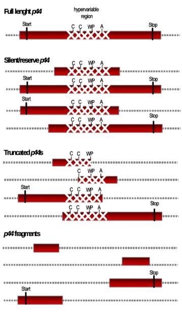
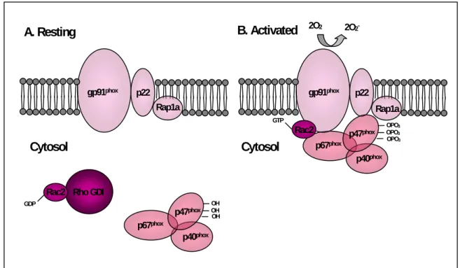
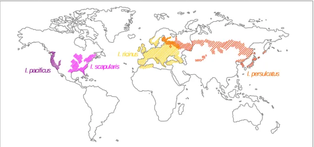
Figure 3. Representation of the intracellular cycle proposed for A. phagocytophilum ……….. 20 Figure 4. Representation of p44 genes ………. 23 Figure 5. Representation of NADPH oxidase ……….. 25 Figure 6. Approximate geographic distributions of four medically important Ixodes
persulcatus/ricinus complex ticks ………..……….... 31
Figure 7. Schematic representation of A. phagocytophilum transmission cycle ……… 34
1.3. Human granulocytic anaplasmosis
Figure 8. Number of HGA cases reported to NETSS, US 1999-2004 ………... 45 Figure 9. Total number of HGA cases reported to NETSS by State, US 1999-2004 ……… 45 Figure 10. Total number of HGA cases reported in each European country since 1997 ... 46 Figure 11. Relative sensitivity of diagnosis tests used for laboratory confirmation of HGA ………. 54 Figure 12. A. phagocytophilum morulae in neuthrophils: A- peripheral blood smear stained by Diff-Quik; B- spleen section stained by immunoalkaline phosphatase/ hematoxylin ………. 55 Figure 13. Immunofluorescence assay (IFA) using HL-60 cells infected with A. phagocytophilum Webster strain as antigen: A- negative sample; B- reactive sample evidenced by green fluorescence of A. phagocytophilum morulae ……….. 59
Chapter II. Exchange technology in Anaplasmataceae field
2.1. Distribution of “Ehrlichia walkeri” in Ixodes ricinus (Acari:Ixodidae) from the Northern part of Italy
Chapter III. Anaplasma phagocytophilum in ticks (Acari: Ixodidae)
3.1. Detection of Anaplasma phagocytophilum DNA in Ixodes ticks (Acari:Ixodidae) from Madeira Island and Setúbal District, mainland Portugal
Figure 1. Collection sites in Madeira Island and Setúbal District, mainland Portugal ……… 90 Figure 2. Dendogram showing the phylogenetic relationships of the msp2 sequences of the newly identified strains and other representative sequences from North American A.
phagocytophilum strains (Webster strain-Wisconsin and USG3 strain-eastern United States),
and from A. marginale Florida strain (msp2 and msp3) ………. 92
3.3. Ticks and tick-borne rickettsiae surveillance in Montesinho Natural Park, Portugal
Figure 1. Montesinho Natural Park ……… 111
3.5. PCR-based survey of Anaplasma phagocytophilum in Portuguese ticks (Acari: Ixodidae)
Figure 1. Tick species and collection areas on the mainland Portugal and in the Madeira Archipelago (2002-2006) ………... 140 Figure 2. Dendrogram showing the phylogenetic relationships between A. phagocytophilum sequences presented in this study and other Anaplasmataceae based on partial nucleotide sequences of the msp2 (A), rrs (B), and groESL (C) ………... 143
Chapter IV. Anaplasma phagocytophilum in non-human vertebrate hosts
4.1. Detection of antibodies against Anaplasma phagocytophilum in wild-rodents, Portugal
Figure 1. Total number of rodent studied by IFA and/or PCR, according to sampling area
(trapping period) ………... 167
4.2. Serological survey and molecular detection of Anaplasma phagocytophilum infection in Portuguese horses
Figure 1. Number of A. phagocytophilum seropositive cases per total horses studied in each district (origin information was not available for nine animals) ………...……..…... 182 Figure 2. Phylogenetic relationships between A. phagocytophilum sequences detected in this study and other Anaplasmataceae based on partial nucleotide sequences of the groESL (A) and
rrs (B) ……….. 185
4.3. Anaplasmataceae as etiologic agents of canine tick-borne disease in Portugal
Figure 1. Phylogenetic relationships between A. platys sequences detected in this study other Anaplasmataceae based on partial nucleotide sequences of the rrs (A) and groESL (B) …... 201
Figure 2. Dendrogram demonstrating the similarity among GroEL amino acid sequences derived from A. platys-infected Portuguese dogs compared to other Anaplasmataceae species ………… 203
Chapter V. Human exposure to Anaplasma phagocytophilum 5.1. Human exposure to Anaplasma phagocytophilum in Portugal
Figure 1. Western blot results for A. phagocytophilum IFA-positive patients ………... 213
5.2. Six years of Anaplasmataceae serodiagnosis in humans. Data from a state laboratory, Portugal
Figure 1. Number of patients that had at least one sera sample submitted to CEVDI/INSA with a request for A. phagocytophilum and/or E. chaffeensis diagnosis during 2000-2006 ………. 235 Figure 2. Light microscopic appearance of C. burnetti human isolate in HL-60 cells. Cytocentrifuge preparations of culture stained with Diff-Quik (A) and IFA (B) …..…...………….…. 236
TABLES INDEX
Chapter 1. State of the art 1.1. Family Anaplasmataceae
Table 1. Current classification of the family Anaplasmataceae ………. 10 Table 2. Biological, ecological and epidemiological features of medically important Anaplasmataceae species .………... 15
1.2. Anaplasma phagocytophilum
Table 3. Prevalence (%) of questing ticks detected with A. phagocytophilum infections by
PCR-based methods ……….. 32
Table 4. A. phagocytophilum infection in wild mammals detected by PCR-based studies ……….. 35 Table 5. Members of Ixodes persulcatus/ricinus complex as potential and confirmed vectors for other tick-borne agents ………..……... 36 Table 6. Prevalence of A. phagocytophilum coinfections in questing Ixodes ticks detected by
PCR-based methods ……… 38
1.3. Human granulocytic anaplasmosis
Table 7. Meta-analysis of A. phagocytophilum seroprevalence (%) among various human populations based in seropositivity obtained from immunofluorescence assay ... 47 Table 8. Clinical complications of HGA reported in North America and in Europe ……… 52 Table 9. Results of confirmatory laboratory testing for 330 patients with HGA from case series or case reports in North America and Europe ……….. 56 Table 10. DNA targets that have been used for the detection of A. phagocytophilum in samples
from infected patients ………... 57
Table 11. Proposed case definition for human granulocytic anaplasmosis ……… 61 Table 12. Recommended adult and pediatric antibiotic treatment for HGA ……… 62
Chapter II. Exchange technology in Anaplasmataceae field
2.1. Distribution of “Ehrlichia walkeri” in Ixodes ricinus (Acari:Ixodidae) from the Northern part of Italy
Chapter III. Anaplasma phagocytophilum in ticks (Acari: Ixodidae)
3.1. Detection of Anaplasma phagocytophilum DNA in Ixodes ticks (Acari:Ixodidae) from Madeira Island and Setúbal District, mainland Portugal
Table 1. Results of PCR to detect A. phagocytophilum DNA in ticks ……….. 91 Table 2. PCR-positive results of ticks ……… 92
3.2. Ticks Parasitizing wild birds in Portugal: Detection of Rickettsia aeschlimannii, R.
helvetica and R. massiliae
Table 1. List of wild birds parasitized by ticks ……….. 100 Table 2. List of wild birds parasitized with ticks infected with rickettsial agents ………. 101
3.3. Ticks and tick-borne rickettsiae surveillance in Montesinho Natural Park, Portugal
Table 1. Number of ticks by species and stage collected in MNP and detection assay results ….. 109 Table 2. Rickettsia isolation results ……… 111
3.4. Detection of Rickettsia helvetica and other spotted fever group rickettsiae in Ixodes
ricinus from Tapada Nacional de Mafra, Portugal.
Table 1. Tick samples detected with Rickettsia DNA ………..………... 123
3.5. PCR-based survey of Anaplasma phagocytophilum in Portuguese ticks (Acari: Ixodidae)
Table 1. A. phagocytophilum PCR results from questing ticks, according to sampling area, species and gender ………... 141 Table 2. A. phagocytophilum PCR results from ticks collected parasitizing vertebrate hosts, according to sampling area, species and gender ……… 142
Chapter IV. Anaplasma phagocytophilum in non-human vertebrate hosts
4.1. Detection of antibodies against Anaplasma phagocytophilum in wild-rodents, Portugal
Table 1. A. phagocytophilum results obtained from mainland mice tested by both IFA and PCR and Madeira Island rodents tested by PCR alone ………... 168
4.2. Serological survey and molecular detection of Anaplasma phagocytophilum infection in Portuguese horses
Table 1. Breed, geographic origin, gender, age, and other infectious disease tests for Portuguese horses with A. phagocytophilum antibodies ……… 183 Table 2. Univariate statistical analysis of A. phagocytophilum seropositive horses according to
4.3. Anaplasmataceae as etiologic agents of canine tick-borne disease in Portugal
Table 1. Dogs presented with active infections detected by IFA and PCR ………... 200
Chapter V. Human exposure to Anaplasma phagocytophilum 5.1. Human exposure to Anaplasma phagocytophilum in Portugal
Table 1. Serologic testing results for A. phagocytophilum and E. chaffeensis …………... 211 Table 2. Epidemiological and clinical data from patients with confirmed exposure to A.
phagocytophilum ………... 212
5.2. Six years of Anaplasmataceae serodiagnosis in humans. Data from a state laboratory, Portugal
Table 1. Patients with positive results for A. phagocytophilum in serologic tests (2000-2006) …… 237 Table 2. Patients with positive results for E. chaffeensis in serologic tests (2000-2006) ……… 239 Table 3. Patients with positive results for both A. phagocytophilum and E. chaffeensis in serologic tests (2000-2006) ………... 240
LIST OF ABBREVIATURES AND ABBREVIATIONS
ankA Gene encoding AnkA
AnkA The 153-160 kDa protein with several tandemly repeated ankyrin motifs ATCC American Type Culture Collection
AT-rich Adenine-timine rich
BF Boutonneuse fever [also referred as Mediterranean spotted fever (MSF)]
bp Base pair
CDC Centers for Disease Control and Prevention
CEVDI Centro de Estudos de Vectores e Doenças Infecciosas
CICT Canine Infectious cyclic thrombocytopenia
CSF Cerebrospinal fluid
DH82 Canine macrophage-like cell line
DNA Desoxiribonucleic acid
EDTA Ethylenediaminetetraacetic acid EGA Equine granulocytic anaplasmosis ELISA Enzyme-linked Immunosorbent assay
FBS Fetal bovine serum
FICT Fluorescein isothocyanate
flgB Flagellin gene
GA Granulocytic anaplasmosis
G+C Guanine + cytosine
gltA Citrate synthase gene
groESL Heat shock operon
GroESL Heat shock proteins, including 10-20 kDa GroES and 58-65 kDa GroEL (also named GroL) that act as chaperonins
HB Human babesiosis
HGA Human granulocytic anaplasmosis
HGE Human granulocytic ehrlichiosis (presently human granulocytic anaplasmosis) HL-60 Human promyelocytic leukaemia cell line
rHL-60 HL-60 cells induced to differentiate into functional granulocytes HME Human monocytic ehrlichiosis
IFA Immunofluorescence assay IgG, IgM Immunoglobulin G, immunoglobulin M
INSA Instituto Nacional de Saúde Dr. Ricardo Jorge
kDa Kilodalton
LB Lyme borreliosis
LPS Lipopolysaccharide MEM Minimum essential medium
MSF Mediterranean spotted fever [also referred as boutonneuse fever (BF)]
msp2 Surface proteins gene family (also p44) MSP2 Surface proteins family (also P44)
NETSS National Electronic Telecommunications Systems for Surveillance
OmpA Outer membrane protein A gene
OmpB Outer membrane protein B gene
p44 Surface proteins gene family (also msp2) P44 Surface proteins family (also MSP2) PBS Phosphate buffered saline
PCR Polymerase chain reaction RMSF Rocky Mountain spotted fever
rrs 16S rRNA gene
Salp Ticks salivary gland protein
s.l. Sensu lato
s.s. Sensu stricto
TBE Tick-borne encephalitis
TBF Tick-borne fever (also referred as pasture fever)
TNM Tapada Nacional de Mafra
US The United States of America (also abbreviated as USA)
vero E6 Epithelial-like cell line obtained from kidney epithelial cells of the african green monkey (Cercopithecus aethiops)
OUTLINE AND OBJECTIVES
Ticks are obligate hematophagous acarines that present widespread distribution and parasitize a broad range of terrestrial vertebrates, including amphibians, reptiles, birds, domestic and wild mammals and humans. Its implication in infectious disease was first evident in veterinary medicine when Boophilus annulatus was linked to Texas cattle fever via transmission of Babesia
bigemina (Smith & Kilbourne, 1893). In the beginning of the 20th century, ticks were additionally
implicated in human disease with the description of tick-borne relapsing fever, caused by Borrelia
duttonii, and its association with Ornithodoros moubata bites (Dutton & Todd, 1905), as well as
recognition of the role that Dermacentor andersoni plays as vectors of Rickettsia rickettsii, the agent of Rocky Mountain spotted fever (Ricketts, 1909). Since then, ticks were linked to several other protozoal, bacterial and viral infections. The last three decades in particular have revealed the emergence of an increasing number of human tick-borne diseases, mostly due to the expanding knowledge of ticks, their pathogenic potential and the development of more reliable diagnostic tools.
Human infections caused by bacteria belonging to the family Anaplasmataceae represent some of the best examples of newly emergent tick-borne diseases. Some of these agents have been long known as veterinary pathogens but their implication in human cases has only recently been recognized, such as for Anaplasma phagocytophilum the etiologic agent of human granulocytic anaplasmosis (HGA). Disease caused by this microorganism was first recognized in 1932 in European sheep and cattle (Gordon et al., 1932), and a similar agent was also later found to be the cause of equine pathology in the United States of America (US) (Gribble, 1969; Stannard
et al., 1969). However, only in the 1990’s were the first cases of human infection described in US
(Bakken et al., 1994; Chen et al., 1994) and in Europe (Petrovec et al, 1997). At present, HGA stands out as the most prevalent human disease caused by an Anaplasmataceae member, due to the number of cases that are reported annually in US; in some areas of the country HGA is the second most frequent vector-borne disease following Lyme borreliosis (LB)1. Moreover, it is suggest that A. phagocytophilum is found broadly across several countries in Europe, Middle East and Asia, raising the possibility of human and animal infections over the same geographic regions.
Although the number of HGA cases outside the US is still limited, the possibility that this zoonosis is under-diagnosed owing to a lack of physician awareness or inavailability of current laboratory diagnostics is controversial and of great concern.
As HGA has now been readily recognized in other European countries, this thesis was undertaken to contribute to the fuller understanding of A. phagocytophilum ecobiology, with an emphasis on its potential importance to human health in Portugal.
In summary, the general aims of this thesis are:
- To investigate the role of Portuguese tick species as vectors of A. phagocytophilum; - To search for potential reservoirs and affected vertebrate hosts in Portugal;
- To ascertain the importance of A. phagocytophilum in human disease;
- To develop methodological and technical approaches applied to HGA laboratory diagnosis; - To isolate and/or characterize A. phagocytophilum strains circulating in the country.
DISSERTATION PLAN
The thesis is organized in six chapters:
The first chapter is the introduction to the theme providing general information about tick-borne bacteria belonging to the family Anaplasmataceae and a broad updated perspective of A.
phagocytophilum and its role in HGA.
Chapter two reflects the collaborative work focused on Anaplasmataceae field, developed during the technology exchange and the acquisition of tools and knowledge that provided the basis for developing the proposed research plan.
Chapter three is devoted to the study of A. phagocytophilum vectors. It describes molecular-based analyses of Anaplasmataceae within several tick species obtained either questing or parasitizing vertebrates in several areas of the country.
Chapter four explores the possibility of non-human vertebrate exposure to A. phagocytophilum supported by serological and molecular-based analyses of biological samples obtained from both wild and domestic animals.
Chapter five addresses the epidemiological basis of HGA in Portugal based on serologic studies of patients with suspected or confirmed tick-borne diseases and in the application of serologic testing in routine diagnosis performed at the Centro de Estudos de Vectores e Doenças Infecciosas,
Instituto Nacional de Saúde Dr. Ricardo Jorge (CEVDI/INSA).
Finally, chapter six summarises the results presented and discussed in previous chapters in an integrated analysis, highlighting their implications in Public Health and delineating future research objectives.
CHAPTER I
STATE OF THE ART
1.1. FAMILY ANAPLASMATACEAE
1.1.1. DEFINITION
Members of the family Anaplasmataceae are non-motile, obligatory intracellular Gram-negative bacteria that reside in a membrane-bound cytoplasmic vacuole of the host cell, either singly or more often in characteristic microcolonies resembling mulberries, termed morulae (Latin
morum = mulberry, Figure 1). They are represented by fifteen or sixteen species, classified in five
distinct genera: Aegyptianella, Anaplasma, Ehrlichia, Neorickettsia and Wolbachia (Table 1). (Garrity et al., 2004; Dumler et al., 2005b).
Most Anaplasmataceae are etiologic agents of worldwide veterinary diseases, some of which are now also regarded as emerging human pathogens. The only exception is the genus
Wolbachia that includes arthropod and nematode endosymbionts, but that may be involved in the
pathogenesis of filiariasis (Hoerauf et al., 2001; Taylor et al., 2005). In general, the biological cycle of Anaplasmataceae involves the infection of invertebrates (Wolbachia), and incidentally also vertebrates (Neorickettsia) or both invertebrate and vertebrate hosts (Aegyptianella, Anaplasma and Ehrlichia). Unique cell tropisms are displayed by these agents and depending on bacterial species, endothelial and/or hematopoietic lineage cells (erythrocytes, monocytes/ macrophages, granulocytes or platelets) are specifically infected in vertebrate hosts. The most important biological, ecological and epidemiological features of Anaplasmataceae with medical importance are showed in table 2.
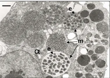
FIGURE 1. Electron photomicrograph of a canine macrophage-like cell (DH82, ATCC CRL-10389) infected with Ehrlichia canis (adapted from Popov et al., 1998).
e
e
m
Ct
The cytoplasm (Ct) contains seven morulae (m) filled with ehrlichiae (e), showing coccoid to ellipsoidal, often pleomorphic shape. Bar 1µm.
TABLE 1. Current classification of the family Anaplasmataceae (adapted from Garrity et al., 2004).
Family Anaplasmataceae Genus Anaplasma Aegyptianella
(incertae sedis) Ehrlichia Neorickettsia Wolbachia
A. bovis A. caudatum A. centrale A. marginale A. platys A. phagocytophilum A. pullorum E. canis E. chaffeensis E. ewingii E. muris E. ruminantium N. helminthoeca N. risticii N. sennetsu W. pipientis
1.1.2. PHYLOGENY AND TAXONOMY
The family Anaplasmataceae is taxonomically classified in the Domain Bacteria, Phylum Proteobacteria, Class "Alphaproteobacteria" and Order Rickettsiales. For many years, Anaplasmataceae were categorized based solely on morphological, ecological, epidemiological and clinical characteristics such as infected host cells, infected mammalian species, geographic location, and antigenic cross-reactivity (Philip, 1957; Moulder, 1974; Weiss & Moulder, 1984). The development of molecular tools for phylogenetic studies and the improvement of methods for the cultivation of obligate intracellular bacteria has helped clarify the exact phylogenetic positions of Anaplasmataceae, which in turn has lead to profound reorganization of this Family and other closely related taxa (Garrity et al., 2004) (Figure 2).
The family Anaplasmataceae was first proposed in the seventh edition of Bergey's Manual of Determinative Bacteriology to include the genus Anaplasma composed of three species, A.
centrale, A. marginale and A. ovis (Philip, 1957). Four additional genera, Aegyptianella (A. pullorum), Eperythrozoon (E. coccoides, E. ovis, E. parvum, E. suis and E. wenyonii), Haemobartonella (H. canis, H. felis and H. muris) and Paranaplasma (P. caudatum and P. discoides) were included in the revision that followed (Moulder, 1974). In the first edition of
Bergey’s Manual of Systematic Bacteriology Paranaplasma was assigned to the genus
Anaplasma, and P. caudatum was renamed as A. caudatum, which represented the last
In the following years the analysis of conserved genes such as 16S rRNA (rrs), heat shock operon (groESL), and citrate synthase (gltA) sequences started to outline phylogenetic relationships of Anaplasmataceae and other closely related bacteria (Weisburg et al., 1989, 1991; Dame et al., 1992; O’Neill et al., 1992; Wen et al., 1995, 1996; Pretzman et al., 1995; Rikihisa et
al., 1997; Zhang et al., 1997; Sumner et al., 2000; Inokuma et al., 2001a, 2001b; Yu et al., 2001).
This approach has been widely used to both identify newly discovered bacteria as well as to redefine existing taxonomy, thus showing that the taxonomic classification of Anaplasmataceae needed to be restructured.
Molecular analysis has shown that Eperythrozoon and Haemobartonella species did not cluster with the other Anaplasmataceae, so their removal from the order Rickettsiales to Mollicutes was proposed (Rikihisa et al., 1997). It has also been shown that some agents classified in the family Rickettsiaceae, especially those of the tribe Ehrlichieae and Wolbachieae, were closely related to Anaplasma (O’Neill et al., 1992; Drancourt & Raoult, 1994; Wen et al., 1995; Walker & Dumler, 1996; Sumner et al., 2000; Inokuma et al., 2001a, 2001b; Yu et al., 2001). Based on an exhaustive analysis of rrs, groESL and surface protein gene sequences deposited in GenBank, Dumler and coworkers (2001) proposed a reorganization of the Order Rickettsiales. Concerning Anaplasmataceae, it was proposed: i) to include the closely related species of the family Rickettsiaceae (Ehrlichia, Neorickettsia, Cowdria, Wolbachia); ii) to expand the genus Anaplasma and incorporate Ehrlichia bovis, Ehrlichia platys and Anaplasma phagocytophilum (including
Ehrlichia phagocytophila, Ehrlichia equi and human granulocytic ehrlichiosis [HGE] agent). The
unification of E. phagocytophila, E. equi and HGE agent as a single species confirmed previous studies that suggested a close molecular (Chen et al., 1994; Pretzman et al., 1995; Goodman et
al., 1996; Sumner et al., 1997; Zhang et al., 1997; Inokuma et al., 2001a, 2001b) and antigenic
(Barlough et al., 1995; Dumler et al., 1995; Madigan et al., 1995) similarity between those agents;
iii) to fuse Cowdria with the genus Ehrlichia and place the sole species, Cowdria ruminantium, with
the existing species Ehrlichia canis, Ehrlichia chaffensis, Ehrlichia ewingii and Ehrlichia muris; iv) to expand the genus Neorickettsia to include Ehrlichia risticii and Ehrlichia sennetsu; v) to exclude
Wolbachia persica and W. melophagi from the genus Wolbachia and to include the genus Wolbachia with its sole member Wolbachia pipientis; vi) to retain provisional Aegyptianella as Genus incertae sedis due to the lack of molecular information.
Almost all the proposed reorganizations in family Anaplasmataceae were introduced in the second edition of Bergey’s Manual of Systematic Bacteriology (Garrity et al., 2001, 2002a, 2002b,
2003, 2004; Dumler et al., 2005b). The heterogeneity of W. pipientis might also justify the revision of species concept with creation of additional taxa to include the distinct host-associated clades that have been described (Lo et al., 2002; Fen et al., 2006). Moreover, the incertae sedis classification of Aegyptianella has been called to question owing to its phenotypic similarities with
Anaplasma spp., and later reinforced by genetic data (Rikihisa et al., 2003). In future revisions new
agents might be included in Anaplasmataceae, such as Candidatus Neoehrlichia mikurensis (Ehrlichia walkerii), E. shimanensis, and Xenohaliotis californiensis (Friedman et al., 2000 ;Brouqui
et al., 2003; Kawahara et al., 2004, 2006; Rikihisa, 2006a).
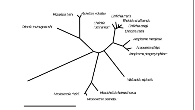
FIGURE 2. Current phylogeny and taxonomic classification of genera in the family Anaplasmataceae. The distance bar represents substitutions per 1,000 bp (adapted from Dumler et
al., 2005a).
1.1.3. HISTORICAL PERSPECTIVE OF HUMAN INFECTION
Our current knowledge of Anaplasmataceae started at the beginning of the 20th century with the descriptions of Theiler (1910) implicating Anaplasma marginale as the etiologic agent of anaplasmosis, a tick-borne disease of ruminants that is still associated to enormous worldwide annual economic losses. In the following years several other agents causing disease in domestic and wild animals were identified, such as Anaplasma centrale (Theiler, 1911); Cowdria (now
Ehrlichia canis Rickettsia rickettsii Rickettsia typhi Ehrlichia chaffeensis Ehrlichia muris Ehrlichia Ehrlichia ewigii Orientia tsutsugamushi ruminantium
Anaplasma marginale Anaplasma platys Anaplasma phagocytophilum Wolbachia pipientis Neorickettsia helminthoeca Neorickettsia risticii Neorickettsia sennetsu
Ehrlichia) ruminantium (Cowdry, 1925), Ehrlichia (now Anaplasma) ovis (Lestoquard, 1924), Ehrlichia phagocytophila (now Anaplasma phagocytophilum) (Gordon et al., 1932; Foggie 1951), Ehrlichia canis (Donatien & Lestoquard, 1935), Ehrlichia (now Anaplasma) bovis (Donatien &
Lestoquard, 1936), Neorickettsia helminthoeca (Philip et al., 1953), Ehrlichia equi (now Anaplasma
phagocytophilum) (Gribble, 1969; Stannard et al., 1969), Ehrlichia (now Anaplasma) platys (Harvey et al., 1978), Ehrlichia (now Neorickettsia) risticii (Holland et al., 1985; Rikihisa & Perry, 1985), Ehrlichia ewingii (Ewing et al., 1971; Anderson et al., 1992), Ehrlichia muris (Wen et al., 1995)
(Table 2).
In spite of abundant reports in the veterinary field, no case of human infection by Anaplasmataceae was described before the 1950s. Sennetsu fever, documented in Japan in 1954 (Misao & Kobayashi, 1955), was formally the first known human disease caused by an Anaplasmataceae member, although it now commands little medical significance. In fact, the mononucleosis-like illness caused by Neorickettsia (formerly Ehrlichia) sennetsu occurs only in limited areas of the Far East, rarely outside Japan and Malaysia and is usually mild, with no deaths having ever been reported. However, in the last decade of the 20th century, the scientific community’s view of Anaplasmataceae infections changed dramatically with the description of two new severe to fatal forms of disease in United States (US). In 1986, human monocytic ehrlichiosis (HME) was reported from Central Arkansas (Maeda et al., 1987). This disease was caused by a new species named Ehrlichia chaffeensis, closely related to the veterinary pathogens E. canis and
E. ewingii (Anderson et al., 1991; Dawson et al., 1991) and later proven to be also infective for
animals, as summarized in Paddock and Childs (2003). In the early 1990’s, cases of human granulocytic anaplasmosis (HGA) (formerly human granulocytic ehrlichiosis [HGE]) were reported from Minnesota and Wisconsin (Bakken et al., 1994; Chen et al., 1994). The exact nature of the causative agent, originally named “HGE agent”, remained uncertain for several years until its unification with the animal pathogens E. phagocytophila and E. equi into a single species designation, Anaplasma phagocytophilum (Dumler et al., 2001; Garrity et al., 2003). Additionally, two other Anaplasmataceae of well known veterinary importance were also described as a cause of human infection. In 1996, E. canis - the etiologic agent of canine monocytic ehrlichiosis was isolated from an apparently healthy man (Perez et al., 1996; Unver et al., 2001b), and later associated with several symptomatic cases (Perez et al., 2006) in Venezuela. More recently in 1999 E. ewingii, the etiologic agent of canine granulocytic ehrlichiosis, was also identified as a human disease agent in US, mostly in immunocompromised patients (Buller et al., 1999).
HME and HGA are now considered the most important human diseases caused by Anaplasmataceae members. In the US, 1,223 cases were reported by 30 state health departments from 1986 to 1997, 742 (60.7%) categorized as HME, 449 (36.7%) as HGA and 32 (2.6%) not ascribed to a specific agent (McQuiston et al., 1999; Paddock & Childs, 2003). In 1999, both diseases had become nationally notifiable and official reports show that HGA is more frequently recognized than HME (CDC, 2001, 2002, 2003, 2004, 2005, 2006). As an example, the average reported annual incidences during 2001-2002 were 14 and 6 cases per 105 inhabitants for HGA and HME, respectively (Demma et al., 2005). Moreover, worldwide investigation has revealed increasing evidence of a potential role of E. chaffeensis and especially A. phagocytophilum in human disease also outside the US. Serological studies have suggested the occurrence of HME in febrile illness patients from Europe (David de Morais et al., 1991; Pierard et al., 1995; Nutti et al., 1998), Africa (Uhaa et al., 1992), and South America (Da Costa et al., 2006), but so far neither E.
chaffeensis isolation nor DNA detection has been achieved in human cases to reinforce these
data. Concerning HGA, the first patient with confirmed disease outside the US was reported from Slovenia in 1997 (Petrovec et al., 1997) and more than fifty cases have been reported in Europe since then. The majority occurred in Northern and Central Europe, especially Slovenia (Lotric-Furlan et al., 2006), but there are also individual case reports from other European countries, later discussed in detail. Polymerase chain reaction (PCR)-based studies of potential tick vectors and reservoir animals have also shown the wide distribution of A. phagocytophilum across Europe and in some parts of Middle East and Asia, which will be object of further revision.
It seems that human A. phagocytophilum-associated infections rather than E. chaffeensis are more commonly recognized over various continents. Thus, HGA possibly represents a widespread and significant public health problem of increasing but undefined magnitude in most countries, and that should be better investigated.
Anaplasma
A. bovis Ixodid ticks/ Bite Ruminants Monocytes Bovine “ehrlichiosis” Africa, Middle East and Asia Donatien & Lestoquard, 1936
A. caudatum Ixodid ticks / Bite Ruminants Erythrocytes Anaplasmosis Worldwide Ristic & Kreier,1984
A. centrale Ixodid ticks / Bite Ruminants Erythrocytes Anaplasmosis Worldwide Theiler, 1911
A. marginale Ixodid ticks / Bite Ruminants Erythrocytes Anaplasmosis Worldwide Theiler, 1910
A. ovis Ixodid ticks / Bite Ruminants Monocytes Asia Lestoquard, 1924
A. platys Ixodid ticks / Bite Canids Platelets Infectious cyclic thrombocytopenia Worldwide Harvey et al., 1978 A. phagocytophilum Ixodid ticks / Bite Ruminants
Equines Humans Canines Felines
Neutrophils Tick-borne fever
Equine granulocytic anaplasmosis Human granulocytic anaplasmosis
Europe
America and Europe United States and Europe United States and Europe United States and Europe
Gordon et al., 1932
Gribble, 1969; Stannard et al., 1969 Bakken et al., 1994; Chen et al., 1994 Madewell & Gribble, 1982
Bjoersdorff et al., 1999c Aegyptianella
A. pullorum Argasid ticks /Bite Birds Erythrocytes Worldwide Carpano, 1928
Ehrlichia
E. canis Ixodid ticks/Bite Canines
Humans Monocytes, macrophages Canine monocytic ehrlichiosis Worldwide; Venezuela Donatien & Lestoquard, 1935 Perez et al., 1996 E. chaffeensis Ixodid ticks / Bite Humans
Canines Monocytes, macrophages Human monocytic ehrlichiosis United States Maeda et al., 1987 Dawson et al., 1996 E. ewingii Ixodid ticks / Bite Canines
Humans
Neutrophils Canine granulocytic ehrlichiosis United States Ewing et al., 1971 Buller et al., 1999
E. muris Ixodid ticks / Bite Rodents Monocytes, macrophages Japan Wen et al., 1995
E. ruminantium Ixodid ticks / Bite Ruminants Endothelial cells, neutrophils Heartwater disease Africa and Caribbean region Cowdry, 1925 Neorickettsia
N. helminthoeca Trematodes/ Ingestion Canines Monocytes, macrophages Salmon poisoning disease United States Philip et al., 1953
N. risticii Trematodes/ Ingestion Equines Monocytes, macrophages Potomac horse fever America and Europe Holland et al., 1985; Rikihisa & Perry, 1985 N. sennetsu Trematodes/ Ingestion Humans Monocytes, macrophages Sennetsu fever Japan and Malaysia Misao & Kobayashi, 1955
1.2. ANAPLASMA PHAGOCYTOPHILUM
1.2.1. GENERALITIES
A. phagocytophilum is a small coccoid to ellipsoidal, often pleomorphic bacterium with a
cell diameter ranging from 0.2 to 2.0 µm. Ultrastructurally, it contains a nucleoid with electron-dense DNA strands and ribosomes surrounded by two limiting membranes, an inner cytoplasmic membrane and a rippled outer cell wall. Like the majority of Anaplasmateaceae, this microorganism forms characteristic microcolonies (morulae) in the cytoplasm of infected cells, as the result of the agent’s binary fission in a confined area defined by membrane-bounded vacuole (Popov et al., 1998).
The natural history of A. phagocytophilum involves both the infection of arthropods, especially Ixodes ticks, and several mammalian species, in which man is included as an incidental dead-end host. Depending on the infected host, different cell types are targeted by this agent in a replication cycle that is still not entirely understood. In vertebrates the microorganism has a marked tropism for polymorphonuclear leucocytes, preferentially infecting neutrophils, a characteristic feature often noted with disease. A. phagocytophilum causes a pathologic process commonly known as granulocytic anaplasmosis (GA). The designation based on cell tropism may become less useful as additional Anaplasmataceae members are recognized as pathogens and since more than one species may be responsible for the broad category of "granulocytic" disease. However, this nomenclature is firmly established in the literature, and to avoid confusion here it will be used to designate disease caused by A. phagocytophilum.
GA affects humans as well as several domestic animals including ruminants, horses, dogs and cats (Gordon et al., 1932; Gribble, 1969; Madewell & Gribble, 1982; Bakken et al., 1994; Chen
et al., 1994; Greig et al., 1996; Bjoerdorf et al., 1999c). The disease is generically characterized as
a non-specific febrile illness accompanied by hematological abnormalities and hepatic injury. Historically, GA has been recognized by different designations, each of them associated with fingdings thought to be caused by a distinct etiologic agent. According to the affected host and implicated pathogen, the disease was named ruminant tick-borne fever (or pasture fever), equine granulocytic ehrlichiosis and human granulocytic ehrlichiosis as the result of infections by E.
single species proposed by Dumler and coworkers (2001) expanded the dimensions and broadened the perspective of this tick-borne disease.
Not cultivable in cell-free media or chicken embryos, the in vitro manipulation of A.
phagocytophilum was initially based on direct purification from infected blood or short-term cultures
of blood cells collected from infected animals. The development of cell culture systems able to mimic in vivo situations made the continuous propagation of the pathogen possible and allowed a more detailed analysis of its biological features. The first in vitro isolation of A. phagocytophilum was achieved by Goodman and coworkers (1996) after inoculation of a patient’s blood in a human promyelocytic leukemia cell line (HL-60, ATCC CCL-240) developed by Gallagher and coworkers (1979). In addition to undifferentiated cell lines, other cells are also currently used for A.
phagocytophilum cultivation, including HL-60 cells induced to differentiate into functional
granulocytes (rHL-60), as well as embryonic Ixodes scapularis tick cell lines IDE8 and ISE6 (Goodman et al., 1996; Munderloh et al., 1996, 1999; Heimer et al., 1997). Furthermore, the development of murine models that in part reproduced the infection process has greatly expanded the understanding of A. phagocytophilum pathogenesis. The use of inbred and targeted gene-deleted (knockout) mice has allowed insight into precise mechanisms of disease and host cell-pathogen interactions in the context of a whole organism, complemented studies based on in vitro tissue culture, ex vivo human and ruminant neutrophil cultures and in vivo large animal experiments (Borjesson & Barthold, 2002).
The complete genome sequence of A. phagocytophilum HZ strain has been recently achieved by Hotopp and coworkers (2006), illustrating the existence of a single circular chromosome of 1,471,282 bp with an average G+C content of 41%. Comparison analysis has shown that most genomic features of A. phagocytophilum are typical of other sequenced Anaplasmataceae, but also reveals new species-specific genes indicating particular niche adaptations (Hotopp et al., 2006).
1.2.2. LIFE-CYCLE
The study of naturally- and experimentally-infected neutrophils as well as human and tick-derived cell cultures has allowed a detailed analysis of the intracellular replication cycle of A.
Popov et al., 1998; Webster et al., 1998). This bacterium undergoes a simple cycle involving three fundamental steps: invasion of host cells likely through receptor-mediated endocytosis; proliferation by binary fission in a membrane vacuole that does not fuse with lysosomes (parasitophorous vacuoles); exit via rupture of both A. phagocytophilum-containing vacuole and the adjacent host cell membrane when the morula is located at the periphery of the infected cell.
In general, morulae tend to be much larger and contain more microorganisms in tick cells than in neutrophils or HL-60 cells. Pathogen morphology is also more variable presenting a higher degree of pleomorphism (Munderloh et al., 1996, 1999). Genome analysis has shown that A.
phagocytophilum lacks genes for both lipopolysaccharide and peptidoglycan biosynthesis (Lin &
Rikihisa, 2003; Hotopp et al., 2006). The peptiglycan layer is a polymer consisting of sugars and amino acids that form a homogeneous layer between the plasma membrane and the cell-wall of eubacteria, conferring structural strength to bacteria. The lack of this layer justifies the morphologic pleomorphism of A. phagocytophilum that should be expected to be higher in environments with less osmotic pressure than those experienced inside the body of invertebrate vector (Rikihisa, 2006b).
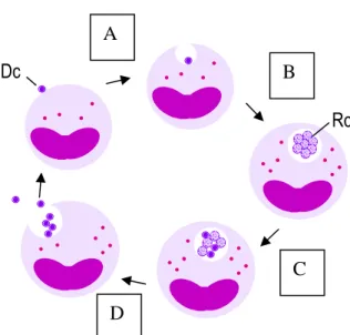
In both mammalian and tick cells, A. phagocytophilum presents two distinct ultrastructural forms: reticulate cells (RC) characterized by a loosely packed and randomly dispersed nucleoid and dense-cored cells (DC) with a condensed electron-dense protoplast. By temporal examination of A. phagocytophilum development in HL-60 cells, Webster and coworkers (1998) concluded that the early forms of the agent have the round reticular appearance while later structures are small and dense. In tick-cells the agent also presented the same temporal pattern; A. phagocytophilum colonies comprise appreciable numbers of condensing organisms in older tick-cell cultures (Munderloh et al., 1996, 1999). This data suggests the existence of an intracellular biphasic development that undergoes from RC towards DC cells, similar to that observed for other closely related agents such as A. marginale (Blouin & Kocan, 1998) and E. chaffeensis (Zhang et al., 2007), in which DC are the infective form and RC the metabolic active form that multiplies by binary fission (Figure 3). However, both the dense core and reticulate forms undergo binary fission in
vitro, suggesting plasticity to any potential developmental or differentiation cycle (Popov et al.,
1998).
Depending on the infected host, A. phagocytophilum replication takes place in different cells types. In naturally infected ticks the agent has been detected in midgut cells, hemocytes and salivary gland cells (Magnarelli et al., 1995b; Telford et al., 1996; Alberti et al., 1998; 2000;
FIGURE 3. Representation of the intracellular cycle proposed for A. phagocytophilum. Rc A Dc D B C
A- Dense core (Dc) microorganism starts the cycle by attaching and entering into a susceptible host cell; B- Once inside the cell it remains within the vacuole that does not fuse with lysossomes. The microorganism differentiates into a metabolic active reticulated form (Rc) and multiplies by binary fission, forming characteristic dense packed microcolonies known as morulae; C- After multiple rounds of division Rc starts to differentiate into DC forms; D- Finally, by exocytosis or host cell lysis the infectious DC are released to initiate new cycles in new host cells (Blouin & Kocan, 1998; Zhang et al., 2007).
Kim et al., 2003; Ohashi et al., 2005). These different locations are presumed to represent part of a life-cycle similar to that observed for A. marginale (Ge et al., 1996; Kocan et al., 2004). It is assumed that following ingestion in the blood meal by feeding ticks, A. phagocytophilum primarily infects midgut epithelial cells, passes through this barrier and, via circulating hemocytes, subsequently colonizes salivary gland cells from where it is transmitted to vertebrates via salivary emissions released during the next feeding. The infection of salivary glands seems to be an early event that may take place during or soon after the infective blood meal, since questing ticks already present A. phagocytophilum in acinus cells (Telford et al., 1996; Ohashi et al., 2005). Up to now, salivary glands are regarded as the most important organ for the agent’s maintenance in vector ticks and subsequent transmission to vertebrate hosts. Other organs might also be infected by A. phagocytophilum, which may contribute to the agent’s ability to persist in the vector tick, but it does not seem to include ovarian tissue. In fact, infected ticks show persistent bacteremia, with the agent surviving arthropod molts and being maintained in successive life stages (transstadial transmission) but with no, or inefficient offspring infection (transovarial transmission) (Richter et al., 1996; Telford et al., 1996; Des Vignes & Fish 1997; Hodzic et al., 1998; Goethert & Telford, 2003). Of interest, Sukumaran and coworkers (2006) described an Ixodes tick salivary gland protein, Salp16, that in part determines successful colonization of salivary glands by A. phagocytophilum after acquisition of a blood meal from an infected mammalian host.
Once inside a mammalian host the agent targets cells derived from bone marrow precursors, most often neutrophils and band neutrophils. The infection is not localized, affecting circulating peripheral blood cells, but A. phagocytophilum has occasionally been detected in
spleen, lung, liver, bone marrow, lymph node and heart (Madigan et al., 1995; Walker & Dumler, 1997; Bunnel et al., 1999; Lepidi et al., 2000; Martin et al., 2000, 2001; Trofe et al., 2001; Remy et
al., 2003; Bayard-Mc Neeley et al., 2004). Both ex vivo and in vitro data have emphasized that
pathogen tropism is especially directed to the mature granulocyte population (Heimer et al., 1997; Klein et al., 1998; Zeman et al., 2002a; Bayard-Mc Neeley et al., 2004) and its detection in several organs seems to reflect vascular perfusion or focal sequestration of infected neutrophils (Lepidi et
al., 2000). However, Walker and Dumler (1997) also discussed the possibility that the ligand
recognized by A. phagocytophilum adhesion is broadly distributed over a wide variety of cells from different lineages, justifying the occasional detection of this agent in endothelial cells, fibroblasts and macrophages. Recent experimental studies have reinforced the hypothesis that other cell types might be involved in A. phagocytophilum cycle by the demonstration that the bacterium is capable of infecting endothelial cells and passing from the infected endothelium into neutrophils (Munderloh et al., 2004; Herron et al., 2005). As conjectured in those studies an endothelial reservoir cell may mediate the infection of blood cells and A. phagocytophilum spreads from the site of tick feeding, and offers opportunities for ongoing, direct cell-to-cell infection of neutrophils, avoidance of host immune effectors, and completion of the pathogen life-cycle by infection of circulating leukocytes available for transfer to blood-feeding ticks (Munderloh et al., 2004; Herron et
al., 2005).
1.2.3. PATHOGENESIS
A. phagocytophilum undergoes substantial selection pressure to be able to survive and
replicate in different environments during its life-cycle. In vector ticks, pathogens must survive extreme fluctuations in temperature, pH, osmotic pressure, and other factors related to the physiological status of these arthropods. In vertebrate hosts, they must overcome the highly developed inflammatory and immunologic defences of mammals. Moreover, A. phagocytophilum possesses an unusual intracellular lifestyle in that it colonizes neutrophils, the key cells in innate immunity and the first line of defence against invading pathogens. These short-lived, terminally differentiated cells ingest invading microorganisms and destroy them by various means, which include fusing the bacteria-containing phagosome with acidic lysosomes as well as directing toxic oxidative and proteolytic compounds into the phagosomal lumen (Mayer-Scholl et al., 2004). The