www.cbpv.org.br/rbpv ISSN 0103-846X (Print) / ISSN 1984-2961 (Electronic)
Doi: http://dx.doi.org/10.1590/S1984-29612017042
Detection of
Anaplasma
sp. phylogenetically related to
A.
phagocytophilum
in a free-living bird in Brazil
Detecção de
Anaplasma
sp. filogeneticamente associado a
A. phagocytophilum
em uma
ave de vida livre no Brasil
Anna Claudia Baumel Mongruel1; Jyan Lucas Benevenute2; Priscila Ikeda2; Marcos Rogério André2;
Rosangela Zacarias Machado2; Adriano de Oliveira Torres Carrasco1; Meire Christina Seki1*
1 Laboratório de Doenças Infecciosas e Parasitárias, Departamento de Medicina Veterinária, Universidade Estadual do Centro-Oeste –
UNICENTRO, Guarapuava, PR, Brasil
2 Laboratório de Imunoparasitologia Veterinária, Departamento de Patologia Veterinária, Faculdade de Ciências Agrárias e Veterinárias
– FCAV, Universidade Estadual Paulista “Júlio de Mesquita Filho” – UNESP, Jaboticabal, SP, Brasil
Received May 28, 2017 Accepted June 19, 2017
Abstract
Wild animals play an important role in carrying vectors that may potentially transmit pathogens. Several reports highlighted the participation of wild animals on the Anaplasma phagocytophilum cycle, including as hosts of the agent. The aim of this study was to report the molecular detection of an agent phylogenetically related to A. phagocytophilum isolated from a wild bird in the Midwest of the state of Paraná, Brazil. Fifteen blood samples were collected from eleven different bird species in the Guarapuava region. One sample collected from a Penelope obscura bird was positive in nested PCR targeting the 16S rRNA gene of Anaplasma spp. The phylogenetic tree based on the Maximum Likelihood analysis showed that the sequence obtained was placed in the same clade with A. phagocytophilum isolated from domestic cats in Brazil. The present study reports the first molecular detection of a phylogenetically related A. phagocytophilum bacterium in a bird from Paraná State.
Keywords: Anaplasmosis, wild animals, birds, hemoparasites, PCR.
Resumo
Animais selvagens possuem participação importante como carreadores dos vetores responsáveis por transmitir doenças e vários relatos destacam a participação de animais silvestres no ciclo do Anaplasma phagocytophilum, inclusive como hospedeiros do agente. O presente trabalho tem por objetivo relatar pela primeira vez a detecção molecular da infecção por um agente filogeneticamente associado a A. phagocytophilum em uma ave silvestre no interior do Paraná, Brasil. Foram colhidas 15 amostras de sangue originadas de onze espécies diferentes de aves, todas provenientes da região de Guarapuava. Apenas uma amostra pertencente a uma ave da espécie Penelope obscura foi positiva para o ensaio de nested PCR baseado no gene 16S rRNA. A árvore filogenética baseada na análise por máxima verossimilhança demonstrou que a sequência obtida no presente estudo se posicionou no mesmo clado com cepas de A. phagocytophilum isoladas de gatos domésticos no Brasil. O presente trabalho relata pela primeira vez a detecção molecular de Anaplasma sp. filogeneticamente relacionado à A. phagocytophilum, em um animal da espécie P. obscura, assim como a presença do parasita em uma ave silvestre do Estado do Paraná, Brasil.
Palavras-chave: Anaplasmose, animais selvagens, aves, hemoparasitas, PCR.
Several studies about the incidence of hemoparasites among birds have been conducted in Brazil, especially focusing in parasites such as Plasmodium spp., Haemoproteus spp., and Trypanosoma spp. (BENNETT & LOPES, 1980; ADRIANO & CORDEIRO, 2001; LOBATO et al., 2011; SEBAIO et al., 2010, 2012 ; TOSTES et al., 2015).
The genus Anaplasma comprises Gram-negative and obligatory intracellular bacteria that infect cytoplasmic vacuoles of eukaryote cells (DUMLER et al., 2001). The main clinical symptom of anaplasmosis caused by Anaplasma phagocytophilum is unspecific fever in humans (DUMLER & WALKER, 2001). However, the majority of infected patients may present not only fever, but also headache, myalgia, thrombocytopenia, and elevation in the levels of hepatic enzymes (DUMLER et al., 2005). Nevertheless, anaplasmosis is still considered a disease of difficult diagnosis when no laboratory tests are carried out, as clinical symptoms are unspecific (MASSUNG et al., 2003). The course of infection in humans can be mild or severe, depending on the health condition of the patient, and it has been reported that about 40% of the cases may require hospitalization (RYMASZEWSKA & GRENDA, 2008). Human granulocytic anaplasmosis caused by A. phagocytophilum was reported for the first time in 1994 (BAKKEN et al., 1994; CHEN et al., 1994), and this condition has motivated studies on the characteristics of the causative agent.
A. phagocytophilum can naturally infect sheep, cattle, horses, dogs, cats, and humans (MASSUNG et al., 2002) by invading granulocytes, especially neutrophils (EBANI et al., 2008). Although A. phagocytophilum may have worldwide distribution, reports on its occurrence are concentrated in the United States, especially in the Midwestern, northeastern, and western regions of the country (CFSPH, 2013). Wild animals play an important role in carrying vectors that may potentially transmit pathogens (EBANI et al., 2008). Several reports highlighted the participation of wild mammals, such as deer, foxes, rodents, and carnivores in A. phagocytophilum cycles, including their role as hosts of these bacteria (LIZ et al., 2000; FUENTE et al., 2008; VERONESI et al., 2011; ANDRÉ et al., 2012, JAHFARI et al., 2014). Wild and domestic animals may share pathogens, as described by Zhan et al. (2010), who found goats, sheep, and wild rodents in the same area infected by the same strain of A. phagocytophilum. Such occurrences can bring the agent in closer contact with humans.
Some studies indicated that wild birds may be responsible for the distribution of vectors infect by A. phagocytophilum (DANIELS et al., 2002; OGDEN et al., 2008; HILDEBRANDT et al., 2010; PALOMAR et al., 2012). However, only a few of them considered that birds might have direct participation in the epidemiology of the disease (SKORACKI et al., 2006; IOANNOU et al., 2009; JAHFARI et al., 2014). In 2012, Machado et al. (2012) showed, for the first time, that Brazilian wild birds can act as hosts for Anaplasma sp. The authors reported detection of Anaplasma sp. phylogenetically associated with A. phagocytophilum in blood samples from Southern carcaras (Caracara plancus) and American black vultures (Coragyps atratus) using molecular methods.
The transmission of the bacteria from birds to ticks was observed in previous studies (KEESING et al., 2012), and although bird infection is not believed to lead to the development of significant clinical signs, specific antibodies, or bacteremia (JOHNSTON et al.,
2013), a correlation between the participation of birds in the dispersion of the pathogen, and increases in the number of cases of human and animal granulocytic anaplasmosis over the past decade have been described in the literature (IOANNOU et al., 2009).
Analysis conducted in Europe showed that A. phagocytophilum genotypes could be clustered in at least four major groups called ecotypes. Each ecotype has unique geographical and host characteristics. One of these ecotypes, interestingly, was associated only with birds (JAHFARI et al., 2014).
Currently, there are no reports that elucidate the epidemiological situation of Anaplasma spp. in wild animals in the state of Paraná, southern Brazil. The aim of the present study was to report the first molecular isolation of Anaplasma sp. from a blood sample of a Penelope obscura (native bird species) found in the Guarapuava region, in Midwestern state of Parana.
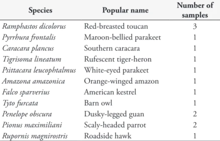
Between August and November 2014, 15 blood samples were collected from eleven different bird species. The animals were sent to the Wildlife Care Center of the State University of Midwest (UNICENTRO), situated in Guarapuava, state of Paraná, Brazil. Blood samples were collected during physical examination for clinical care. The number of samples collected from each bird species is shown in Table 1.
None of the birds presented tick infestations at physical inspection. Blood samples were collected by venipuncture, were placed in tubes containing 5% EDTA, and were then stored at -20 °C until they were used. Molecular assays were performed at the Immunoparasitology Laboratory of Universidade Estadual Paulista “Júlio de Mesquita Filho” (UNESP, Jaboticabal). DNA extraction was performed using the QIAamp DNA Blood and Tissue Mini Kit (QIAGEN), according to the manufacturer’s
recommendations. A nested cPCR assay based on the 16S rRNA gene (MASSUNG et al., 1998) was performed to determine the presence of Anaplasma sp. DNA in the samples.
The amplified products were purified and sequenced in an automatic sequencer (ABI Prism 310 genetic analyzer - Applied
Biosystems/Perkin-Elmer) for confirmation. Sequences obtained were used for subsequent phylogenetic analysis. Trimming was performed using the Phred software (EWING & GREEN, 1998), which evaluates electropherograms produced during
Table 1. Number of birds sampled, according to the species and popular name.
Species Popular name Number of
samples
Ramphastos dicolorus Red-breasted toucan 3
Pyrrhura frontalis Maroon-bellied parakeet 1
Caracara plancus Southern caracara 1
Tigrisoma lineatum Rufescent tiger-heron 1
Psittacara leucophtalmus White-eyed parakeet 1
Amazona amazonica Orange-winged amazon 1
Falco sparverius American kestrel 1
Tyto furcata Barn owl 1
Penelope obscura Dusky-legged guan 2
Pionus maximiliani Scaly-headed parrot 2
the sequencing. The quality of the peaks corresponding to each sequenced base was observed, and a probability of error was attributed to each of the samples. Bases with quality scores above 20 were considered to be used in the present study. The CAP3 software (http://pbil.univ-lyon1.fr/cap3.php) was used for alignment of the consensus sequence. BLAST software was used to analyze nucleotide sequences (BLASTn) in order to compare the sequences obtained with similar ones found in international databases (GenBank) (BENSON et al., 2002). Sequences saved in “FASTA” mode were aligned with other homologous sequences from the same sequenced gene retrieved from the GenBank databank using Multiple Alignment with Fast Fourier Transform (MAFFT) software v.7 (KATOH & STANDLEY, 2013). Maximum likelihood analysis was performed using the RaxML cluster black box (STAMATAKIS et al., 2008), via the CIPRES portal (MILLER et al., 2010) with 1,000 bootstrapping replicates. The proportion of invariable sites was estimated using the GTRGAMMA+I evolution model. Phylogenetic tree editing and rooting (via an external group) was performed using the Treegraph 2.0.56-381 beta software (STÖVER & MÜLLER, 2010).
Amplification of a fragment of the 16S rRNA gene of Anaplasma spp. was obtained by nested cPCR in a single sample (6.66%) that was collected from a P. obscura. Phylogenetic inference based on Maximum Likelihood showed that the genotype detected in the P. obscura specimen from the Gurarapuava region, Brazil, was close to that of Anaplasma spp. that was detected in domestic cats in Brazil (KF964049) (Figure 1).
Ehrlichia spp. detected in wild birds by Machado et al. in 2012 was phylogenetically related to Ehrlichia sp. detected in wild Felidae by André et al. (2010), suggesting that wild birds may participate in the transmission cycle of hemoparasites between species. Machado et al. (2012) detected Anaplasma sp. phylogenetically associated with A. phagocytophilum, also by amplification of the 16S rRNA gene, in 14.28% of 21 bird samples, a higher percentage than the one found in the present study (6.66%). The possible reasons for this variation may be the use of fewer samples, the difference among species sampled,
or lower incidence of the pathogen in the area where the present study was conducted.
Ixodes sp. ticks, which are more commonly found in the northern hemisphere, are considered the main vectors of A. phagocytophilum (RYMASZEWSKA & GRENDA, 2008; WOLDEHIWET, 2010). However, in Brazil, this pattern has not been established. Although no ticks at any phase of life were found in the P. obscura specimen described in the present study, this bird species that belongs to the order galliformes remains on the ground most of the time, and this habit may facilitate tick infestation. A relationship between the occurrence of ticks in certain bird species and ground-feeding habits has already been reported (HASLE, 2013; DIAKOU et al., 2016).
Amblyomma longirostre ticks parasitizing P. obscura in the state of Paraná were described by Arzua et al. (2005), and this tick species was also reported parasitizing others wild bird species in southern and southeastern Brazil (SOARES et al., 2009; SANCHES et al., 2013). Moreover, Rickettsia was detected in A. longirostre found in wild birds from Paraná (PACHECO et al., 2012), suggesting a potential participation of this tick species in the transmission of hemoparasites among wild birds in this state.
According to Doan et al. (2013), evaluation of 16S rRNA gene is a sensitive molecular tool for the detection of circulating Anaplasma spp. genotypes. However, Paulauskas et al. (2012) stated that the use of msp4 gene shows greater reliability in terms of phylogenetic positioning, as gene 16S rRNA is highly conserved, with few polymorphic positions. These findings reinforce the results obtained in the present study, once we reported the infection by an Anaplasma sp. genotype in a free-living bird specimen using the 16S rRNA gene. The sample was negative to additional target genes.
We believe that new genotypes of Ehrlichia and Anaplasma circulate in wild and domestic animals in Brazil. The pre-established PCR protocols targeting genes other than 16S rRNA have shown low accuracy in the identification of new Anaplasmataceae genotypes in Brazil (MURPHY et al., 1998; MASSUNG et al., 1998; KOCAN et al., 2000).
This pattern of positive results to Anaplasmataceae 16S rRNA gene in PCR assays and negative results to other target genes was already described in previous studies that aimed at detecting Anaplasmataceae in domestic and wild animals in Brazil (ANDRÉ et al., 2012, 2014, 2015; SACCHI et al., 2012). Blood samples showing low bacteremia, and an inadequate PCR protocol for a variant of Anaplasma sp. species may explain why variable results were obtained with different target genes (ANDRÉ et al., 2012).
The samples analyzed in this study were collected during the clinical inspection of birds sent to the Wildlife Care Center of UNICENTRO for medical care. Because of this, few birds were sampled, and only one sample from each animal could be collected. The detection threshold for genetic material of the pathogen in nested PCR, associated with possible low levels of bacteremia may also explain why we obtained negative results. These limitations reinforce the need for testing more samples, besides using more sensible techniques to elucidate the hypothesis raised in our study.
Further information regarding the vector is necessary. Moreover, other genes should be used for more concise results about the possible Anaplasma variant that may be found in wild animals in the state of Paraná. As information on infection by Anaplasma spp. in animals from Paraná State is lacking, data of the present study may contribute to the knowledge about the geographic distribution of Anaplasmataceae agents.
References
Adriano EA, Cordeiro NS. Prevalence and intensity of Haemoproteus
columbae in three species of wild doves from Brazil. Mem Inst Oswaldo
Cruz 2001; 96(2): 175-178. PMid:11285493. http://dx.doi.org/10.1590/ S0074-02762001000200007.
André MR, Adania CH, Teixeira RHF, Vargas GH, Falcade M, Sousa L, et al. Molecular detection of Hepatozoon spp. in Brazilian and exotic wild carnivores. Vet Parasitol 2010; 173(1-2): 134-138. PMid:20630658. http://dx.doi.org/10.1016/j.vetpar.2010.06.014.
André MR, Denardi NCB, Sousa KCM, Goncalves LR, Henrique PC, Ontivero CRGR, et al. Arthropod-borne pathogens circulating in free-roaming domestic cats in a zoo environment in Brazil. Ticks Tick Borne Dis 2014; 5(5): 545-551. PMid:24889035. http://dx.doi.org/10.1016/j. ttbdis.2014.03.011.
André MR, Dumler JS, Scorpio DG, Teixeira RH, Allegretti SM, Machado RZ. Molecular detection of tick-borne bacterial agents in Brazilian and exotic captive carnivores. Ticks Tick Borne Dis 2012; 3(4): 247-253. PMid:22749737. http://dx.doi.org/10.1016/j.ttbdis.2012.04.002.
André MR, Herrera HM, Fernandes SJ, Sousa KC, Gonçalves LR, Domingos IH, et al. Tick-borne agents in domesticated and stray cats from the city of Campo Grande, state of Mato Grosso do Sul, midwestern Brazil. Ticks Tick Borne Dis 2015; 6(6): 779-786. PMid:26187416. http:// dx.doi.org/10.1016/j.ttbdis.2015.07.004.
Arzua M, Onofrio VC, Barros-Battesti DM. Catalogue of the tick collection (Acari, Ixodida) of the Museu de História Natural Capão da Imbuia, Curitiba, Paraná, Brazil. Rev Bras Zool 2005; 22(3): 623-632. http://dx.doi.org/10.1590/S0101-81752005000300015.
Bakken JS, Dumler JS, Chen S, Eckman MR, Van Etta LL, Walker DH. Human Granulocytic Ehrlichiosis in the Upper Midwest United States:
a new species emerging? JAMA 1994; 272(3): 212-218. PMid:8022040. http://dx.doi.org/10.1001/jama.1994.03520030054028.
Bennett GF, Lopes OS. Blood parasites of some birds from São Paulo State, Brazil. Mem Inst Oswaldo Cruz 1980; 75(1-2): 117-134. http:// dx.doi.org/10.1590/S0074-02761980000100012.
Benson DA, Karsch-Mizrachi I, Lipman DJ, Ostell J, Rapp BA, Wheeler DL. GenBank. Nucleic Acids Res 2002; 30(1): 17-20. PMid:11752243. http://dx.doi.org/10.1093/nar/30.1.17.
Chen SM, Dumler JS, Bakken JS, Walker DH. Identification of a granulocytotropic Ehrlichia species as the etiologic agent of human disease.
J Clin Microbiol 1994; 32(3): 589-595. PMid:8195363.
Daniels TJ, Battaly GR, Liveris D, Falco RC, Schwartz I. Avian reservoirs of the agent of human granulocytic ehrlichiosis? Emerg Infect Dis 2002; 8(12): 1524-1525. PMid:12498679. http://dx.doi.org/10.3201/ eid0812.010527.
Diakou A, Norte AC, Carvalho IL, Núncio S, Nováková M, Kautman M, et al. Ticks and tick-borne pathogens in wild birds in Greece. Parasitol Res 2016; 115(5): 2011-2016. PMid:26847630. http://dx.doi.org/10.1007/ s00436-016-4943-3.
Doan HTT, Noh JH, Choe SE, Yoo MS, Kim YH, Reddy KE, et al. Molecular detection and phylogenetic analysis of Anaplasma bovis from
Haemaphysalis longicornis feeding on grazing cattle in Korea. Vet Parasitol
2013; 196(3-4): 478-481. PMid:23602362. http://dx.doi.org/10.1016/j. vetpar.2013.03.025.
Dumler JS, Barbet AF, Bekker CP, Dasch GA, Palmer GH, Ray SC, et al. Reorganization of genera in the families Rickettsiaceae and Anaplasmataceae
in the order Rickettsiales: unification of some species of Ehrlichia with
Anaplasma, Cowdria with Ehrlichia and Ehrlichia with Neorickettsia,
descriptions of six new species combinations and designation of Ehrlichia equi and ‘HGE agent’ as subjective synonyms of Ehrlichia phagocytophila.
Int J Syst Evol Microbiol 2001; 51(6): 2145-2165. PMid:11760958.
http://dx.doi.org/10.1099/00207713-51-6-2145.
Dumler JS, Choi KS, Garcia-Garcia JC, Barat NS, Scorpio DG, Garyu JW, et al. Human Granulocytic Anaplasmosis and Anaplasma phagocytophilum.
Emerg Infect Dis 2005; 11(12): 1828-1834. PMid:16485466. http://
dx.doi.org/10.3201/eid1112.050898.
Dumler JS, Walker DH. Tick-borne ehrlichioses. Lancet Infect Dis
2001; 1(1): 21-28. PMid:11871406. http://dx.doi.org/10.1016/S1473-3099(09)70296-8.
Ebani V, Cerri D, Fratini F, Ampola M, Andreani E. Seroprevalence of
Anaplasma phagocytophilum in domestic and wild animals from central
Italy. New Microbiol 2008; 31(3): 371-375. PMid:18843892.
Ewing B, Green P. Base-calling of automated sequencer traces using phred. II. Error probabilities. Genome Res 1998; 8(3): 186-194. PMid:9521922. http://dx.doi.org/10.1101/gr.8.3.186.
Fuente J, Ruiz-Fons F, Naranjo V, Torina A, Rodríguez O, Gortázar C. Evidence of Anaplasma infections in European roe deer (Capreolus capreolus) from southern Spain. Res Vet Sci 2008; 84(3): 382-386. PMid:17655893. http://dx.doi.org/10.1016/j.rvsc.2007.05.018.
Hasle G. Transport of ixodid ticks and tick-borne pathogens by migratory birds. Front Cell Infect Microbiol 2013; 3: 48. PMid:24058903. http:// dx.doi.org/10.3389/fcimb.2013.00048.
Hildebrandt A, Franke J, Meier F, Sachse S, Dorn W, Straube E. The potential role of migratory birds in transmission cycle of Babesia spp.,
2010; 1(2): 105-107. PMid:21771516. http://dx.doi.org/10.1016/j. ttbdis.2009.12.003.
Ioannou I, Chochlakis D, Kasinis N, Anayiotos P, Lyssandrou A, Papadopoulos B, et al. Carriage of Rickettsia spp., Coxiella burnetti and
Anaplasma spp. by endemic and migratory wild birds and their ecotparasites
in Cyprus. Europ Soc Clin Microbiol Infect Dis 2009;15(S2): 158-160. PMid:19281460. http://dx.doi.org/10.1111/j.1469-0691.2008.02207.x.
Jahfari S, Coipan EC, Fonville M, van Leeuwen AD, Hengeveld P, Heylen D, et al. Circulation of four Anaplasma phagocytophilum ecotypes in Europe. Parasit Vectors 2014; 7(1): 365. PMid:25127547. http://dx.doi. org/10.1186/1756-3305-7-365.
Johnston E, Tsao JI, Muñoz JD, Owen J. Anaplasma phagocytophilum
infection in American robins and gray catbirds: an assessment of reservoir competence and disease in captive wildlife. J Med Entomol 2013; 50(1): 163-170. PMid:23427666. http://dx.doi.org/10.1603/ME12141.
Katoh K, Standley DM. MAFFT multiple sequence alignment software 4 version 7: improvements in performance and usability. Mol Biol Evol
2013; 30(4): 772-780. PMid:23329690. http://dx.doi.org/10.1093/ molbev/mst010.
Keesing F, Hersh MH, Tibbetts M, McHenry DJ, Duerr S, Brunner J, et al. Reservoir competence of vertebrate hosts for Anaplasma phagocytophilum.
Emerg Infect Dis 2012; 18(12): 2013-2016. PMid:23171835. http://
dx.doi.org/10.3201/eid1812.120919.
Kocan AA, Levesque GC, Whitworth LC, Murphy GL, Ewing SA, Barker RW. Naturally occurring Ehrlichia chaffeensis infection in coyotes from Oklahoma. Emerg Infect Dis 2000; 6(5): 477-480. PMid:10998377. http://dx.doi.org/10.3201/eid0605.000505.
Liz JS, Anderes L, Sumner JW, Massung RF, Gern L, Rutti B, et al. PCR detection of Granulocytic Ehrlichiae in Ixodes ricinus Ticks and wild small mammals in Western Switzerland. J Clin Microbiol 2000; 38(3): 1002-1007. PMid:10698987.
Lobato DNC, Braga EM, Belo NO, Antonini Y. Hematological and parasitological health conditions of the Pale-breasted Thrush (Turdus leucomelas) (Passeriformes: Turdidae) in southeastern Brazil. Zoologia 2011; 28(6): 771-776. http://dx.doi.org/10.1590/S1984-46702011000600010.
Machado RZ, André MR, Werther K, Souza E, Gavioli FA, Alves RF Jr. Migratory and Carnivorous Birds in Brazil: reservoirs for Anaplasma
and Ehrlichia Species? Vector Borne Zoonotic Dis 2012; 12(8): 705-708.
PMid:22607070. http://dx.doi.org/10.1089/vbz.2011.0803.
Machado RZ, Teixeira MMG, Rodrigues AC, André MR, Gonçalves LR, Silva JB, et al. Molecular diagnosis and genetic diversity of tick-borne Anaplasmataceae agents infecting the African buffalo Syncerus caffer
from Marromeu Reserve in Mozambique. Parasit Vectors 2016; 9(1): 454. PMid:27531003. http://dx.doi.org/10.1186/s13071-016-1715-y.
Massung RF, Mauel MJ, Owens JH, Allan N, Courtney JW, Stafford KC 3rd, et al. Genetic variants of Ehrlichia phagocytophila, Rhode Island and Connecticut. Emerg Infect Dis 2002; 8(5): 467-472. PMid:11996680. http://dx.doi.org/10.3201/eid0805.010251.
Massung RF, Priestley RA, Miller NJ, Mather TN, Levin ML. Inability of a Variant Strain of Anaplasma phagocytophilum to Infect Mice. J
Infect Dis 2003; 188(11): 1757-1763. PMid:14639548. http://dx.doi.
org/10.1086/379725.
Massung RF, Slater K, Owens JH, Nicholson WL, Mather TN, Solberg VB, et al. Nested PCR assay for detection of Granulocytic Ehrlichiae. J
Clin Microbiol 1998; 36(4): 1090-1095. PMid:9542943.
Miller MA, Holder MT, Holder MT, Vos R, Midford PE, Liebowitz T, et al. The CIPRES Portals [online]. 2010 [cited 2015 Nov 22]. Available from: http://www.phylo.org/sub_sections/portal
Murphy GL, Ewing SA, Whitworth LC, Fox JC, Kocan AA. A molecular and serologic survey of Ehrlichia canis, E. chaffeensis, and E. ewingii in dogs and ticks from Oklahoma. Vet Parasitol 1998; 79(4): 325-339. PMid:9831955. http://dx.doi.org/10.1016/S0304-4017(98)00179-4.
Ogden NH, Lindsay LR, Hanincová K, Barker IK, Bigras-Poulin M, Charron DF, et al. Role of migratory birds in introduction and range expansion of Ixodes scapularis Ticks and of Borrelia burgdorferi and Anaplasma
phagocytophilum in Canada. Appl Environ Microbiol 2008; 74(6):
1780-1790. PMid:18245258. http://dx.doi.org/10.1128/AEM.01982-07.
Pacheco RC, Arzua M, Nieri-Bastos FA, Moraes J Fo, Marcili A, Richtzenhain LJ, et al. Rickettsial infection in ticks (Acari: Ixodidae) collected on birds in Southern Brazil. J Med Entomol 2012; 49(3): 710-716. PMid:22679880. http://dx.doi.org/10.1603/ME11217.
Palomar AM, Santibáñez P, Mazuelas D, Roncero L, Santibáñez S, Portillo A, et al. Role of birds in dispersal of etiologic agents of Tick-Borne Zoonoses, Spain, 2009. Emerg Infect Dis 2012; 18(7): 1188-1191. PMid:22709801. http://dx.doi.org/10.3201/eid1807.111777.
Paulauskas A, Radzijevskaja J, Rosef O. Molecular detection and characterization of Anaplasma phagocytophilum strains. Comp Immunol
Microbiol Infect Dis 2012; 35(2): 187-195. PMid:22305337. http://
dx.doi.org/10.1016/j.cimid.2012.01.001.
Rymaszewska A, Grenda S. Bacteria of the genus Anaplasma – characteristics
of Anaplasma and their vectors: a review. Vet Med 2008; 53(11): 573-584.
Sacchi AB, Duarte JM, André MR, Machado RZ. Prevalence and molecular characterization of Anaplasmataceae agents in free-ranging Brazilian marsh deer (Blastocerus dichotomus). Comp Immunol Microbiol Infect Dis
2012; 35(4): 325-334. PMid:22381686. http://dx.doi.org/10.1016/j. cimid.2012.02.001.
Sanches GS, Martins TF, Lopes IT, Costa LFS, Nunes PH, Camargo-Mathias MI, et al. Ticks infesting birds in Atlantic Forest fragments in Rio Claro, State of São Paulo, Brazil. Rev Bras Parasitol Vet 2013; 22(1): 6-12. PMid:24252949. http://dx.doi.org/10.1590/S1984-29612013000100003.
Sebaio F, Braga EM, Branquinho F, Fecchio A, Marini MA. Blood parasites in passerine birds from the Brazilian Atlantic Forest. Rev Bras Parasitol Vet 2012; 21(1): 7-15. PMid:22534938. http://dx.doi.org/10.1590/ S1984-29612012000100003.
Sebaio F, Braga EM, Branquinho F, Manica LT, Marini MA. Blood parasites in Brazilian Atlantic Forest birds: effects of fragment size and habitat dependency. Bird Conserv Int 2010; 20(4): 432-439. http:// dx.doi.org/10.1017/S0959270910000110.
Skoracki M, Michalik J, Skotarczak B, Rymaszewska A, Sikora B, Hofman T, et al. First detection of Anaplasma phagocytophilum in quill mites (Acari: Syringophilidae) parasitizing passerine birds. Microbes Infect
2006; 8(2): 303-307. PMid:16293433. http://dx.doi.org/10.1016/j. micinf.2005.06.029.
Soares JF, Soares CDM, Gallio M, Silva AS, Moreia JP, Barros-Battesti DM, et al. Occurrence of Amblyomma longirostre in Ramphastos dicolorus
in Southern Brazil. Cienc Rural 2009; 39(3): 930-932. http://dx.doi. org/10.1590/S0103-84782009005000012.
Stöver BC, Müller KF. TreeGraph 2: combining and visualizing evidence from different phylogenetic analyses. BMC Bioinformatics 2010; 11(1): 7. PMid:20051126. http://dx.doi.org/10.1186/1471-2105-11-7.
The Center for Food Security & Public Health – CFSPH. Ehrlichiosis
and Anaplamosmosis: zoonotic species [online]. Iowa: Iowa State University;
2013 [cited 2015 Nov 22]. Available from: http://www.cfsph.iastate.edu/ Factsheets/pdfs/ehrlichiosis.pdf
Tostes R, Vashist U, Scopel KKG, Massard CL, Daemon E, D’Agosto
M. Plasmodium spp. and Haemoproteus spp. infection in birds of the
Brazilian Atlantic Forest detected by microscopy and polymerase chain reaction. Pesq Vet Bras 2015; 35(1): 67-74. http://dx.doi.org/10.1590/ S0100-736X2015000100014.
Veronesi F, Galuppi R, Tampieri MP, Bonoli C, Mammoli R, Fioretti DP. Prevalence of Anaplasma phagocytophilum in fallow deer (Dama dama) and feeding ticks from an Italy preserve. Res Vet Sci 2011; 90(1): 40-43. PMid:20561656. http://dx.doi.org/10.1016/j.rvsc.2010.05.019.
Woldehiwet Z. The natural history of Anaplasma phagocytophilum.Vet
Parasitol 2010; 167(2-4): 108-122. PMid:19811878. http://dx.doi.
org/10.1016/j.vetpar.2009.09.013.
Zhan L, Cao W, Jiang J, Zhang X, Liu Y, Wu X, et al. Anaplasma
phagocytophilum from Rodents and Sheeps, China. Emerg Infect Dis