DOI: 10.5935/2359-4802.20180068
REVIEW ARTICLE
Mailing Address: Rob S.B. Beanlands
University of Ottawa Heart Institute - 40 Ruskin Street, Ottawa, Ontario, K1Y 4W7 - Canadá E-mail: [email protected]
Myocardial Viability: From PARR-2 to IMAGE HF - Current Evidence and Future Directions
Fernanda Erthal, Christiane Wiefels, Steven Promislow, Riina Kandolin, Ellamae Stadnick, Lisa Mielniczuk, Terrence
Ruddy, Gary Small, Rob Beanlands
University of Ottawa Heart Institute, Ontário - Canadá
Manuscript received July 05, 2017, revised manuscript October 03, 2017, accepted October 15, 2018.
Heart Failure; Myocardial Stunning; Positron Emission
Tomography Computed Tomography; Hybernating.
Keywords
Abstract
Ischemic heart failure is a growing disease with high
morbidity and mortality. Several studies suggest the
benefit of viability imaging to assist revascularization
decision, but there is controversy. Multiple imaging
modalities can be used to accurately define hibernating
myocardium; however, the best approach remains
uncertain. This review will highlight current evidence
and future directions of viability imaging assessment.
Introduction
Ischemic heart failure (HF) is the leading cause of
HF and an epidemic disease worldwide with growing
prevalence and high mortality rate.
1,2In 2011, 1 in 9
death certificates in the United States listed HF.
1In 2015
in Brazil, 27,434 deaths occurred due to HF.
3Medical
treatment, cardiac rehabilitation, revascularization and
the increased understanding of its pathophysiology have
improved the overall prognosis and survival of patients
with HF over the last years, but, despite that, around 50%
of the patients diagnosed with HF will die 5 years after
the initial diagnosis.
2Accumulated evidence of the past years has suggested
that individualized-target therapy with viability imaging
assessment may improve outcome.
4,14,15This review will
focus on the understanding of the viability concept and
current evidence.
What is viable myocardium?
A simplistic way to describe viable myocardium
is all tissue that is not scar/fibrosis (non-viable
myocardium). Naturally, normal myocardium is
viable. Dysfunctional myocardium that is viable has the
potential to recover from an injury.
4,14,15Meanwhile, two
concepts under the umbrella of “viable myocardium” can
be often misunderstood. “Stunned” and “hibernating”
myocardium are conditions in which function is impaired
but is potentially reversible. Stunned myocardium is
characterized by the persistent dysfunction that follows
an episode of ischemia. Hence, there is normal rest flow
and impaired function. The severity and duration of
the stunning (post-ischemic dysfunction) depend on
duration, extent and severity of the preceding ischemic
insult. So long as there is no infarction during such
ischemia, full recovery is expected, the timing of which
also depends on the duration, extent and severity of
the preceding ischemia. If stunning occurs repeatedly,
the myocardium must adapt to the repetitive injury.
It does so by reducing contractile function and flow in
response to these events.
15Repetitive stunning is believed
to be the precursor to hibernating myocardium, where
both measured perfusion and function are reduced
but restorable in whole or in part if blood flow can be
adequately restored before irreversible injury occurs. This
is the area of focus for viability imaging (Table 1).
4,14,15Imaging modalities for viability assessment
Several imaging modalities can be used to assess
hibernating myocardium, and each has different
metabolic/cellular targets and findings to detect
viable and hibernating myocardium. Cardiac positron
emission tomography (PET) with
18Fluorodeoxyglucose
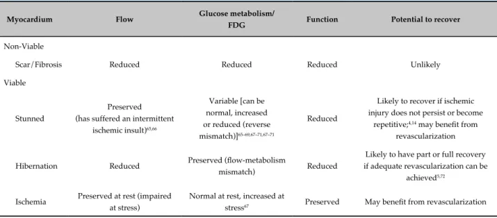
Table 1 - Viable and non-viable myocardium
Myocardium Flow Glucose metabolism/
FDG Function Potential to recover
Non-Viable
Scar/Fibrosis Reduced Reduced Reduced Unlikely
Viable
Stunned
Preserved (has suffered an intermittent
ischemic insult)65,66
Variable [can be
normal, increased or reduced (reverse mismatch)]65–69,67–71,67–71
Reduced
Likely to recover if ischemic
injury does not persist or become repetitive;4,14 may benefit from
revascularization
Hibernation Reduced Preserved (flow-metabolism
mismatch) Reduced
Likely to have part or full recovery if adequate revascularization can be
achieved5,72
Ischemia Preserved at rest (impaired at stress)
Normal at rest, increased at
stress67 Preserved May benefit from revascularization
glucose uptake. Single-photon emission computed
tomography (SPECT) with thallium-201 (
201Tl),
a potassium analogue, has the sarcolemma membrane
integrity as its target (sodium/potassium ATPase pump
activity).
16SPECT with technetium-99m (
99mTc)-based
tracers test the mitochondrial membrane integrity.
17,18Dobutamine echocardiogram (ECHO) and dobutamine
magnetic resonance imaging (MRI) measure myocardial
contractile reserve. Delayed enhancement MRI and
computed tomography target the amount of fibrotic
tissue, and myocardial contrast ECHO targets the
microvascular integrity.
19,20In a meta-analysis by Schinkel et al.
5reviewing 24
studies (756 patients) comparing all available imaging
modalities,
18FDGPET was shown to be the most sensitive
to predict regional function recovery, and dobutamine
ECHO was the most specific (92%, 63%, 74% and 87% and
80%, 78%, 75% and 83% of sensitivity, specificity, positive
and negative predictive value for PET and ECHO,
respectively).
5Cardiac MRI, which was underrepresented
in this meta-analysis, had sensitivity, specificity, positive
and negative predictive values of 74%, 82%, 78% and 78%
for dobutamine stress MRI and 84%, 63%, 72% and 78%
for delayed enhancement MRI.
5In this same meta-analysis, a total of 721 patients
underwent
99mTc-tracer-based SPECT and 1,119 had
201Tl SPECT to assess viability.
201Tl was more sensitive
and
99mTc-tracer-based SPECT more specific to predict
recovery, with sensitivity, specificity, positive and
negative predictive values of 87%, 54%, 67% and 79% and
83%, 65%, 74% and 76% for
201Tl and
99mTc, respectively.
5Comparisons between nuclear techniques suggest
18FDG
PET is the superior technique to detect the amount
of hibernating myocardium,
21–25except for one study
directly comparing
201Tl and
18FDG PET, which suggested
similar viability detection for both methods.
26More recent data analyzing MRI performance in
detecting viable myocardium have supported its
high sensitivity.
27–30Romero et al.
27have conducted a
meta-analysis of MRI prospective trials including 24
studies (698 patients) and found a sensitivity of 95% for
predicting functional recovery for MRI with delayed
enhancement. Dobutamine MRI was the most specific
(91%) when compared to delayed enhancement and
end-diastolic wall thickness techniques.
27Kühl et al.
29have studied 29 patients with chronic ischemic HF and
mean ejection fraction of 32% who had both MRI and
PET/SPECT (
18FDG for metabolism and
99mTc SPECT
for perfusion) performed at baseline and at 6-month
follow-up after revascularization.
29The group found
MRI to have higher sensitivity and PET/SPECT to be
more specific (97%
versus
87% sensitivity and 68%
versus
76% specificity for MRI and PET/SPECT, respectively).
29A more recent study has analyzed the feasibility of PET/
MRI scanners in evaluating segment functional recovery
in 28 patients post-acute myocardial infarction (MI) and
percutaneous revascularization.
30All patients underwent
PET/MRI with contrast for delayed enhancement and
18
FDG injection for uptake assessment 5-7 days after the
Figure 1 – “Images illustrating different combinations of FDG uptake and LGE transmurality. First column: FDG ≥ 50%/LGE non-transmural (‘PET viable/MRI viable); second column: FDG < 50%/LGE non-transmural (‘PET non-viable/MRI non-viable’); third column: FDG < 50%/LGE non-transmural (‘PET non-viable/MRI viable’). White arrows indicate the respective area of ischaemically affected
myocardium.” – With permission from Rischpler et al, Eur Heart J Cardiovasc Imaging.30
assessment at 6 months.
30The study has concluded that
simultaneous assessment of glucose metabolism and scar
assessment using a hybrid PET/MRI scanner is feasible.
Moreover, the agreement between the techniques was
high (82% of the segments were either non-viable or
viable for both PET and MRI, k = 0.65). In only 18% of the
segments was there disagreement, and, in all of them, PET
suggested non-viability while MRI suggested viability.
30The recovery was higher in the segments in which there
was agreement between the techniques (78%
versus
41%
for PET viable/MRI viable and PET non-viable/MRI
viable, respectively). Recovery was similar between PET
viable/MRI viable and PET viable/MRI
non-viable segments, suggesting PET better dichotomized
the degree of recovery between viable and non-viable
myocardium. In the PET non-viable/MRI viable segments,
there was some recovery (41%), suggesting a lower
threshold for % FDG uptake cutoff (40-45% instead of
50%) may have detected some viable segments identified
by MRI. Overall the techniques appear complementary.
Their combined use as PET/MR may offer comprehensive
tissue characterization of metabolism, scar and function
and may refine our ability to define viable myocardium.
Further studies are warranted (Figures 1 and 2).
30Clinical relevance of viability assessment: PARR-2
and STICH
Figure 2 - “Regional wall motion abnormality and functional recovery in the long-term course. The wall motion abnormality early
after AMI and at follow-up as well as the resulting functional recovery of the 95 dysfunctional segments were evaluated regarding
different patterns of LGE transmurality and FDG uptake [LGE transmurality (A and D), FDG uptake (B and E), combination of LGE transmurality and FDG uptake (C and F)] “- With permission from Rischpler et al, Eur Heart J Cardiovasc Imaging.30
AMI: acute myocardial infarction; LGE: late gadolinium enhancement; PET: positron emission tomography; MR: magnetic resonance imaging.
with ischemic HF.
9,31–34Allman et al.
9have conducted
a meta-analysis with 24 studies, and their analysis has
shown the benefit of revascularization only in patients
with viable myocardium as opposed to scar.
9More
recently, a meta-analysis including 29 studies by Inaba
et al. has documented the benefit of revascularization
over medical therapy in patients with dysfunctional
viable myocardium.
31To date, there have been two major prospective
randomized trials comparing outcome in patients with
ischemic HF who underwent viability assessment:
PARR-2 (Positron emission tomography And Recovery
following Revascularization phase 2)
6and STICH
(Surgical Treatment for Ischemic Heart Failure) viability
substudy
35trials.
PARR-2 has randomized 430 patients from 9 centers,
to have either viability assessment with
18FDG PET
or standard care without
18FDG PET, before decisions
regarding revascularization.
6A trend toward benefit
for the primary outcome (cardiac death, MI and cardiac
hospitalization at 1 year) has been observed in the arm
that underwent FDG PET to assist with clinical
decision-making [36% of events in the standard care arm and 30%
in the PET arm, relative risk 0.82; p = 0.16 and hazard
ratio (HR) 0.78; p = 0.15].
6However, not all patients
Figure 3 - “Risk-adjusted, event-free survival curves for time-to-composite event for patients who adhered to PET imaging
recommendations (FDG PET Adhere) versus standard care (STD) in patients randomized at sites participating in long-term follow-up.
Hazard ratio = 0.73 (95% CI 0.54–0.99; p = 0.042). No at risk = number of patients who had not died, not had transplant, not dropped out, and not had events. Seven patients whose last follow-up date was within 10 days of 1,825 days (5 years) were included in the 5-year total. CI indicates confidence interval; FDG PET, F-18-fluorodeoxyglucose positron emission tomography; and STD, standard
care”. – With permission from Mc Ardle et al, Circ Cardiovasc Imaging.11
management adhered to the imaging recommendations
may have an impact on patient outcome.
6A PARR-2 substudy has supported the importance of
adherence to PET findings and that of teamwork of: i)
revascularization (surgeons, interventional cardiology);
ii) HF; and iii) imaging specialists.
8This along with iv)
access to FDG and v) the cardiac PET imaging experience
of a centre has the potential to impact outcome. The
Ottawa-FIVE (i.e. i-v above) study has had 111 patients
from an experienced center in which PET was easily
available and physicians were comfortable with the
technology and its interpretation. In this scenario, patients
in the FDG PET arm had clear benefit when compared to
standard care (19% of cumulative proportion of events
in the PET arm versus
41% in the standard care group)
and multivariable analysis showed benefit (HR 0.34; 95%
confidence interval 0.16-0.72; p = 0.005).
8In long-term (5 years) follow-up, the PARR-2
population in which PET recommendations were
followed had improved primary outcome (HR 0.73, 95%
confidence interval 0.54-0.99, p = 0.042) (Figure 3).
11In addition, PARR-2 has shown that the amount of
hibernating myocardium also plays an important role
in patient outcome.
7With increasing extent of mismatch
(hibernating myocardium), the likelihood of benefit with
revascularization also increases. In this substudy of the
PARR-2 trial involving 182 patients in the PET arm, a
cutoff of 7% was able to distinguish between patients
who would or would not benefit from revascularization,
which is in accordance with previous values reported by
Di Carli el at.
10(5%), Lee et al.
12(7.6%) and Ling et al.
36(10%) (Figure 4).
The STICH trial has observed conflicting results
compared to previous studies regarding the benefit of
revascularization for patients with viable myocardium.
35A total of 1,212 patients were randomized to receive
optimal medical therapy alone or medical therapy
plus revascularization.
35,37Of these, 601 patients
Figure 4 - Interaction between mismatch on 18FDG PET (hibernating myocardium) and clinical outcome. A) Various levels of mismatch
and its hazards ratios and 95% CI. For patients with mismatch > 7% there was improvement in the primary outcome (cardiac death,
myocardial infarction and cardiac hospitalization at 1 year) (with permission from D’Egidio et al.7). B) Relationship between % of
mismatch and adjusted HR for all cause of death in patients who received medical therapy or early revascularization. Greater amounts of hibernating myocardium related with increased risk of medical therapy (with permission from Ling et al.12). C) Relationship between amount of mismatch and improvement in functional status after revascularization (with permission from Di Carli et al., Circulation10). CI: confidence interval; HR: hazard ratio.
adjustment for baseline characteristics.
35More recently,
the 10-year follow-up of the original trial, STICHES
(STICH Extension Study)
37has shown the benefit of
revascularization for all-cause death, cardiovascular
death and cardiovascular hospitalization over optimal
medical therapy alone.
37In the STICH viability substudy, while viability
did predict outcome, it was not independent of other
parameters and did not predict outcome benefit from
revascularization, leaving questions yet to be answered.
The greater long-term benefit in the revascularization arm
in the main trial indeed highlights the need for a careful
assessment of patients with ischemic HF, balancing the
risks and benefits in short and long term.
Although the ISCHEMIA trial (NCT01288560) does
not specifically evaluate viability, its results may
assist in understanding the role of ischemia imaging
in guiding revascularization. Currently, more than
5,000 patients have been randomized worldwide to an
invasive strategy +/- revascularization
versus optimal
medical management.
Table 2 - Comparison of the STICH Viability Substudy
with PARR-2 - Adapted with permission from
Mielniczuk et al., JACC Cardiovasc Imaging
43STICH
substudy PARR-2
Patient population
Randomized? Not the substudy Yes
Mean age, years 60.7 63
Male sex 85 84
Previous CABG 3 19
Diabetes mellitus 39 39
Estimated
GFR < 60 mL/min/1.73m2 7.5 34
Mean serum creatinine 108 µmol/L
Viability testing
SPECT or dobutamine echocardiography
PET
Prevalence of viability 81 22
Values are % unless otherwise indicated. CABG: coronary artery bypass grafting; GFR: glomerular filtration rate; SPECT: single-photon emission computed tomography.
Beyond clinical events, there is evidence that patients
undergoing FDG PET have improved quality of life versus
standard care (not undergoing FDG PET) at least in the short
term.
40Other studies have also reported revascularization
directed by FDG PET improves HF symptoms and quality
of life.
10,41There is also evidence to support that viability
imaging with PET is cost-effective when hibernation data
are used to guide revascularization.
42Comparing PARR2 and STICH
It is important to understand the differences between
PARR-2 and STICH in order to appreciate their respective
significance.
6,35,40,43First, in STICH, patients had to be
acceptable for revascularization. While patients were
randomized to coronary artery bypass graft surgery
versus
optimal medical therapy, imaging was not randomized
nor did it direct the therapy decision. Conversely,
in PARR-2, patients in whom decisions regarding
revascularization was uncertain were randomized to
FDG PET viability imaging versus standard care with
no FDG PET imaging. The tests for viability assessment
were also different:
18FDG PET in PARR-2 and SPECT or
dobutamine ECHO in STICH. Compared to the STICH
population, PARR-2 patients had more renal dysfunction
(7.5%
versus
34%), had more prior coronary artery
bypass graft surgery (3%
versus 19%), more multivessel
coronary artery disease (75%
versus 90%) and less viable
myocardium (81% versus 22%), suggesting these patient
cohorts were not the same (Table 2).
40,43From those
studies, it is safe to conclude that viability imaging is not
needed in all patients with ischemic heart disease and
left ventricular dysfunction who are being considered
for revascularization. However, there may be high-risk
patients whose decisions are particularly difficult where
viability imaging has a role.
40,43Viability tests: when should we use it?
Current evidence and guidelines support the use of
viability imaging to assist decision-making in patients
with ischemic HF (Table 3).
44–50The imaging modality of
choice for viability assessment needs to be individualized
according to each clinical scenario, technology availability
and institution expertise.
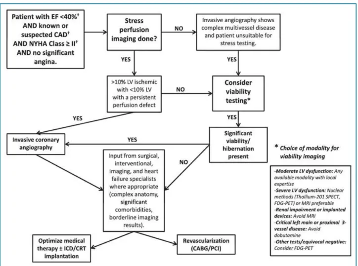
14,40,41,49–52In our experience, viability imaging is appropriate in
patients with known or strongly suspected ischemic HF,
New York Heart Association (NYHA) ≥ II, moderate to
severe left ventricular dysfunction (left ventricular ejection
fraction < 40%), moderate to large perfusion defects and no
significant ischemia, significant comorbities and/or poor
vessel targets (Figure 5).
49,51,52On the other hand, viability
is not (or less) useful in patients with predominantly
angina CCS > II, those with normal or mild left ventricular
dysfunction, critical left main coronary artery disease,
patients with good revascularization targets, those with
already-demonstrated moderate to severe ischemia and
those with minimal or no comorbidities.
51,52Figure 6
illustrates two examples of viability imaging.
When viability imaging is needed, the choice of
which test depends on specific advantages of the
different modalities, availability and local expertise. Until
comparative evidence is available (see “Future Directions”),
the following is an approach to select which test for viability
in which circumstance as suggested by the authors:
51,521. Normal or mild left ventricular dysfunction –
viability imaging is rarely needed.
2. Moderate left ventricular dysfunction – any
method can be considered depending on availability
and local expertise.
Table 3 - Guidelines, Appropriate Use Criteria and Position Statements for the use of viability imaging in patients with
ischemic heart failure. With permission from Wiefels et al., Curr Cardiovasc Imaging Rep.
73Recommendation Grade Level Organization
Nuclear imaging for assessment of myocardial viability for
consideration of revascularization in patients with CAD and LV
dysfunction who do not have angina
I B ACC/AHA/ASNC
Radionuclide Imaging 200344
Cardiac PET and CMR should be used in the evaluation and
prognostication of patients with ICM and LV dysfunction I B
CCS/CAR/CANM/CNCS/
Can SCMR 200747
Noninvasive imaging to detect myocardial ischemia /viability in HF
and CAD IIa C ACCF/AHA CHF 2013
48
Viability assessment is reasonable before revascularization in HF
patients with CAD IIa B ACCF/AHA CHF 2013
48
Non-invasive stress imaging (CMR, echo, SPECT, PET) may be
considered for the assessment of myocardial ischemia and viability in patients with HF and CAD (considered suitable for coronary
revascularization) before the decision on revascularization.
IIb B ESC CHF 201644
Myocardial viability testing should be considered in patients with
ischemic CM and reduced LV EF eligible for revascularization Appropriate use score: 9
AACF/ASNC/ACR/ASE/ SCCT/SCMR/ SNM 200945
gadolinium enhanced MRI which are more sensitive
than contractile reserve.
5,27,534. Renal failure (GFR < 30) or implanted devices –
avoid MRI.
5. Left main coronary artery disease or severe proximal
3-vessel disease – avoid dobutamine.
6. Equivocal results on another viability test or negative
results on another viability test, where certainty is needed
to completely rule [in or] out viability – consider FDG
PET or MRI as highly sensitive methods.
5,27,51,53Future directions
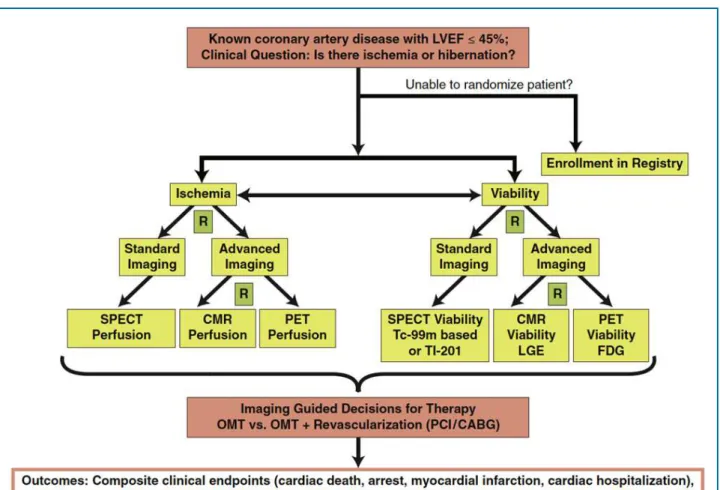
The IMAGE HF (Imaging Modalities to Assist with
Guiding therapy in the Evaluation of patients with Heart
Failure) project includes a group of clinical trials, one of
which is the AIMI-HF trial (Alternative Imaging Modalities
in Ischemic Heart Failure) (NCT01288560)
54(Figure 7).
AIMI-HF is a multicenter randomized trial and registry
study involving centers from Canada, United States,
Finland, Brazil and Argentina. It compares the impact of
standard of care investigation (SPECT) versus advanced
imaging (PET and MRI) for viability and ischemia
assessment. Composite outcomes are cardiac death,
resuscitated cardiac arrest, MI and cardiac hospitalization.
In cases where the patient is not randomized to one or the
other arm, they are included in a clinical registry.
54This
study will help us understand the impact of the advanced
cardiac imaging modalities for the viability assessment
and their impact on patient outcome.
PET and MRI viability targets are different and may be
complementary. The availability of PET/MRI scanners
is growing, and an initial study suggests the feasibility
of simultaneous assessment of FDG uptake and delayed
enhancement.
30Indeed, analysis per segment showed
increased accuracy for predicting wall motion recovery in
segments of accordance between the modalities.
30Further
trials are needed to show its reproducibility.
Cardiac biomarkers (troponin T and brain natriuretic
peptide) are used for patient assessment and as prognostic
tools.
54–58A recent study has demonstrated their correlation
with hibernating myocardium independently of ejection
fraction, age and kidney function (Figure 8).
58Future
paradigm shifts in the work-up of patients with ischemic
HF could involve the use of biomarkers to optimize
image-guided therapy or in some cases be independent
of imaging to decide revascularization therapy, but this
theoretical approach requires specific study.
Figure 5 - Flow diagram illustrating a potential algorithm for use of viability imaging. – With permission from Mc Ardle et al.,
Can J Cardiol52
CABG: coronary artery bypass grafting; CAD: coronary artery disease; CRT: cardiac resynchronization therapy; EF: ejection fraction; ICD: implantable cardiac defibrillator; LV: left ventricle; MRI: magnetic resonance imaging; NYHA: New York Heart Association; PCI: percutaneous coronary intervention; SPECT: single-photon emission computed tomography. †Denotes the required indications for 18F-fluorodeoxyglucose positron
emission tomography (FDG-PET) imaging in Ontario.
evidence of reduced MIBG uptake reflecting the high SN
signal. The PARAPET study (Prediction of ARrhythmic
Events with Positron Emission Tomography) has shown
that sympathetic denervation measured by
11C-meta-hydroxyephedrine (HED), a PET tracer able to quantify
sympathetic denervation, could predict sudden cardiac
death independently of ejection fraction and infarct
size.
61A novel F-18 PET tracer (LMI1195) is under
initial evaluation and may be able to also measure
myocardial innervation.
62–64Its main advantage over
HED is its longer half-life, which could enable wide
distribution and hence potential for wider use of SN
function imaging in the future.
Conclusion
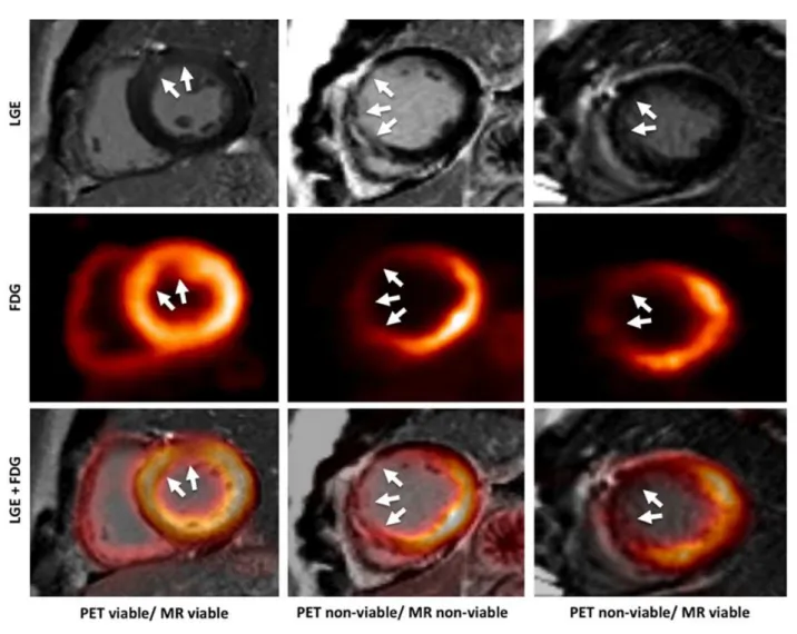
Figure 6 - (A) 13N perfusion PET and 18FDG metabolism PET in short axis (SAO), horizontal long axis (HLA) and vertical long axis
(VLA) showing extensive area of mismatch in the mid to distal anterior wall and apex (white arrow). (B) Polar map with quantitative analysis of the scar amount (7%) on the top (match defect) and hibernating myocardium (22%) on the bottom (mismatch). “Given the
significant amount of hibernating myocardium, it was recommended that the patient proceed with coronary artery bypass grafting.” (adapted from Weifels et al., with permission).73 (C) Cardiac MRI showing subendocardial scar involving > 75% of the myocardium
from the basal to apical anteroseptal wall, mid to apical anterior wall and apex, suggesting no viability in the LAD territory in a
patient with a history of previous anterior myocardial infarction and coronary angiogram showing occluded mid LAD. (D) Cardiac
MRI of a patient with occluded proximal LAD with collaterals, 95% stenosis ostial LCx and occluded OM1 showing subendocardial scar from the basal to apical anterior wall, mid to apical anteroseptal wall, and basal to mid lateral wall involving < 50% myocardium, suggesting viability in the LAD and LCx territories. Given these findings, the patient went on to have CABG (LITA->LAD, left radial->OM1, SVG->right PIV). He is clinically doing well one year post-CABG.
Figure 7 - “The AIMI-HF (Alternative Imaging Modalities in Ischemic Heart Failure) trial algorithm”. “The primary endpoint is a
composite of cardiac death, MI, resuscitated cardiac arrest, or cardiac rehospitalization.” – With permission from Mielniczuk et al., JACC Cardiovasc Imaging43
CMR: cardiac magnetic resonance; LGE: late gadolinium enhancement; LVEF: left ventricular ejection fraction; SPECT: single-photon emission computed tomography; PET: positron emission tomography; R: randomization.
revascularization or at least to guide imaging to guide
revascularization? Further research is needed here.
In the meantime, clinicians, surgeons, interventional
cardiologists and imaging specialists must work
together as a team to enable the best decisions for each
individualized patient in order to optimize the patient’s
desired outcomes.
Acknowledgments and disclosures
RSB is a career investigator supported by the Heart
and Stroke Foundation of Ontario, a Tier 1 Research
Chair supported by the University of Ottawa, and
the University of Ottawa Heart Institute Vered Chair
in Cardiology. He receives research support and
honoraria from Lantheus Medical Imaging, Jubilant
DraxImage and GE.
Author contributions
Acquisition of data: Erthal F, Wiefels C, Promislow S.
Analysis and interpretation of the data: Erthal F, Wiefels
C, Promislow S. Writing of the manuscript: Erthal F,
Wiefels C, Promislow S, Kandolin R, Stadnick E. Critical
revision of the manuscript for intellectual content: Erthal
F, Wiefels C, Promislow S, Stadnick E, Mielniczuk L,
Ruddy T, Small G, Beanlands R.
Potential Conflict of Interest
No potential conflict of interest relevant to this article
was reported.
Sources of Funding
Figure 8 - NT-proBNP (A) and hs-cTnT (B) concentrations in “patients with and those without significant (> 10%) hibernation. Median (interquartile range) values of (A) serum NT-proBNP and (B) hs-cTnT levels are shown at the top of each corresponding
bar.” – Adapted with permission from Zelt et al., Can J Cardiol58
NT-proBNP (Log of serum N-terminal pro b-type natriuretic peptide), hs-cTnT (high-sensitivity cardiac troponin T).
1. Mozaffarian D, Benjamin EJ, Go AS, Arnett DK, Blaha MJ, Cushman M, et al; American Heart Association Statistics Committee and Stroke Statistics Subcommittee. Heart disease and stroke statistics--2015 update: a report from the American Heart Association. Circulation. 2015;131(4):e29322. Erratum in: Circulation. 2016;133(8):e417. Circulation. 2015;131(24):e535.
2. Levy D, Kenchaiah S, Larson MG, Benjamin EJ, Kupka MJ, Ho KK, et al. Long-term trends in the incidence of and survival with heart failure. N Engl J Med. 2002;347(18):1397-402.
3. Brasil. Ministério da Saúde. Mortalidade TabNet Win32 3.0. [Acesso em 2017 nov 16]. Disponível em: http://tabnet.datasus.gov.br/cgi/tabcgi. exe?sim/cnv/obt10uf.de.
4. Ghosh N, Rimoldi OE, Beanlands RS, Camici PG. Assessment of myocardial ischaemia and viability: role of positron emission tomography. Eur Heart J. 2010;31(24):2984-95.
5. Schinkel AF, Bax JJ, Poldermans D, Elhendy A, Ferrari R, Rahimtoola SH. Hibernating myocardium: diagnosis and patient outcomes. Curr Probl Cardiol. 2007;32(7):375-410.
6. Beanlands RS, Nichol G, Huszti E, Humen D, Racine N, Freeman M, et al; PARR-2 Investigators. F-18-fluorodeoxyglucose positron emission tomography imaging-assisted management of patients with severe left ventricular dysfunction and suspected coronary disease. a randomized, controlled trial (PARR-2). J Am Coll Cardiol. 2007;50(20):2002-12.
7. D’Egidio G, Nichol G, Williams KA, Guo A, Garrard L, deKemp R, et al; PARR-2 Investigators. Increasing benefit from revascularization is associated with increasing amounts of myocardial hibernation: a substudy of the PARR-2 trial. JACC Cardiovasc Imaging. 2009;2(9):1060-8.
8. Abraham A, Nichol G, Williams KA, Guo A, deKemp RA, Garrard L, et al; PARR 2 Investigators. 18F-FDG PET imaging of myocardial viability
in an experienced center with access to 18F-FDG and integration with clinical management teams: the Ottawa-FIVE substudy of the PARR 2 trial. J Nucl Med. 2010;51(4):567-74.
9. Allman KC, Shaw LJ, Hachamovitch R, Udelson JE. Myocardial viability testing and impact of revascularization on prognosis in patients with coronary artery disease and left ventricular dysfunction: a meta-analysis. J Am Coll Cardiol. 2002;39(7):1151-8.
10. Di Carli MF, Asgarzadie F, Schelbert HR, Brunken RC, Laks H, Phelps ME, et al. Quantitative relation between myocardial viability and improvement in heart failure symptoms after revascularization in patients with ischemic cardiomyopathy. Circulation. 1995;92(12):3436-44.
11. Mc Ardle B, Shukla T, Nichol G, deKemp RA, Bernick J, Guo A, et al; PARR 2 Investigators. Long-term follow-up of outcomes with F-18-fluorodeoxyglucose positron emission tomography imaging–assisted management of patients with severe left ventricular dysfunction secondary to coronary disease. Circ Cardiovasc Imaging. 2016;9(9). pii: e 004331.
12. Ling LF, Marwick TH, Flores DR, Jaber WA, Brunken RC, Cerqueira MD, et al. Identification of therapeutic benefit from revascularization in patients with left ventricular systolic dysfunction: inducible ischemia versus hibernating myocardium. Circ Cardiovasc Imaging. 2013;6(3):363-72.
13. Di Carli MF, Davidson M, Little R, Khanna S, Mody FV, Brunken RC, et al. Value of metabolic imaging with positron emission tomography for evaluating prognosis in patients with coronary artery disease and left ventricular dysfunction. Am J Cardiol. 1994;73(8):527-33.
14. Lim SP, McArdle BA, Beanlands RS, Hessian RC. Myocardial viability: it is still alive. Semin Nucl Med. 2014;44(5):358-74.
References
Study Association
This study is not associated with any thesis or
dissertation work.
Ethics approval and consent to participate
15. Fallavollita JA, Malm BJ, Canty JM Jr. Hibernating myocardium retains metabolic and contractile reserve despite regional reductions in flow, function, and oxygen consumption at rest. Circ Res. 2003;92(1):48-55.
16. Sinusas AJ, Watson DD, Cannon JM, Beller GA. Effect of ischemia and postischemic dysfunction on myocardial uptake of technetium-99m-labeled methoxyisobutyl isonitrile and thallium-201. J Am Coll Cardiol 1989;14(7):1785-93.
17. Freeman I, Grunwald AM, Hoory S, Bodenheimer MM. Effect of coronary occlusion and myocardial viability on myocardial activity of technetium-99m-sestamibi. J Nucl Med Off Publ Soc Nucl Med. 1991;32(2):292-8.
18. Beanlands RS, Dawood F, Wen WH, McLaughlin PR, Butany J, D'Amati G, et al. Are the kinetics of technetium-99m methoxyisobutyl isonitrile affected by cell metabolism and viability? Circulation. 1990;82(5):1802-14.
19. Kaul S. Myocardial Contrast Echocardiography a 25-year retrospective. Circulation. 2008;118(3):291-308.
20. Camici PG, Prasad SK, Rimoldi OE. Stunning, hibernation, and assessment of myocardial viability. Circulation. 2008;117(1):103-14.
21. Marzullo P, Parodi O, Reisenhofer B, Sambuceti G, Picano E, Distante A, et al. Value of rest thallium-201/technetium-99m sestamibi scans and dobutamine echocardiography for detecting myocardial viability. Am J Cardiol. 1993;71(2):166-72.
22. Marzullo P, Sambuceti G, Parodi O. The role of sestamibi scintigraphy in the radioisotopic assessment of myocardial viability. J Nucl Med. 1992;33(11):1925-30.
23. Sawada SG, Allman KC, Muzik O, Beanlands RS, Wolfe ER Jr, Gross M, et al. Positron emission tomography detects evidence of viability in rest technetium-99m sestamibi defects. J Am Coll Cardiol. 1994;23(1):92-8.
24. Cuocolo A, Pace L, Ricciardelli B, Chiariello M, Trimarco B, Salvatore M. Identification of viable myocardium in patients with chronic coronary artery disease: comparison of thallium-201 scintigraphy with reinjection and technetium-99m-methoxyisobutyl isonitrile. J Nucl Med. 1992;33(4):505-11.
25. Dilsizian V, Arrighi JA, Diodati JG, Quyyumi AA, Alavi K, Bacharach SL, et al. Myocardial viability in patients with chronic coronary artery disease. Comparison of 99mTc-sestamibi with thallium reinjection and
[18F]fluorodeoxyglucose. Circulation. 1994;89(2):578-87. Erratum in: Circulation. 1995;91(12):3026.
26. Bonow RO, Dilsizian V, Cuocolo A, Bacharach SL. Identification of viable myocardium in patients with chronic coronary artery disease and left ventricular dysfunction. Comparison of thallium scintigraphy with reinjection and PET imaging with 18F-fluorodeoxyglucose. Circulation. 1991;83(1):26-37.
27. Romero J, Xue X, Gonzalez W, Garcia MJ. CMR imaging assessing viability in patients with chronic ventricular dysfunction due to coronary artery disease: a meta-analysis of prospective trials. JACC Cardiovasc Imaging. 2012;5(5):494-508.
28. Schvartzman PR, Srichai MB, Grimm RA, Obuchowski NA, Hammer DF, McCarthy PM, et al. Nonstress delayed-enhancement magnetic resonance imaging of the myocardium predicts improvement of function after revascularization for chronic ischemic heart disease with left ventricular dysfunction. Am Heart J. 2003;146(3):535-41.
29. Kühl HP, Lipke CS, Krombach GA, Katoh M, Battenberg TF, Nowak B, et al. Assessment of reversible myocardial dysfunction in chronic ischaemic heart disease: comparison of contrast-enhanced cardiovascular magnetic resonance and a combined positron emission tomography–single photon emission computed tomography imaging protocol. Eur Heart J. 2006;27(7):846-53.
30. Rischpler C, Langwieser N, Souvatzoglou M, Batrice A, van Marwick S, Snajberk J, et al. PET/MRI early after myocardial infarction: evaluation of viability with late gadolinium enhancement transmurality vs. 18F-FDG uptake. Eur Heart J Cardiovasc Imaging. 2015;16(6):661-9.
31. Inaba Y, Chen JA, Bergmann SR. Quantity of viable myocardium required to improve survival with revascularization in patients with ischemic cardiomyopathy: a meta-analysis. J Nucl Cardiol. 2010;17(4):646-54.
32. Mule JD, Bax JJ, Zingone B, Martinelli F, Burelli C, Stefania A, et al. The beneficial effect of revascularization on jeopardized myocardium: reverse remodeling and improved long-term prognosis. Eur J Cardio-Thorac Surg. 2002;22(3):426–30.
33. Tamaki N, Yonekura Y, Yamashita K, Saji H, Magata Y, Senda M, et al. Positron emission tomography using fluorine-18 deoxyglucose in evaluation of coronary artery bypass grafting. Am J Cardiol. 1989;64(14):860-5.
34. Haas F, Haehnel CJ, Picker W, Nekolla S, Martinoff S, Meisner H, et al. Preoperative positron emission tomographic viability assessment and perioperative and postoperative risk in patients with advanced ischemic heart disease. J Am Coll Cardiol. 1997;30(7):1693-700.
35. Bonow RO, Maurer G, Lee KL, Holly TA, Binkley PF, Desvigne-Nickens P, et al; STICH Trial Investigators. Myocardial viability and survival in ischemic left ventricular dysfunction. N Engl J Med. 2011;364(17):1617-25.
36. Lee KS, Marwick TH, Cook SA, Go RT, Fix JS, James KB, et al. Prognosis of patients with left ventricular dysfunction, with and without viable myocardium after myocardial infarction. Relative efficacy of medical therapy and revascularization. Circulation. 1994;90(6):2687-94.
37. Velazquez EJ, Lee KL, Jones RH, Al-Khalidi HR, Hill JA, Panza JA, et al; STICHES Investigators. Coronary-artery bypass surgery in patients with ischemic cardiomyopathy. N Engl J Med. 2016;374(16):1511-20.
38. Siebelink HM, Blanksma PK, Crijns HJ, Bax JJ, van Boven AJ, Kingma T, et al. No difference in cardiac event-free survival between positron emission tomography-guided and single-photon emission computed tomography-guided patient management: a prospective, randomized comparison of patients with suspicion of jeopardized myocardium. J Am Coll Cardiol. 2001;37(1):81-8.
39. Beanlands RS, Ruddy TD, Freeman M, Nichol G. Patient management guided by viability imaging. J Am Coll Cardiol. 2001;38(4):1271-3.
40. Mielniczuk LM, Beanlands RS. Does imaging-guided selection of patients with ischemic heart failure for high risk revascularization improve identification of those with the highest clinical benefit?: Imaging-guided selection of patients with ischemic heart failure for high-risk revascularization improves identification of those with the highest clinical benefit. Circ Cardiovasc Imaging. 2012;5(2):262-70.
41. Shukla T, Nichol G, Wells G, deKemp RA, Davies RA, Haddad H, et al. Does FDG PET-assisted management of patients with left ventricular dysfunction improve quality of life? A substudy of the PARR-2 trial. Can J Cardiol. 2012;28(1):54-61.
42. Jacklin PB, Barrington SF, Roxburgh JC, Jackson G, Sariklis D, West PA, et al. Cost-effectiveness of preoperative positron emission tomography in ischemic heart disease. Ann Thorac Surg 2002;73(5):1403-9.
43. Mielniczuk LM, Toth GG, Xie JX, De Bruyne B, Shaw LJ, Beanlands RS. Can functional testing for ischemia and viability guide revascularization? JACC Cardiovasc Imaging. 2017;10(3):354-64.
44. Klocke FJ, Baird MG, Lorell BH, Bateman TM, Messer JV, Berman DS, et al; American College of Cardiology; American Heart Association Task Force on Practice Guidelines; American Society for Nuclear Cardiology. ACC/AHA/ASNC Guidelines for the Clinical Use of Cardiac Radionuclide Imaging—Executive Summary A Report of the American College of Cardiology/American Heart Association Task Force on Practice Guidelines (ACC/AHA/ASNC Committee to Revise the 1995 Guidelines for the Clinical Use of Cardiac Radionuclide Imaging). Circulation. 2003;108(11):1404-18.
Tomography, the Society for Cardiovascular Magnetic Resonance, and the Society of Nuclear Medicine. Circulation. 2009;119(22):e561-87.
46. Anavekar NS, Chareonthaitawee P, Narula J, Gersh BJ. Revascularization in patients with severe left ventricular dysfunction: is the assessment of viability still viable? J Am Coll Cardiol. 2016;67(24):2874-87.
47. Beanlands R, Chow B, Dick A, Friedrich MG, Gulenchyn KY, Kiess M, et al; Canadian Cardiovascular Society; Canadian Association of Radiologists; Canadian Association of Nuclear Medicine; Canadian Nuclear Cardiology Society; Canadian Society of Cardiac Magnetic Resonance. CCS/CAR/ CANM/CNCS/CanSCMR joint position statement on advanced noninvasive cardiac imaging using positron emission tomography, magnetic resonance imaging and multidetector computed tomographic angiography in the diagnosis and evaluation of ischemic heart disease – executive summary. Can J Cardiol. 2007;23(2):107-19.
48. Yancy CW, Jessup M, Bozkurt B, Butler J, Casey DE Jr, Drazner MH, et al; American College of Cardiology Foundation/American Heart Association Task Force on Practice Guidelines. 2013 ACCF/AHA Guideline for the Management of Heart Failure: a report of the American College of Cardiology Foundation/American Heart Association Task Force on practice guidelines. Circulation 2013;128(16):e240-327.
49. McMurray JJ, Adamopoulos S, Anker SD, Auricchio A, Böhm M, Dickstein K, et al; ESC Committee for Practice Guidelines. ESC Guidelines for the diagnosis and treatment of acute and chronic heart failure 2012: The Task Force for the Diagnosis and Treatment of Acute and Chronic Heart Failure 2012 of the European Society of Cardiology. Developed in collaboration with the Heart Failure Association (HFA) of the ESC. Eur Heart J. 2012;33(14):1787-847.
50. Arnold JM, Liu P, Demers C, Dorian P, Giannetti N, Haddad H, et al; Canadian Cardiovascular Society. Canadian Cardiovascular Society consensus conference recommendations on heart failure 2006: diagnosis and management. Can J Cardiol. 2006;22(1):23-45. Erratum in: Can J Cardiol. 2006;22(3):271.
51. Erthal F, Chow B, Heller G, Beanlands R. Nuclear Cardiology Procedures in the Evaluation of Myocadial Viability. In: Heller G, Hendel R. Nuclear cardiology: practical applications. 3rd ed. Philadelphia: Mc Graw Hill; 2017. chap. 21.
52. Mc Ardle BA, Beanlands RS. Myocardial viability: whom, what, why, which, and how? Can J Cardiol 2013;29(3):399-402.
53. Pagano D, Bonser RS, Townend JN, Ordoubadi F, Lorenzoni R, Camici PG. Predictive value of dobutamine echocardiography and positron emission tomography in identifying hibernating myocardium in patients with postischaemic heart failure. Heart. 1998;79(3):281-8.
54. O’Meara E, Mielniczuk LM, Wells GA, deKemp RA, Klein R, Coyle D, et al; IMAGE HF investigators. Alternative Imaging Modalities in Ischemic Heart Failure (AIMI-HF) IMAGE HF Project I-A: study protocol for a randomized controlled trial. Trials. 2013 Jul 16;14:218.
55. de Boer RA, Daniels LB, Maisel AS, Januzzi JL. State of the art: newer biomarkers in heart failure. Eur J Heart Fail. 2015;17(6):559-69.
56. Beaudoin J, Singh JP, Szymonifka J, Zhou Q, Levine RA, Januzzi JL, et al. Novel heart failure biomarkers predict improvement of mitral regurgitation in patients receiving cardiac resynchronization therapy-The BIOCRT Study. Can J Cardiol. 2016;32(12):1478-84.
57. Moe GW, Ezekowitz JA, O’Meara E, Lepage S, Howlett JG, Fremes S, et al; Canadian Cardiovascular Society. The 2014 Canadian Cardiovascular Society Heart Failure Management Guidelines Focus Update: anemia, biomarkers, and recent therapeutic trial implications. Can J Cardiol. 2015;31(1):3-16. Erratum in: Can J Cardiol. 2016;32(3):394.
58. Zelt JG, Liu PP, Erthal F, deKemp RA, Wells G, O'Meara E, et al. N-terminal pro B-type natriuretic peptide and high-sensitivity cardiac
troponin T levels are related to the extent of hibernating myocardium in patients with ischemic heart failure. Can J Cardiol. 2017;33(11):1478-88.
59. Canty JM Jr, Suzuki G, Banas MD, Verheyen F, Borgers M, Fallavollita JA. Hibernating myocardium: chronically adapted to ischemia but vulnerable to sudden death. Circ Res. 2004;94(8):1142-9.
60. Luisi AJ Jr, Suzuki G, Dekemp R, Haka MS, Toorongian SA, Canty JM Jr, et al. Regional 11C-hydroxyephedrine retention in hibernating myocardium: chronic inhomogeneity of sympathetic innervation in the absence of infarction. J Nucl Med Off Publ Soc Nucl Med. 2005;46(8):1368-74.
61. Fallavollita JA, Heavey BM, Luisi AJ, Michalek SM, Baldwa S, Mashtare TL Jr, et al. Regional myocardial sympathetic denervation predicts the risk of sudden cardiac arrest in ischemic cardiomyopathy. J Am Coll Cardiol 2014;63(2):141-9.
62. Higuchi T, Yousefi BH, Reder S, Beschorner M, Laitinen I, Yu M, et al. Myocardial kinetics of a novel [(18)F]-labeled sympathetic nerve PET tracer LMI1195 in the isolated perfused rabbit heart. JACC Cardiovasc Imaging. 2015;8(10):1229-31.
63. Sinusas AJ, Lazewatsky J, Brunetti J, Heller G, Srivastava A, Liu YH, et al. Biodistribution and radiation dosimetry of LMI1195: first-in-human study of a novel 18F-labeled tracer for imaging myocardial innervation. J Nucl Med. 2014;55(9):1445-51.
64. Zelt JG, Renaud J, Mielniczuk L, Garrard L, Nalter O, Guo A, et al. Positron emission tomography provides accurate measure of cardiac sympathetic innervation compared to Carbon-11 hydroxyephedrine. [poster]. In: ACC. 18-Non invasive imaging [echocardiography, nuclear PET, MR and CT]. J Am Coll Cardiol. 2018;71(11):1482.
65. Canty JM Jr, Fallavollita JA. Hibernating myocardium. J Nucl Cardiol. 2005;12(1):104-19.
66. Camici PG, Dutka DP. Repetitive stunning, hibernation, and heart failure: contribution of PET to establishing a link. Am J Physiol Heart Circ Physiol. 2001;280(3):H929–36.
67. Abramson BL, Ruddy TD, deKemp RA, Laramee LA, Marquis JF, Beanlands RS. Stress perfusion/metabolism imaging: a pilot study for a potential new approach to the diagnosis of coronary disease in women. J Nucl Cardiol. 2000;7(3):205-12.
68. Abbott BG, Liu YH, Arrighi JA. [18F]Fluorodeoxyglucose as a memory marker of transient myocardial ischaemia. Nucl Med Commun. 2007;28(2):89-94.
69. Dou KF, Xie BQ, Gao XJ, Li Y, Yang YJ, He ZX, et al. Use of resting myocardial 18F-FDG imaging in the detection of unstable angina. Nucl Med Commun. 2015;36(10):999-1006.
70. Anselm DD, Anselm AH, Renaud J, Atkins HL, de Kemp R, Burwash IG, et al. Altered myocardial glucose utilization and the reverse mismatch pattern on rubidium-82 perfusion/F-18-FDG PET during the sub-acute phase following reperfusion of acute anterior myocardial infarction. J Nucl Cardiol. 2011;18(4):657-67.
71. Schwaiger M, Pirich C. Reverse flow-metabolism mismatch: what does it mean? J Nucl Med. 1999;40(9):1499-502.
72. Fukuoka Y, Nakano A, Tama N, Hasegawa K, Ikeda H, Morishita T, et al. Impaired myocardial microcirculation in the flow-glucose metabolism mismatch regions in revascularized acute myocardial infarction. J Nucl Cardiol. 2017;24(5):1641-50.
73. Wiefels C, Erthal F, de Kemp RA, et al. Radionuclide imaging in decision-making for coronary revascularization in stable ischemic heart disease. Curr Cardiovasc Imaging Rep. 2018. (in press).