Outubro de 2011
Universidade do Minho
Escola de Engenharia
Filipa Alexandra Baltar Lobo Coelho
Effect of Diocleinae lectins on bacteria
and fungi planktonic and sessile cells
UMinho|20
11
Filipa Ale
xandra Baltar Lobo Coelho
Ef
Dissertação de Mestrado
Mestrado em Engenharia Clínica
Trabalho realizado sob a orientação da
Professora Doutora Mariana Henriques
e co-orientação da
Professora Doutora Maria Olívia Pereira
Outubro de 2011
Universidade do Minho
Escola de Engenharia
Filipa Alexandra Baltar Lobo Coelho
Effect of Diocleinae lectins on bacteria
and fungi planktonic and sessile cells
É autorizada a reprodução parcial desta dissertação, apenas para efeitos de
investigação, mediante declaração escrita do interessado, que a tal se
compromete.
Universidade do Minho, / / .
Acknowledgements
iii
Acknowledgements
Despite this work have an individual character, it involved several persons whose knowledge and cooperation was essential for its development. Here, I want to say thanks to all this people.
To Dra. Olívia Pereira and Dra. Mariana Henriques, I want to thank for all the knowledge they shared with me, for the sympathy and encouragement, for the opportunities that they offered me and the availability of guidance at all stages of the development of this work.
To Margarida, for the time she dedicated to me in the initial phase of my work, for the experience and knowledge that shared with me, for the sympathy and availability demonstrated which proved to be essential for my self-confidence, autonomy and organization.
I am also thankful to Susana because she was always available to helping me.
I thank the Center of Biological Engineering of University of Minho for having made available the Laboratory of Applied Microbiology, where I developed this work, and all the ones that I met in the Laboratory, for making me feel part of the group, for the glad and relaxed environment, the availability and aid that all had always demonstrated.
Finally, I am thankful, to my parents, to Tozé and my friends. To my parents for all the financial support, affection and unconditional love and for the continuous incentive that they gave me at the end of one more step in my life. To Tozé by affection, love and patience that has always held for me, especially in the most difficult hours, and my friends for the affection and good moments they have compelled me to have, even in times of a lot of work.
Abstract
v
Abstract
Effect of Diocleinae lectins on bacteria and fungi planktonic and sessile cells
Microorganisms have been showing augmented resistance towards antimicrobials, being microbial resistance, nowadays, one of the biggest problems of public health. Thus, there is an increasing interest in the development of new strategies of microbial control, namely the ones based on natural products, especially from plants, such as lectins. So, it is of utmost importance to test new antimicrobial compounds, especially with a wide range, against bacteria and fungi. Therefore, the main aim of this thesis was to evaluate the effect of new lectins against both Gram-negative and Gram-positive bacteria, as well, as yeast. Moreover, these lectins were assessed against planktonic and sessile cells.
Four Diocleinae lectins: Canavalia ensiformis, Canavalia brasiliensis, Canavalia maritima and Canavalia boliviana, were used in this study. Their effect was assessed against two positive bacteria (Staphylococcus epidermidis, Staphylococcus aureus), two Gram-negative bacteria (Pseudomonas aeruginosa, Klebsiella oxytoca) and two yeasts (Candida albicans and Candida tropicalis). The effect was evaluated on microbial planktonic growth, early-stage adhesion, on biofilm biomass and biofilm viable cells.
The four lectins showed different activities (inhibitory or stimulatory effect) on planktonic growth, early-stage adhesion and biofilm formation, for the same microorganism. In general, the inhibitory effect of lectins was most notable on planktonic growth than on biofilm formation. Although there are few differences in the inhibitory capacity of lectins, K. oxytoca can be considered the microorganism that suffered lower inhibition. Interestingly, the results demonstrated that activities of lectins tested were species-dependent, namely, their action was different between the two Gram-negative species, as well as the two Gram-positive and the two yeasts. This highlights that the interaction between the lectin and the cell is of high specificity.
So, in conclusion, due to the specificity of the lectins assayed, it could be of major interest to improve their potential (synergic effect) by using more than one at the same time or combined with conventional antimicrobial agents.
Resumo
vii
Resumo
Efeito de lectinas da subfamília Diocleinae em células bacterianas e fúngicas, no estado planctónico e séssil
A resistência exibida pelos microrganismos a agentes anti-microbianos representa um dos maiores problemas que a Saúde pública enfrenta na atualidade. Desta forma, tem-se denotado um aumento do interesse pelo desenvolvimento de novas estratégias para o controlo microbiano que passam pela procura de produtos naturais, especialmente oriundos de plantas, como as lectinas. A utilização de novos compostos antimicrobianos que possam ser usados contra uma vasta gama de bactérias e fungos, revela-se de extrema importância. Assim, o objectivo desta dissertação consistiu na verificação do efeito da aplicação de novas lectinas em bactérias Gram-positivas e Gram-negativas, bem como em leveduras. Além disso, estas lectinas foram testadas em células, quer no estado planctónico e quer no estado séssil.
Neste estudo foram utilizadas quatro lectinas da subfamília Diocleinae: Canavalia ensiformis, Canavalia brasiliensis, Canavalia maritima and Canavalia boliviana. O seu efeito foi avaliado em duas bactérias Gram-positivas (Staphylococcus epidermidis, Staphylococcus aureus), duas bactérias Gram-negativas (Pseudomonas aeruginosa, Klebsiella oxytoca) e em duas leveduras (Candida albicans and Candida tropicalis). A acção das lectinas foi analisada no crescimento planctónico, na adesão inicial, na biomassa de biofilme e no número de células cultiváveis dos biofilmes.
As quatro lectinas demonstraram atividades distintas (efeito inibitório e de estimulação) no crescimento planctónico, na adesão inicial e na formação de biofilme, para o mesmo microrganismo. Em geral, o efeito inibitório das lectinas foi mais notório no crescimento planctónico do que na formação de biofilme. Apesar da capacidade inibitória das lectinas não diferir muito entre os microrganismos, a bactéria K. oxytoca foi a menos sensível aos compostos usados.
Estes resultados demonstraram que a atividade das lectinas testadas se encontra dependente da espécie, sendo a sua ação diferente entre as duas espécies Gram-positivas, as duas Gram-negativas e as duas leveduras utilizadas. Corrobora-se, assim, a elevada especificidade existente na interação entre as lectinas e as células.
Em suma, dada a especificidade das lectinas testadas, pensa-se que estas poderão ter um potencial acrescido se utilizadas em conjunto ou combinadas com agentes antimicrobianos convencionais.
Contents ix
Contents
Acknowledgements ... iii Abstract ... v Resumo ... vii Contents ... ix List of Figures ... xiList of Tables ... xiii
List of Abbreviations ... xv
Chapter 1 | Introduction ... 1
1.1. Contextualization and objectives ... 2
1.2. Literature review ... 3
1.2.1. Microorganisms ... 3
Staphylococcus epidermidis and Staphylococcus aureus ... 3
Klebsiella oxytoca and Pseudomonas aeruginosa ... 4
Candida albicans and Candida tropicalis... 4
1.2.2. Structure and components of the cell wall ... 5
1.2.2.1. Bacterial cell wall ... 5
Gram-positive bacteria ... 5
Gram-negative bacteria... 6
1.2.2.2. Fungal cell wall ... 7
1.2.3. Cell adhesion as virulence factor... 7
1.2.4. Biofilms ... 8
1.2.4.1. Biofilm development ... 10
1.2.4.2. Biofilm resistance to antimicrobial agents ... 12
Restricted penetration ... 12
Slow growth rate ... 12
Persisters and phenotypic variants ... 13
1.2.5. Natural antimicrobial agents ... 13
1.2.5.1. Lectins... 14
Lectin sources ... 15
Contents
x
Chapter 2 | Materials and Methods ... 21
2.1. Lectins origin ... 22
2.2. Lectins stock solution ... 22
2.3. Microorganisms and culture conditions ... 22
2.4. Lectin antimicrobial activity ... 23
2.5. Lectin effect on early-stage adhesion ... 23
2.6. Biofilm prevention ... 24
2.6.1. Biofilm biomass quantification ... 24
2.6.2. Biofilm viable cells enumeration ... 25
2.7. Statistical analysis... 25
Chapter 3 | Results ... 27
3.1. Bacterial and yeast planktonic growth ... 28
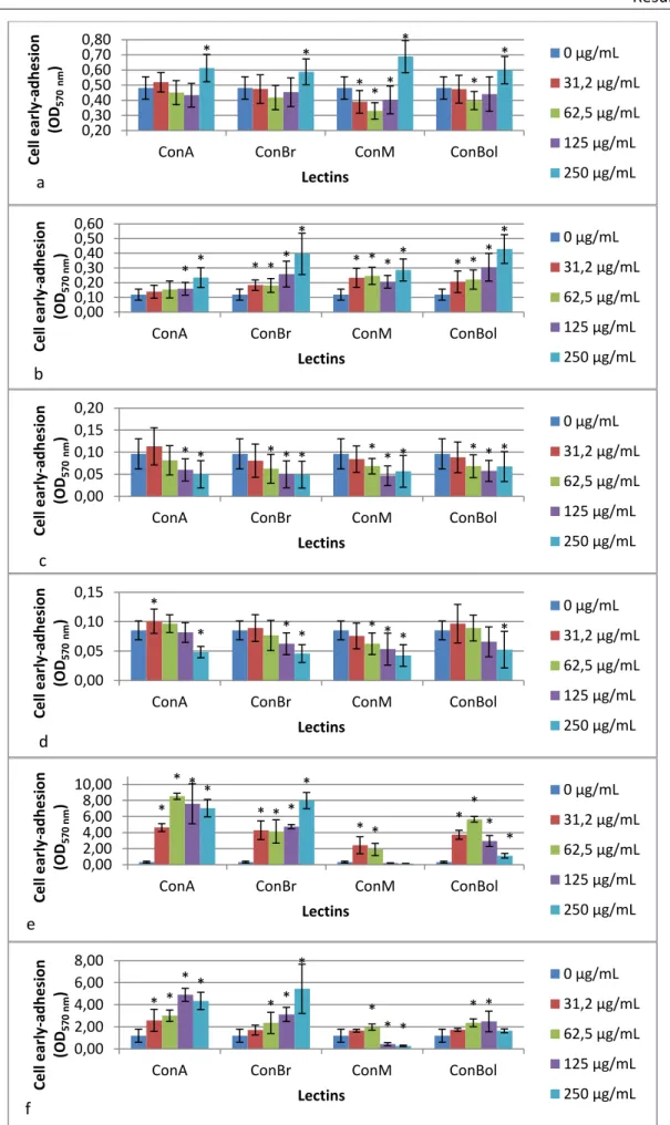
3.2. Bacteria and yeast early-stage adhesion... 30
3.3. Bacteria and yeast biofilm mass ... 30
3.4. Bacteria and yeast viable biofilm-entrapped cells ... 33
3.5. Summary table of results ... 33
Chapter 4 | Discussion ... 37
Chapter 5 | Conclusion and suggestions for future research ... 43
List of Figures
xi
List of Figures
Figure 1. Representative scheme of Gram-positive cell envelope (cytoplasmic membrane and the cell wall (7)). ... 5 Figure 2. Representative scheme of Gram-negative cell envelope (cytoplasmic membrane and the cell wall) (7). ... 6 Figure 3. Representative scheme of fungal cell envelope (7). ... 7 Figure 4. Schematic model of biofilm development processes (95). ... 10 Figure 5. Schematic representation of three types of plant lectins: merolectins, hololectins and chimerolectins (64). ... 14 Figure 6. The X-ray structure of ConA (adapted from (70)). ... 15 Figure 7. Exogenous lectin attached to the T cell targeting the altered glycoprotein present on tumor cell surface, promoting the lyses of the cell (65). ... 19 Figure 8. Effect of lectins on bacteria and yeast planktonic growth (a – Pseudomonas aeruginosa; b - Klebsiella oxytoca; c – Staphylococcus aureus; d - Staphylococcus epidermidis; e – Candida albicans; f – Candida tropicalis). *Statistically different from the control, 0 µg/mL (p < 0.05)... 29 Figure 9. Effect of lectins on bacteria and yeast early-stage adhesion (a – Pseudomonas aeruginosa; b - Klebsiella oxytoca; c – Staphylococcus aureus; d - Staphylococcus epidermidis; e – Candida albicans; f – Candida tropicalis). *Statistically different from the control, 0 µg/mL (p < 0.05)... 31 Figure 10. Bacteria and yeast biofilm formation (quantification of biofilm mass) in the presence of lectins (a – Pseudomonas aeruginosa; b - Klebsiella oxytoca; c – Staphylococcus aureus; d - Staphylococcus epidermidis; e – Candida albicans; f – Candida tropicalis). *Statistically different from the control, 0 µg/mL (p < 0.05). ... 32 Figure 11. Bacteria and yeast biofilm formation (determination of the number of viable biofilm-entrapped cells) in the presence of lectins (a – Pseudomonas aeruginosa; b - Klebsiella oxytoca; c – Staphylococcus aureus; d - Staphylococcus epidermidis; e – Candida albicans; f – Candida tropicalis). *Statistically different from the control, 0 µg/mL (p < 0.05). ... 34
List of Tables
xiii
List of Tables
Table 1. Microorganisms frequently associated with biofilms on medical devices ... 9 Table 2. Common applications of plant lectins as tools in basic and medical sciences ... 16 Table 3. Summary table of results... 35
List of Abbreviations
xv
List of Abbreviations
Aal Lectin from Araucaria angustifolia
AHL N-acyl homoserine lactone
AIDS Acquired Immune Deficiency Syndrome
ATCC American Type Culture Collection
CECT Spanish Type Culture Collection
CFU Colony forming unit
ConA Concanavalin A from Canavalia ensiformis
ConBol Lectin from Canavalia boliviana
ConBr Lectin from Canavalia brasiliensis
ConM Lectin from Canavalia maritima
CV Crystal violet
DGL Lectin from Dioclea grandiflora
DVL Lectin from Dioclea violacea
EPS Extracellular polymeric substance
ER Endoplasmic reticulum
EuniSL Lectin from Eugenia uniflora
GlcNAc N-acetil-D-glucosamine
HHA Lectin from Hippeastrum hybrid
LTA Lipoteichoic acids
MIC Minimum inhibitory concentration
MOA Lectin from Marasmium oreades
MRSA Methicillin-resistant Staphylococcus aureus
OD Optical density
PBS Phosphate buffered solution
PHA Lectin from Phaseolus vulgaris
POL Lectin from Pleurotus ostreatu
PS Polystyrene
Psl Polysaccharide synthesis locus
PWM Pokeweed mitogen
RIPs Ribosome inactivating proteins SBA Soybean lectin
List of Abbreviations
xvi
SCID Severe Combined Immunodeficiency
SDA Sabouraud Dextrose Agar
SDB Sabouraud Dextrose Broth
SSTI Skin and Soft tissue infections
TFA Lectin from Trigonella foenumgraecum
TSA Tryptic Soy Agar
TSB Tryptic Soy Broth
WGA Wheat germ agglutinin from Triticum vulgaris
Introduction
2
1.1. Contextualization and objectives
Today, bacterial and fungal infectious diseases are becoming hard to treat with conventional antimicrobial agents due to the development of resistance, being a serious health problem. This situation has promoted the search for new alternatives to treat infections and to control surface contamination.
Natural products, mainly plant-derived, are the most searched and studied compounds. Lectins are one of the many compounds extracted from plants, being proteins without catalytic function or immunological characteristics that bind specifically to carbohydrates. One of the largest and most studied families of lectins is those derived from leguminous plants.
Several lectins have been revealing to be able to inhibit bacterial and yeast pathogens, so, lectins may be important tools to control bacterial and fungal infectious diseases.
Until now, plant-based research has almost exclusively focused on planktonic microorganism, while biofilm forms, that are more resistant to antimicrobial agents and, consequently, more difficult to control, stay largely unexplored.
In this study, four plant lectins of Diocleinae subtribe (Canavalia ensiformis - ConA; Canavalia brasiliensis - ConBr; Canavalia maritima - ConM; Canavalia boliviana - ConBol) were screened for their effect on the growth, early-stage adhesion and subsequent biofilm formation of Staphylococcus epidermidis and Staphylococcus aureus (both gram positive bacteria), Pseudomonas aeruginosa and Klebsiella oxytoca (both gram negative bacteria) and for Candida albicans and Candida tropicalis.
Introduction
3
1.2. Literature review
It is common that microorganisms attach to wet surfaces and form structures named biofilms. Biofilms are structured communities of surface-associated microbial cells enclosed in a self-produced polymeric matrix, forming sessile populations (1).
Biofilms still a concern in a broad range of areas such as food, environmental and biomedical fields. They can play important roles in bioremediation and fermentation facilities, facilitating stable operations, but on the other hand they are very problematic, particularly in food and health sector (2) (3). Many persistent and chronic microbial infections are related with biofilms, and occur essentially in hospital settings by contamination of indwelling medical devices. Medical devices are contaminated by the flora from the patient or health care personnel after touching contaminated environmental surfaces (2) (4). Such infections are difficult to diagnose and treat with conventional approaches, because biofilms have intrinsic mechanisms responsible for their highly resistance to antimicrobial agents (5).
The widespread use, and sometimes misuse, of antimicrobial agents leads to the development of microorganism resistance against conventional drugs (6). Microbial resistance is, nowadays, one of the biggest problems of public health. So, it is important to limit the spread of these microorganisms, limiting contamination of surfaces and medical devices, as well as finding appropriate antimicrobial agents for biofilm eradication (7).
Currently, the search for new environmental friendly and more effective strategies to control both planktonic and sessile microorganisms is becoming a priority. The research communities and pharmaceutical industries are searching new alternatives based on natural products with antimicrobial activity and low toxicity (8-11).
1.2.1. Microorganisms
Bacterial and fungal infectious diseases are a serious health problem being the lead cause of premature deaths all over the world (12-14).
Bacteria, such S. aureus, S. epidermidis, P. aeruginosa and K. oxytoca, and yeasts from Candida genus, are some of the microorganisms responsible for human infections and important agents of nosocomial infections.
Staphylococcus epidermidis and Staphylococcus aureus S. epidermidis and S. aureus are both Gram-positive bacteria.
Introduction
4
S. epidermidis is a commensal skin bacterium and an opportunistic pathogen, being frequently isolated from infected medical devices, such as catheters (15) (16). Its infections promote an increase of global morbidity and mortality, especially in neonates (4).
S. aureus is the main etiological agent of numerous skin and soft tissue infections (SSTI). It remains the major human pathogen in hospital- and community- acquired infections causing diseases with high mortality rate (17). It is a common target for natural product drug screenings, because there has been observed an increase of antibiotics resistance, like methicillin-resistant S. aureus (MRSA) that presents a significant threat to public health (18) (19).
Klebsiella oxytoca and Pseudomonas aeruginosa
One of the most clinically important species of the genus Klebsiella is K. oxytoca. Klebsiellae are Gram-negative bacteria and opportunistic pathogens, being a frequent cause of severe diseases, such as urinary tract infections and pneumonia. Klebsiella infections are, essentially, associated with hospital care units (20) (21).
Another opportunistic Gram-negative bacterium, which is capable of causing fatal systemic diseases in humans, is P. aeruginosa. This pathogen infects patients, for example, when cutaneous barriers are breached and/or when immunologic system is compromised. Patients with respiratory diseases are commonly infected with P. aeruginosa and more than 90 % of them eventually die. P. aeruginosa is responsible for innumerous diseases, like pneumonia, SSTI, endocarditis, septicemia, among others, being the fourth leading cause of nosocomial infection and the main cause of hospital-acquired pneumonia (22) (23).
Candida albicans and Candida tropicalis
Candida species are the most common fungi involved in invasive human diseases, especially in immunocompromised patients (24).
C. albicans is generally considered to be an endogenous pathogen, but exogenous transmission (transmission from patient to patient by hands of health care personnel) is also verified (25). It is the main fungal pathogen agent found in humans, being responsible for both mucosal and deep tissue infections, such as infections of the skin, oral cavity, gastrointestinal tract, vagina and vascular system of humans (26) (27). Vaginal infections caused by C. albicans affect up to 75 % of women, at least, once in their life time (28).
C. tropicalis is the second most frequent Candida species, being the main cause of bloodstream and urinary tract infections, especially in hospitalized patients (29). It is
Introduction
5 considered highly virulent and an important cause of infections in patients with cancer, especially leukemia (25) (26) (29).
1.2.2. Structure and components of the cell wall
Cell wall acts as a permeability barrier, maintains the cell shape and mediates the initial interaction between microorganism and the environment (24). Moreover, it is characteristic of both bacteria and fungi and has a protective function but, on the other hand, it is vulnerable to some antimicrobial agents which have the capacity to disturb the processes by which the wall is synthesized. During cell growth and proliferation, cell wall is continuously undergoing biosynthesis and extension, because it is a dynamic structure. This feature makes bacteria and fungi susceptible to antimicrobial agents which disturb the biosynthesis of the cell wall (6).
Cell wall structure of bacteria and fungi is different from each other, resulting in different susceptibilities to antimicrobial agents (6).
1.2.2.1. Bacterial cell wall
Gram-positive bacteria
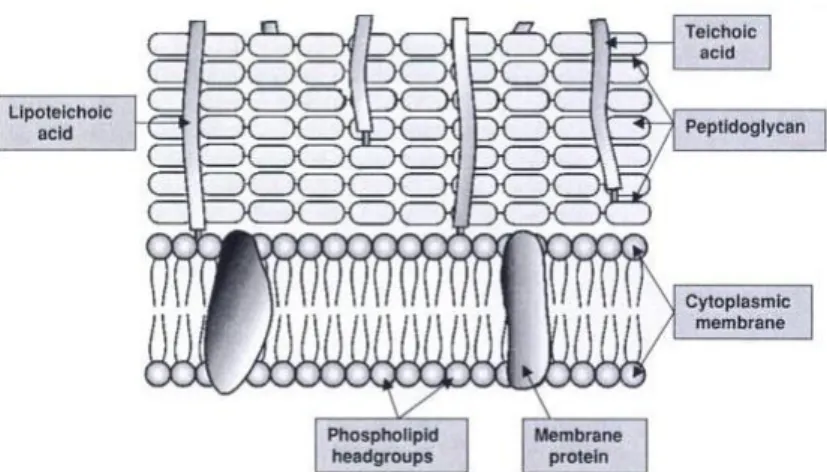
As demonstrated in Figure 1, the basic structure of Gram-positive bacteria wall is quite simple. At least 50 % of wall mass is peptidoglycan, present in several layers. Peptidoglycan is organized in a cross-linked structure that provides a tough and fibrous fabric, giving shape and strength to the cell. This structure enables bacteria wall to resist a high internal osmotic pressure. Additionally, 30 to 40 % of the wall mass is due to an acidic polymer which is linked to peptidoglycan. This polymer is often a teichoic acid. There are two types of teichoic acids, namely wall teichoic acid (WTA) and lipoteichoic acid (LTA). Its acidic character guarantees that the cell surface is polar and carries a negative charge (6) (30).
Introduction
6
Teichuronic acid is synthesized, replacing teichoic acids, when phosphate supply to the cell is limited (6) (30).
Moreover, 5 to 10 % of the wall mass is related with the proteins present on the structure. Cell wall also contains neutral sugars, such as mannose, arabinose, rhamnose and glucosamine, and acidic sugars, such as glucuronic acid and mannuronic acid, which are present on the cell wall as subunits of polysaccharides. These proteins and polysaccharides often occur in the outer layers of the wall, being almost responsible for antigenic properties of bacteria (6) (30).
Gram-negative bacteria
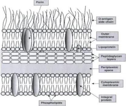
The Gram-negative cell wall is far more complex than Gram-positive cell wall, as observed in Figure 2. Peptidoglycan is no longer the major component of the cell wall, being present only one or two layers of peptidoglycan (representing 5 to 10 % of the wall mass) (6) (30).
Just outside the cytoplasmic membrane, there is a layer called periplasmic space. This layer contains biochemical compounds, such enzymes, transport proteins, secreted materials and components of peptidoglycan. Immediately outside the periplasm, there is the peptidoglycan layer of the wall. It is much thinner than in Gram-positive bacteria, but still important to assure the wall strength (6) (31).
After peptidoglycan layer, the cell wall contains three main components, such as lipoprotein, outer membrane and lipopolyssacharides (30).
Introduction
7 Lipoprotein is an important constituent of the outer membrane proteins. Only about one-third is linked to peptidoglycan, and the remaining, despite of being unattached, forms part of the membrane (6) (30) (31).
The outer membrane is composed by various compounds. It is basically a lipid bilayer with protein components. Lipopolysaccharide, phospholipids, fatty acids and proteins are the main constituents of this layer. The structure of lipopolysaccharide is complex and can vary from one bacterial strain to another. Lipopolysaccharides are exclusively on the outer surface of the outer membrane (6) (30) (32).
1.2.2.2. Fungal cell wall
Polysaccharides are the main component of fungal cell wall representing approximately 80 % of the cell wall dry weight (Figure 3). Proteins, lipids and various inorganic salts compose the others 20 % (33). The main polysaccharides found in diverse fungi cell wall are various forms of glucans and
chitin, such as branched polymers of glucose containing β-1,3 and β-1,6 linkages (β-glucans), unbranched polymers of N-acetil-D-glucosamine (GlcNAc) containing β-1,4 bonds
(chitin) and polymers of mannose covalently associated with proteins (24) (33) (34).
Chitin and β-glucans (microfibrillar polymers) form the external skeleton representing the structural components of the cell wall. The skeleton provides strength to the cell (24) (33). Although yeast cell wall composition is similar to the filamentous forms, hyphal cells contain at least three times more chitin than yeast cells (24) (33).
Proteins of the fungal cell wall are involved in permeability control, in recognition of other fungi, regulation of several processes and interaction with hosts (33).
1.2.3. Cell adhesion as virulence factor
Attachment of microorganisms, like bacteria, to tissues is the first step in the establishment of infection (18). Microorganism ability to adhere and consequently form a biofilm, which can cause an infection, is one of the major virulence factors (35).
Adhesion is a distinct process, but essential for biofilm development, that can be divided into two stages, namely the primary adhesion (docking stage) and the secondary adhesion (locking phase) (36).
Introduction
8
Primary adhesion between bacteria/fungi and abiotic surfaces is usually mediated by nonspecific interactions, and between microorganism and living or devitalized tissues is mediated through specific molecular mechanisms (36).
This stage is reversible and dependent on the interaction between the microorganism cell surface and the conditioned surface. At an early stage, the organism has to achieve a critical proximity to the surface (generally < 1 nm). Then, adhesion depends on the sum of attractive or repulsive forces existent between the two surfaces, such as electrostatic and hydrophobic interactions, steric hindrance, Van der Waals forces, temperature, and hydrodynamic forces, among others. Electrostatic interactions are, generally, repulsive forces, because most bacteria and inert surfaces are negatively charged. Hydrophobic interactions are frequently the strongest of all long-range non-covalent forces, playing an important role in adhesion and consequently in microbial infections (3) (35) (36). The hydrophobicity of the conditioned surface can vary a lot, because it is dependent on the molecules in the conditioning film (15) (36).
Repulsion existent between two surfaces can be override by specific molecular interactions mediated by adhesins present on extracellular filamentous appendages (for example, pili and flagella) (3) (36). Pili are responsible to make cells more adhesive, so cells with these filamentous appendages adhere strongly to other cells or inorganic surfaces. Probably pili are capable to overcome the initial electrostatic repulsion barrier existent, being important to surface colonization (3).
Molecular mediated binding between specific adhesins and the surface is employed in the second stage of adhesion. Microorganisms consolidate the adhesion process producing exopolysaccharides. At the end, adhesion becomes irreversible being the microorganism firmly attached to the surface, without any physical or chemical interference (37) (38).
1.2.4. Biofilms
A biofilm is a well organized cooperating community of surface-associated microbial cells enclosed in a self-produced polymeric matrix (36) (37).
In response to environmental changes, microorganisms switch between planktonic growth and sessile form of life (2). Biofilm-associated microorganisms have a different behavior from planktonic organisms. They have reduced growth rates and ability to resist to antimicrobial treatments and host defenses, being a significant factor in microorganism virulence (4) (38) (39).
Introduction
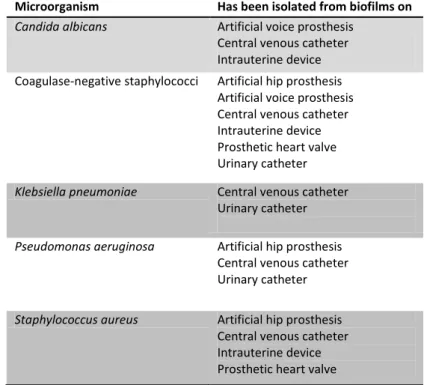
9 About 60 % of human infections results from biofilm formation on human mucosa (40). Biofilm are able to develop into implants and medical devices, such as catheters, cardiac valves, artificial joints, plates and screws, causing nosocomial infections (Table 1). These biofilms present in medical devices are usually a source of recurrent infections, which are notoriously more difficult to eradicate (2) (41). So, biofilm associated infections account for high morbidity and mortality rates (8) (42).
Depending on the device and its duration of action, biofilms can be formed by single or multiple species of microorganisms. For example, urinary catheter biofilm can be composed of single species at the beginning, but longer exposures lead to multispecies biofilms (38) (43). These biofilms on indwelling medical devices can include either yeast, positive or Gram-negative bacteria (38).
It is obviously that microorganisms have benefits from the biofilm mode of growth, such as a protective environment against harmful conditions in the host, ability to survive a nutrient limitation, osmotic stress, pH changes, anoxia, mechanical stress and exposure to antibiotics (2).
Microorganism Has been isolated from biofilms on Candida albicans Artificial voice prosthesis
Central venous catheter Intrauterine device Coagulase-negative staphylococci Artificial hip prosthesis
Artificial voice prosthesis Central venous catheter Intrauterine device Prosthetic heart valve Urinary catheter
Klebsiella pneumoniae Central venous catheter Urinary catheter
Pseudomonas aeruginosa Artificial hip prosthesis Central venous catheter Urinary catheter
Staphylococcus aureus Artificial hip prosthesis Central venous catheter Intrauterine device Prosthetic heart valve
Introduction
10
1.2.4.1. Biofilm development
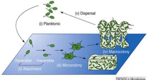
Biofilm formation process may be divided into initial attachment, microcolony formation, maturation and dispersion, as shown in Figure 4 (2).
On the second stage of adhesion, microorganisms are firmly attached to the surface, producing extracellular polymeric substance (EPS). Adhesion is closely followed by cell division and proliferation, raising microcolonies (1). This stage is a crucial step for biofilm development being the foundation upon which the mature biofilm will be built (36).
After establishment of microcolonies, the maturation process begins (2). The density and complexity of biofilm increase. Surface-adhered microorganisms actively replicate and extracellular components are generated through interaction between cells and molecules in immediate environment, raising glycocalyx (36).
Depending on environmental conditions, biofilms, for example from P. aeruginosa, can take a flat or mushroom-shaped structure (Figure 4). A flat structure is formed by means of twitching motility bacteria spread on the surface forming a confluent mat of cells. On the other hand, the migration of motile subpopulations of biofilm to the surface of a non-motile subpopulation (stalk) originates the caps, forming the mushroom-shaped structure (2). The growth potential and shape of a biofilm is dependent of the availability of nutrients, the perfusion of those nutrients to the cells within the biofim, the removal of waste, the internal pH, oxygen perfusion, osmolarity and carbon source (1) (36). Studies revealed that, when glucose is used as a carbon source, P. aeruginosa PAO1 forms biofilms with a mushroom-shaped structure, but when citrate, benzoate or casamino acids are used P. aeruginosa forms flat biofilms (44).
Fungi like C. albicans have the ability to grow with three different morphologies, such as yeast, pseudohyphae and hyphae. Hyphal growth is believed to be one of the most
Introduction
11 important virulence factors (27) (28). Commonly, Candida species biofilms consist of two distinct layers, a thin, basal dense region of yeast cells, and an overlying thicker, but more open, hyphal layer, which raise a mushroom-shaped structure (41) (45). Dimorphism is not absolutely necessary for biofilm formation, but might be essential for the development of a spatially organized structure (41).
The last stage of biofilm development is dispersion (2). When a biofilm reaches a critical mass, a dynamic equilibrium is reached with the detachment of a subpopulation of biofilm cells (outermost layer of growth) which revert to a planktonic lifestyle. These organisms are free to escape from mature biofilm and colonize other surfaces (2) (36).
Cell to cell signaling, known as quorum sensing, has been demonstrated to play a role in cell attachment and detachment from biofilms, influencing biofilm maturation (37) (43).
Intracellular communication between microorganisms is usually carried out by bacteria/fungi products that are able to diffuse away from one cell and enter into another cell (43). Extracellular accumulation of signaling molecules synchronize expression of a set of genes which allow monitoring a factor such as cell population density (2).
Beyond preventing overpopulation, cell to cell signaling may alter distribution of microorganisms in the biofilm, alter protein expression and introduce new genetic in neighborhood cells, and incorporate microorganisms in biofilm (43) (45).
Studies revealed that farnesol acts as a quorum-sensing molecule inhibiting filamentation in C. albicans. Biofilm formation was almost completely inhibited when C. albicans cells was preincubated with high concentrations of farnesol. Moreover, supernatants recovered from mature biofilms inhibited filamentation of planktonic C. albicans, indicating that a morphogenetic autoregulatory compound (probably farnesol) is produced in biofilms (46).
On Gram-negative bacteria, such as P. aeruginosa, the best studied quorum sensing system is the LuxR-LuxI system, which uses an N-acyl homoserine lactone (AHL) as quorum sensing signal (47). Hentzer and colleagues (48) reported that a halogenated furanone compound (derivative of the secondary metabolites produced by Delisea pulchra) was able to interfere with AHL in P. aeruginosa. This interference affected the architecture of the biofilm and enhanced the process of bacterial detachment, leading to a loss of bacterial biomass from the substratum.
Introduction
12
1.2.4.2. Biofilm resistance to antimicrobial agents
Biofilms are commonly highly resistant to antimicrobial agents (2). Several studies revealed that bacterial cells in a biofilm can become 10-1000 times more resistant to antibiotics than corresponding planktonic growing bacteria (1).
However, the mechanisms responsible for the resistance of biofilms to antimicrobial agents are not fully understood (41). Several mechanisms were proposed to explain the resistance of biofilms, including restricted penetration of drugs through biofilm matrix; the slow growth rates of biofilm; the appearance of resistant strains and phenotypic variants within biofilm populations, and expression of biofilm-specific genes (1) (2) (41).
Restricted penetration
Microorganisms in biofilms tend to be within the EPS matrix rather than on its surface. So, they are less accessible to antimicrobial agents. Thus, poor penetration of antimicrobial agents is suggested as one of the factors accounting for the resistance of biofilms (2) (41).
A reaction of the compound or its adsorption by the components of the biofilm matrix, result in slow diffusion rates and delayed penetration of antimicrobial agents within the biofilm. So, higher concentrations of antimicrobial agents are needed to kill biofilm cells regarding their planktonic counterparts (1) (2) (41).
For instance, P. aeruginosa biofilm formed on dialysis membranes delayed piperacillin diffusion suggesting that the biofilm matrix acts like a diffusion barrier to that antibiotic (2).
Slow growth rate
Biofilm cells grow slowly, mainly in mature biofilms, because of the limited availability of nutrients and the anaerobic conditions, particularly at the base of the biofilm (2) (36) (41). For example, beta-lactams and imipenem show low activity against mature biofilms, but they become more effective with younger biofilms (2). It is believed that oxygen limitations slow down microbial growth as it restricts the metabolic activity of the cells. Slow growth rate, oxygen limitation and low metabolic activity may be responsible for the antimicrobial resistance in biofilms. A slow growth rate is frequently accompanied by modifications in cell surface composition that could, in turn, affect the cell susceptibility to antimicrobial agents (2) (36) (41).
Introduction
13
Persisters and phenotypic variants
Brooun and colleagues (49) observed that most of the P. aeruginosa biofilm cells were killed by a low concentration of antibiotics, but a subpopulation of cells survived even after a further antibiotic treatment with higher concentrations. These cells were called ‘persisters’ and it is thought that their programmed cell dead is disable. It is believed that physiological adaptive changes in persisters cells are responsible for the extraordinary survival ability of the biofilms (1) (2).
When microorganisms adhere to a surface and develop a biofilm they express an altered phenotype (1) (2) (41). Some researchers (49) (50) have started to identify genes that are repressed or activated in biofilms relatively to planktonic cells, with special interest in genes that may contribute to antimicrobial resistance. A recent study (51) demonstrates that genes encoding the two types of efflux pump present in C. albicans are upregulated during biofilm formation and development.
In conclusion, antimicrobial resistance in bacterial/fungal biofilms is a complex process that cannot be explained by a single molecular mechanism (41).
1.2.5. Natural antimicrobial agents
Natural products have been used since ancient times (52). For instances, penicillin was discovered in 1938, but its entirely development took place in twentieth century. With the discovery of penicillin, a new era began, being natural products replaced by synthetic or semi-synthetic substances. Antibiotics had an enormous success in controlling acute bacterial infections (6) (39) (52). However, bacteria, as well as fungal pathogens, have developed antimicrobial resistance against conventional antimicrobial agents due to widespread and indiscriminate use of these drugs (6).
The development of microbial drug resistance and drug-related toxicity has promoted the search for new alternatives, as natural drugs, to control, mainly, healthcare-associated infections (8-11). So, currently, there has been a widespread interest in the study of plant-derived compounds as alternatives for microbial control (11) (40) (53).
Natural compounds are generally accepted because of the perception that they are safe (as they have been used in folk medicine, since many years ago, for the prevention and treatment of infections and diseases) (40). Many components can be extracted from plants, such as lectins, extracts, essential oils and secondary metabolites (10) (54) (52).
Introduction
14
1.2.5.1. Lectins
Lectins are proteins or glycoproteins with, at least, one carbohydrate or derivate binding site without catalytic function enzimatic) or immunological characteristics (non-immune) (55-58). They have the capacity to bind, reversibly and with high specificity, to mono- and oligosaccharides without suffer any chemical modification. This distinguishes lectins from other enzymes and carbohydrate binding proteins, and makes them important tools for biotechnological and biomedical applications (56) (59-61).
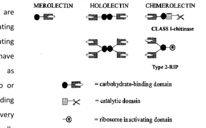
Peumans and Van Damme (62) distinguished lectins in three major types based on their structure, namely “merolectins”, “hololectins” and “chimerolectins”. Merolectins (Figure 5) are small, single polypetide proteins, which are built exclusively of a single carbohydrate-binding domain. Because of its
monovalent nature, they are incapable of precipitating glycoconjugates or agglutinating cells. Hololectins (Figure 5) have similar structural pattern as merolectins, but contain two or more carbohydrate-binding domains, which are very homologous. The majority of all known plant lectins are
comprised in this group, because they have multiple binding sites, being capable of agglutinating cells or precipitating glycoconjugates (62) (63). Chimerolectins (Figure 5) are a group of fusion proteins which possess a carbohydrate-binding domain and an unrelated domain with another biological activity (for example catalytic activity) that acts independently of carbohydrate-binding domain. Chimerolectins can behave as merolectins or hololectins, depending of the number of sugar-binding sites (62) (63). For example, type 2 ribosome-inactivating proteins (RIPs) with two carbohydrate-binding sites on their B chain (e.g. ricin) agglutinate cells, whereas class I plant chitinases with a single chitin-binding domain do not (62) (64).
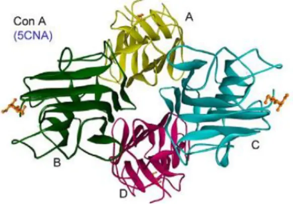
The first lectin discovered was ricin, a very toxic protein from Ricinus communis. It was extracted by Stillmark in 1888 (65). The first lectin isolated, sequenced and with tree-dimensional structure determined by x-ray crystallography, was ConA from Canavalia ensiformis (Figure 6). ConA is the best characterized plant lectin, with a high number of studies
Figure 5. Schematic representation of three types of plant lectins: merolectins, hololectins and chimerolectins (62).
Introduction
15 (biochemical, biophysical and structural) (65) (66). ConA is a Diocleinae (Leguminosae) lectin and all Diocleinae lectins studied have many
chemical, physic-chemical and structural properties in common. They share the same monosaccharide specificity for glucose and D-mannose and, despite their high similarity, they have distinct biological activities (9) (67) (68). The different biological activities are mainly due to changes in three parameters, such as binding specificity to complex carbohydrates, pH-dependent oligomerization state and the
relative orientation of carbohydrate-binding sites. These differences in the described parameters among the lectins may result from specific amino acid replacements at key positions along their primary structures (66).
Lectins may interact with carbohydrates through hydrogen bounds, Van der Waals forces, metal coordination and hydrophobic interactions. Essentially, hydroxyl groups on sugar molecules can serve both as a donor and an acceptor to cooperate in hydrogen bonds (69-71). Most plant lectins studied are biosynthesized via the secretory pathway. Lectins are synthesized at ribosomes attached to the endoplasmic reticulum (ER). Then, lectins enter the lumen of the ER and are further transported through the Golgi apparatus (where lectins are modified by the glycosylational process), ending up in the vacuole (65) (72).
Lectin sources
Lectins are widely distributed in nature as they can be found in all kingdoms of life, ranging from animals, microorganisms (bacteria, yeasts and viruses) and plants (55) (58) (73).
Plants were the first discovered source of lectins, and remain the most frequently used (57).
Plant lectins can be framed in, at least, seven distinct families. The mainly four families of structurally and evolutionary related proteins are the legume lectins, the type 2 RIPs, the chitin-binding lectins, and the monocot mannose-binding lectins (74) (75). Legume lectins are the largest and most studied family of plant lectins. Some of the best known legume lectins are jackbean (ConA), soybean (SBA) and phytohemagglutinin (PHA) (76).
Additionally, smaller families (Cucurbitaceae phloem lectins, amaranthins and jacalin-related lectins) have also been characterized (74) (75).
Figure 6. The X-ray structure of ConA (adapted from (68)).
Introduction
16
Different families of plants, as well as different tissues of the same plant, can contain different lectins with different bioactivities and different carbohydrate-binding specificities (57) (77).
The richest sources of most lectins are the storage organs of plants, especially seeds. But also roots (Urtica, Calystegia), tubers or bulbs (Allium, Tulipa), bark (Sophora, Robinia) or leaves (Aloe, Lactuca) can contain significant amounts of lectins. The amount of lectin can be different from one plant species to other (65).
Biological activities and applications of lectins
Monosaccharides, such as mannose, galactose, acetylgalactosamine, N-acetylglucosamine, fucose and N-acetylneuraminic acid, are typical constituents of surfaces of eukaryotic cells. Almost all lectins have high specificity for the sugars describes above, being this fact relevant for biological activities of lectins. The binding of lectins is dynamic and reversible. The association constant of lectin-monosaccharide interactions is relative low (in millimolar range) and lower than lectin-oligosaccharide interactions, however it is often highly selective. This is due to the fact that lectins exhibit an exquisite specificity for di-, tri-, and tetrasaccharides (76).
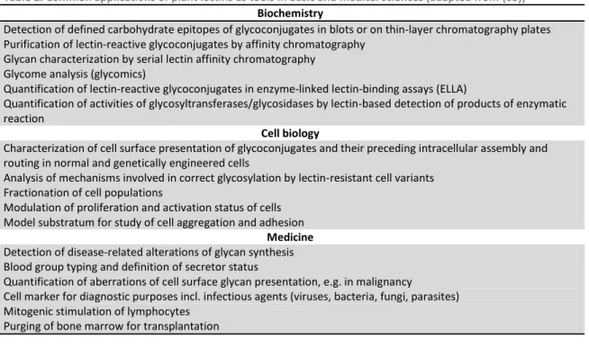
Lectins ability to bind carbohydrates with considerable specificity makes them valuable tools in scientific research, particularly in clinical, biochemical and biotechnology studies (76) (78). Some examples of common applications of plant lectins as tools in basic and medical sciences are demonstrated in Table 2.
Table 2. Common applications of plant lectins as tools in basic and medical sciences (adapted from (65))
Biochemistry
Detection of defined carbohydrate epitopes of glycoconjugates in blots or on thin-layer chromatography plates Purification of lectin-reactive glycoconjugates by affinity chromatography
Glycan characterization by serial lectin affinity chromatography Glycome analysis (glycomics)
Quantification of lectin-reactive glycoconjugates in enzyme-linked lectin-binding assays (ELLA)
Quantification of activities of glycosyltransferases/glycosidases by lectin-based detection of products of enzymatic reaction
Cell biology
Characterization of cell surface presentation of glycoconjugates and their preceding intracellular assembly and routing in normal and genetically engineered cells
Analysis of mechanisms involved in correct glycosylation by lectin-resistant cell variants Fractionation of cell populations
Modulation of proliferation and activation status of cells Model substratum for study of cell aggregation and adhesion
Medicine
Detection of disease-related alterations of glycan synthesis Blood group typing and definition of secretor status
Quantification of aberrations of cell surface glycan presentation, e.g. in malignancy
Cell marker for diagnostic purposes incl. infectious agents (viruses, bacteria, fungi, parasites) Mitogenic stimulation of lymphocytes
Introduction
17
Blood typing
Lectins are frequently used for applications based on agglutination or precipitation reaction (76). ConA is capable of agglutinates erythrocytes and yeasts, as consequence of lectin interaction with carbohydrates on the surface of the cells. Lectins with ability to agglutinate erythrocytes are named as hemagglutinins (79).
Most legume lectins have hemagglutinating activity and most of them also exhibit blood type specificity, so they can recognize the difference in blood group antigens having the carbohydrate epitopes (ABO, Le, Ii, T, Tn, Forssman antigens, etc.). Studies revealed that extracts of the asparagus pea (Lotus tetragonolobus) only agglutinated blood type O erythrocytes, whereas crude extracts of the lima bean (Phaseolus limensis) agglutinated only blood type A erythrocytes. The lectin from Dolichos biflorus is used to distinguish between A1
and A2 blood subgroups (63) (76) (79).
Hemagglutinins which exhibit blood type specificity played a key role in early investigations on the structural basis of the specificity of the antigens associated with the ABO blood group system (79).
Antibacterial and antifungal activity
Owing to the physiological functions of plant lectins in nature (plant’s defense), antibacterial and antifungal activity of lectins has been intensively discussed (62). Interactions between lectins and human pathogenic microorganisms were reported since many years ago (58). Lectins are extensively used in microbiology and parasitology domain for identification of the agent structures and with protective effect against infection (66).
Recently, several studies were carried out to verify whether lectins have some antibacterial and/or antifungal activity. Cavalcante and colleagues (80) verify that Staphylococcus mutans growth was affected by ConBol, ConBr and ConM lectins. Oliveira and colleagues (58) demonstrated that a lectin from Eugenia uniflora (EuniSL) strongly inhibited the growth of S. aureus, P. aeruginosa and Klebsiella sp. and moderately inhibited the growth of Bacillus subtilis, Streptococcus sp. and Escherichia coli. Santi-Gadelha and colleagues (77) used a lectin from Araucaria angustifolia (Aal) against Gram-positive and Gram-negative bacteria concluding that this lectin had stronger antimicrobial activity. They observed, through electron microscopy, the presence of pores and severe disruption of the Gram-positive bacteria membrane and substantial bubbling in Gram-negative cell wall, besides some destroyed bacteria.
Lectins from the seeds of Phaseolus vulgaris and Pisum sativum (81), from the mushroom Astragalus mongholicus (82), and from the pepper seed Capsicum frutescens (83),
Introduction
18
have been reported to have antifungal activity. Antifungal properties have also been found from the pepper seeds Capsicum annuum by Kuku and colleagues (83), and from a chitin-binding lectin from Talisia esculenta (84). This latter lectin had antifungal activity against Fusarium oxysporum, Saccharomyces cerevisiae, etc.
Charungchitrak and colleagues (57) demonstrated that a lectin from the seeds of Archidendron jiringa had antifungal activity against, for example, Exserohilum turcicum and Colletotrichum cassiicola, and antibacterial activity against Gram-positive and Gram-negative bacteria, such as P. aeruginosa and S. aureus. These results indicated that some lectins are able to inhibit both bacteria and yeasts pathogens (57).
Mitogenic stimulation
The first lectin found with mitogenic activity was PHA (lectin from Phaseolus vulgaris), but nowadays there are several lectins with this capacity. A mitogenic lectin possesses the ability to stimulate lymphocytes to undergo mitosis. The activation of lymphocytes occurs as a result of binding of lectins to cell surface sugars (79) (85).
Lectins such as PHA and ConA are able to stimulate T lymphocites (T cells), while pokeweed mitogen (PWM) is able to stimulate both T and B cells. This stimulation by lectins provides an easy and simple means to assess the immunocompetence of patients which suffer from a diversity of diseases, including AIDS (Acquired Immune Deficiency Syndrome), and to monitor the effects of various immunosuppressive and immunotherapeutic manipulations (76).
Antiproliferative activity and cancer diagnosis
Lectins have been shown to be of great interest since they have been reported as potential anticancer agents. They have been tested as adjuvants in tumor treatment and in diagnosis (66).
For example, lectins like Agaricus bisporus, Tricholoma mongolicum and Volvariella volvacea have antiproliferative and antitumor action. Wang and colleagues (86) demonstrated that lectin from Pleurotus ostreatus (POL) when administrated in mice promotes a dramatic shrinkage of sarcoma S-180 and hepatoma H-22 cells, having a potent antitumor activity. A similar situation was observed when three cancer cell lines (HeLa, L929 and EATC) were treated with ConA and PHA-P lectins. These lectins inhibited cell proliferation and caused damages on cell lines used, being the effect of PHA-P stronger than ConA. Cells showed morphological changes (damages), such as dilatation of mitochondria, cytoplasmic vacuolation, rounded cells and cell aggregation (87).
Introduction
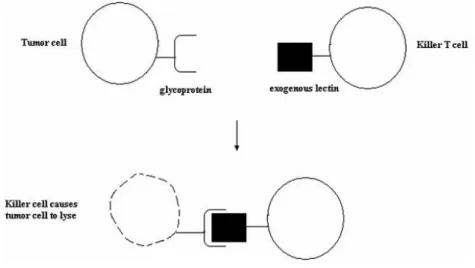
19 Development of cancer cells seems to be associated with changes in cell surface sugars which lead to the assumption that high susceptibility to agglutination by lectins was a property shared by malignant cells. Wheat germ agglutinin (WGA) and ConA have the ability to selectively agglutinate neoplastic cells (79). Because malignant cells display altered surface glycoproteins, exogenous lectins tagged with the killer T cells can promote the lyses of tumor cells, controlling tumor (Figure 7). Also, lectins present on the surface of malignant cells are able to binding exogeneous sugars which contain molecules and are capable of internalizing them by endocytosis, having a potential use in cancer treatment strategies (63).
Bone marrow transplantation
Some lectins have been used in organ transplantation applications. Selective agglutination by SBA allows separation of B and T mouse splenocytes (bone marrow). Since 1981 SBA lectin is used for purging human bone marrow for transplantation. It is employed frequently for transplantations into children born with severe combined immune deficiency (SCIDs or “bubble children” – they are highly susceptible to microbial infections). About 75 % of the hundreds of children transplanted after SBA purging of bone marrow have been cured and lead a normal life. SBA-purged bone marrow was also used experimentally for treatment of end stage leukemia patients, as an alternative to other techniques for T cell depletion, such as monoclonal antibodies (76) (85).
Figure 7. Exogenous lectin attached to the T cell targeting the altered glycoprotein present on tumor cell surface, promoting the lyses of the cell (63).
Materials and Methods
22
2.1. Lectins origin
The lectins Canavalia ensiformis - ConA; Canavalia brasiliensis - ConBr; Canavalia maritima – ConM and Canavalia boliviana - ConBol were gently provided by Professor Benildo Cavada from BioMol-Lab (Laboratory of Biologically Actives Molecules, Department of Biochemistry and Molecular Biology, Federal University of Ceará - Brazil).
2.2. Lectins stock solution
Phosphate buffered solution (PBS) was prepared in sterilized water being added 8 g/L of sodium chloride (NaCl), 0.2 g/L of potassium chloride (KCl), 0.3 g/L of potassium dihydrogen phosphate (KH2PO4) and 1.15 g/L of disodium phosphate (Na2HPO4). The pH was adjusted to
7.0. Before being used, this solution was autoclaved at 121 ˚C for 15 minutes and reserved at room temperature.
The Diocleinae lectins were solubilised in PBS previously prepared, at 37 °C and 120 rpm for 3 h, in order to obtain stock solutions with a concentration of 1 mg/mL. After that, the solutions were filtered through a 0.22 µm cellulose filter and preserved at 4 °C.
Immediately prior to experiments, the lectin stock solution was diluted in Tryptic Soy Broth
(
TSB; Liofilchem) for bacteria, and in Sabouraud Dextrose broth (SDB; Liofilchem) for yeasts, to obtain the concentrations of 62.5, 125, 250 and 500 µg/mL of lectin. Then, bacteria and yeasts suspensions (after its concentrations has been adjusted) were added to the diluted solutions of lectins, obtaining the final concentrations of 31.2, 62.5, 125 and 250 µg/mL of lectins used in experiments.2.3. Microorganisms and culture conditions
The microorganisms used in the experiments are all reference strains from American Type Culture Collection (ATCC) and Spanish Type Culture Collection (CECT). This included two Gram-positive bacteria (Staphylococcus epidermidis CECT231 and Staphylococcus aureus ATCC), two Gram-negative bacteria (Klebsiella oxytoca ATCC13182 and Pseudomonas aeruginosa ATCC10145), and two yeasts (Candida albicans ATCC1472 and Candida tropicalis ATCC750).
Bacteria were preserved at – 80 ˚C in TSB supplemented with 20 % glycerol. Yeasts were preserved at – 80 ˚C in SDB supplemented with 10 % glycerol.
Prior to each experiment, bacteria and fungi cells were grown, respectively, on Tryptic Soy Agar (TSA; Merck) and on Sabouraud Dextrose Agar (SDA; Liofilchem) plates for 24 h, at 37
Materials and Methods
23 ˚C. These media were previously prepared and autoclaved at 121 ˚C for 15 minutes and reserved at room temperature.
After 24 h of growth on solid media, an isolated colony of each microorganism was removed and used to inoculate 20 mL of TSB or SDB, and incubated for 18 h, at 37 °C, under constant agitation of 120 rpm.
Prior to use, the bacteria and yeasts suspensions were adjusted to a final concentration of 2 x 10⁸ cells/mL (for early adhesion characterization) and 1 x 10⁶ cells/mL (for planktonic and biofilm growth tests). Bacterial suspensions were adjusted through the measurement of the optical density (OD) at 640 nm and calibration curves, previously determined for each bacterium. The yeasts inocula were adjusted using a Neubauer chamber.
2.4. Lectin antimicrobial activity
All bacteria and yeast suspensions were adjusted to 1 x 10⁶ cells/mL, and then 100 µL of each suspension were transferred to a 96-well tissue culture plates (Orange Scientific), previously prepared with 100 µL of TSB or SDB supplemented with a dose of each lectin (described on section 2.2). These plates were incubated during 24 h at 37 °C and 120 rpm.
Microbial growth was determined through measurement of optical density at 640 nm of the liquid content of each well using a microtiter plate reader, being the bacteria and yeast planktonic growth results presented as OD640 nm.
Negative control, wells contained culture medium only, was also performed in order to determine whether the tested medium interfere with the measurement of planktonic growth.
2.5. Lectin effect on early-stage adhesion
To verify the effect of lectins on bacteria and yeast early-stage adhesion, the cells were put in contact with a range of concentrations of all lectins, during 2 h at 37 °C and 120 rpm. Each bacteria and yeast suspensions were previously adjusted to 2 x 10⁸ cells/mL. Then, 500 µL of each cell suspension was transferred to a 24-well tissue culture plates (Orange Scientific), containing 500 µL of TSB or SDB per well, supplemented with a dose of each lectin (described on section 2.2).
After 2 h of contact, the content of each well was removed, the adhered cells were washed twice with sterilized water, being the biomass quantified by crystal violet (CV) staining, according to Stepanovic and colleagues (88). For that, the plates containing the adhered cells were left to air dry for 30 min, and 1 mL of 98 % methanol were transferred to each well in
Materials and Methods
24
order to fix the remaining attached cells, for 15 min. Afterwards, the plates were emptied and left to air dry. The fixed cells were stained with 1 mL of CV (Gram’s staining; Merck) per well, for 5 min. After this staining step, plates were washed with running tap water, air dried, and filled with 1 mL of 33 % (v/v) of acetic acid (Merck) in order to resolubilize the CV bound to the adherent cells. After that 200 µL of each well was transferred to a 96-well tissue culture plates and the quantitative analysis of adhered cells was performed through the measurement of optical density at 570 nm of each well using a microtiter plate reader, being the results presented as OD570nm. Negative control, wells contained culture medium only, was also
performed in order to determine whether the tested media and the plate material could adsorb CV and interfere with the adhered cells quantification.
2.6. Biofilm prevention
To assess whether the presence of the lectins could interfere with the establishment of biofilms by bacteria and yeasts on polystyrene (PS) surfaces, biofilms (24 h) were allowed to form in the presence of a range of concentrations of all lectins. Biofilms were developed using the microtiter plate test developed by Stepanovic and colleagues (88). Briefly, 100 μL of each bacteria and fungi suspensions, previously adjusted to 1 x 10⁶ cells/mL, were transferred to each well of a 96-well tissue culture plates (Orange Scientific) containing 100 μL of TSB or SDB per well supplemented with a dose of each lectin (described on section 2.2).
After 24 h of biofilm growth, the content of each well was removed; biofilms were washed twice with sterilized water and reserved for further characterization.
2.6.1. Biofilm biomass quantification
The biomass of the biofilms was quantified by a staining method, as described for early adhesion. For quantification of the biofilm mass, the quantities of methanol, CV and acetic acid were 200 μL per well. After resolubilize the CV bound to the adherent biofilm, the quantitative analysis of biofilm mass was performed through the measurement of optical density at 570 nm of each well using a microtiter plate reader, being the biofilm mass presented as OD570nm.
Negative control, wells contained culture medium only, was also performed in order to determine whether the tested media and the plate material could adsorb CV and interfere with the biofilm biomass quantification.
Materials and Methods
25
2.6.2. Biofilm viable cells enumeration
In order to determine the number of viable biofilm-entrapped cells, biofilm suspensions were prepared.
For the bacterial biofilms, 200 µL of sterilized water were added to each well, being the biofilms detached by ultrasonic bath in a Sonicor SC-52 (Sonicor Instruments) operating at 50 kHz, during 6 min. The yeasts biofilm cells suspensions were obtained by completely scraping the adhered biomass from the wells, through the use of a yellow tip, into 200 µL of sterilized water. Afterwards, the biofilm suspensions of each 2 wells per condition were collected and gently vortexed for 1 min to disrupt possible cell aggregates (these parameters were previously optimized in order to promote the complete removal of all the biofilm-attached cells without lysis).
Biofilm suspensions were then serially diluted, plated on TSA (bacterial suspensions) or on SDA (yeasts suspensions), and incubated at 37 °C in an aerobic incubator for 24 h. The number of colony forming units (CFU) was enumerated, being the biofilm cell numbers presented as log10 (CFU/cm2).
2.7. Statistical analysis
Statistical analyses were performed by PASW Statistics. The method used was one-way ANOVA with Tukey post hoc test. Data were obtained in triplicates from, at least, three separate experiments. The results were presented as mean ± standard deviation (SD). A P-value 0.05 was considered indicative of statistical significance.
Results
28
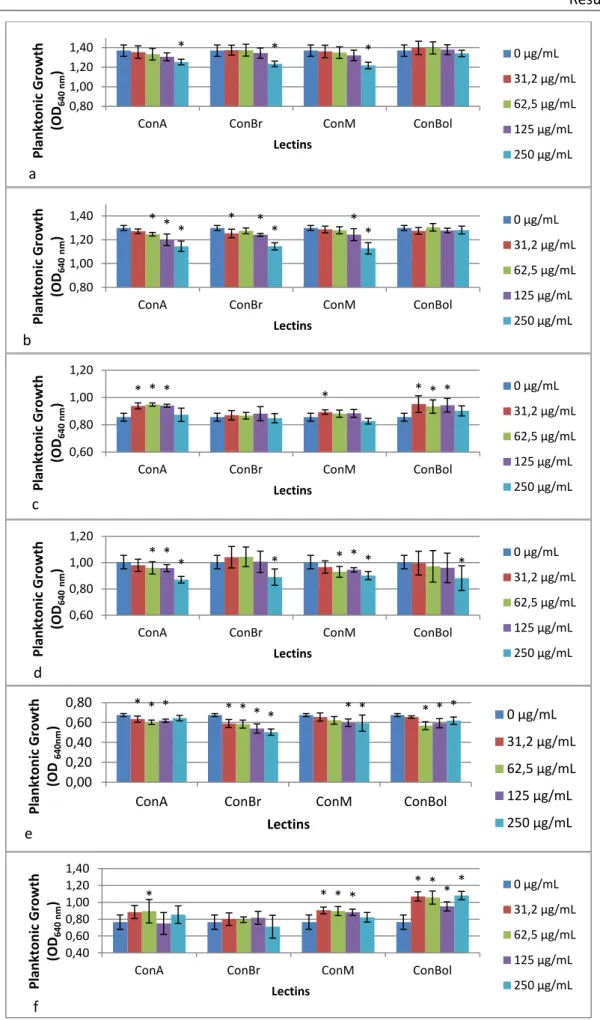
3.1. Bacterial and yeast planktonic growth
After 24 h in contact with lectins, bacteria and yeast growth was determined through measurement of optical density at 640 nm. The effect of lectins on microbial planktonic growth is shown in Figure 8.
For P. aeruginosa (Figure 8a) and K. oxytoca (Figure 8b), both Gram-negative bacteria, the higher concentration of the all lectins, with the exception of ConBol, showed inhibitory activity (p<0.05).
Curiously, ConA and ConBol stimulated (p<0.05) the growth of S. aureus (Figure 8c). ConBr did not demonstrate significant differences compared to the control (p>0.05). In opposition, higher concentrations of all lectins had antimicrobial activity (p<0.05) against S. epidermidis growth (Figure 8d).
All lectins showed inhibitory activity (p<0.05) for C. albicans, but they enhanced the growth (p<0.05) of C. tropicalis, with exception of ConBr which had no significant effect (p>0.05).
Results
29
Figure 8. Effect of lectins on bacteria and yeast planktonic growth (a – Pseudomonas aeruginosa; b - Klebsiella
oxytoca; c – Staphylococcus aureus; d - Staphylococcus epidermidis; e – Candida albicans; f – Candida tropicalis).
*Statistically different from the control, 0 µg/mL (p < 0.05).
* * *
0,80 1,00 1,20 1,40
ConA ConBr ConM ConBol
Pl an kt o n ic Gr o wt h (OD 6 40 nm ) Lectins 0 µg/mL 31,2 µg/mL 62,5 µg/mL 125 µg/mL 250 µg/mL a * * * * * * * * 0,80 1,00 1,20 1,40
ConA ConBr ConM ConBol
Plan kt o n ic G ro w th (OD 640 nm ) Lectins 0 µg/mL 31,2 µg/mL 62,5 µg/mL 125 µg/mL 250 µg/mL b * * * * * * * 0,60 0,80 1,00 1,20
ConA ConBr ConM ConBol
Pl an kt o n ic Gr o wt h (OD 6 4 0 nm ) Lectins 0 µg/mL 31,2 µg/mL 62,5 µg/mL 125 µg/mL 250 µg/mL c * * * * * * * * 0,60 0,80 1,00 1,20
ConA ConBr ConM ConBol
Pl an kt o n ic Gr o wt h (OD 640 nm ) Lectins 0 µg/mL 31,2 µg/mL 62,5 µg/mL 125 µg/mL 250 µg/mL d * * * * * * * * * * * * 0,00 0,20 0,40 0,60 0,80
ConA ConBr ConM ConBol
Pl an kt o n ic Gr o wt h (OD 6 4 0 nm ) Lectins 0 µg/mL 31,2 µg/mL 62,5 µg/mL 125 µg/mL 250 µg/mL e * * * * * * * * 0,40 0,60 0,80 1,00 1,20 1,40
ConA ConBr ConM ConBol
Pl an kt o n ic Gr o wt h (OD 6 4 0 nm ) Lectins 0 µg/mL 31,2 µg/mL 62,5 µg/mL 125 µg/mL 250 µg/mL f