UNIVERSIDADE DO ALGARVE
Departamento de Ciências
Biomédicas e Medicina
Polyphenols neuroprotective effect in a
Parkinson’s disease yeast model: phosphoproteome
alterations
Vítor Hugo da Silva Gonçalves
Dissertação para obtenção do Grau de Mestre em
Ciências Biomédicas
Trabalho efetuado sob orientação de:
Cláudia dos Santos, Ph.D., IBET/ITQB-UNL
Sandra Tenreiro, Ph.D., IMM-UNL
Inês Araújo, Ph.D., CBME-UAlg
ii
Polyphenols neuroprotective effect in a Parkinson’s disease yeast model:
phosphoproteome alterations
Declaração de autoria de trabalho
Declaro ser o(a) autor(a) deste trabalho, que é original e inédito. Autores e trabalhos consultados estão devidamente citados no texto e constam da listagem de referências incluída.
Copyright
“A Universidade do Algarve tem o direito, perpétuo e sem limites geográficos, de arquivar e publicitar este trabalho através de exemplares impressos reproduzidos em papel ou de forma digital, ou por qualquer outro meio conhecido ou que venha a ser inventado, de o divulgar através de repositórios científicos e de admitir a sua cópia e distribuição com objetivos educacionais ou de investigação, não comerciais, desde que seja dado crédito ao autor e editor.”
iii
“A lesson without pain is meaningless. For you cannot gain anything without sacrificing something else in return.”
iv
Acknowledgments
I would never have been able to finish my dissertation without the guidance of my supervisors, help from friends, and support from my family. All words will be few to express my gratitude to all who helped me in this stage of my life.
First of all I would like to thank to Inês Araújo, Ph.D., my Neuroscience teacher and supervisor, who always opened the doors of neuroscience and for all the help over my scientific career.
In special, would like to express my deepest gratitude to Cláudia dos Santos, Ph.D., for her excellent guidance, caring, patience, and providing me with an excellent atmosphere for doing research.
To Sandra Tenreiro, Ph.D., a special thanks for accepting to be my supervisor and for introducing me to “the yeast world”.
My sincerely gratitude to Professor Ricardo Boavida Ferreira for being an admirable teacher, and for allowing me to developed this work on his laboratory and for all the knowledge transmitted during this year.
I cannot forget to express my gratitude to all the Disease and Stress Biology lab team, for all the support and cooperation gave during this year. My research would not have been possible without their helps.
Last but not the least, I would like to thank my family, especially my mother, father and my little brothers. They were always supporting me and encouraging me with their best wishes.
v
Abstract
Parkinson’s disease (PD) is a massive chronic progressive disorder characterized by the intracellular depositions known as Lewy Bodies (LBs), mainly composed of alpha-synuclein protein (aSyn). These proteinaceous aggregates are associated with the loss of dopaminergic neurons. aSyn aggregation is thought to be a key event in the PD pathogenesis leading to the formation of LBs.
Thus is extremely important to search and explore therapeutic agents that prevent or reduce aSyn toxicity. The consumption of Polyphenols-rich foods and beverages has been suggested to limit the neurodegeneration associated with a variety of neurological disorders and to prevent or reverse abnormal deteriorations in cognitive performance. These metabolites are consider to exert modulatory actions on cellular systems through direct action on various signalling pathways.
In this study it was analysed the effects mediated by a polyphenol-enriched fraction (PEF) from leaves of Corema album in a yeast model of PD expressing aSyn protein. In an effort to obtain new insights into the protective mechanisms of C. album leaf PEFs, as well as the signaling pathways potentially affected by their bioactivity and by aSyn expression, we performed a two-dimensional electrophoresis based quantitative and phosphoproteomic analysis.
It was possible to find several proteins that were either differentially expressed or differentially phosphorylated in response to aSyn expression or due to the incubation of yeast cells with C. album leaf PEFs. Also, the results obtained from growth and immunodetection assays suggest that C. album PEFs are able to protect yeast cells from the aSyn induced toxicity increasing their viability.
These results imply C. album polyphenols from leaves, as potential therapeutic agent directed towards preventing the aSyn induced toxicity. The understanding of the molecular effects of polyphenols will open novel opportunities for the exploration of their molecular effects and for drug development.
vi
Resumo
A doença de Parkinson (DP) é uma desordem neurodegenerativa, progressiva e crónica caracterizada por deposições intracelulares denominadas corpos de Lewy (CL), que são maioritariamente compostos pela proteína alfa-sinucleína (aSyn). Estes agregados proteináceos estão associados com a degeneração dos neurónios dopaminérgicos.
Assim é extremamente importante investigar possíveis agentes terapêuticos que sejam capazes de reduzir ou prevenir a toxicidade da aSyn. O consumo de alimentos e bebidas ricos em polifenóis estão associados a uma limitação na neurodegeneração associada a várias desordens neurológicas e a prevenir ou reverter deteriorações na performance cognitiva. Estes metabolitos exercem alterações nos sistemas celulares através do interação com várias vias de sinalização.
Neste estudo foi analisado o efeito de frações enriquecidas em polifenoís (FEPs) de folhas de C. album num modelo de PD em levedura com expressão da proteína aSyn. Num esforço para obter novo conhecimento em relação ao mecanismo de proteção dos PEFs da folha de C. album, assim como, potencias vias de sinalização afetadas pela sua bioatividade e pela expressão da aSyn, foi realizada uma análise quantitativa e fosfoproteomica através da eletroforese bidimensional.
Foi possível encontrar várias proteínas que são diferencialmente expressas ou fosforiladas em resposta à expressão da aSyn ou devido à incubação das leveduras com PEFs da folha de C. album. Além disso, os resultados obtidos a partir dos crescimentos celulares e ensaios de imunodetecção sugerem que os PEFs da folha de C. album são capazes de proteger as leveduras a partir da toxicidade induzida pela aSyn e aumentar a viabilidade celular.
Estes resultados implicam os polifenóis de folhas de C. album, como potencial agente terapêutico para prevenir a toxicidade induzida pela aSyn. A compreensão dos efeitos moleculares dos polifenóis abrirá novas oportunidades para a exploração dos seus efeitos moleculares e desenvolvimento de agentes terapêuticos.
vii
Resumo alargado
As doenças neurodegenerativas são caracterizadas por morte neuronal excessiva e prematura em regiões focais do cérebro. Entre elas a doença de Parkinson (DP) é uma das mais comuns.
Em termos patológicos a DP é caracterizada por uma perda profunda e progressiva dos neurónios dopaminérgicos, localizados na SNpc do mesencéfalo, região localizada na porção superior do tronco encefálico, originando reduções de dopamina na via nigroestriatal. Esta perda progressiva dos neurónios dopaminérgicos está deve-se à presença de corpos de Lewy (CL), agregados proteicos compostos maioritariamente pela proteína aSyn. aSyn é uma proteína que está presente nos terminais pré-sinápticos onde se encontra associada ao trafego intracelular.
A agregação desta proteína em CL esta associada a vários processos patológicos como disfunção mitocondrial e consequente stress oxidativo, disfunção do sistema da ubiquitina/proteassoma (UPS) e vias de autofagia, e bloqueio da libertação de dopamina nos terminais pré-sinápticos.
Uma vez que a medicação atual apenas atenua sintomas da DP, sem parar ou retardar a degeneração de neurónios dopaminérgicos, existe uma necessidade urgente para a identificação de novos agentes terapêuticos que sejam capazes de contrariar a toxicidade da aSyn. Os polifenóis ou compostos fenólicos são fitoquímicos amplamente distribuídos no reino vegetal. Os polifenóis estão presentes em plantas, frutas e vegetais, incluindo pequenos frutos, azeite e vinho. O consumo de alimentos ricos em polifenóis tem sido associado a efeitos benéficos para a saúde humana. Estudos indicam que existe uma relação entre o consumo de alimentos ricos em polifenóis e uma diminuição da incidência de doenças neurodegenerativas.
Estes compostos estão envolvidos num largo espectro de mecanismos de ação celulares, como é o caso da regulação de vias de sinalização relacionadas com a modulação de genes envolvidos em mecanismos morte sobrevivência e proliferação celular, regulação da função mitocondrial e ativação de moléculas antioxidantes endógenos. Assim, os compostos fenólicos surgem como candidatos a possíveis agentes terapêuticos na DP.
A levedura Saccharomyces cerevisiae é um dos modelos biológicos mais versáteis para estudar doenças neurodegenerativas, devido ao facto de mecanismos relacionados com a disfunção mitocondrial e proteossomal e regulação transcricional serem
viii
extremamente bem conservados entre leveduras e humanos, possibilitando assim estudar os mecanismos envolvidos neste tipo de doenças.
Neste estudo o principal objetivo consiste em analisar o efeito de frações enriquecidas de polifenoís (FEPs) de folhas de Corema album, uma espécie Portuguesa, no proteoma e fosfoproteoma de um modelo biológico de PD em levedura.Com esta análise, torna-se possível estudar a rede de proteínas do nosso modelo de PD em levedura, bem como a modulação na expressão proteica mediada pelos polifenoís de C. album.
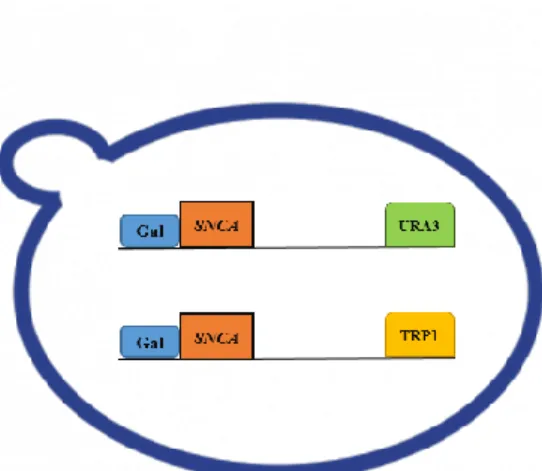
O primeiro passo deste trabalho consistiu na otimização de um modelo de PD em levedura. O modelo e composto por 3 estirpes de S. cerevisiae; aSyn-1 e aSyn-2 ambas com o mesmo genoma (MAT alpha can1-100 his3-11 15 leu2-3 112 ade2-1 GAL1pr-syn
WT::TRP1 GAL1pr-syn WT::URA3) que possuem um plasmídeo integrativo, com 2
cópias do gene SNCA (aSyn). A expressão da proteína aSyn é regulada por um promotor da galactose. Por ultimo, a estirpe controlo não apresenta o gene SNCA (can1-100
his3-11 15 leu2-3 his3-112 pRS304::TRP1 pRS306::URA3 ade2-1).
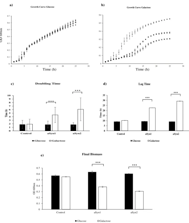
Numa primeira análise foram realizadas curvas de crescimento para as estirpes aSyn-1, aSyn-2 e controlo em diferentes meios de cultura. Os resultados obtidos indicam que a expressão da proteína aSyn nas estirpes aSyn-1 e aSyn-2 é toxica levando a decréscimo na viabilidade celular das leveduras. Contudo, os resultados mostram que a estirpe aSyn-2 é mais sensível aos efeitos tóxicos de expressão aSyn, portanto, esta estirpe foi selecionada para os estudos posteriores.
A próxima etapa permitiu avaliar o efeito exercido por FEPs obtidas a partir de folhas de C. album em leveduras que expressam a proteína aSyn. Assim, usando duas metodologias, curvas de crescimento e “spot assays”, as estirpes controlo e aSyn-2 foram incubadas com várias concentrações de FEPs de folhas de C. album. Em ambos os métodos foi possível observar que várias concentrações de FEPs de folhas de C. album reduzem a toxicidade induzida pela expressão da aSyn na estirpe aSyn-2, e promovem uma melhoria na viabilidade celular.
Numa fase mais avançada do trabalho, a análise do proteoma total e fosfosproteoma por eletroforese bidimensional revelou que a expressão da aSyn e o tratamento das leveduras com FEPs de folhas de C. album promovem alterações na expressão proteica de várias proteínas.A análise do proteoma total permitiu encontrar 22 proteínas que apresentaram variações seu padrão de expressão. Curiosamente foi verificado que a maioria dessas proteínas se encontra “downregulated” devido a expressão da aSyn, mas fascinantemente, o tratamento com PEFs recupera o padrão
ix
normal de expressão dessas proteínas. Estes resultados sugerem que os PEFs de folhas de
C. album possivelmente estão a modular vias sinalização inerentes à aSyn.
Relativamente à análise do fosfoproteoma das estirpes controlo e aSyn-2, foram detetadas 10 proteínas que apresentaram variações no nível de fosforilação. Curiosamente foi observada uma tendência no nível de fosforilação apresentado por estas proteínas, onde a incubação das leveduras da estirpe aSyn-2 com FEPs reduz o nível de fosforilação comparativamente às leveduras da mesma estirpe que não receberam o tratamento. Foi observado que a expressão da aSyn aumenta a fosforilação na estirpe aSyn-2, relativamente estirpe controlo. A modulação dos níveis de fosforilação das proteínas devido ao tratamento com FEPs de folhas de C. album indica que estes compostos provavelmente possuem a capacidade de ativar ou inibir vias de sinalização relacionadas com a fosforilação proteica. Sabendo que a fosforilação da aSyn está implícita na formação dos CL, estes resultados sugerem que os polifenóis provenientes das folhas de
C. album podem ser promissores agentes terapêuticos na DP.
Observou-se ainda através de ensaios de imunodeteção que leveduras da estirpe aSyn-2 que foram incubadas com FEPs de C. album exibem níveis inferiores da proteína aSyn relativamente a leveduras que não tiveram o mesmo tratamento com FEPs. Tendo em conta vários estudos indicando que os polifenóis são agentes capazes de modular os sistemas de autofagia celular, estes resultados sugerem que a proteção mediada pelos FEPs da folha da C. album pode estar relacionada com a degradação da aSyn via autofagia.
Apesar dos resultados promissores obtidos neste trabalho, estudos envolvendo a seleção de subproteomas serão necessários para uma melhor compressão dos mecanismos subjacentes aos efeitos neuroprotetores mediados pelos polifenóis das folhas de C. album.
Palavras-chave: Doença de Parkinson, levedura, polifenóis, aSyn, Frações enriquecidas de polifenóis
x
General index
Acknowledgments ... iv
Abstract ... v
Resumo ... vi
Resumo alargado ... vii
General index ... x
Index of figures ... xii
Index of tables ... xv
Abbreviations ... xvi
Goals ... 1
Introduction ... 2
Neurodegenerative Diseases ... 2
Parkinson’s disease, definition and historical perspective ... 2
Genetic determinants of Parkinson’s disease ... 4
Hallmarks of Parkinson’s disease ... 5
Alpha-Synuclein: structural and functional properties ... 8
Physiological function of Alpha-Synuclein ... 10
Alpha-Synuclein Aggregation ... 11
Alpha-Synuclein Phosphorylation ... 13
Mitochondrial Dysfunction and Oxidative Stress in PD ... 15
aSyn the principal player in PD ... 17
Polyphenols ... 18
Polyphenols in Neurodegeneration ... 19
The Awesome power of the Yeast tool ... 21
Parkinson’s disease Yeast Model ... 22
Proteomics approaches ... 23
Phosphoproteomics ... 24
Materials and Methods ... 26
Plant Material ... 26
Extract Preparation ... 26
Extract Fractionation ... 26
Measurement of Total Phenol Content ... 27
Yeast strains ... 27
Media and growth conditions ... 27
xi
Phenotypic Growth Assays ... 28
Protein extraction ... 29
Protein Quantification ... 29
SDS-PAGE ... 29
Western Blot ... 30
Immunodetection ... 30
Sample preparation for Two-Dimensional Electrophoresis ... 31
Two-Dimensional Gel Electrophoresis ... 31
Pro-Q® Diamond ... 32
Commassie Brilliant Blue G ... 32
2D Gel Analysis ... 32
Results and discussion ... 34
Establishment of the Yeast model of PD ... 34
Growth curves ... 34
Effects of polyphenols on yeast growth ... 37
Phenotypic Growth assays with polyphenols ... 39
Proteomic Approach ... 42
2DE protocol optimization ... 42
Proteome Analysis ... 44
Protein pattern of Control and aSyn-2 yeast strains with C. album leaf PEFs 44 SDS-PAGE-detection of phosphoproteins ... 53
Phosphoproteome of control and aSyn-2 yeast strains with C. album Leaf PEFs 54 C. album leaves PEF effects on aSyn protein ... 59
Final consideration and future perspectives. ... 62
Annex 1 - Western Blot - aSyn Immunodetection ... 65
xii
Index of figures
Figure 1: Immunohistochemical labeling of intraneuronal inclusions, termed LBs, in the SNpc dopaminergic neuron. Immunostaining with an antibody against aSyn
reveals a LB (black arrow) with an intensely immunoreactive central zone surrounded by a faintly immunoreactive peripheral zone (left image). Conversely, immunostaining with an antibody against ubiquitin yields more diffuse immunoreactivity within the LB (right image). (Adapted from reference7) ... 6
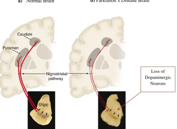
Figure 2: Neuropathology of PD a) Schematic representation of the normal nigrostriatal
pathway (in red). It is composed of dopaminergic neurons whose cell bodies are located in the substantia nigra pars compacta (SNpc; see black arrows). These neurons project (thick solid red lines) to the basal ganglia and synapse in the striatum (i.e., putamen and caudate nucleus). The image demonstrates the normal pigmentation of the SNpc, produced by neuromelanin within the dopaminergic neurons. b) Schematic representation of the diseased nigrostriatal pathway (in red). In PD, the nigrostriatal pathway degenerates. There is a marked loss of dopaminergic neurons that project to the putamen (dashed line) and a much more modest loss of those that project to the caudate (thin red solid line). The image demonstrates depigmentation (i.e., loss of dark-brown pigment neuromelanin; see black arrows) of the SNpc due to the marked loss of dopaminergic neurons. (Adapted from reference7). ... 7
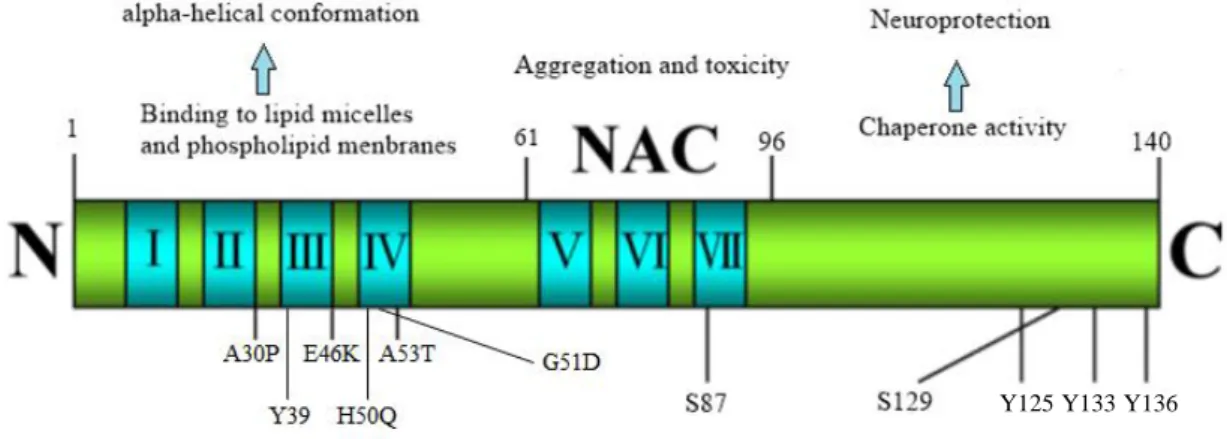
Figure 3: Molecular structure and functional characteristic of aSyn. aSyn is
functionally divided into N-terminal (1–60aa), NAC (61–95aa), and C-terminal (96– 140aa) domains. The N-terminal domain contains four motifs (blue color) and has five point mutation sites linked to PD (A30P, E46K, H50Q, G51D and A53T) and also a tyrosine residue (Y39) which is a phosphorylation target. The NAC domain, which encompasses the most hydrophobic residues, promotes aggregation, with a phosphorylation site (S87). The C-terminal domain exhibits chaperone activity that tends to decrease protein aggregation, has one phosphorylation site (S129) and three tyrosine residues (Y125, Y133, Y136). (Adapted from25,27,28) ... 9
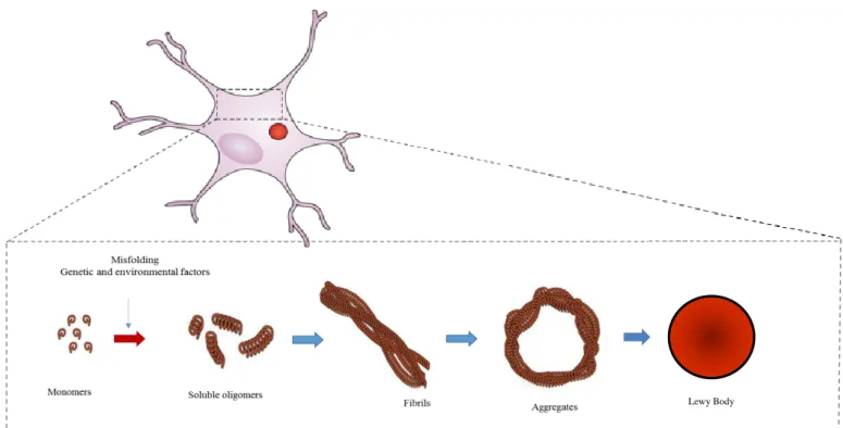
Figure 4: aSyn aggregation. Schematic illustration of the process whereby normal
soluble aSyn misfolds is converted into pathological oligomers and higher aggregates that fibrillize and deposit into Lewy bodies in affected neurons in PD brain. ... 12
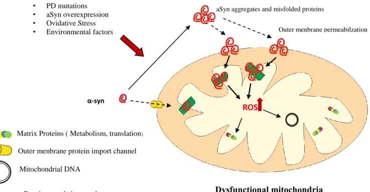
Figure 5: Effect of aSyn in mitochondria. PD mutations; aSyn overexpression;
oxidative stress and environmental factors are able to trigger aSyn aggregation and lead to mitochondrial dysfunction. Impairment of mitochondrial complex I activity by aSyn results in an increased production of ROS and promotes aSyn aggregation. Compromising mitochondrial function results in ATP deficits and ultimately will lead to a disruption of cellular homeostasis, abnormal protein aggregation and apoptosis. (adapted from reference20,49) ... 16
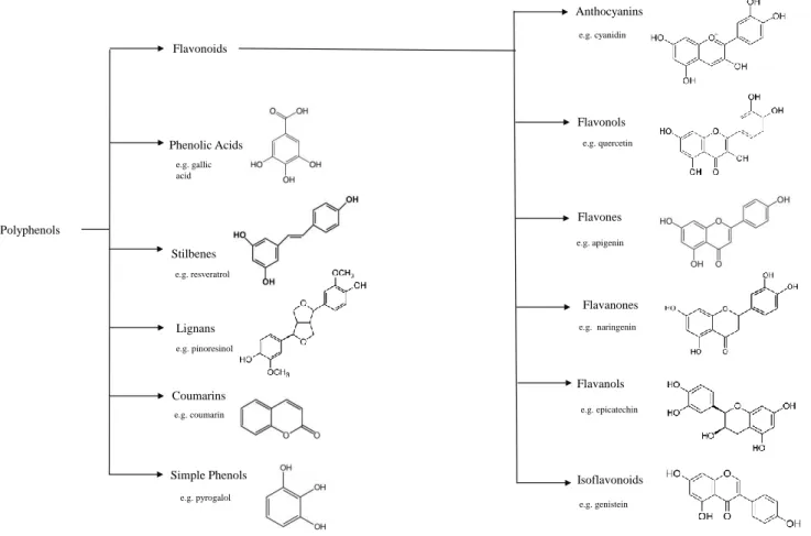
Figure 6: Classification and chemical structures of the main dietary polyphenols classes ... 19 Figure 7: Example of Yeast Construction to study PD related features. Yeast model
xiii
galactose inducible promoter and auxotrophic markers URA3 and TRP1 respectively, which by homologous recombination are integrated into the host genome. ... 23
Figure 8: Growth Curves and growth parameters of S. cerevisiae strains expressing or not aSyn. S. cerevisiae strains Control (♦); aSyn-1 (●); aSyn-2 (×) were grown in SC
liquid medium with a) glucose or b) galactose for 24h at 30ºC. a) and b) Grown was kinetically monitored hourly by OD measurements at 600 nm and growth curve represented. c); d); e) Growth parameters were determined based on the growth curve, c) doubling time, representing the time necessary to cells to duplicate, determined by logarithmic transformation of raw OD 600nm from growth curves. d) Lag time, representing the adaptation time, determined by logarithmic transformation of raw OD 600nm from growth curves. e) Final Biomass (OD 600nm), representing the final OD of each culture, is calculated based on the measurements of OD at 24 hours of growth. Results represent the mean ± SD of three independent biologic replicates. *** indicate statistically significance between Control, aSyn-1 and aSyn-2, for a p<0.001 respectively. ... 35
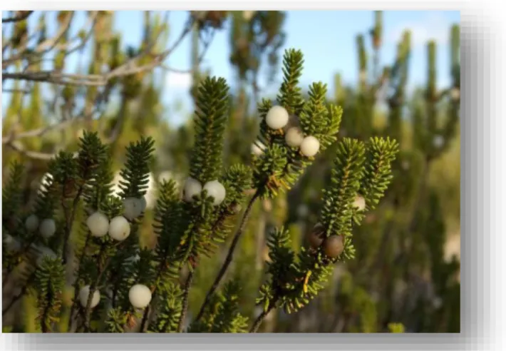
Figure 9: Corema album (Portuguese Crowberry; Camarinha), fruits and leaves 37 Figure 10: Growth Curves and growth parameters of S. cerevisiae strains (Control and aSyn-2) with C. album leaf PEFs. S. cerevisiae strains incubated with different
concentrations of C. album leaf PEFs (μg GAE.mL-1) Control [0] (●); aSyn-2 [0] (▲); aSyn-2 [15] (♦); aSyn-2 [30] (×); aSyn-2 [62.5] (-); aSyn-2 [125] (+); aSyn-2 [500] (■); Yeast cells were grown in SC liquid medium with galactose for 24h at 30ºC. a) Grown was kinetically monitored hourly by OD measurements at 600 nm and growth curve represented. b); c); d) Growth parameters were determined based on the growth curve. b) Final Biomass (OD 600nm), c) Doubling time, d) Lag time. Results represent the mean ± SD of three independent biologic replicates. *, ** and *** indicate statistically significance between Control and aSyn-2, for a p<0.05, p<0.01 and p<0.001 respectively. ... 38
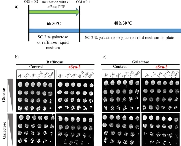
Figure 11: Phenotypic growth assay of control and aSyn-2 strains grown in the presence of polyphenol fractions. a) Schematic representation of cells growth for 6
hours at 30 ºC, with constant shaking in liquid medium containing galactose or raffinose, with several concentrations of PEFs. Cells were serial diluted, subsequently 5 μL of each dilution was spotted in solid medium with glucose or galactose and incubated for 48 hours at 30 ºC. b) Spot assays for control and aSyn-2 strains that grew 6 hours in SC liquid medium containing raffinose, supplemented with several concentrations of C. album leaf PEFs. Spotted in solid medium with galactose or glucose and incubated for 48 hours at 30 ºC. c) Spot assays for control and aSyn-2 strains that grew 6 hours in SC liquid medium containing galactose, supplemented with several concentrations of C. album leaf PEFs. Spotted in solid medium with galactose or glucose and incubated for 48 hours at 30 ºC. Image acquisition was made with Chemidoc XRS and Quantity-one software and the most representative of biological replicates is show. ... 41
Figure 12: Representative 2-DGE gels of BY4741 strain cell proteome (75 μg). The
gels were CBB stained. a) Extraction performed with Tris-HCl buffer. b) Extraction was done with RS buffer. c) The extraction was performed with Tris-HCl buffer, and the protein sample was submitted to cleaning step using 2D Clean-Up kit. d) Extraction was
xiv
performed with RS buffer, with subsequently cleaning step using 2D Clean-Up kit. IEF was performed with 3-10 NL IPG strips. ... 43
Figure 13: Representative 2-DGE gel of control strain cell proteome. (75 μg) The gel
was CBB stained. The normalized volumes were compared to the controls (control strain) in order to estimate variations on protein content. In total 22 spots showing statistical significant differences among the four conditions in the study (control; control+PEF; aSyn-2; aSyn-2+PEF). Spots that were differentially abundant with statistical significance in all replicates were identified by Progenesis SameSpots image analysis. Further numbered spots with differential expression pattern (table 3) will be sent for protein identification by MS. IEF was performed with 3-10 NL IPG strips.Three independent biologic replicates were done for each condition... 45
Figure 14: Protein phosphorylation profile of yeast strains incubated with C. album leaves PEF (control+PEF; aSyn-2+PEF) and without treatment (control; aSyn-2).
10 μg of protein sample of each condition was applied. SDS-PAGE gel was stained with Pro-Q DPS. Images of the gels were scanned in a laser imager with 532-nm excitation and 580 bandpass emission filter FLA-5100 Fuji Photo Film Co, Ltd. A representative image is show with two independent biologic replicates for each condition. ... 54
Figure 15: Representative 2-DGE gel of empty strain phosphoproteome (75 μg). The
gel was stained with Pro Q-DPS. The normalize volumes were compared to the controls in order to estimate variations in phosphorylation levels. In total 10 spots exhibited phosphorylation signal between the four conditions in the study (control; control+PEF; aSyn-2; aSyn-2+PEF). IEF was performed with 3-10 NL IPG strips. ... 55
Figure 16: aSyn expression profile treated with C. album leaf PEF. aSyn expression
levels in yeast cells assessed by western blot analysis of total protein extract. a) aSyn expression levels of control and aSyn-2 yeast cells subjected to the treatment with C.
album leaf PEFs. b) Relative densitometric intensities. PGK was used as an internal
loading control. Protein siganls were detected using the chemiluminescence detection kit FemtoMax Super Sensitive Chemiluminescent HRP Substracte. A representative image is shown. 3 independent biological replicates were performed. ... 59
Figure 17: Schematic model of the role of Curcumin and Resveratrol in autophagy and possible modulation in autophagy mediated by C. album leaf PEFs. Resveratrol
causes activation of Sirtuins, more precisely SIRT-1, followed by the induction of ATG proteins, essentials for the autophagic machinery. This pathway may be affected in the same way due to C .album leaf PEFs (dashes line). Also mTOR is downregulated by curcumin, and potentialy could be downregulated by C. album Leaf PEFs (dashes line). Induced autophagy enhances the clearance of aSyn, thereby may contribute to the neuroprotection on PD. ... 61
Figure 18: aSyn expression profile. aSyn expression levels in yeast cells assessed by
western blot analysis of total protein extract. Different concentrations of galactose were used to optimize the better condition to express high levels of aSyn. PGK was used as an internal loading control. Protein siganls were detected using the chemiluminescence detection kit FemtoMax Super Sensitive Chemiluminescent HRP Substracte. A representative image is shown. ... 65
xv
Index of tables
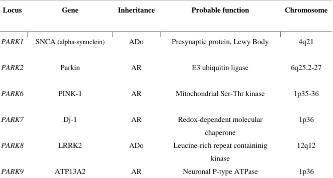
Table 1- Parkinson’s disease symptoms (Adapted from reference6 ) ... 3
Table 2- Genetic factors with PARK status in PD. ADo, autosomal dominant; AR,
autosomal recessive. (Adapted from reference8,14,15) ... 5
Table 3: Normalized volumes for protein spots differentially expressed among the four conditions in the study: control; control+PEF; aSyn-2; aSyn-2+PEF, and the
respective pI and MW (kDa). Differences between treatments are denoted as * p<0.05, **p<0.01 and ***p<0.001. ... 46
Table 4: Number of spots with expression variations in the respective conditions under study: control; control+PEF; aSyn-2; aSyn-2+PEF. ... 52 Table 5: Normalized volumes for protein spots presenting phosphorylation levels among the four conditions in study: contro; control+PEF; aSyn-2; aSyn-2+PEF,
xvi
Abbreviations
2-DGE - Two-dimensional gel electrophoresis
A30P - Alanine to proline substitution at aSyn amino acid residue 30 A53T - Alanine to threonine substitution at aSyn amino acid residue 53 AD - Alzheimer’s disease
aSyn - Alpha-synuclein
ATP - Adenosine tri-phosphate CBB - Commassie Brilliant Blue
CL - corpos de Lewy
CHAPS - 3[(3-Cholamidoproply)dimethylammonio]-propanesulfonic acid CNS - Central Nervous System
CSM - Complete supplement mixture DA - Dopamine
DSB lab - Disease and Stress Biology lab
FEPs - Frações enriquecidas de polifenoís
DT - Doubling time DTT - Dithiothreitol
E46K - Glutamic acid to lysine substitution at aSyn residue 46 ER - Endoplasmatic reticulum
GAE - Gallic acid equivalents IEF - Isoelectric Focusing
IMM - Instituto de Medicina Molecular
ITQB - Instituto de Tecnologia Química e Biológica LB - Lewy bodies
MS - Mass Spectrometry MW - Molecular Wieght NAC - Non-Aβ-component
OD600nm - Optical Density at 600 nm PBS - Phosphate Buffer Saline PD - Parkinson’s disease
xvii
pI - Isoelectric Point
PTM’s - Postranslational Modifications ROS - Reactive oxygen species
SDS-PAGE - Sodium Dodecyl Sulphate-Polyacrylamide Gel Electrophoresis SPE - Solid phase extraction
UPS - Ubiquitin-proteasome system WT - Wild type
YPD - Yeast extract, peptone and dextrose SNpc - “Substancia nigra pars compacta” PINK1- PTEN-induced kinase 1
LRRK2 - Leucine-rich repeat kinase 2 ATP13A2 - ATPase type 13A2
E3 - Ubiquitin ligase ADo - Autosomal dominant AR - Autosomal recessive
NAC - Non-amyloid-β-component TH - Tyrosine hydroxylase
SNARE - Soluble N-ethylmaleimide-sensitive factor attachment protein receptor MPTP - (1- methyl-14-phenyl-1, 2, 3, 6-tetrahydropyridine)
DNA - Deoxyribonucleic acid
EGCG - (-)-epigallocatechin-3-gallate EC - (-)-epicatechin
PVDF - Polyvinylidene Fluoride
BDNF - brain-derived neurotrophic factor DP – Doença de Parkinson
IPG - Immobilized pH-gradient
Pro-Q DPS - Pro-Q Diamond phosphoprotein stain SC - Synthetic complete
MBA - Membrane blocking agent TBS -Tris-buffered saline
xviii
HRP - Horseradish peroxidase NL - Non linear
SD - Standard deviation GAL - Galactose
GFP - Green fluorescent protein RS - Resuspension Buffer MW - Molecular Weight SIRT-1- Sirtuin 1
ATG - Autophagy-related protein
mTOR - Mammalian target of rapamycin
1
Neurodegenerative diseases are multifactorial and currently there is no effective therapy against the progressive neuronal death characteristic of these pathologies. It is therefore very important to find new therapeutic strategies that can prevent and / or delay the neurodegeneration process. Plant polyphenols appear in response to this demand, having been reported to have substantial neuroprotective activity.
S. cerevisiae is one of the most versatile biological systems used as a model for
the study of neurodegenerative diseases. C. album leaf PEFs protective effect was already defined in a yeast PD model.
The main objective of this study is to access the effect of C. album leaf PEFs on proteome and phosphoproteome of the yeast model of PD. This analysis will be done using two dimensional gels of proteins and will require optimization of the technique involved. A detailed analyses of the protein profiles obtained using appropriate software for image analysis will allow to find proteins that respond to effect of aSyn expression and to the treatment with C. album leafs PEFs. The Identification of those proteins will contribute to the overall understanding of our PD yeast model and neuroprotective effects of C. album leaf PEFs.
Polyphenols neuroprotective effect in a Parkinson’s disease yeast model: phosphoproteome alterations was a work developed between the Disease and Stress
Biology Laboratory (DSB), from Instituto de Tecnologia Química e Biológica (ITQB) – Universidade Nova de Lisboa and Cellular and Molecular Neuroscience Unit, from Instituto de Medicina Molecular, from Faculdade de Medicina – Universidade de Lisboa.
2
Introduction
Neurodegenerative Diseases
Human neurodegenerative diseases are characterized by the progressive loss of structure and/or function of neurons, leading to their death.
Neurodegenerative disorders such as Parkinson’s (PD) and Alzheimer‘s (AD) diseases account for a significant and increasing proportion of morbidity and mortality in the recent world. The most common risk factor for developing neurodegenerative diseases, is aging. With the rise in human lifespan around the world in the last years, the incidence of neurodegenerative diseases, especially AD or PD, has increased dramatically. United Nations population projections estimate a world population of 400 million people over 80 years by the year 2050. Therefore, it is expected that over the next generation, the percentage of elderly people will double and consequently the proportion of persons suffering from some kind of neurodegenerative diseases.
As the population ages, an improved understanding of these diseases will be vital to developing more effective therapies and combating the staggering personal, social, and economic costs of these diseases1–3.
Parkinson’s disease, definition and historical perspective
PD was first described in 1817 by James Parkinson in his monograph ''An essay on
the shaking palsy'', where he identifies six cases of PD4–7. Previously referred to as ‘‘paralysis agitans’’, nowadays PD is among the most prevalent neurodegenerative disorders as it affects approximately 6 million individuals worldwide8,9. Generally, the
onset of PD occurs in patients over the age of 50 years and its incidence slowly progresses with increasing age, affecting about 2% of people over 65 years old9–11. Clinical
manifestations of PD consists in a series of severe motor defects produced by resting muscle tremor, muscle rigidity, brandykinesia and postural instability6,8–10. Brandykinesia refers to slowness of movement, and may significantly impair the quality of life. Brandykinesia is considered to be the main feature and the necessary condition for diagnosis of PD6–8. These motor disabilities begin to be felt by patients with about 5-10 years of disease, even when treated with symptomatic medication available7.
3
Table 1- Parkinson’s disease symptoms (Adapted from reference6 )
All these motor symptoms are thought to arise primarily from the loss of dopaminergic (DA) neurons within the “substancia nigra pars compacta” (SNpc), located
in mesencephalon8.
Almost two centuries after the first description of PD, there are only symptomatic treatments available for this pathology, which essentially involves substituting dopamine, or suppressing pathological neuronal oscillations via deep brain stimulation10,12.
The discovery of levodopa in 1960 revolutionized the treatment of PD. Levodopa has the ability to restore dopaminergic transmission deficiency and provides remarkable symptomatic relief to the vast majority of these patients. This remains the most efficacious agent available for PD treatment. However, we soon learned that after several years of treatment most patients develop involuntary movements termed “dyskinesias,” which are difficult to control and significantly impair the quality of life7,9,13.
As a result, in the recent years efforts have been made to develop new therapeutic approaches to treat PD. Interventions such as stem-cell transplantation and gene therapies have been examined as potential treatments for PD. Although early results indicated that patients may obtain long-term clinical benefit from the intrastriatal transplantation of human embryonic mesencephalic tissue, it later became clear that there are several restrictions to stem cell therapy. These include the limited availability of fetal human mesencephalic tissue and the usually very low percentage of viable transplanted cells9. On the other hand, in the case of gene therapy, viral vectors have proven to be an ideal
Motor Symptoms Non-Motor Symptoms
Tremor, Brandykinesia, rigidity, postural
instability
Hypomimia, dysarthria, dysphagia, sialorrhoea
Decreased arm swing, shuffling gait,
festination difficulty arising from chair, turning in bed
Micrographia, cutting food, feeding, hygiene, slow activities of daily living
Glabellar reflex, blespharospasm, dystonia,
strial deformity, scoliosis, camptocormia
Cognitive impairment, brandyphrenia,
tip-of-the-tongue phenomenon
Depression, apathy, anhedonia, fatigue
Sensory symptoms: anosmia, ageusia,
paresthesias
Dysautonomia (orthostatic hypotension,
constipation, urinary and sexual dysfunction, abnormal sweating, seborrhoea), weight loss
Sleep disorders (behavior disorder, vivid
dreams, daytime drowsiness, sleep
4
vehicle to deliver or silence genes of interest in the brain in order to develop genetic strategies. Still none of these approaches could stop the degeneration of dopaminergic neurons9.
Our capacity to discover new PD treatment is limited for several reasons; one of them relates to the complex and multifactorial etiology of PD, which involves an intricate interplay of a large network of factors: genes, environment and gene-environment interactions, lack of knowledge of the specific molecular events that provoke neurodegeneration and aging5,7,12.
Genetic determinants of Parkinson’s disease
In the last decades the discovery of several genes linked to rare familial forms of PD came to prove the role of genetics in disease development and brought new evidence for understanding the pathological mechanisms of PD. The majority of cases are thought to be idiopathic. Nevertheless, in 5-10% of cases, PD presents as a Mendelian form displaying both recessive and dominant models of inheritance8,14–16. Several genetic loci named PARK are identified to cause PD (Table 2) These include two autosomal dominant genes, SNCA encoding alpha-synuclein (aSyn), and leucine-rich repeat kinase 2 (LRRK2), and four autosomal recessive genes, encoding Parkin, DJ-1, PTEN-induced kinase1 (PINK1) and ATPase type 13A2 (ATP13A2)8,14,15.
SNCA gene point mutations (A53T, A30P, E46K, H50Q and G51D), also assigned as PARK1, as well as duplications and triplications, cause autosomal dominant PD16–18. On the other hand Parkin function as an E3 ubiquitin ligase by targeting misfolded proteins to the ubiquitin proteasome pathway for degradation. The loss of its E3 ligase activity lead to autosomal recessive early onset PD15. PINK1 mutations are the second
most-common cause of autosomal recessive PD after Parkin. PINK1 is located in the mitochondria and mutations in the kinase domain of the protein lead to a mitochondrial dysfunction and dopaminergic neuronal degeneration, as they result in impaired phosphorylation of its substrates in mitochondria15,16,19. Loss of function mutations in the DJ-1 gene are associated with forms of autosomal recessive early-onset Parkinsonism. DJ-1 is thought to protect neurons from oxidative stress by acting as redox-dependent chaperone, preventing misfolding and aggregation of oxidized mitochondrial proteins. Accordingly with this DJ-1 knockout mice presents motor impairments and dopaminergic dysfunction15,16,20. Mutations in the LRRK2 gene cause autosomal dominant PD. This
5
protein contains several domains, including RAS/GTPase, tyrosine kinase, MAPKK and leucine-rich-repeat domain, in which mutations can affect signaling pathways relevant in PD5.Finally, mutations in ATP13A2 cause an atypical form of PD with dementia, named Kufor-Rake syndrome8.
Table 2- Genetic factors with PARK status in PD. ADo, autosomal dominant; AR, autosomal recessive.
(Adapted from reference8,14,15)
Hallmarks of Parkinson’s disease
Pathological hallmarks of PD are loss of dopaminergic neurons in “substancia nigra
pars compacta”, and the presence of intraneuronal proteinacious cytoplasmic inclusions
named Lewy Bodies (LBs)5,8,10,11,21–24.
LBs are cellular inclusions that can be visualized by histological analysis and are predominantly constituted by the presynaptic protein aSyn, a small acid protein involved in both sporadic and familial cases of PD5,7,8,10,12,21–23. LBs present a spherical structure with more than 15 μm in diameter and have a organized structure containing a dense hyaline core surrounded by a clear halo7 (Fig.1).
Locus Gene Inheritance Probable function Chromosome
PARK1 PARK2 PARK6 PARK7 PARK8 PARK9 SNCA (alpha-synuclein) Parkin PINK-1 Dj-1 LRRK2 ATP13A2 ADo AR AR AR ADo AR
Presynaptic protein, Lewy Body
E3 ubiquitin ligase
Mitochondrial Ser-Thr kinase
Redox-dependent molecular chaperone
Leucine-rich repeat containinig kinase
Neuronal P-type ATPase
4q21 6q25.2-27 1p35-36 1p36 12q12 1p36
6
Figure 1: Immunohistochemical labeling of intraneuronal inclusions, termed LBs, in the SNpc dopaminergic neuron. Immunostaining with an antibody against aSyn reveals a LB (black arrow) with an
intensely immunoreactive central zone surrounded by a faintly immunoreactive peripheral zone (left image). Conversely, immunostaining with an antibody against ubiquitin yields more diffuse
immunoreactivity within the LB (right image). (Adapted from reference7)
PD is often unremarkable, with mild frontal atrophy in some cases, but there is no significant atrophy of brainstem. However, in the SNpc, it can be found the cell bodies of nigrostriatal neurons and these are projected primarily to the putamen (Fig.2). The loss of these neurons, which normally contain amounts of neuro-melanin, produces the classic neuropathological finding of SNpc depigmentation7,25. Sections of the brainstem usually reveal loss of the normally dark black pigment in the SNpc (Fig. 2).This loss of pigmentation correlates with neuronal loss of dopaminergic neurons in the SNpc and noradrenergic neurons in the locus ceruleus7,25.
Indeed, the spreading of LBs correlates with the progression of the disease. In PD, generally LBs are mainly found at sites of neuronal loss such as the SNpc thus indicating that their presence may be related to nerve cell loss9,26.
7
The aSyn deposition which is the main constituent of the LBs is thought to be central to the pathogenesis of PD. Recent findings have suggested that aSyn deposition at the synapse may be the key issue of the neurodegenerative process. The crucial role of aSyn in PD pathophysiology is further sustained by findings showing that the areas that degenerate in PD (SNpc, striatum and ventral tegmental area) express low levels of aSyn in physiological conditions suggesting that these regions may be more vulnerable to a pathological increase of its levels, which normally occurs during aging4,9.
Loss of Dopaminergic
Neurons
a) Normal Brain b) Parkinson’s Disease Brain
Figure 2: Neuropathology of PD a) Schematic representation of the normal nigrostriatal pathway (in red). It is
composed of dopaminergic neurons whose cell bodies are located in the substantia nigra pars compacta (SNpc; see black arrows). These neurons project (thick solid red lines) to the basal ganglia and synapse in the striatum (i.e., putamen and caudate nucleus). The image demonstrates the normal pigmentation of the SNpc, produced by neuromelanin within the dopaminergic neurons. b) Schematic representation of the diseased nigrostriatal pathway (in red). In PD, the nigrostriatal pathway degenerates. There is a marked loss of dopaminergic neurons that project to the putamen (dashed line) and a much more modest loss of those that project to the caudate (thin red solid line). The image demonstrates depigmentation (i.e., loss of dark-brown pigment neuromelanin; see black arrows) of the
8
Alpha-Synuclein: structural and functional properties
aSyn is an abundant, 140 amino acid long, neuronal cytoplasmic protein. The sequence of aSyn gene (SNCA) has a high degree of conservation in numerous organisms, and mutations or multiplications of this gene have been associated with familial forms of PD4,9.
aSyn is predominantly localized to presynaptic terminals in the central nervous system (CNS), where it is freely associated with synaptic vesicles5. It belongs to the synuclein family, which includes also β- and γ-synuclein. These proteins have a common N-terminal sequence containing a different number of repeated regions while they differ in the C-terminal region4,9.
While all the synucleins are present in human brain, only aSyn is show to be associated with pathological structures in neurodegenerative conditions27. Sequence analysis suggests that aSyn consists of three distinct regions: the N-terminal amphipathic region (residues 1-60), the central hydrophobic Non-amyloid-β-component (NAC) region (residues 61-95), and the C-terminal acidic region (residues 96-140)4,9,22,27.
The N-terminal half of aSyn contains four 11-amino acid imperfect repeats with a highly conserved hexameric motif (KTKEGV), which is involved in the formation of amphipathic α-helices, similar to lipid-biding domain of apolipoprotein-like class A2 and its essential for membrane binding4,9,27. Several lines of evidence support the membrane-binding capacity of the N-terminal region. For example, mutations in this region were show to perturb plasma membrane localization of the protein in yeast27.
This portion of the protein includes the sites of five familial PD mutations: A30P, A53T, E46K and two new mutations discovered H50Q and G51D. Also, N-terminal acetylation is critical for forming α-helical oligomers thus suggesting that this event could have important implications for both the native and pathological structures of aSyn4,17,18,27 (Fig.3).
The central region comprises the NAC sequence which is highly hydrophobic and aggregation prone. This part of aSyn can undergo a conformational change from random coli to β-sheet structure. Moreover, it is able to form cylindrical β-sheets and amyloid-β-like fibrils and protofibrils. The NAC region is indispensable for aSyn aggregation and toxicity; the deletion of large segments within this motif greatly diminished aSyn oligomerization and fibrillogenesis in vitro, and in a cell based-assay. Moreover, mutation of only one single amino acid in this central domain of aSyn can alter the aggregation
9
properties of the protein, emphasizing the importance of the NAC domain in the aggregation process4,22,25,27 (Fig.3).
The last segment of aSyn, the C-terminal region (residue 96 to 140) is highly enriched in proline acidic residues and contains three highly conserved tyrosine residues (Y125; Y133; Y136) that are phosphorylation targets. The C-terminal is responsible for the intrinsically disordered nature of aSyn. This domain also plays a regulatory role in the aggregation and fibril formation of the protein, by shielding the hydrophobic NAC domain from the watery solution, in addition, the C-terminal appears to have chaperone activity4,22,25,27,28.
Deletion or modifications of C-terminal of aSyn, as well as changing the charge or hydrophobicity of the domain, enhances the aggregation rate of aSyn in vitro and in cells. For example, phosphorylation of Serine 129 (S129) or nitration of Y125, Y133 and Y136 promote the formation of aSyn filaments or oligomers in the C-terminal half and enhance tendency to aggregate and fibrilize. These results suggest that the C-terminal can play a role of an intramolecular chaperone by preventing aSyn from fibrillization4,22,25,27,29.
From these characteristics that we see above, it can be inferred that aSyn is a very versatile protein, as it can easily adopt different conformations upon interacting with biological membranes of different compositions, other proteins or protein complexes in physiological conditions9,22.
Figure 3: Molecular structure and functional characteristic of aSyn. aSyn is functionally divided into
N-terminal (1–60aa), NAC (61–95aa), and C-terminal (96–140aa) domains. The N-terminal domain contains four motifs (blue color) and has five point mutation sites linked to PD (A30P, E46K, H50Q, G51D and A53T) and also a tyrosine residue (Y39) which is a phosphorylation target. The NAC domain, which encompasses the most hydrophobic residues, promotes aggregation, with a phosphorylation site (S87). The C-terminal domain exhibits chaperone activity that tends to decrease protein aggregation, has one phosphorylation site (S129) and three tyrosine residues (Y125, Y133, Y136). (Adapted from25,27,28)
10
Physiological function of Alpha-Synuclein
Although aSyn is distributed to almost all subcellular compartments, it is particularly enriched in the presynaptic terminals where it is loosely associated with the distal reserve pool of synaptic vesicles. The high presynaptic concentration of aSyn, and its association with synaptic vesicles, suggest a physiological role of the protein in regulation of synaptic transmission as well as synaptic vesicle recycling22,25,27,29,30.
Neurotransmitters are stored in vesicles after being synthesized. Those vesicles dock and fuse with the plasma membrane to release neurotransmitters into the synaptic cleft and then restock via recycling from the distal vesicle pool27,31.
Overexpression, knockdown and knockout of aSyn lead to deficiencies in synaptic transmissions, supporting that it plays an important role in the regulation of neurotransmitter release, synaptic function and plasticity. For example, SNCA-knockout mice exhibit an impairment in hippocampal synaptic responses to prolonged trains of high-frequency stimulation that deplete the docked and reserve pool of synaptic vesicles22. Moreover depletion and suppression of aSyn induces an impairment of vesicle trafficking between the reserve pool and the ready releasable pool and a deficiency in the neurotransmitter uptake. Experiments with siRNA-mediated knockdown of aSyn resulted
in significant decreases in Vmax for dopamine uptake and the surface density of dopamine
transporter in SH-SY5Y cells. On the other hand transgenic mice overexpressing human
aSyn display impairment in synaptic vesicle exocytosis and reduction in neurotransmitter release22,27. Another study in PC12 cell line show that overexpression of aSyn resulted in
an accumulation of vesicles at the synapse22,27,31, and similar effects have been observed
in genetic rodent models of PD27.In line with this studies overexpression of aSyn in yeast
induces a block of the ER-to-Golgi vesicular traffic leading to defects in endocytosis and exocytosis4.
In transgenic mice, overexpression of aSyn was found to inhibit tyrosine hydroxylase (TH), the enzyme in charge for dopamine synthesis in dopaminergic neurons and consequently lead to a reduction on dopamine synthesis. In an opposite way down-regulation of aSyn increase TH activity and DA synthesis27.
The possible role of aSyn in regulating synaptic homeostasis is not exclusively related to its direct interaction with synaptic vesicles, because aSyn is able to interact with proteins that control synaptic vesicle exocytosis, such as phospholipase D2, the family of
11
RAB small GTPases, the synaptic protein synapsin 1 and soluble N-ethylmaleimide-sensitive factor attachment protein receptor (SNARE), which are crucial modulators of membrane trafficking, exocytosis and synaptic vesicle release in neuronal cells4,9.
These data strongly indicate that aSyn displays an important role in the trafficking of synaptic vesicles and in the regulation of vesicle exocytosis and subsequently in neurotransmitter release4,22. Thereby, aSyn may contribute to a regulatory effect in the synaptic homeostasis and abnormalities in this regulatory function may influence the neurotransmitter release, leading to alterations on short-term and long-term synaptic plasticity4,22,27.
Alpha-Synuclein Aggregation
The abnormal deposition of specific proteins in brain tissues is a feature of several age-related neurodegenerative diseases, including PD7. Several consequences are hypothesized to be associated with protein misfolding and aggregation: the loss of normal function; gain of toxic function, mechanical disruption of cellular compartments and processes such as synaptic, mitochondrial function and proteasomal activity12,23,32,33. The sequence of molecular events that lead to development of neurodegenerative diseases, implicates the accumulation of misfolded protein species, extending from oligomers to higher fibrilar protein aggregates (fig. 4), and once aggregated, these aggregates can grow and enlarge by recruitment of more misfolding proteins. Accumulation of aggregated aSyn affects various functional structures of the nervous system leading to serious cognitive and behavioral alterations33.
12
The levels of aSyn in the CNS depend on the balance between the rates of aSyn synthesis, aggregation and clearance. Abnormal protein structure resulting from genetic alterations or overexpression of aSyn could create an imbalance between these mechanisms and will result in abnormal levels of aSyn that might benefit the formation and/or accumulation of oligomeric and fibrillar species, which will ultimately lead to the formation of large aggregates the LBs8,22,26. For example, four missense mutations on the gene SNCA (A30P, E46K H50Q, and A53T) have been shown to accelerate aSyn aggregation compared to wild-type17,18,25,29. These higher rates of aggregation displayed by the mutant forms can be explained by changes in the structure of protein, because these mutations have the ability to disturb the α-helical structure of the protein in the N-terminal region, increasing the possibility of generating pathogenic conformation25,29. Many other factors and events have been reported to influence the aggregation of aSyn, such as: i) posttranslational modification of aSyn (phosphorylation; truncation). Phosphorylation of S129 promote the formation filaments and oligomers27,28; ii) Incubation of aSyn protein
with metals (cooper, iron) and pesticides (paraquat). In yeast, Fe3+ ions increase the
Lewy Body
Figure 4: aSyn aggregation. Schematic illustration of the process whereby normal soluble aSyn misfolds is converted
into pathological oligomers and higher aggregates that fibrillize and deposit into Lewy bodies in affected neurons in PD brain.
13
number of cells containing aSyn inclusions34. Overexpression of aSyn in GT1-7
hypothalamic cells generate aSyn inclusions like structures, and also alterations in mitochondria function, increasing the production of free radicals29.
Another dysfunction associated with the aggregation of aSyn and its abnormal levels in the brain, is the impairment of the ubiquitin-proteosome system (UPS)7,8,35. The UPS is one important pathway for protecting cells against misfolded proteins, clearing misfolded proteins from the cytosol, endoplamatic reticulum and nucleus36. Overexpression of wild-type or mutant aSyn in cultures cells and in the brains of transgenic mice is known to impair UPS function. Proteossomal dysfunction and consequent accumulation of misfolded proteins may provoke a vicious cycle, which probably defies the capacity of the proteossomal clearance systems promoting accumulation and development of a self-propagating cycle7,33,35. When the misfolded aSyn begin to propagate and reach the acceptor cells, they begin to seeding the aggregation of endogenous protein in a prion–like fashion way37–39. This prion-like transmission of aSyn pathology relies on the premise that a sick neuron could release its misfolded aSyn to the extracellular space, or they gain access to the extracellular space when the neuron dies. Once out of the neuron the aSyn could be free to enter an adjacent neuron by endocytosis and act as a template, seeding the aggregation and initiating the formation of a LBs37. Studies with neuronal stem cells and embryonic neurons grafted
into the brain of aSyn overexpressing transgenic mice can take up host aSyn and develop inclusions, supporting this theory40. The accumulation of aggregated aSyn affects various functional structures of the nervous system leading to serious cognitive and behavioral alterations33.
Alpha-Synuclein Phosphorylation
Posttranslational modifications (PTMs), such as phosphorylation, ubiquitination or sumoylation, which alter the protein size, charge, structure conformation and/or biological function, can also affect protein folding and aggregation, and thereby play a critical role in neurodegenerative disorders28,41. Beyond all this PTMs, protein phosphorylation is the most widespread type of modification used in signal transduction affecting every basic cellular process42.
aSyn protein presents several phosphorylation sites (4 serine, 10 threonine, 4 tyrosine residues) that are highly conserved among numerous species. Nevertheless,
14
approximately 90 % of aSyn deposited in LBs is phosphorylated at S129. In contrast, only 4 % or less of total aSyn is phosphorylated at this residue in normal brain, suggesting a close relationship between aSyn phosphorylation at S129 and its aggregation14,41,43–46. Several studies performed in cell lines associate aSyn phosphorylation with formation of oligomers, cytoplasmic and nuclei aggregates and cytoplasmic inclusions28, and in transgenic mouse models of PD, aSyn phosphorylation caused accelerated neuronal loss, suggesting a toxic effect of S129 phosphorylation21. Even so, some studies in yeast using a S129A mutation that blocks the phosphorylation in S129 residue, resulted in more toxic forms, and increased the number of inclusions and oligomeric species compared with the WT protein28. One hypothesis to explain this result lies in the fact that for some proteins phosphorylation acts as a signal for degradation by UPS and/ or autophagy. If this happens with aSyn protein, phosphorylated aSyn might accumulate in LBs due to proteosomal impairment leading to aggregation28,46.
Multiple kinases have been implicated in aSyn phosphorylation, namely, G-protein coupled receptor kinases (GRK2, GRK3, GRK5 and GRK6), casein kinases 1 and 2 (CK1, CK2) and Polo-like kinase 2 (PLK2) and PLK328,44,47. All these kinases could play an important role in the modulation of aSyn physiology. For example, knockdowns of GKR3, GKR5 and GRK6 significantly decrease levels of aSyn phosphorylated at S12944.
Overexpression of aSyn increased GRK5 protein expression in SH-SY5Y cells and in brain extracts of transgenic mice expressing human aSyn, additionally GRK5 was found to colocalize with aSyn in LBs of PD patients28. Another study showed that the overexpression of PLK2 is protective by mediating selective autophagy clearance of phosphorylated S129 aSyn48.This show that these kinases decide the fate of aSyn protein, thus, inhibition or overexpression of relevant kinases could be important in order to generate a potential therapeutic strategy28,43,48. Gathering all these data, it is clear that phosphorylation of aSyn is important in the context of its aggregation and toxicity, yet no conclusive agreement of this precise contribution this PTM has toward the disease process. For example, there is still no consensus on whether phosphorylation is neurotoxic or neuroprotective28.
15
Mitochondrial Dysfunction and Oxidative Stress in PD
Mitochondria, the power house of living cells and regulators of cell survival and death are especially complex and delicate organelles19. The first connection between
Parkinson’s and mitochondria became apparent in the early 1980s with the discovery that a neurotoxin, MPTP (1- methyl-14-phenyl-1, 2, 3, 6-tetrahydropyridine) causing parkinsonian syndrome inhibits the mitochondrial respiratory complex I (NADH-quinone oxidoreductase)19,20,40,49. MPTP is converted to MPP+ by glial cells in brain and taken up by the dopamine transporter expressed in dopamine neurons. MPP+ is able to bind and inhibit mitochondrial complex I causing mitochondrial dysfunction and increasing the levels of ubiquitinydated proteins20,50.
Consistent deficits in the subunits and activity of mitochondrial complex I of the electron transport chain in SNpc of PD patients is a prominent phenomenon. The activity of complex I has been reported to be reduced (in the range of 30%) in the CNS and frontal cortex of PD patients at autopsy. In mitochondrial preparations from PD frontal cortex samples, complex I subunits, derived from mitochondrial genome, were found to be oxidatively damaged15,40. Taking in to account this data, aSyn can be found in the mitochondria of the striatum and SNpc of PD patients contributing to impairing the complex I activity50. Moreover, in transgenic mouse models of aSyn overexpression, several mitochondrial anomalies were observed. These include oxidation of mitochondria associated proteins, mitochondrial DNA damage, increased oxidative stress, and bioenergetics defects40. Oxidative damage to mitochondrial DNA (mtDNA) may
compromise encoding of respiratory chain subunits, thus initiating a vicious cycle of oxidative stress and bioenergetics failure49. Recently, it has been shown that the
N-terminal 32 amino acids of aSyn function as a targeting sequence for import aSyn into mitochondria20. This targeting sequence drives aSyn to the inner mitochondrial membrane where it associates with complex I, leading to a decrease in its activity and increasing reactive oxygen species (ROS) production in human dopaminergic neuronal cultures overexpressing wild-type aSyn. In these models overexpression of A53T aSyn enhance these effects (Fig.5)19,49. ROS are chemically reactive molecules containing oxygen and are produced in all aerobic cells. Oxidative stress occurs when the generation of ROS overwhelms cells capacity (enzymatic and non-enzymatic antioxidants defenses) to scavenging them24.
16
In yeast cells expression of wild-type or A53T mutant aSyn accelerates apoptosis and ROS production. Remarkably these effects did not occur in yeast cells lacking mtDNA. Also, in other model C. elegans the overexpression of aSyn reduces the fusion rate of mitochondria40. At last, aSyn protofibrils are known to form annular pores in membranes, suggesting that permeabilization of mitochondrial membranes may be involved in aSyn toxicity20.
As a consequence of impaired electron flux through complex I, mitochondrial ATP production decrease, creating a deficit of energy supply while the generation of ROS is increased24. Excessive production of ROS by mitochondria lead to impairment of cellular
redox balance pushes the oxidation of biological macromolecules, such as DNA, proteins and lipids leading to failure of biological functions24,49. Another study in mice lacking aSyn presented resistance to MPTP, showing that aSyn is an essential mediator of the toxic effects of complex I inhibitors. Remarkably, mitochondrial fragmentation another effect of overexpression of aSyn, can be prevented by PINK1, parkin, or DJ-1 but not by their pathogenic mutants, and the mitochondrial chaperone TRAP1 moderate the aSyn
Outer menbrane permeabilzation
α-syn ROS • PD mutations • aSyn overexpression • Ovidative Stress • Environmental factors
aSyn aggregates and misfolded proteins
Respitatory chain complex I
Mitochondrial DNA
Outer menbrane protein import channel Matrix Proteins ( Metabolism, translation)
Dysfunctional mitochondria
Figure 5: Effect of aSyn in mitochondria. PD mutations; aSyn overexpression; oxidative stress and environmental factors
are able to trigger aSyn aggregation and lead to mitochondrial dysfunction. Impairment of mitochondrial complex I activity by aSyn results in an increased production of ROS and promotes aSyn aggregation. Compromising mitochondrial function results in ATP deficits and ultimately will lead to a disruption of cellular homeostasis, abnormal protein aggregation and apoptosis. (adapted from reference20,49)
17
toxicity in cellular models, supporting the notion that aSyn affects mitochondrial function40. Taking in to account all the data above, the association of aSyn with
mitochondria complex I raises the possibility that aSyn controls respiratory activity, and may mediate complex I inhibition generating mitochondrial alterations that will lead to an imbalance in cellular oxidative status inducing proteossomal deregulation. This may exacerbate protein aggregation and consequently degenerative events intensifying PD progression20,50.
aS
yn the principal player in PDAt largest level, aSyn and its abnormal accumulation in LBs is a key event in PD7. As seen aSyn is an extremely flexible protein that can adopt different conformations and interact with different types of membranes and proteins. aSyn accumulation, pathological modifications, aggregation and transmission can significantly impair synaptic functions causing defects in intracellular traffic and release of dopamine by dopaminergic neurons of the SNpc affecting memory and cognitive function4,7,51. Moreover elevated levels of aSyn overwhelm the ability of normal quality-control system to prevent or reverse protein misfolding or eliminate proteins that have assembled into pathological aggregates5. Mitochondrial dysfunction and oxidative stress are also characteristics of the abnormal expression and deposition of aSyn, causing deficits in ATP and may also promote misfolded protein conformations. The activation of the programmed cell death machinery are also believed to be factors that start the death of dopaminergic neurons in PD7,15.
Together PD is an extremely complex neurodegenerative disease associated with different types of cellular dysfunctions.
18
Polyphenols
Polyphenolic compounds, or polyphenols, are secondary metabolites of plant metabolism and are widely distributed in the plant kingdom. Polyphenols are present in plants, fruits, and vegetables, including berries, olive oil, red wine, and tea52,53. They constitute a large group of phytochemicals with more than 8000 identified compounds. Examples of polyphenolic families include, stilbenes, coumarins, phenolic acids and lignans, being, flavonoids are the largest group of polyphenols53(Fig. 6).
In terms of structure, phenolic compounds are characterized by the presence of at least one hydroxyl functional group (-OH) linked to a benzene ring. (Fig.6) For example, all flavonoids share a basic structure consisting of two or more benzene rings.
The primary functions of these compounds are protection of plants against ROS, produced during photosynthesis52. Thus, phenolic compounds like flavonoids have the ability to quenching free radicals and interrupting the propagation of new free radical species54. In terms of consumption, the main dietary sources of polyphenols are fruits, vegetables and beverages, and the dietary intake of polyphenols has been estimated at about 1 g/day by Scalbert and Williamson55,56. Due to their biological properties including
antioxidant, anti-inflammatory, antitumorigenic, antianxiety, anticarcinogenic, antiviral, anti-mutagenic and cardio-protective, polyphenols are currently receiving much attention, emerging as health promoting compounds55,56. Several pathways have been reported as
being targets of phenolic compounds, thereby demonstrating the broad spectrum of targets and strengthening their usefulness in addressing multifactorial diseases52,57.