United States of America,3School of Life Sciences, École Polytechnique Fédérale de Lausanne, Lausanne, Switzerland,4Swiss Institute of Bioinformatics, Lausanne, Switzerland,5Infections and
Immunoepidemiology Branch, Division of Cancer Epidemiology and Genetics, National Cancer Institute, Bethesda, Maryland, United States of America,6Rho, Inc., Chapel Hill, North Carolina, United States of America,7San Francisco Department of Public Health, San Francisco, California, United States of America, 8Department of Epidemiology, Johns Hopkins School of Public Health, Baltimore, Maryland, United States of America,9Institute of Virology and AIDS Research, First Hospital of Jilin University, Changchun, China, 10Department of Molecular Microbiology and Immunology, Johns Hopkins School of Public Health, Baltimore, Maryland, United States of America
*ping.an@nih.gov(PA);winklerc@mail.nih.gov(CAW)
Abstract
Human APOBEC3 cytidine deaminases are intrinsic resistance factors to HIV-1. However, HIV-1 encodes a viral infectivity factor (Vif) that degrades APOBEC3 proteins.In vitro APO-BEC3F (A3F) anti-HIV-1 activity is weaker than A3G but is partially resistant to Vif degrada-tion unlike A3G. It is unknown whether A3F protein affects HIV-1 diseasein vivo. To assess the effect ofA3Fgene on host susceptibility to HIV- acquisition and disease progression, we performed a genetic association study in six well-characterized HIV-1 natural cohorts. A common six-Single Nucleotide Polymorphism (SNP) haplotype ofA3Ftagged by a codon-changing variant (p. I231V, with allele (V) frequency of 48% in European Americans) was associated with significantly lower set-point viral load and slower rate of progression to AIDS (Relative Hazards (RH) = 0.71, 95% CI: 0.56, 0.91) and delayed development of pneumocystis pneumonia (PCP) (RH = 0.53, 95% CI: 0.37–0.76). A validation study in the International Collaboration for the Genomics of HIV (ICGH) showed a consistent associa-tion with lower set-point viral load. An in vitro assay revealed that theA3FI231V variant may influence Vif mediated A3F degradation. Our results provide genetic epidemiological evi-dence that A3F modulates HIV-1/AIDS disease progression.
Author Summary
Cytidine deaminases of the human APOBEC3 (A3) gene family serve as intrinsic resis-tance factors to HIV-1 and other retroviruses. HIV-1 encodes the viral infectivity factor OPEN ACCESS
Citation:An P, Penugonda S, Thorball CW, Bartha I, Goedert JJ, Donfield S, et al. (2016) Role of APOBEC3FGene Variation in HIV-1 Disease Progression and Pneumocystis Pneumonia. PLoS Genet 12(3): e1005921. doi:10.1371/journal. pgen.1005921
Editor:Giorgio Sirugo, Ospedale San Pietro Fatebenefratelli, ITALY
Received:August 11, 2015
Accepted:February 16, 2016
Published:March 4, 2016
Copyright:This is an open access article, free of all copyright, and may be freely reproduced, distributed, transmitted, modified, built upon, or otherwise used by anyone for any lawful purpose. The work is made available under theCreative Commons CC0public domain dedication.
Data Availability Statement:All relevant data are within the paper and its Supporting Information files.
Funding:The Hemophilia Growth and Development
(Vif) protein that degrades APOBEC3 proteins via the ubiquitination-proteosomal path-way. APOBEC3F (A3F), unlike APOBEC3G (A3G), is partially resistant to Vif-mediated degradation. The antiviral activity of the A3 family has largely been demonstrated inin vitroexperiments, and there is mounting evidence thatA3Ggenetic variants influence HIV disease progression. It is not resolved if A3F protein affects HIV diseasein vivo. To assess thein vivoeffect of A3F, we performed a genetic association study of genetic vari-ants inA3Ffor their influence on HIV- acquisition and HIV disease progression. A com-monA3Fhaplotype was associated with a 30% reduced rate of AIDS disease progression, lower set-point viral load and delayed development of pneumocystis pneumonia (PCP) in European Americans. This study provides the first epidemiological evidence that A3F might modify HIV-1/AIDS pathogenesis.
Introduction
The apolipoprotein B mRNA-editing enzyme catalytic polypeptide-like 3 (APOBEC3, A3) pro-teins are a family of cellular cytidine deaminases that defend against a diverse set of retrovi-ruses, endogenous retroelements and DNA viretrovi-ruses, including human immunodeficiency virus type I (HIV-1) [1–5]. Humans A3 proteins are encoded by sevenA3genes (A3A,A3B,A3C,
A3D,A3F,A3G, andA3H) tandemly arrayed on chromosome 22. Inin vitroexperimental sys-tems, several members of the human APOBEC3 family are capable of inhibiting HIV-1 replica-tion to some degree (A3G, A3F, A3D and some A3H haplotypes), with A3G and A3F showing evidence of strong inhibitory activity. A3G protein catalyzes deamination of cytosine bases on the DNA minus strand during reverse transcription, inducing guanosine (G)-to-adenosine (A) hypermutation in the HIV-1 provirus [2,6–8]. A3G causes GG-to-AG transitions, while APO-BEC3F causes GA-to-AA nucleotide changes. However, HIV-1 encodes an accessory protein, viral infectivity factor (Vif), to counteract APOBEC3 proteins by mediating the proteasomal degradation of A3 proteins [9–13]. It is unknown whether the anti-HIV-1 activity of A3 pro-teins is completely neutralized by Vif or if A3 still exerts meaningful antiviral effect in vivo. While G to A hypermutation of the HIV genome is deleterious to the virus [10], [16–18], a recent in vitro study showed sub-lethal levels of A3G induced G to A mutations may contribute to viral diversity, which consequently may contribute to immune escape and drug resistance [14]. How A3 proteins’opposing antiviral and viral mutation properties contribute to HIV-1 pathogenesis in vivo is a critical question.
The antiviral strength of A3F is unresolved byin vitroexperiments as some reported that A3F activity is as strong as A3G [7,15–17] while others indicated that it is weaker [18–21], pos-sibly due to varied experimental settings. A3F is highly expressed in CD4+ T-cells, the cells infected by HIV-1 [22,23]. Unlike A3G, A3F is partially resistant to Vif-mediated degradation [15]. A3F also has a favored deamination sequence target that differs from A3G (50-TC for A3F and 50-CC for A3G) [24]. Patterns of HIV DNA hypermutation observed in HIV-1 infected individuals are consistent with the notion that both A3G and A3F might be acting on HIV-1in vivo[25].
Testing thein vivorole of intrinsic host restriction factors such as A3 proteins are challeng-ing due to a lack of a good animal model [26,27]. The genetic associations between natural polymorphisms inA3genes and resistance to infection to HIV-1or restriction to HIV-1 disease progression would provide critical evidence supporting anin vivorole for A3 proteins in restricting HIV. Genetic variation inA3GandA3Hhave been shown to associate with HIV-1 disease progression [28–30], supporting thein vivoactivity of A3G and A3H’s. However, it
on Drug Abuse (NIDA), and the National Institute of Mental Health (NIMH). The MACS data in this manuscript were collected by the Multicenter AIDS Cohort Study Collaborative Study Group with centers (Principal Investigators) located at: Johns Hopkins University Bloomberg School of Public Health (Joseph Margolick), U01-AI35042; Northwestern University (Steven Wolinsky), U01-AI35039; University of California, Los Angeles (Roger Detels), U01-AI35040; University of Pittsburgh (Charles Rinaldo), U01-AI35041; the Center for Analysis and Management of MACS, Johns Hopkins University Bloomberg School of Public Health (Lisa Jacobson), UM1-AI35043. This project has been funded in whole or in part with federal funds from the National Cancer Institute, National Institutes of health, under contract HHSN26120080001E. This Research was supported in part by the Intramural Research Program of the NIH, National Cancer Institute, Center for Cancer Research. The funders had no role in study design, data collection and analysis, decision to publish, or preparation of the manuscript.
Competing Interests:PA, EBR and CAW are
remains unknown whether A3F confers any appreciable effect on HIV acquisition or HIV pathogenesis. We investigated the association of variants in theA3Fgene with susceptibility to HIV-1 acquisition, HIV-1 viral load, and progression to AIDS in six clinically well-character-ized, natural history HIV cohorts.
Results
Linkage disequilibrium, haplotype structure and characteristics of
A3F
variants
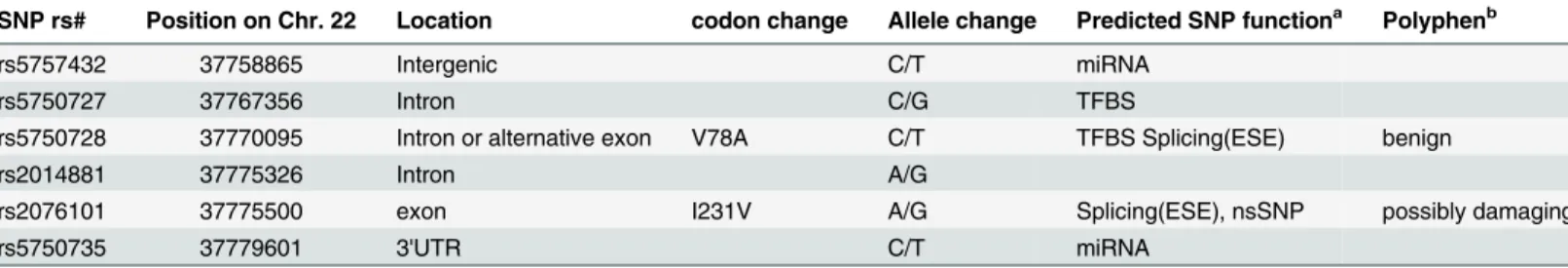
A3Fis about 17 kb in length, containing 7 exons. Six potentially functional SNPs, including the missense SNP rs2076101 ATC>GTC, coding p.I231V, were genotyped in the AIDS cohorts (Fig 1andTable 1). The allele rs2076101G (231V) had a frequency of 48% in European
Fig 1. SNPs analyzed in theA3Fgene.(A) Gene structure and SNP locations. The colored blocks indicate exons, empty blocks, untranslated regions (UTR), horizontal arrows, the direction of transcription and vertical arrows, the positions of SNPs. (B) Linkage disequilibrium matrix of SNPs in theA3Fgene region in European Americans, as illustrated by Haploview [74]. Red block indicates D’= 1.0, and the number in the blocks indicates the value of D’. The linkage disequilibrium block depicted by black triangle was based on the 95% Confidence interval criteria. (C) Haplotypes in theA3Fgene region in European Americans.
Americans (EA) and 76% in African Americans (AA) in our cohorts. Genotype distributions of the 6 SNPs conformed to Hardy-Weinberg equilibrium expectations (P>0.05) in European Americans and African Americans. The 6 SNPs were in near-absolute linkage disequilibrium (LD) and highly correlated (D’= 1 andr2>0.95, respectively) in EA. SNP rs2076101A/G (p. I231V) tags the most frequent A3F haplotype comprising the variant alleles of all 6 SNPs (Fig 1); we therefore used rs2076101A/G (p.I231V) to represent theA3Fhaplotype in the associa-tion analyses.
Based on HapMap LD map covering the APOBEC3 gene family region (S1 Fig),A3F,3G,
3Heach gene forms a distinct haplotype block but are not in strong LD with each other, as reported previously [29].
The possible functional consequence of the six SNPs, as evaluated by the Variant Effect Pre-dictor (VEP) function from Ensembl (www.ensembl.org/), is listed inTable 1. The missense SNP rs2076101 (p.I231V) results in a change from isoleucine to valine, has a Polyphen score of 0.90, reflecting a non-conservative change with possible deleterious consequence. SNP rs5750728 is located in the intron of A3F encoding the major transcript; however, the shorter transcript encoding 101 amino acids (NM_0010066666) would contain p.V78A, a benign change. Whether this isoform is functionalin vivois still unknown. SNP rs5750727 in intron 1 is located within an experimentally identified regulatory feature in CD4+ T-cells
(ENSR00001532770, based on Ensembl database).
Impact of
A3F
231V haplotype on HIV-1 progression
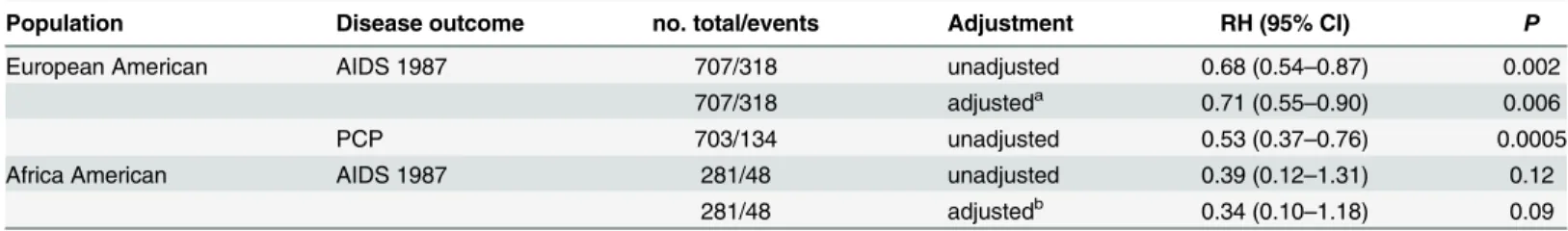
To assess the impact of theA3F231V haplotype on disease progression from the estimated date of seroconversion to clinical AIDS, we performed time-to-event analysis for 707 European American seroconverters. The seroconversion date was estimated as the midpoint between the last seronegative and the first seropositive HIV-1 antibody test date (mean seroconversion interval 0.89 years, range 0.06–3.0 years). In the Kaplan-Meier survival curve analysis, p. 231V was associated with delayed progression to clinical AIDS (Fig 2A) (P= 0.02, in both log-rank and Wilcoxon test) in the dominant genetic model. To account for covariates (age, sex, and cohort) and genetic confounders (CCR5andHLAvariants), we employed adjusted Cox pro-portional hazards regression models to evaluate progression rates to clinical AIDS in 707 Euro-pean American seroconverters. After stratifying by age group, sex and cohort, carriers of one or two copies of p. 231V showed significantly delayed progression to clinical AIDS (dominant genetic model, RH = 0.68, 95% CI, 0.54–0.87,P= 0.002); In the model adjusted for the known genetic factors ofHLAalleles (HLA-C,B27,B57,B35Px,HLAclass I homozygosity, andCCR5
(p1 andΔ32)[31,32], the association remained significant (RH = 0.71, 95% CI, 0.56–0.91,
Table 1. Characteristics ofA3Fgenetic variants.
SNP rs# Position on Chr. 22 Location codon change Allele change Predicted SNP functiona Polyphenb
rs5757432 37758865 Intergenic C/T miRNA
rs5750727 37767356 Intron C/G TFBS
rs5750728 37770095 Intron or alternative exon V78A C/T TFBS Splicing(ESE) benign
rs2014881 37775326 Intron A/G
rs2076101 37775500 exon I231V A/G Splicing(ESE), nsSNP possibly damaging
rs5750735 37779601 3'UTR C/T miRNA
aPredicated by SNPinfo web server [http://snpinfo.niehs.nih.gov]); TFBS, transcription factor-binding site; miRNA, MicroRNA-binding sites; nsSNP,
non-synonymous SNP; ESE, exonic splicing enhancer.
bPredicated by Variant Effect Predictor from Ensembl (www.ensembl.org/)
Padj.= 0.007,Table 2). Further adjustment for potential population stratification using the first 2 eigenvalues led to similar result (RH = 0.71, 95% CI, 0.55–0. 90;Peigen= 0.006). TheA3F
asso-ciation was not attenuated (RH = 0.68, 95% CI, 0.51–0. 90) with additional adjustment for the
Fig 2. Genetic effect ofA3F231V haplotype on AIDS progression.Kaplan-Meier survival curves ofA3F-231I/V genotype carriers progression to AIDS since HIV-1 infection in the (A) combined cohorts. (B) MACS cohort. (C) other cohorts. (D). Kaplan-Meier survival curves ofA3F-231I/V genotype carriers for progression to PCP since HIV-1 infection in 703 seroconverter European Americans. RH and adjustedPvalues were obtained from a Cox proportional hazards model.Pvalues for survival curves were obtained from a log-rank test.
doi:10.1371/journal.pgen.1005921.g002
Table 2. Impact ofA3F231V haplotype on AIDS progression assessed by Cox proportional hazards regression model.
Population Disease outcome no. total/events Adjustment RH (95% CI) P
European American AIDS 1987 707/318 unadjusted 0.68 (0.54–0.87) 0.002
707/318 adjusteda 0.71 (0.55
–0.90) 0.006
PCP 703/134 unadjusted 0.53 (0.37–0.76) 0.0005
Africa American AIDS 1987 281/48 unadjusted 0.39 (0.12–1.31) 0.12
281/48 adjustedb 0.34 (0.10
–1.18) 0.09
Dominant genetic model (231V/V or V/I vs. I/I) was tested and was stratified by sex, age and cohort.
aadjusted for covariatesHLAhomozygosity,HLA-C,HLA-B
*57,HLA-B*35PX,HLA-B*27,CCR5-Δ32,CCR5-59029 and thefirst 2 eigenvalues
badjusted for covariatesHLAhomozygosity,HLA-B
*57,HLA-B*35PX,HLA-B*27, andCCR5-59029.
known HIV progression modifiersA3Bgene deletion [33] andA3G199376C [30]. The effect was not affected by the potential seroconversion estimate errors; RHs were changed from 0.71 to 0.72, 0.72, respectively, when seroconversion intervals were shortened to<2.0 (n = 649) or to<1.5 years (n = 585). The additive genetic model also showed significant association (RH = 0.80, 95% CI, 0.68–0.95,Peigen= 0.009). Bayesian analysis using the Gibbs sampler with
50,000 iterations provided the same results, indicating the p values are robust. Separated analy-ses conducted in the MACS cohort alone (Fig 2B) or in the other cohorts (Fig 2C) showed sig-nificant association in the same direction. These results provide evidence that the haplotype carrying the 6 variant alleles including p. 231V was associated with a 30% reduced rate of AIDS disease in European Americans. In 281 African Americans seroconverters, p. 231V trended in the same direction in a Cox model analysis (RH = 0.35, 95% CI, 0.10–1.19,Padj.= 0.09; RH = 0.30, 95% CI, 0.07–1.37,P= 0.12 with further adjustment of population stratification).
The protective effect of p. 231V for AIDS progression was also observed in a defined disease category analysis (S2 Fig), which allows the addition of seroprevalent patients who had not progressed to AIDS for>10 years (time from seroconversion to AIDS) to the slow progressor category of seroconverters; only seroconverters were included in the fast category (10 years) to avoid frailty bias. The prevalence of AIDS protective genotypes 231V/V and 231 I/V was ele-vated in the slow progressors (Odds ratio (OR) = 0.62,P= 0.004,chi2test,S2 Fig). The results of both the survival (Fig 2AandTable 2) and defined disease category analyses (S2 Fig) sup-port a strong dominant 231V association with delayed progression to clinical AIDS outcome.
A3F
231V haplotype association with pneumocystis pneumonia (PCP)
Multiple different AIDS-defining conditions including opportunistic infections and malignan-cies are AIDS-defining [34]. In an explanatory data analysis, we tested whether the common AIDS defining condition, pneumocystis pneumonia was influenced by p.231V. In a Kaplan-Meier survival curve analysis of 703 seroconverters, carriers of 1 or 2 copies of the p. 231V hap-lotype showed a significantly slower progression to pneumocystis pneumonia, compared with 231I/I carriers (log-rank test,P= 0.003; Wilcoxon test,P= 0.005,Fig 2D). A Cox model analy-sis also showed an association of p.231V with delayed progression to PCP (dominant model, RH = 0.53, 95% CI 0.37–0.76, Wald testPdom= 0.0005,Table 2; additive model, RH = 0.69,
95% CI 0.54–0.89,P= 0.004; both adjusted for age, sex and cohort). A sensitivity test in the MACS cohort revealed no impact of PCP prophylaxis usage on the association. In a sensitivity test performed in 410 MACS seroconverters, adjustment of PCP prophylaxis (n = 138) did not attenuate the association of p.231V with PCP (RH changed from 0.637 to 0.633). These results demonstrated that p.231V has a protective association with PCP development. In 278 Africa American seroconverters, a nonsignificant protective trend to PCP was seen for p.231V homo-zygote carriers (OR = 0.55, 95% CI, 0.28–1.12,P= 0.10;S3 Fig)
A3F
231V haplotype association with HIV-1 viral load
Among 183 MACS seroconverters with available viral load set-point data, individuals carrying the p.231V (n = 141) had significantly lower mean viral load than those homozygous for the p.231I (n = 42) (mean 4.46 ± 0.69 versus 4.72 ± 0.46 log10 copies/ml;P= 0.02). The p.231V carriers were associated with a 0.27 ± 0.65 log10lower set-point viral load (equivalent of 1.86-fold copies per ml of plasma). For comparison, heterozygousity forCCR5Δ32 [35], had a
0.23 ± 0.65 lower viral load (P= 0.04) in this dataset, supporting the validity of the analysis. To replicate the viral load association result, we obtained the association results of theA3F
13 Europe- or USA-based cohorts in ICGH including 6 cohorts used in this study with viral load data available. For independent confirmation, we removed 183 MACS seroconverter sub-jects used in the above viral load analysis from the ICGH. A meta-analysis of 13 cohorts com-prising 10,395 seropositives showed a consistent association ofA3Frs2076101 (231V) with lower set-point viral load (β= -0.04, 95% CI, -0.07, -0.01,P= 0.01) in EA (Fig 3A). The associa-tion was mainly driven by the cohorts used in this study (comprising cohorts MACS, MHCS, DCG, HGDS, SFCCC and ALIVE), and most other European-ancestry cohorts showed nonsig-nificant trends in the same direction. In African American subjects, a meta-analysis showed no association with viral load but a significant protective association was seen for the ALIVE cohort (95% CI ofβ, -0.32, -0.02,Fig 3B). Together, these results indicate a modest but consis-tent association between p.231V and lower viral load.
Impact of
A3F
231V on HIV-1 acquisition
No differences in theA3Fp.231V frequencies were observed between African- or European Ameri-can HIV-1 seronegative groups and HIV-1-infected group (S1 Table), indicating that theA3F
genetic variation assessed herein has no obvious effect on susceptibility to HIV-1 acquisition.
A3F
rs5750728 alters transcriptional factor binding
The intronic SNP rs5750728 is in near absolute LD with 231V and is predicted to have regula-tory function (Table 1). We used electrophoretic mobility shift assays (EMSA) to determine if the variant alters transcription factor binding. The rs5750728 T containing oligonucleotide probe formed a major DNA-protein complex with HeLa cell nuclear extract, which was nearly absent when using rs5750728 C containing oligonucleotide (Fig 4A, lanes 3 and 4). The DNA-protein complex was abolished by the addition of excessive unlabeled competitor probes
Fig 3. HIV-1 viral load levels ofA3F231V allele carriers in multiple HIV-1 cohorts.Forest plot of effect estimates of HIV-1 viral load for theA3F
rs2076101 G allele with 95% confidence intervals per study group (box and whiskers) and after meta-analysis (diamond) in the International Collaboration for the Genomics of HIV (ICGH) consortium [36]. (A) Meta-analyses for European or European-descents (eur). (B) Meta-analyses for African Americans (aam). A description of the study groups and cohorts used in the meta-analysis is presented inS3 Table.
(Fig 4A,lanes 5 and 6), but was not affected by non-specific competitor probes (Fig 4A,lanes 7 and 8), demonstrating the binding specificity. These results indicate that the rs5750728 C var-iant allele caused a loss of binding to certain transcription factors, although the identity of the transcription factors is not yet known.
Genomatix software SNPInspector predicted that rs5750728 T/C change generates tran-scription factor binding sites for E2FF (myc activator/cell cycle regulator), KLFS (Krueppel like transcription factors), and ZKSCAN3 (zinc finger with KRAB and SCAN domains 3), and loss of WHNF (winged helix binding sites) (Fig 4B). Another SNP rs5750727 C/G causes lost site for MRF2 (modulator recognition factor 2).
Fig 4. Allelic-specific protein binding ofA3Frs5750728T/C.(A) For the EMSA, twoA3FDNA fragments with rs5750728 T or C allele were incubated with HeLa nuclear extract. The specificity of the intense bands present in the rs5750728 T lane (lane 3) was demonstrated by adding 200-fold excess of unlabeled T oligo (lane 5) and unrelated TFIID oligos (lane 7) as competitors. (B) Transcription factor binding sites affected by the rs5750728 T/C change based on SNPInspector. The C allele of rs5750728 generates E2FF, ZKSCAN3 (matching the positive (+) strand of the rs5750728 residingA3FDNA fragment), KLFS and loss of WHNF (matching the negative—strand) sites. The rs5750728 site is in red and underlined.
Influence of
A3F
231V haplotype on HIV-1 Vif mediated degradation
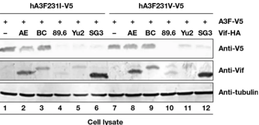
HIV-1 Vif recruits Cul5-based CRL E3 ligase to target A3F for ubiquitination and degradation [37]. To determine whether A3F variants could alter A3F sensitivity to Vif mediated degrada-tion, A3F I231-V5 and A3F V231-V5 expression vectors were co-transfected with several HIV-1 Vif expression vectors in HEK293T cells. A3F expression was determined by immunoblotting using anti-V5 antibodies. A3F I231-V5 and A3F V231-V5 were sensitive to strains 89.6, Yu2, and SG3 HIV-1 Vif mediated degradation but resistant to AE and BC HIV-1 Vif mediated deg-radation (Fig 5). Nevertheless, A3F V231-V5 was approximately 39% more resistant (based on normalized intensity) to HIV-1 AE Vif mediated degradation (Fig 5, lane 8) compared to A3F I231-V5 (Fig 5, lane 2). A3F V231-V5 was 30% more resistant to HIV-1 Yu2 Vif (Fig 5, lane 11) mediated degradation compared to A3F I231-V5 (Fig 5, lane 5). These data suggest that A3F variations at position 231 may influence the sensitivity of A3F to certain HIV-1 Vif medi-ated inactivation.Influence of
A3F
231V haplotype on HIV-1 hypermutation
The effect of rs2076101 on the level of HIV hypermutation was tested using paired human genotypes and viral sequences from 421 patients in the Swiss HIV Cohort Study. Counts of GA-to-AA hypermutations likely induced by A3F were quantified as in the Hypermut2 tool [38]. We observed 8.11(out of 144.95 potential A3F specific G->A mutations, 5.5% hypermu-tation rate), 8.23 (5.6%) and 8.63 (5.3%) A3F specific G->A mutations in three rs2076101 genotype groups, respectively; the log odds ratio of hypermutation was not higher in individu-als carrying the A3F 231V than those who don’t (coefficient = 0.0047,P= 0.88,S2 Table).
Discussion
In vitrostudies have identified A3F as one of the human APOBEC3 proteins with intrinsic anti-HIV-1 activity, and A3F unlike A3G is partially resistant to degradation by HIV-1 Vif [7,15–17]. In this population-based genetic study, we assessed the impact ofA3Fvariation on HIV-1 disease progression in longitudinal HIV-1/AIDS natural disease cohorts. We found that the haplotype encodingA3Fp. 231V and bearing 5 other variant alleles was significantly asso-ciated with slower progression to AIDS and lower viral load set-point. This protective effect is most pronounced for PCP, the most common AIDS-defining disease in the era before effective
Fig 5. Influence of HIV-1 Vif on A3F variant protein expression.HEK293T cells in 12-well plate were co-transfected with 1μg of Vif expression vector or a control vector, plus 0.3μg of A3F-231I-V5 expression vector encoding a V5-tagged A3F231I protein (lanes 1–6), or plus 0.3μg of A3F231V-V5 expression vector encoding a V5-tagged A3F-231V protein (lanes 7–12). At 48 h after transfection, cell lysates were harvested
for immunoblot analysis with indicated antibodies.
antiretroviral therapy was available. Our results provide genetic evidence supporting an active role of human A3F in modulating HIV-1 diseasein vivo. The observed population level associ-ations point toward the physiological relevance of A3F, which has long been debated [7,15– 21].
A3F suppresses HIV-1 replication through deaminase-induced G-to-A hypermutations in viral DNA, as well as deamination-independent impairment of viral reverse transcription and prevention of HIV DNA integration [15–17,39,40]. The mechanism by which theA3Fvariant haplotype influences AIDS pathogenesis is not clear. Mechanistically, the expectation would be that A3F variant protein isoforms have different abilities in inhibiting HIV replication, either by change of protein function or abundance. The association of p.231V with lower viral load supports the idea that A3F ancestral protein isoform containing p. 231V is more effective in restricting HIV replication compared to the derived protein isoforms. In anin vitroHIV-1 infectivity experiment, comparable or slight stronger inhibitory activity against an HIV-1 X4 strain with the wild type Vif was observed for A3F 231V (45% infectivity) compared with A3F 231I (57% infectivity) [21]. It is possible that over time the inhibitory effects of the variant pro-tein may lead to detectable differences in disease outcomes. In a HIV-1 vif-A3F degradation assay, we have also observed that A3F I231V can influence A3F sensitivity to certain HIV-1 Vif proteins. Altered Vif sensitivity of A3F variation may be a contributing factor for the observed differences in HIV-1 viral loads in our study population. Direct Sanger sequencing of HIV-1 viral RNA in plasma of patients in the Swiss HIV cohort identified very low levels of G to A hypermutation, which may represent a small subset of the variation present in the integrated proviral HIV. Although HIV G to A variation mediated by A3F was not associated with the
A3FI231V genotype, deeper sequencing provirus may be needed to accurately access the role ofA3Fgenetic variants on HIV editing [41,42]. Further studies are also needed to determine if
A3Fvariants affects A3F protein function in blocking reverse transcription and or HIV integration.
Pneumocystis pneumonia, or PCP, caused by the fungus Pneumocystis jirovecii, is the most common opportunistic infection in untreated AIDS patients [43,44]. PCP often occurs in those with low CD4+ cell counts<200/mm3; in the MACS cohort, 30% of those patients developed PCP within 3 years after CD4+ cell counts dropping to<200/mm3[43].A3Fvariation impact on PCP may be related with altered activity of A3F in the pulmonary inflammatory environ-ment after CD4+ cell decline. The pathogenesis of PCP is marked with the immune-mediated pulmonary inflammation involving chemokines and cytokines [44]. Inflammatory stimuli such as lipopolysaccharide and interferon-αpotently induce A3G and A3F protein expression
[45]. Research will be needed to determine if A3F protein is activated in lung inflammation sites, affecting local viral replication and immune response.
51], but not in the more biologically relevant T-cell lines [52,53]. The impact of theA3Bgene deletion on HIV-1 infection and progression is inconsistent, possibly due to differences in study designs and case-control selection. We previously reported that the homozygousA3Bdeletion (D/D) was associated with increased risk to HIV-1 acquisition by comparing HIV-1 exposed but uninfected individuals to HIV-1 incident seroconverters, and further that seroconverters pro-gressed more rapidly to clinical outcomes[33]. In support of this, one study found that the D/D carriers had a six-fold greater likelihood of having lower CD4+ T cell levels [54]. Other studies found no associations for infection or progression, but these were suffered from frailty bias which might have inflated type 2 errors. For example, one study used healthy controls and long-term non-progressors but not fast progressors [55,56], and the other study used a control group of HIV-1 negative MSM who were nearly a decade younger than infected cases[57]; given equal exposure time, many of the control group may have become infected. This study did show ten-dency to lower CD4+ T cell depletion with theA3Bdeletion during 88 day follow-up [57]. The role of A3B and theA3Bdeletion in HIV disease warrants further clarification as their role in multiple virus, pathogens and cancers increasingly has been recognized [54,58–61].
In this study, we identified a common haplotype tagged byA3F231V variant as a novel AIDS-modifying genetic factor in European Americans, which shows a similar trend in African Americans with HIV infection.A3GH186R is only found in individuals with African ancestry and is almost absent in European population [30], while theA3Bgene deletion is more com-mon in individuals from Asia and is less frequent in Europeans, and is nearly absent in popula-tions with West African ancestry. The differences inAPOBEC3variant frequencies and haplotype structure among continental populations might have arisen from population-spe-cific demographic events and or from viral selective pressure acting on theAPOBEC3genes. This study adds new evidence that A3F plays an important role in restricting pathogenesis of HIV-1 in its natural host. These data support the development of therapeutics targeting the Vif-A3F axis, along with current efforts on A3G-Vif.
Materials and Methods
Study participants
The study group includes 707 European American HIV-1 incident seroconverters, 281 Afri-can AmeriAfri-can incident seroconverters, 1135 HIV-uninfected at-risk individuals and 2076 HIV seroprevalent individuals who were HIV infected at study entry. The censoring date was the earliest of the date of the last recorded visit, or July 31, 1997 for the ALIVE cohort, or Decem-ber 31, 1995 for all other cohorts, to avoid the confounding effect of highly active anti-retrovi-ral therapy (HAART). A later censoring date was used for ALIVE cohort because few ALIVE participants received HAART prior to July 31, 1997 [69]. PCP prophylaxis status was only available for a subset of the MACS cohort; the first self-reported drugs used included Trimeth-oprim/sulfamethoxazole, aerosolized Pentamidine and Dapsone; the combination or switch-over of these drugs were used switch-over the clinical course.
Ethics statement
Ethical approval for the study was obtained from the National Institute of Health Office of Human Subjects Research Protections with OHSRP #3314. Ethical approval was obtained from institutional review boards for each of the respective contributing centers in the International Collaboration for the Genomics of HIV. Written informed consent was obtained from all study participants.
Selection of
A3F
genetic variants
We selected six SNPs that were codon-changing, in a predicted splicing or regulatory site (based on SNPinfo web server [http://snpinfo.niehs.nih.gov]), considering SNP spacing and gene coverage. Several other nonsynonymous variants in A3F are reported in the 1000 genome project but were not genotyped here either because they were rare or tagged by other SNPs: rs2020390 (p. A108S) is in near-absolute LD (r2= 0.94) with rs2076101 (p.I231V) in Europe-ans. rs34182094 (p. A178T), rs12157816 (p. Y307C), rs13056825 (p.N346S) were absent or infrequent in Utah Europeans (CEU) and Yoruba Africans (YRI).
Genotyping of
A3F
genetic variants
SNPs were genotyped using the TaqMan allele discrimination assays on an ABI 7900HT sequencer detector system (Applied Biosystems), according to the manufacturer’s protocol. For quality control, water controls were included on each plate and 10% of samples were dupli-cated. No water contamination or genotype mismatches between duplicates was observed. SNP rs2076101 was also genotyped by a second in-house designed TaqMan assay (sequences avail-able upon request); the resulted genotypes were identical by two assays.
Electrophoretic mobility shift assays (EMSA)
EMSA was performed in nuclear extracts from HeLa cell stimulated with PMA using oligonu-cleotides carryingA3Frs5750728 T/C with an Infrared EMSA Kit (LI-COR), as previously described [70]. The double-stranded oligonucleotides used were (SNP allele underlined): 5’-CC
CTTTGGCCAGTGCGTCCCACCACAT-3’and 5’-CCCTTTGGCCAGTGCGCCCCACCAC
AT-3’.
Cell culture, transfection and immunoblot analysis
Detection of HIV-1 hypermutation
HIV-1 sequences covering thepolgene alone (in half of the patients) or thepolgene with addi-tional other genes were obtained by the Sanger sequencing from plasma of patients in the Swiss HIV Cohort Study. A3F specific hypermutation was quantified as in the Los Alamos Hyper-mut2 tool [38]. Consensus nucleotide sequences of the samples and of the hxb2 reference sequence were translated to amino acid sequence from which a multiple alignment was created with muscle [71]. Using the amino acid sequence alignment we derived a codon-aware nucleo-tide alignment, which was used to count the number of possible hypermutation sites. We counted the number of trinucleotides matching the“GA[A or T or G]”pattern in the reference sequence as the number of potential A3F hypermutation sites. The number of actual A3F induced mutations in each sample was determined as a subset of potential A3F hypermutation sites where the first nucleotide was an A. It should be noted that GA!AA editing can be mediated by A3D/F/H [39,41]. To quantify the background mutation level we used the follow-ing regular expression, which is the same as in the Hypermut2 tool:“G(([CT][ACGT])|([AG] C))”. The level of hypermutation of a patient sample was determined as an odds ratio: (number of A3F induced mutations/number of potential A3F specific sites) / (number of background mutations / number of potential background sites). Statistical tests were carried out on the log-arithm of the latter quantity using linear regression. The regression included host PCA axes as covariates, the allele dosage of rs2076101, and an indicator variable indicating the SNP geno-typing platform.
Statistical analysis
We performed analyses using SAS version 9.12 (SAS Institute, Cary, NC).
We assessed the influences of theA3Fvariants on disease progression to AIDS outcomes by the Cox proportional hazards model (Cox model) and Kaplan-Meier survival curve analyses. We only included seroconverters with known infection dates (midpoint estimation between last seronegative and first seropositive HIV test) in our analysis. The endpoint was clinical AIDS diagnosis using the Centers for Disease Control and Prevention (CDC) 1987 definition of AIDS [34], i.e. HIV-1 infection plus AIDS-defining illness or AIDS-related death. The median time from seroconversion to AIDS was 10 years in European American seroconverters. As the Kaplan-Meier survival analysis indicated that the haplotype tagged by rs2076101 best fitted a dominant genetic model, we compared the heterozygous (231I/V) and homozygous genotype (231V/V) state to the reference group of homozygotes (231I/I) in a Cox proportional hazards model. European American and African American groups were analyzed separately because the allele frequencies were different between the two groups. We included known genetic factors modifying AIDS progression as covariates in the adjusted Cox model analysis:
CCR5Δ32,CCR5-59029 (CCR5-2459, rs1799987),HLA-B27,HLA-B57,HLA-B35Px group (includingHLA-B3502,
B3503,
B3504, and
B5301),
homozygosity for European American (reviewed in [72];HLA-B57 andHLAClass I homozy-gosity for African American. We stratified the analyses by sex and by age at seroconversion: 0–20,>20–40, and>40 years. P values for Cox model analysis were from the Wald test. Although our previous GWAS for association with HIV phenotypes in EA indicated minimal population substructure (genomic inflation factorλ= 1.01), we conservatively include the first
two eigenvalues from a PCA analysis (EigenSoft) to correct for population stratification [73]. HIV-1 viral load set-point was defined as the mean log10-transfromed copies of HIV-1 RNA in plasma measured between months 6–33 after seroconversion (2–5 measurements). Viral load measurements exceeding three-fold (0.5 log10) from the average of all remaining points were excluded, as previously suggested [35]. We used thettest for analysis of differences between viral load means of the group carrying 0 or 1 231V allele versus the 231I homozygote group.
The International Collaboration for the Genomics of HIV (ICGH) combined the GWAS data from 25 cohorts from Europe and North America, including cohorts used in this study [36]. The genotypes of theA3FSNPs from ICGH were obtained from the previous GWAS data [36]. The impact of theA3FSNPs on HIV-1 viral load in ICGH was assessed using a fixed-effect inverse-variance weighted meta-analysis as previously described [36]. A summary of the selected study groups and cohorts used in the meta-analysis is presented inS3 Table. Viral load data from a total of 10395 (7266 European Americans, 3129 African Americans) HIV-1 seropositives was used in the meta-analysis.
The impact ofA3Fvariants on HIV-1 infection susceptibility was assessed by comparing allelic frequencies between the HIV-1 infected group comprising seroconverter and seropreva-lent persons and the HIV-1 uninfected group composed of persons at risk for HIV. Odds ratios (OR) andPvalues were obtained by using a conditional logistic regression test. AllPvalues were 2-tailed.
Supporting Information
S1 Table. Distribution ofA3F-231I/V in the HIV-1 negative and positive groups.
(DOCX)
S2 Table. HIV-1 hypermutations detected from patients in the Swiss HIV Cohort Study, stratified by theA3F- rs2076101 genotypes.
(DOCX)
S3 Table. Summary of cohorts in the International Collaboration for the Genomics of HIV (ICGH) consortium used for HIV-1 viral load analysis.
(DOCX)
S1 Fig. Linkage disequilibrium in theAPOBEC3gene region.Data was based on HapMap
phase III data in the CEU (Utah Caucasian) population and was plotted with Haploview. The intensity of the box reflects the r2level and haplotype block was defined by 95% CI.
(TIFF)
S2 Fig. Frequency distribution ofA3F-231I/V genotypes in European Americans of fast
progressors and slow progressors.
(TIFF)
S3 Fig. Kaplan-Meier survival curves ofA3F-231I/V genotype carriers for progression to
PCP since HIV-1 infection in 278 African American seroconverters.RH and adjustedP val-ues were obtained from the Cox proportional hazards model.Pvalues for survival curves were obtained from the log-rank test.
Conceived and designed the experiments: PA CAW. Performed the experiments: PA EBR WZ. Analyzed the data: PA CWT IB. Contributed reagents/materials/analysis tools: SP JJG SD SB JF GDK XFY. Wrote the paper: PA CAW. Critically read, contributed to and commented on the manuscript: PA SP JJG SD GDK JF XFY CAW.
References
1. Sheehy AM, Gaddis NC, Choi JD, Malim MH (2002) Isolation of a human gene that inhibits HIV-1 infec-tion and is suppressed by the viral Vif protein. Nature 418: 646–650. PMID:12167863
2. Mangeat B, Turelli P, Caron G, Friedli M, Perrin L, et al. (2003) Broad antiretroviral defence by human APOBEC3G through lethal editing of nascent reverse transcripts. Nature 424: 99–103. PMID: 12808466
3. Yu Q, Chen D, Konig R, Mariani R, Unutmaz D, et al. (2004) APOBEC3B and APOBEC3C are potent inhibitors of simian immunodeficiency virus replication. J Biol Chem 279: 53379–53386. PMID: 15466872
4. Suspene R, Guetard D, Henry M, Sommer P, Wain-Hobson S, et al. (2005) Extensive editing of both hepatitis B virus DNA strands by APOBEC3 cytidine deaminases in vitro and in vivo. Proc Natl Acad Sci U S A 102: 8321–8326. PMID:15919829
5. Chiu YL, Greene WC (2008) The APOBEC3 cytidine deaminases: an innate defensive network oppos-ing exogenous retroviruses and endogenous retroelements. Annu Rev Immunol 26: 317–353. doi:10. 1146/annurev.immunol.26.021607.090350PMID:18304004
6. Harris RS, Bishop KN, Sheehy AM, Craig HM, Petersen-Mahrt SK, et al. (2003) DNA deamination mediates innate immunity to retroviral infection. Cell 113: 803–809. PMID:12809610
7. Bishop KN, Holmes RK, Sheehy AM, Davidson NO, Cho SJ, et al. (2004) Cytidine deamination of retro-viral DNA by diverse APOBEC proteins. Curr Biol 14: 1392–1396. PMID:15296758
8. Zhang H, Yang B, Pomerantz RJ, Zhang C, Arunachalam SC, et al. (2003) The cytidine deaminase CEM15 induces hypermutation in newly synthesized HIV-1 DNA. Nature 424: 94–98. PMID:12808465
9. Conticello SG, Harris RS, Neuberger MS (2003) The Vif protein of HIV triggers degradation of the human antiretroviral DNA deaminase APOBEC3G. Curr Biol 13: 2009–2013. PMID:14614829 10. Marin M, Rose KM, Kozak SL, Kabat D (2003) HIV-1 Vif protein binds the editing enzyme APOBEC3G
and induces its degradation. Nat Med 9: 1398–1403. PMID:14528301
11. Sheehy AM, Gaddis NC, Malim MH (2003) The antiretroviral enzyme APOBEC3G is degraded by the proteasome in response to HIV-1 Vif. Nat Med 9: 1404–1407. PMID:14528300
12. Yu X, Yu Y, Liu B, Luo K, Kong W, et al. (2003) Induction of APOBEC3G ubiquitination and degradation by an HIV-1 Vif-Cul5-SCF complex. Science 302: 1056–1060. PMID:14564014
13. Zhang W, Du J, Evans SL, Yu Y, Yu XF (2012) T-cell differentiation factor CBF-beta regulates HIV-1 Vif-mediated evasion of host restriction. Nature 481: 376–379.
14. Sadler HA, Stenglein MD, Harris RS, Mansky LM (2010) APOBEC3G contributes to HIV-1 variation through sublethal mutagenesis. Journal of virology 84: 7396–7404. doi:10.1128/JVI.00056-10PMID: 20463080
15. Liddament MT, Brown WL, Schumacher AJ, Harris RS (2004) APOBEC3F properties and hypermuta-tion preferences indicate activity against HIV-1 in vivo. Curr Biol 14: 1385–1391. PMID:15296757 16. Wiegand HL, Doehle BP, Bogerd HP, Cullen BR (2004) A second human antiretroviral factor,
17. Zheng YH, Irwin D, Kurosu T, Tokunaga K, Sata T, et al. (2004) Human APOBEC3F is another host fac-tor that blocks human immunodeficiency virus type 1 replication. J Virol 78: 6073–6076. PMID:
15141007
18. Gillick K, Pollpeter D, Phalora P, Kim EY, Wolinsky SM, et al. (2013) Suppression of HIV-1 infection by APOBEC3 proteins in primary human CD4(+) T cells is associated with inhibition of processive reverse transcription as well as excessive cytidine deamination. Journal of virology 87: 1508–1517. doi:10.
1128/JVI.02587-12PMID:23152537
19. Chaipan C, Smith JL, Hu WS, Pathak VK (2013) APOBEC3G restricts HIV-1 to a greater extent than APOBEC3F and APOBEC3DE in human primary CD4+ T cells and macrophages. Journal of virology 87: 444–453. doi:10.1128/JVI.00676-12PMID:23097438
20. Miyagi E, Brown CR, Opi S, Khan M, Goila-Gaur R, et al. (2010) Stably expressed APOBEC3F has negligible antiviral activity. Journal of virology 84: 11067–11075. doi:10.1128/JVI.01249-10PMID:
20702622
21. Mulder LC, Ooms M, Majdak S, Smedresman J, Linscheid C, et al. (2010) Moderate influence of human APOBEC3F on HIV-1 replication in primary lymphocytes. Journal of virology 84: 9613–9617.
doi:10.1128/JVI.02630-09PMID:20592068
22. Refsland EW, Stenglein MD, Shindo K, Albin JS, Brown WL, et al. (2010) Quantitative profiling of the fullAPOBEC3mRNA repertoire in lymphocytes and tissues: implications for HIV-1 restriction. Nucleic Acids Res 38: 4274–4284. doi:10.1093/nar/gkq174PMID:20308164
23. Koning FA, Newman EN, Kim EY, Kunstman KJ, Wolinsky SM, et al. (2009) Defining APOBEC3 expression patterns in human tissues and hematopoietic cell subsets. J Virol 83: 9474–9485. doi:10.
1128/JVI.01089-09PMID:19587057
24. Russell RA, Smith J, Barr R, Bhattacharyya D, Pathak VK (2009) Distinct domains within APOBEC3G and APOBEC3F interact with separate regions of human immunodeficiency virus type 1 Vif. J Virol 83: 1992–2003. doi:10.1128/JVI.01621-08PMID:19036809
25. Albin JS, Harris RS (2010) Interactions of host APOBEC3 restriction factors with HIV-1 in vivo: implica-tions for therapeutics. Expert Rev Mol Med 12: e4. doi:10.1017/S1462399409001343PMID: 20096141
26. Sheehy AM, Erthal J (2012) APOBEC3 versus Retroviruses, Immunity versus Invasion: Clash of the Titans. Molecular biology international 2012: 974924. doi:10.1155/2012/974924PMID:22720156 27. Ross SR (2009) Are viruses inhibited by APOBEC3 molecules from their host species? PLoS
patho-gens 5: e1000347. doi:10.1371/journal.ppat.1000347PMID:19390611
28. Valcke HS, Bernard NF, Bruneau J, Alary M, Tsoukas CM, et al. (2006)APOBEC3Ggenetic variants and their association with risk of HIV infection in highly exposed Caucasians. AIDS 20: 1984–1986. PMID:16988524
29. Cagliani R, Riva S, Fumagalli M, Biasin M, Caputo SL, et al. (2011) A positively selectedAPOBEC3H haplotype is associated with natural resistance to HIV-1 infection. Evolution 65: 3311–3322. doi:10. 1111/j.1558-5646.2011.01368.xPMID:22023594
30. An P, Bleiber G, Duggal P, Nelson G, May M, et al. (2004)APOBEC3Ggenetic variants and their influ-ence on the progression to AIDS. J Virol 78: 11070–11076. PMID:15452227
31. An P, Winkler CA (2010) Host genes associated with HIV/AIDS: advances in gene discovery. Trends Genet 26: 13.
32. Fellay J, Shianna KV, Ge D, Colombo S, Ledergerber B, et al. (2007) A whole-genome association study of major determinants for host control of HIV-1. Science 317: 944–947. PMID:17641165
33. An P, Johnson R, Phair J, Kirk GD, Yu XF, et al. (2009)APOBEC3BDeletion and Risk of HIV-1 Acquisi-tion. J Infect Dis 200: 1054–1058. doi:10.1086/605644PMID:19698078
34. CDC(1987) Revision of the CDC surveillance case definition for acquired immunodeficiency syndrome. MMWR 36 Suppl 1: 1S–15S.
35. Fellay J, Ge D, Shianna KV, Colombo S, Ledergerber B, et al. (2009) Common genetic variation and the control of HIV-1 in humans. PLoS Genet 5: e1000791. doi:10.1371/journal.pgen.1000791PMID: 20041166
36. McLaren PJ, Coulonges C, Ripke S, van den Berg L, Buchbinder S, et al. (2013) Association study of common genetic variants and HIV-1 acquisition in 6,300 infected cases and 7,200 controls. PLoS Pathog 9: e1003515. doi:10.1371/journal.ppat.1003515PMID:23935489
37. Liu B, Sarkis PT, Luo K, Yu Y, Yu XF (2005) Regulation of Apobec3F and human immunodeficiency virus type 1 Vif by Vif-Cul5-ElonB/C E3 ubiquitin ligase. J Virol 79: 9579–9587. PMID:16014920 38. Rose PP, Korber BT (2000) Detecting hypermutations in viral sequences with an emphasis on G—>A
monia among men infected with human immunodeficiency virus type 1. Multicenter AIDS Cohort Study Group. N Engl J Med 322: 161–165. PMID:1967190
44. Thomas CF Jr., Limper AH (2004) Pneumocystis pneumonia. N Engl J Med 350: 2487–2498. PMID: 15190141
45. Mehta HV, Jones PH, Weiss JP, Okeoma CM (2012) IFN-alpha and lipopolysaccharide upregulate APOBEC3 mRNA through different signaling pathways. J Immunol 189: 4088–4103. doi:10.4049/ jimmunol.1200777PMID:22972924
46. Jarmuz A, Chester A, Bayliss J, Gisbourne J, Dunham I, et al. (2002) An anthropoid-specific locus of orphan C to U RNA-editing enzymes on chromosome 22. Genomics 79: 285–296. PMID:11863358 47. Reddy K, Winkler CA, Werner L, Mlisana K, Abdool Karim SS, et al. (2010)APOBEC3Gexpression is
dysregulated in primary HIV-1 infection and polymorphic variants influence CD4+ T-cell counts and plasma viral load. AIDS 24: 195–204. doi:10.1097/QAD.0b013e3283353bbaPMID:19996938 48. Singh KK, Wang Y, Gray KP, Farhad M, Brummel S, et al. (2013) Genetic variants in the host restriction
factorAPOBEC3Gare associated with HIV-1-related disease progression and central nervous system impairment in children. Journal of acquired immune deficiency syndromes 62: 197–203. doi:10.1097/ QAI.0b013e31827ab612PMID:23138837
49. Bogerd HP, Wiegand HL, Doehle BP, Cullen BR (2007) The intrinsic antiretroviral factor APOBEC3B contains two enzymatically active cytidine deaminase domains. Virology 364: 486–493. PMID: 17434555
50. Doehle BP, Schafer A, Cullen BR (2005) Human APOBEC3B is a potent inhibitor of HIV-1 infectivity and is resistant to HIV-1 Vif. Virology 339: 281–288. PMID:15993456
51. Rose KM, Marin M, Kozak SL, Kabat D (2005) Regulated production and anti-HIV type 1 activities of cytidine deaminases APOBEC3B, 3F, and 3G. AIDS Res Hum Retroviruses 21: 611–619. PMID: 16060832
52. Hultquist JF, Lengyel JA, Refsland EW, LaRue RS, Lackey L, et al. (2011) Human and rhesus APO-BEC3D, APOBEC3F, APOBEC3G, and APOBEC3H demonstrate a conserved capacity to restrict Vif-deficient HIV-1. J Virol 85: 11220–11234. doi:10.1128/JVI.05238-11PMID:21835787
53. Refsland EW, Hultquist JF, Harris RS (2012) Endogenous origins of HIV-1 G-to-A hypermutation and restriction in the nonpermissive T cell line CEM2n. PLoS Pathog 8: e1002800. doi:10.1371/journal. ppat.1002800PMID:22807680
54. Prasetyo AA, Sariyatun R, Reviono, Sari Y, Hudiyono, et al. (2015) TheAPOBEC3Bdeletion polymor-phism is associated with prevalence of hepatitis B virus, hepatitis C virus, Torque Teno virus, and Toxo-plasma gondii co-infection among HIV-infected individuals. J Clin Virol 70: 67–71. doi:10.1016/j.jcv. 2015.07.009PMID:26305823
55. Itaya S, Nakajima T, Kaur G, Terunuma H, Ohtani H, et al. (2010) No evidence of an association between theAPOBEC3Bdeletion polymorphism and susceptibility to HIV infection and AIDS in Japa-nese and Indian populations. J Infect Dis 202: 815–816; author reply 816–817. doi:10.1086/655227
PMID:20684727
56. Winkler CA, An P, Buchbinder S, Donfield S, Goedert J, et al. (2010) Reply to Itaya et al. Journal of Infectious Diseases 202: 816–817.
57. Imahashi M, Izumi T, Watanabe D, Imamura J, Matsuoka K, et al. (2014) Lack of association between intact/deletion polymorphisms of the APOBEC3B gene and HIV-1 risk. PLoS One 9: e92861. doi:10. 1371/journal.pone.0092861PMID:24667791
59. Jha P, Sinha S, Kanchan K, Qidwai T, Narang A, et al. (2012) Deletion of the APOBEC3B gene strongly impacts susceptibility to falciparum malaria. Infect Genet Evol 12: 142–148. doi:10.1016/j.meegid.
2011.11.001PMID:22108670
60. Zhang T, Cai J, Chang J, Yu D, Wu C, et al. (2013) Evidence of associations of APOBEC3B gene dele-tion with susceptibility to persistent HBV infecdele-tion and hepatocellular carcinoma. Hum Mol Genet 22: 1262–1269. doi:10.1093/hmg/dds513PMID:23213177
61. Burns MB, Temiz NA, Harris RS (2013) Evidence for APOBEC3B mutagenesis in multiple human can-cers. Nat Genet 45: 977–983. doi:10.1038/ng.2701PMID:23852168
62. Phair J, Jacobson L, Detels R, Rinaldo C, Saah A, et al. (1992) Acquired immune deficiency syndrome occurring within 5 years of infection with human immunodeficiency virus type-1: the Multicenter AIDS Cohort Study. J Acquir Immune Defic Syndr 5: 490–496. PMID:1560346
63. Buchbinder SP, Katz MH, Hessol NA, O'Malley PM, Holmberg SD (1994) Long-term HIV-1 infection without immunologic progression. AIDS 8: 1123–1128. PMID:7986410
64. Vlahov D, Graham N, Hoover D, Flynn C, Bartlett JG, et al. (1998) Prognostic indicators for AIDS and infectious disease death in HIV-infected injection drug users: plasma viral load and CD4+ cell count. JAMA 279: 35–40. PMID:9424041
65. Hilgartner MW, Donfield SM, Willoughby A, Contant CF Jr., Evatt BL, et al. (1993) Hemophilia growth and development study. Design, methods, and entry data. Am J Pediatr Hematol Oncol 15: 208–218. PMID:8498644
66. Goedert JJ, Kessler CM, Aledort LM, Biggar RJ, Andes WA, et al. (1989) A prospective study of human immunodeficiency virus type 1 infection and the development of AIDS in subjects with hemophilia. N Engl J Med 321: 1141–1148. PMID:2477702
67. Goedert JJ, Biggar RJ, Melbye M, Mann DL, Wilson S, et al. (1987) Effect of T4 count and cofactors on the incidence of AIDS in homosexual men infected with human immunodeficiency virus. JAMA 257: 331–334. PMID:3491911
68. An P, Duggal P, Wang LH, O'Brien SJ, Donfield S, et al. (2007) Polymorphisms ofCUL5are associated with CD4+ T cell loss in HIV-1 infected individuals. PLoS Genet 3: e19. PMID:17257057
69. Celentano DD, Galai N, Sethi AK, Shah NG, Strathdee SA, et al. (2001) Time to initiating highly active antiretroviral therapy among HIV-infected injection drug users. AIDS 15: 1707–1715. PMID:11546947
70. An P, Goedert JJ, Donfield S, Buchbinder S, Kirk GD, et al. (2014) Regulatory Variation in HIV-1 Dependency Factor ZNRD1 Associates with Host Resistance to HIV-1 Acquisition. The Journal of infectious diseases.
71. Edgar RC (2004) MUSCLE: multiple sequence alignment with high accuracy and high throughput. Nucleic Acids Res 32: 1792–1797. PMID:15034147
72. O'Brien SJ, Nelson GW (2004) Human genes that limit AIDS. Nat Genet 36: 565–574. PMID: 15167933
73. Troyer JL, Nelson GW, Lautenberger JA, Chinn L, McIntosh C, et al. (2011) Genome-wide association study implicatesPARD3B-based AIDS restriction. The Journal of infectious diseases 203: 1491–1502. doi:10.1093/infdis/jir046PMID:21502085