Cross Dimerization of Amyloid-
b
and
a
Synuclein Proteins
in Aqueous Environment: A Molecular Dynamics
Simulations Study
Jaya C. Jose, Prathit Chatterjee, Neelanjana Sengupta*
Physical Chemistry Division, CSIR-National Chemical Laboratory, Pune, Maharashtra, India
Abstract
Self-assembly of the intrinsically unstructured proteins, amyloid beta (Ab) and alpha synclein (aSyn), are associated with Alzheimer’s Disease, and Parkinson’s and Lewy Body Diseases, respectively. Importantly, pathological overlaps between these neurodegenerative diseases, and the possibilities of interactions between AbandaSyn in biological milieu emerge from several recent clinical reports andin vitrostudies. Nevertheless, there are very few molecular level studies that have probed the nature of spontaneous interactions between these two sequentially dissimilar proteins and key characteristics of the resulting cross complexes. In this study, we have used atomistic molecular dynamics simulations to probe the possibility of cross dimerization betweenaSyn1–95and Ab1–42, and thereby gain insights into their plausible early assembly pathways
in aqueous environment. Our analyses indicate a strong probability of association between the two sequences, with inter-protein attractive electrostatic interactions playing dominant roles. Principal component analysis revealed significant heterogeneity in the strength and nature of the associations in the key interaction modes. In most, the interactions of repeating Lys residues, mainly in the imperfect repeats ‘KTKEGV’ present inaSyn1–95were found to be essential for cross
interactions and formation of inter-protein salt bridges. Additionally, a hydrophobicity driven interaction mode devoid of salt bridges, where the non-amyloid component (NAC) region ofaSyn1–95came in contact with the hydrophobic core of
Ab1–42 was observed. The existence of such hetero complexes, and therefore hetero assembly pathways may lead to
polymorphic aggregates with variations in pathological attributes. Our results provide a perspective on development of therapeutic strategies for preventing pathogenic interactions between these proteins.
Citation:Jose JC, Chatterjee P, Sengupta N (2014) Cross Dimerization of Amyloid-bandaSynuclein Proteins in Aqueous Environment: A Molecular Dynamics Simulations Study. PLoS ONE 9(9): e106883. doi:10.1371/journal.pone.0106883
Editor:Paolo Carloni, German Research School for Simulation Science, Germany
ReceivedApril 8, 2014;AcceptedAugust 7, 2014;PublishedSeptember 11, 2014
Copyright:ß2014 Jose et al. This is an open-access article distributed under the terms of the Creative Commons Attribution License, which permits unrestricted use, distribution, and reproduction in any medium, provided the original author and source are credited.
Data Availability:The authors confirm that all data underlying the findings are fully available without restriction. All relevant data are within the paper and its Supporting Information files.
Funding:Funding for this project was received from CSIR 12th FYP Multi-Scale Simulation and Modeling project (or MSM; project number CSC0129), and from Center of Excellence for Sustainable Polymer Industry through Research Innovation & Training (CoE-SPIRIT), established through a grant from the Department of Chemical and Petrochemicals. The funders had no role in study design, data collection and analysis, decision to publish, or preparation of the manuscript.
Competing Interests:The authors have declared that no competing interests exist.
* Email: [email protected]
Introduction
Misfolding and aggregation of amyloidogenic proteins in the intra- or extra-cellular regions of the human brain are associated with multiple neurodegenerative diseases (ND) [1–6]. Although these diseases differ in their pathological attributes, the toxic transformations of the proteins are associated with similar pathways characterized initially by the formation of soluble oligomers, followed progressively by the emergence and elongation of protofibrillar and fibrillar aggregates [7–11]. Interestingly, recent clinical studies indicate that the symptoms associated with different ND can occur synergistically, leading to the worsening of overall prognosis [12,13]. Recent experimental and theoretical studies have found that the abnormal cross interactions between different misfolded proteins could lead to such mixed pathologies [14–16].
Among different NDs, Alzheimers Disease (AD), Lewy Body Disease (LBD), and Parkinson’s Disease (PD) are the leading cause of dementia and moving disorders in the elderly. While oligomerisation and fibrillisation of Ab has been identified as a
toxic event in AD [2], progressive accumulation ofaSyn has been linked to PD [3]. Recent studies suggest thataSyn may also have a crucial role to play in pathology of AD [3]. A large fraction of AD patients exhibitaSyn positive Lewy bodies associated with LBD in their brains [5,17]. Evidences suggest that AbandaSyn interact directlyin vivoandin vitro[14,15,18]. Transgenic mouse models demonstrate AbenhancesaSyn accumulation and neuronal deficit [15]. Multi-dimentional NMR studies in membrane mimicking environment reported that the molecular interaction ofaSyn with Ab40and Ab42are site-specific, and that membrane boundaSyn
induced structural alterations that are more profound in Ab42
compared to those in Ab40[14]. The same study also suggests that
the oligomerization pathways foraSyn with Ab42and Ab40in the
vicinity of cellular membranes are different [14]. Short MD simulations showed that AbandaSyn localized on a lipid bilayer surface are capable of forming ring-like hybrid structures that can porate the membrane [18]. Interestingly, recent kinetic study suggest that the fibrils and oligomers of Ab40, Ab42andaSyn can
Both AbandaSyn are intrinsically unstructured proteins (IUPs) whose pathological transformations are fundamentally dependent on their primary sequences. Although Ab is an amphiphilic peptide, it has distinctive hydrophobic patches, particularly the central hydrophobic core L17VFFA21and the C-terminal hydro-phobic region A30–A42. The intra- and intermolecular interactions in these regions are known to lead to the compactification of this peptide in its monomeric state followed by its aggregation to form toxic species [8,20–23]. In addition, the charged residues E22, D23, K28of the Abpeptide, that can form intra- and intermolecular salt bridges in the N-terminal fragment and at the central region play important roles in the peptide’s the pathological transformations [24–27]. aSyn is composed of three distinct regions; an N-terminal lipid binding domain (residues 1–60), a continuous hydrophobic domain (residues 61–95) and a highly acidic C terminal region. Among these, the hydrophobic segment is the non amyloid component (NAC) of the amyloid plaques found in AD [3]. The first two regions of aSyn is composed of six imperfect repeat sequence motifs KTKEGV, but the role of these repeats in the toxicity of the protein has not yet been understood.
We note that despite increasing evidences of overlapping pathologies of AD and PD and accelerated neurodegeneration arising from cross influences of AbandaSyn, there are relatively few molecular level studies that directly probe the interactions between these two dissimilar IUPs. To the best of our knowledge, molecular details of their spontaneous associations in regimes that resemble the aqueous cytoplasmic conditions remain uncharacter-ized. In this study, we have used microsecond scale unbiased molecular dynamics (MD) simulations to discern the early inter-molecular associations between the monomeric forms of Aband aSyn in aqueous environment. We mention here that interactions with surfaces can hinder the translation diffusion of proteins and affect the rates of their assembly and aggregation [28–30]. The initial diffusive regime has been noted to play important roles in self-assembly of amyloidogenic peptides [31]. Our simulations are performed such that restrictions on the initial diffusive regime due to surface tethering or adsorption are avoided. Our results indicate a high probability of cross-dimerization between the two sequentially dissimilar proteins leading to the formation of metastable complexes that may have the potential to further co-fibrillize. Principal component analysis revealed distinct asso-ciation modes with variations in the strength and nature of inter-protein interactions, salt bridge propensities and extents of conformational disorder. The majority of cross-interactions were found to be driven electrostatically, with the Lys repeats ofaSyn playing important roles in enhancing stability via inter-protein salt bridge formation. Remarkably, however, we also found the existence of an interaction mode that was predominantly stabilized via hydrophobic interactions. Our study provides evidence of marked heterogeneity in the cross interactions responsible for primary association of the two disease-associated IUPs. The data strongly suggest the existence of multiple pathways of cross-fibrillization between AbandaSyn, and therefore high degrees of polymorphism in the resultant cross aggregates.
Methods
Generation of Initial Monomer Conformations
We generated putative monomeric conformations of Ab and aSyn monomers in aqueous environment by employing the accelerated molecular dynamics simulations (AMD) method with torsional boost [32] to suitably alter the predominantly helical, solution-state NMR structures of Ab (1Z0Q) [33] and aSyn (2KWW) [34], available in the PDB database. The Abstructure
was experimentally reported via solution NMR studies in a 3:7 mixture of hexafluoro-2-propanol and water, while the aSyn structure was reported in the micellar environment. AMD as implemented in the NAMD2.8 package [35] was used with the CHARMM all atom force field with CMAP correction [36,37]. The theoretical details of the AMD method can be found in other reports [38–40]. Briefly, AMD ensures enhanced barrier crossing and sampling within shorter durations by altering the potential energy surface (V(r)) with the boost energy, Eb, and the acceleration parameter,a. The modified potentialV*(r)is given as,
V(~rr)§Eb ð1Þ
V(~rr)~V(~rr)zDV(~rr), V(~rr)vEb ð2Þ
Here, the bias potentialDV(r)is obtained as,
DV(~rr)~ðEb{V(~rr)Þ
2
Eb{V(~rr)za
ð3Þ
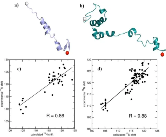
Increasing values ofEbandaresult, respectively, in enhancing and reducing the extent of acceleration. In accordance with the optimized AMD methods [40] preliminary, short unbiased simulations were performed to obtain the mean dihedral energies (Vdih), andEbwas set such that their difference was 4 kcal mol21 times the number of residues in the protein. The acidic tail region 96–140 ofaSyn was excluded as the C-terminal truncated aSyn has been shown to have higher propensity for aggregation [41– 43]. Aband aSyn are intrinsically disordered proteins with wide conformational ensembles [44–46]. However, the Ab conforma-tion obtained towards the end of our 17 ns long AMD simulaconforma-tions had marked similarities with important conformational members reported before, in terms of the emergence of anti-parallel C-terminal beta sheets and reduced N-C-terminal helicities [45,47]. We generated an ensemble of the free peptide monomers with the conformations thus obtained, and calculated the average 15N chemical shift values using the SHIFTS program, [48] and compared them with the experimentally determined values for Ab [49] and aSyn [50]. The mean chemical shift values were positively correlated with the experimental values. The Pearson Correlation Coefficients (PCC) for Ab and aSyn were 0.86 and 0.88, respectively. The selected conformations, and the corre-sponding chemical shift correlation plots are shown in Figure 1.
System Setup and Simulation Protocols
temperature of 310 K was maintained with Langevin dynamics with a collision frequency of 1 ps21, and the Langevin piston Nose´-Hoover method, was used to maintain a constant pressure of 1 atm [52,53]. The cutoff radius for Lennard Jones interaction was set to 12 A˚ . SHAKE [54] was used for constraining bonds involving hydrogen atoms. Electrostatic interactions were calcu-lated with particle-mesh Ewald method [55]. A time step of 2 fs was used. A total of 1.3ms of unbiased simulations were generated.
Pymol [56] and the VMD [57] tools were used for the generation of snapshots and visualization of the trajectories.
Principal Component Analysis
In order to capture the most significant modes of cross-monomer interactions, clustering based on Cartesian Principal component analysis (PCA) was conducted on combined snapshots of the interacting trajectories using the program Carma [58]. PCA has been widely recognized as a reliable starting point to identify important modes of interacting systems produced by MD simulations [45,59–61]. The heavily populated clusters are identified by analysing the distribution of first three principal components using an rmsd cutoff of 2.4 A˚ . The probability density of the distribution of first two principal components corresponding to the fluctuations of the Ca atoms in the bound system is
calculated and converted into a free energy function using the following equation,
DG~{kBTln
p pmax
ð4Þ
where,kBis Boltzmann’s constant,Tis the temperature in absolute
units,pis probability obtained from the distribution of the first two principal components, andpmaxis the corresponding maximum
probability.
Configurational Entropy
We have calculated the configurational entropy per heavy atoms of AbandaSyn peptides in bound and unbound state using Schlitter’s method [62] as implemented in Carma program [58]. This method has been widely used to calculate the degree of change in internal conformation of bio-systems using MD trajectories [63–65]. Here the initial structure of each peptide is used as reference, to remove the translations and rotations with respect to the center of mass of the systems. According to Schlitter’s method the absolute entropy can be approximated as follows,
SabsvS~ 1
2kBln det 1z kBTe2
B2 M 1 2sM12
ð5Þ
Where kB is the Boltzmann’s constant, h-is Planck’s constant
divided by2p, eis Euler’s number,Mis the mass matrix of3N
dimension containingNatomic masses of the system andsis the covariance matrix. The elements in the covariance matrix can be expressed as,
sij~Sðxi{SxiTÞ xj{SxjT
T ð6Þ
Figure 1. Snapshots of starting monomeric structures of a) Ab1–42and b)aSyn1–95used in the unbiased simulations in the study.
Correlation of average theoretical15N chemical shifts with experimentally determined15N chemical shifts for c) Ab1–42and d)aSyn1–95. The linear regressions (straight lines) and the corresponding Pearson Correlation Coefficients (R) are provided. See text for details.
doi:10.1371/journal.pone.0106883.g001
where, xi and xj are the Cartesian coordinates of the selected atoms.
Results
I. Evaluation of inter-protein association
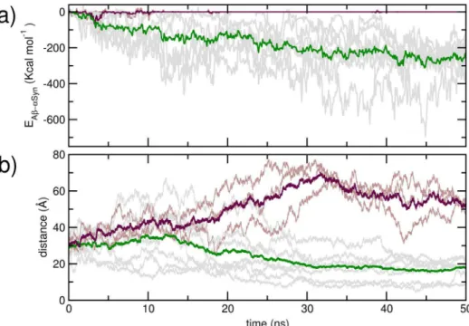
The initial inter-peptide center of mass distance, as well as their distances and relative orientations at 10 ns are provided in Table 1. In Figure 2 a), we present evolution of the peptide-peptide interaction strength over the first 50 ns of simulation for the trajectories. While three trajectories indicate no inter-protein interaction at the end of 50 ns, AbandaSyn in seven trajectories demonstrate strong interaction. The mean inter-protein interac-tion strength at the end of 50 ns is2172.96 (672.8) kcal mol21.
We have shown corresponding evolution of the center of mass distances in Figure 2b. The mean inter-monomer center of mass distance at 50 ns of the seven trajectories where Ab and aSyn interact are 17.5 A˚ , while the corresponding mean distance obtained from the non-interacting ones are 53.0 A˚ . The interaction energy, center of mass distances and relative orienta-tions at 50 ns have also been provided in Table 1. The interacting trajectories were each further propagated for at least an additional 100 ns; evolutions of corresponding inter-peptide interaction strength and center of mass distances of these trajectories over 150 ns are provided in Figure S1 in File S1. We have further compared the residue-wise backbone mean squared fluctuation (MSF) in the AbandaSyn obtained at 0–50 ns of the simulations, with that obtained over 100–150 ns, from the peptides in the interacting trajectories (Figure S2 in File S1). An overall sharp reduction in the MSF is noted upon the formation of complexes from the two dissimilar peptides, with comparatively greater decrease in the middle regions. The interaction of AbandaSyn is thus commensurate with a decrease in the structural disorder.
The discussion above shows that despite the early diffusive regime, AbandaSyn have a marked, enthalpy driven propensity to interact and form dimeric complexes in aqueous solution. In Figure 3, we provide a residue-wise breakdown of the total inter-peptide interaction. Interestingly, we found that the charged residues of each peptide exhibit significantly stronger interactions compared to the hydrophobic and polar residues. This was manifestly clear when we considered the strongest interaction arising from each residue (Figure 3 b and d). Interestingly,
interactions arising from the repeating Lys residues of the repeating units in the N- and C-terminii of aSyn give rise to distinctly strong interactions.
II. Interaction heterogeneity
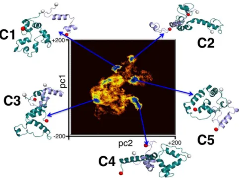
Principal component analysis as described in Methods was performed with snapshots of the dimerized complexes where the proteins’ centers of mass were closer than 30 A˚ . In Figure 4, we present the free energy landscape as a function of the first (pc1) and the second (pc2) principal components. Five distinct clusters were obtained from the PCA and named C1, C2, C3, C4 and C5 in order of decreasing cluster population. Snapshots corresponding to structures residing at the cluster centers have been shown in Figure 4. In order to decipher distinguishing traits of the individual complexes in each cluster, these representative struc-tures were individually simulated for 4 ns under the same conditions as the original simulations.
In Table 2, we have reported mean values of the number of inter-protein contacts; the radii of gyration (Rg) of the dimeric
complexes; and electrostatic and van der Waals components of the Ab-aSyn interaction strengths of all five clusters. As in a previously reported study [20], two residues are taken to form a contact if the centers of mass of their sidechains approach within a distance of 7 A˚ . The five clusters are found to have significant variation in the number of inter-protein contacts, the level of compactness of the complexes as well as of the individual protein units (reported in Table S2 in File S1), and the strength of the inter-protein interactions. C5 has the highest average number of inter-protein residue contacts commensurate with the strongest inter-protein interaction (Etot) of 2485.2 kcal/mol. C2 and C4 have a
comparable number of inter-protein contacts, which are margin-ally lower than the contacts in C5. Interestingly, however, while the inter-protein interaction strength in C2 is comparable to that of C5, the interaction strength in C4 is significantly weaker, being only282.7 kcal/mol in its mean value. Clusters C3 and C1 have
markedly lower mean inter-protein contacts, with values of only 23 and 14.6, respectively. However, the inter-protein interactions in C3 and C1 are stronger than that in C4.
Interestingly, we note that in C1, C2, C3 and C5, the inter-protein interaction is dominated by electrostatic energy. In contrast, in the cluster C4, the dominant non-bonded contribution arises from van der Waals interactions. However, despite the
Table 1.Inter-protein orientations and interaction energies along the simulation trajectories.
Traj. No. d0 d10 h10 d50 h50 Eint
1 33.2 31.8 79.6 12.8 101.8 2177.3
2 33.2 53.0 27.5 19.7 65.6 2173.2
3 33.2 56.8 29.8 19.1 136.3 2213.1
4 33.2 23.0 134.2 19.1 99.1 2275.0
5 33.2 38.7 135.9 55.9 60.9 0.0
6 33.2 51.7 157.8 53.2 68.5 0.0
7 18.9 13.7 162.1 21.6 109.4 294.6
8 18.3 43.2 133.3 22.0 76.0 2403.3
9 24.3 39.4 116.9 50.1 97.4 0.0
10 24.3 26.7 119.1 8.3 158.2 2297.5
The inter-protein center of mass distances (in A˚) at the start of the unbiased simulations is denoted as d0, at 10 ns is denoted as d10, and at 50 ns is denoted as d50. The
relative orientations of the proteins are specified by the angle (in degrees) between the vectors joining the N- and C-terminii of each protein, at 10 ns (h10) and at 50 ns
(h50). Eintdenotes the total inter-protein interaction at 50 ns (in kcal mol21).
weaker inter-protein interaction, the number of residue-residue contacts in C4 is comparable to that of C5 and C2.
We have compared the inter-protein side-chain contact probability maps for all five clusters in Figure 5. The contact
maps reveal high degrees of contact heterogeneity amongst the various clusters. In C1, contacts were predominantly formed between the N-terminii of Ab and aSyn. Relatively weaker contacts were noted between residues 35 to 50 ofaSyn with the
Figure 2. Evolution of the a) total inter-peptide interaction strength, and b) inter peptide distance over the first 50 ns of the unbiased simulation.Data for the dimerising trajectories are shown ingray, and averages shown ingreen; the data for non-dimerising trajectories are inbrownand the averages shown is inmaroon.
doi:10.1371/journal.pone.0106883.g002
Figure 3. Non-bonded interaction energies in kcal mol21.Residue wise: average interaction energy of Ab
1–42withaSyn1–95(a), maximum interaction energy of Ab1–42withaSyn1–95(b), average interaction ofaSyn1–95with Ab1–42(c), and maximum interaction energy ofaSyn1–95with Ab1–42(D). The residues with strong interactions are labeled with one letter code of the respective amino acids.
doi:10.1371/journal.pone.0106883.g003
Abhydrophobic domain comprising of residues 30 to 35. In C2, the N-terminal residues of Ab made contacts with two distinct domains of aSyn, namely the segments 32 to 53, and 63 to 85, while the Ab C-terminal residues I41 and A42 made weaker contacts with the region A50–E63ofaSyn. C3 was predominantly characterized by N-N and C-C terminal contacts between two peptides. It is very interesting to notice that in system C4, the hydrophobic NAC region ofaSyn came in close proximity of the segment 10–42 of Abcontaining hydrophobic regions 17–21, 30– 35 and 39–42. In system C5, we could observe high contact probability at the terminus of both the peptides. Residues from 1– 35 region ofaSyn were seen to make contact with segments 1–5, 15–24 and 27–42 of Ab. Similarly, the C-terminal residues 80–91 ofaSyn made contact with the C-terminal segment 25–35 of Ab. We have provided inter-protein residue-residue contact energy maps corresponding to the maximum interaction strength in Figure S3 in File S1. This has been done separately for the electrostatic and the van der Waals interaction energies. We note that the strong inter-residue contact probabilities in C1, C2, C3 and C5 (as observed in Figure 5) are reflected sharply in the
maximum electrostatic interactions. In contrast, the maximum contact probabilities in cluster C4 are reflected clearly in the van der Waals interactions, distinguishing this cluster from the others in the nature of interactions responsible for the dimeric complex. We note here that in every cluster except C4, the contact points were non-contiguous, and the repeating Lys residues in theaSyn sequence made significant contributions to the interaction strength.
III. Interfacial salt bridge propensities
The significant electrostatic contribution to the inter-peptide interaction in the majority of clusters lead us to investigate the possible role of salt bridges in stabilizing the hybrid Ab-aSyn complexes. We point out that inter-protein salt bridges are known to play important roles in intra- and inter-protein interactions [11,24–27,61,66–69]. We utilized the VMD software for analyzing salt bridge propensities. While VMD reported no inter-peptide salt bridges in the cluster C4, multiple salt bridges were detected in clusters C1, C2, C3 and C5. In Figure 6, we present distributions
Figure 4. The clusters evolved during Cartesian principal component analysis (PCA) of Ab1–42andaSyn1–95cross dimer system.The
two-dimensional representation of the distribution of density functionDG,corresponding to the fluctuations of the Caatoms on the plane of the top two principal components, pc1 and pc2 is shown. TheDGvalues spread in the range of 0 to 4.2 kcal mol21. The representative structures from five distinct clusters are shown.
doi:10.1371/journal.pone.0106883.g004
Table 2.Details of cluster heterogeneity.
Cluster Ncont Rg Etot ECoul EvdW
C1 14.6 (4.5) 17.6 (0.4) 2146.7 (47.0) 2124.1 (48.4) 222.6 (8.4)
C2 41.0 (3.6) 18.8 (0.6) 2361.4 (97.0) 2297.7 (96.9) 263.8 (7.1)
C3 23.0 (4.8) 16.3 (0.3) 2158.7 (57.3) 2131.2 (55.0) 227.6 (6.6)
C4 43.4 (3.5) 22.5 (0.7) 282.7 (17.4) 223.5 (16.0) 259.2 (4.7)
C5 49.1 (5.6) 18.6 (0.5) 2485.2 (54.7) 2428.3 (55.3) 256.9 (9.2)
The number of inter-protein contacts (Ncont), radius of gyration of the dimer complex (Rg), total interaction strength (Etot), and the electrostatic (Ecoul) and the van der
Waals components (EvdW) of the total interaction. The units for distances and energies are A˚ and kcal mol21, respectively.
of the inter-residue distances between the salt bridging pairs in C1, C2, C3 and C5. In each of these clusters, we found that the repeating Lys units ofaSyn participated in all or a majority of the observed salt bridges. In C1, two salt bridges of high stability are formed between residuesaSynK21-AbE11, and betweenaSynE28 -AbR5, while a transient salt bridge is noted between aSynK6 -AbD7. Five salt bridges were noted in C2, out of which two (aSynK80-AbE3 and aSynE80-AbD7) were highly stable, while
three (aSynK6-AbD1, aSynK32-AbD1 and aSynK32-AbE3) were relatively more transient. The cluster C3 was found to have just two transient salt bridges, betweenaSynK6-AbE3andaSynK12 -AbE22. Five salt bridges were observed in cluster C5, of which the aSynK6-AbE22,aSynK10-AbD23andaSynK12-AbD1were stable and the rest (aSynK6-AbE23 and aSynE83-AbK28) transient. In Table S1 in File S1, we have reported the mean and standard deviations of the inter-residue center of mass distance between the
Figure 5. Residue specific side chain contact probability ofaSyn1–95with Ab1–42in different interaction sub modes.
doi:10.1371/journal.pone.0106883.g005
salt bridging pairs. In clusters C2 and C5, we note the propensity to form salt bridges involving more than two charged residues. Several previous studies have highlighted important roles of such ‘complex’ salt bridges in influencing protein stabilities [70–72]. In C2, K32 ofaSyn transiently forms salt bridges with D1 and E3 of Ab, while K80 ofaSyn forms stable salt bridges with D7 and E3 of Ab. In C5, both K6 and K10 of aSyn are found to form salt bridges with D23 of Ab; while the former is transient, the latter is stable. The K6 ofaSyn is also noted to form a transient salt bridge with E22 of Ab.
In Figure 7, we report, for each cluster, the radial distribution functions (RDFs) calculated between oxygens of the solvent water molecules, and the Caas well as the heavy atoms of residues that
take display inter-residue contact. The first solvation shell of water oxygens is located at a distance of about 3.9 A˚ for Ca, and at about
2.8 A˚ when all protein heavy atoms are considered for all clusters. For each cluster, we first note a sharp reduction in the first solvation shell of the interfacial residues compared to the full complex. Interestingly, however, the interfacial RDFs describe significant variation in the extent of hydration at the inter-protein contacts. Both Caas well as the heavy atom RDFs show that the
interface corresponding to cluster C4 has the least hydration, reiterating the hydrophobicity driven stability of this particular protein-protein interaction mode. Amongst the remaining clusters, we find the inter-protein interfaces of C1 and C3 to be relatively
more hydrated than those of C2 and C5. It is to be noted here that salt bridge formation is often associated with a desolvation barrier [73–75]. Thus, the observation of a relatively drier interface in C2 and C5, compared to C1 and C3, is consistent with the observation of a greater number of interfacial salt bridges in the former clusters.
IV. Conformational disorder
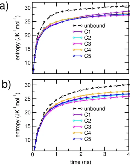
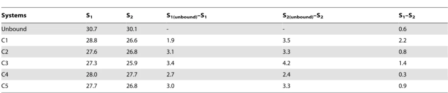
To compare the relative extents of disorder in each cluster, we estimated the cumulative configurational entropy per heavy atom in the individual protein units using Schlitter’s method described earlier. For comparison, we also obtained the corresponding cumulative entropies in the unbound states of the proteins. The results are plotted in Figure 8. Individual protein units in each dimerizing cluster displayed marked decrease in the net config-urational entropies over the corresponding unbound state. The configurational entropies of the Ab units had greater overlap between the clusters compared toaSyn units. In Table 3, we have listed the saturation values of the entropies and the entropy loss upon dimerization for each cluster. The configurational entropy per atom was higher for Ab, both in the free as well as in the dimerized states. However, for a given cluster, entropy losses on the average were greater for atoms belonging to theaSyn unit. The entropy per heavy atom foraSyn and Abwas closest in the C4 cluster, indicating the closest level of conformational disorder
in the two peptides. Further, C4 was also characterized by the least overall entropy loss. However, for the clusters with stronger electrostatic interactions, we noted the absence of clear correla-tions between the strength of inter-protein interaction and the extent of entropy loss upon dimerization. Particularly the cluster C3, which displayed largest entropy loss, ranked third in the
strength of inter-protein interactions. However, it is observed that the cluster C3 has a relatively high number of internal atom-atom contacts, particularly in theaSyn protein; this is reflected in the smaller Rg values (Table S2 in File S1). In comparison, the
strongly associated clusters C2 and C5 had fewer internal contacts, and marginally higher configurational entropy than C3. These
Figure 7. Radial distribution functions of water oxygens, around a) backbone Caatoms, b) all heavy atoms of residues that make inter-protein contacts.A minimum contact probability of 0.7 has been considered.
doi:10.1371/journal.pone.0106883.g007
data suggest that the internal compactness of the protein units, particularly of aSyn, can be a contributing factor to the overall rigidity of the associated complexes.
Discussion and Conclusion
Recentin vitroandin vivostudies report that cross interactions between dissimilar IUPs can play significant roles in clinically observed mixed pathological traits in ND patients [14–19,76–80]. Notably, significant experimental evidence exists to suggest that Ab, whose assembly can trigger AD, andaSyn, whose assembly is responsible for PD, can co-associate in biological milieu [14,15,17,18,76,78,79]. However, to the best of our knowledge, there exist no molecular level studies probing their unrestricted associations in aqueous environments. In this study, we reported the heterogeneous interactions of Ab1–42 and aSyn1–95 from a
large ensemble of the dimeric complex obtained from unbiased MD simulations of the protein sequences in explicit water.
In four out of the five interaction modes discerned with Principal component analysis, electrostatic forces are seen to dominate over van der Waals interactions. Residue specific investigations revealed the importance of the Lys residues, especially those in the imperfect repeating units ofaSyn, during cross dimerisation. We note here that Lys specific molecular tweezers have been reported to be capable of inhibiting the aggregation of various amyloidogenic peptides [81–84]. 1,4-napthoquinone based inhibitors were also found to interact with Lys residues and efficiently reduced the fibrilisation propensity of aSyn [85]. Thus, our observation of the importance of the Lys repeats in the cross dimerization may be used for designing drugs targeted at inhibiting Ab-aSyn co-assembly.
Clusters with dominant electrostatic interactions were charac-terized by the presence of multiple inter-protein salt bridges.
Figure 8. Cumulative configuration entropy per heavy atom for a) Ab1–42protein and b)aSyn1–95protein.The entropy of the unbound
Interestingly, the majority of salt bridges were formed between Lys residues ofaSyn and Asp or Glu of Ab.Studies suggest that the disruption of salt bridges is likely to affect the structure, toxicity and oligomerisation ofaSyn [11]. Similarly, in Abaggregates, the salt bridge between D23 and K28 is crucial for stability of the hairpin form and formation of fibrillar aggregates [17,24–27]. Further studies would reveal if the inter-protein salt bridges have any disruptive effects on the ones crucial for self-assembly, and the extent to which this may result in structural dissimilarities between the self-aggregates and the co-aggregates.
Importantly, hydrophobic interactions were also found to play crucial roles in the hetero dimerisation process. In a single interaction mode devoid of inter-protein salt bridges, the van der Waals interactions dominated over the average electrostatic interactions. In this particular system, the hydrophobic core regions comprising of 17–21, 30–35 and 39–42 of Abwere found to be in contact with the NAC ofaSyn. Additionally, we observed inter peptide contact of Ab with residues of the NAC in all the electrostatically stabilized clusters except C1. We point out that the hydrophobic core regions in Ab play crucial roles in its early dynamics, oligomerization and fibril formation [8,20–22,86,87]. Similarly, in aSyn the central hydrophobic NAC region is necessary for its aggregation and this fragment is clinically observed in amyloid plaques found in patients with LBD [3,5,14,88–91]. Earlier solid phase binding studies as well as NMR studies indicate that the NAC interacts with Ab,particularly with residues G67, G73 and V74 and proposed a mechanism for the overlapping pathogenesis that the cleavage of NAC is catalyzed by Ab oligomer [14]. It is worthwhile to mention here that a major strategy in the drug design against for amyloidogenic peptides is to target regions that drive hydrophobic interactions [86,91–93]. Thus, the results of our analyses demonstrating NAC interaction with Ab hydrophobic regions, along with the experimental reports, indicate that these regions could represent other plausible therapeutic targets.
Before concluding, we note that it is important to study secondary structural details of the peptide monomers during hetero assembly, and this requires careful comparison of results obtained from multiple force fields with experimental data. The clear evidence of complex formation without the emergence of strand motifs indicates that the complexes are metastable and could be prone to further assembly. Longer, millisecond timescale simulations may reveal more modes of Ab-aSyn associations. Nevertheless, the evidence of significant heterogeneity in the nature of interactions leading to cross dimerization revealed by our microsecond simulations is strongly suggestive of heterogeneity during the seeding phase and along the early assembly pathways. This may result in the emergence of hetero oligomers and thus
significant levels of polymorphism in higher ordered aggregates. In further studies, the interactions of preformed hybrid systems with lipid bilayers would greatly facilitate identification of the level of toxicity of each species. This information, along with the specific inter-residue interactions, could significantly aid the development of therapeutics against synergistic ND.
Supporting Information
File S1 Supporting files. Figure S1, Evolution of the a) total inter-peptide interaction strength, and b) inter peptide distance over 150 ns for the dimerising trajec-tories. Figure S2, The backbone mean square fluctua-tion (MSF) for the a) N-terminal residues, b) middle regions, and c) C-terminal residues of Ab1–42,and the d) N-terminal residues, e) middle regions, and f) C-terminal residues of aSyn1–95. The data for the last 50 ns of the dimerising trajectories are shown ingray, with the averages in green (solid line). Corresponding average data for the same systems for the initial 50 ns is provided in green (broken line). Average data for the non-dimerising systems is shown inmaroon
(broken line) for comparison. Figure S3, Residue wise maximum electrostatic (left column) and van der Waal (right column) interaction energies (in kcal mol21) of aSyn1–95with Ab1–42for clusters C1, C2, C3, C4 and C5. Table S1, Mean value of the inter-residue sidechain distances (dSB, in A˚ ) between the residues that form salt bridges in the clusters a) C1, b) C2, c) C3, d) C5.Standard deviations are provided in braces. The first residue belongs to aSyn1–95; the second residue belongs to Ab1–42.Table S2, Mean
values of the total number of internal contacts formed in the Ab1–42(NintAb) andaSyn1–95(NintaS) proteins in the five clusters.The corresponding radii of gyration (in A˚ ) have been denoted as RgAband RgaS.
(PDF)
Acknowledgments
The authors thank Professor M. G. Zagorski and Professor D. Eliezer for providing chemical shift values for AbandaSyn, respectively. J.C.J. and P.C thank CSIR for their Senior Research Fellowship. CDAC, Pune is thanked for generously providing additional computing resources. J.C.J. would like to thank Mr. Xavier Prasanna for the help with graphics.
Author Contributions
Conceived and designed the experiments: NS. Performed the experiments: JCJ PC. Analyzed the data: JCJ. Contributed to the writing of the manuscript: NS JCJ.
Table 3.Configurational entropy calculations.
Systems S1 S2 S1(unbound)–S1 S2(unbound)–S2 S1–S2
Unbound 30.7 30.1 - - 0.6
C1 28.8 26.6 1.9 3.5 2.2
C2 27.6 26.8 3.1 3.3 0.8
C3 27.3 25.9 3.4 4.2 1.4
C4 28.0 27.7 2.7 2.4 0.3
C5 27.7 26.8 3.0 3.3 0.9
Configurational entropy per heavy atoms (in J K21mol21) for Ab
1–42(S1) and theaSyn1–95(S2) proteins in the unbound states and in the clusters C1, C2, C3, C4 and C5.
The entropy differences between the unbound and bound states, as well as the difference between the entropies of Ab1–42andaSyn1–95are also provided.
doi:10.1371/journal.pone.0106883.t003
References
1. Grundke-Iqbal I, Iqbal K, Quinlan M, Tung YC, Zaidi MS, et al. (1986) Microtubule-associated protein tau. A component of Alzheimer paired helical filaments. J Biol Chem 261: 6084–6089.
2. Hardy J, Selkoe DJ (2002) The amyloid hypothesis of Alzheimer’s disease: Progress and problems on the road to therapeutics. Science 297: 353. 3. Iwai A, Masliah E, Yoshimoto M, Ge N, Flanagan L, et al. (1995) The precursor
protein of non-A beta component of Alzheimer’s disease amyloid is a presynaptic protein of the central nervous system. Neuron 14: 467–475.
4. Prusiner SB (1998) Prions. Proc Natl Acad Sci USA 95: 13363–13383. 5. Spillantini MG, Schmidt ML, Lee VMY, Trojanowski JQ , Jakes R, et al. (1997)
a-Synuclein in Lewy bodies. Nature 388: 839–840.
6. Hu X, Crick SL, Bu G, Frieden C, Pappu RV, et al. (2009) Amyloid seeds formed by cellular uptake, concentration, and aggregation of the amyloid-beta peptide. Proc Natl Acad Sci USA 106: 20324–20329.
7. Dobson CM (2003) Protein folding and misfolding. Nature 426: 884–890. 8. Bernstein SL, Wyttenbach T, Baumketner A, Shea JE, Bitan G, et al. (2005)
Amyloidb-Protein: Monomer Structure and Early Aggregation States of Ab42 and Its Pro19 Alloform. J Am Chem Soc 127: 2075–2084.
9. Cappai R, Barnham K (2008) Delineating the Mechanism of Alzheimer’s Disease AbPeptide Neurotoxicity. Neurochem Res 33: 526–532.
10. Liao MQ , Tzeng YJ, Chang LYX, Huang HB, Lin TH, et al. (2007) The correlation between neurotoxicity, aggregative ability and secondary structure studied by sequence truncated Abpeptides. Febs lett 581: 1161–1165. 11. Winner B, Jappelli R, Maji SK, Desplats PA, Boyer L, et al. (2011) In vivo
demonstration thata-synuclein oligomers are toxic. Proc Natl Acad Sci USA 108: 4194–4199.
12. Jellinger KA (2011) Interaction between alpha-Synuclein and Other Proteins in Neurodegenerative Disorders. ScientificWorldJournal 11: 1893–1907. 13. Irwin DJ, Lee VMY, Trojanowski JQ (2013) Parkinson’s disease dementia:
convergence ofa-synuclein, tau and amyloid-bpathologies. Nat Rev Neurosci 14: 626–636.
14. Mandal P, Pettegrew J, Masliah E, Hamilton R, Mandal R (2006) Interaction between AbPeptide andaSynuclein: Molecular Mechanisms in Overlapping Pathology of Alzheimer’s and Parkinson’s in Dementia with Lewy Body Disease. Neurochem Res 31: 1153–1162.
15. Masliah E, Rockenstein E, Veinbergs I, Sagara Y, Mallory M, et al. (2001) b-Amyloid peptides enhancea-synuclein accumulation and neuronal deficits in a transgenic mouse model linking Alzheimer’s disease and Parkinson’s disease. Proc Natl Acad Sci USA 98: 12245–12250.
16. Morales R, Moreno-Gonzalez I, Soto C (2013) Cross-Seeding of Misfolded Proteins: Implications for Etiology and Pathogenesis of Protein Misfolding Diseases. PLoS Pathog 9: e1003537.
17. Resende R, Marques SF, Ferreiro E, Simo˜es I, Oliveira C, et al. (2013) Effect of a-Synuclein on AmyloidbInduced Toxicity: Relevance to Lewy Body Variant of Alzheimer Disease. Neurochem Res 38: 797–806.
18. Tsigelny IF, Crews L, Desplats P, Shaked GM, Sharikov Y, et al. (2008) Mechanisms of Hybrid Oligomer Formation in the Pathogenesis of Combined Alzheimer’s and Parkinson’s Diseases. PLoS ONE 3: e3135.
19. Ono K, Takahashi R, Ikeda T, Yamada M (2012) Cross-seeding effects of amyloidb-protein anda-synuclein. J Neurochem 122: 883–890.
20. Lee C, Ham S (2010) Characterizing amyloid-beta protein misfolding from molecular dynamics simulations with explicit water. J Comput Chem: 349–355. 21. Jana AK, Jose JC, Sengupta N (2012) Critical Roles of Key Domains in Complete Adsorption of AbPeptide on Single-Walled Carbon Nanotubes: Insights with Point Mutations and MD Simulations. Phys Chem Chem Phys 15: 837–844.
22. Jana AK, Sengupta N (2012) Adsorption Mechanism and Collapse Propensities of the Full-Length, Monomeric Ab1–42 on the Surface of a Single-Walled Carbon Nanotube: A Molecular Dynamics Simulation Study. Biophys J 102: 1889–1896.
23. Zhang S, Iwata K, Lachenmann MJ, Peng JW, Li S, et al. (2000) The Alzheimer’s Peptide AbAdopts a Collapsed Coil Structure in Water. J Struct Biol 130: 130–141.
24. Anand P, Nandel FS, Hansmann UHE (2008) The Alzheimerb-amyloid (Ab-39) dimer in an implicit solvent. J Chem Phys 129: 1–7.
25. Ma B, Nussinov R (2002) Stabilities and conformations of Alzheimer’sb-amyloid peptide oligomers (Ab16–22, Ab16–35, and Ab10–35): Sequence effects. Proc Natl Acad Sci USA 99: 14126–14131.
26. Reddy G, Straub JE, Thirumalai D (2009) Influence of Preformed Asp23-Lys28 Salt Bridge on the Conformational Fluctuations of Monomers and Dimers of Ab Peptides with Implications for Rates of Fibril Formation. J Phys Chem B 113: 1162–1172.
27. Tarus B, Straub JE, Thirumalai D (2006) Dynamics of Asp23-Lys28 Salt-Bridge Formation in Ab10–35 Monomers. J Am Chem Soc 128: 16159–16168. 28. Chatelier RC, Minton AP (1996) Adsorption of globular proteins on locally
planar surfaces: models for the effect of excluded surface area and aggregation of adsorbed protein on adsorption equilibria. Biophys J 71: 2367–2374. 29. Minton AP (1999) Adsorption of Globular Proteins on Locally Planar Surfaces.
II. Models for the Effect of Multiple Adsorbate Conformations on Adsorption Equilibria and Kinetics. Biophys J 76: 176–187.
30. Minton AP (2001) The Influence of Macromolecular Crowding and Macromo-lecular Confinement on Biochemical Reactions in Physiological Media. J Biol Chem 276: 10577–10580.
31. Chong SH, Ham S (2012) Impact of chemical heterogeneity on protein self-assembly in water. Proc Natl Acad Sci USA 109: 7636–7641.
32. Hamelberg D, Mongan J, McCammon JA (2004) Accelerated molecular dynamics: A promising and efficient simulation method for biomolecules. J Chem Phys 120: 11919–11929.
33. Tomaselli S, Esposito V, Vangone P, van Nuland NA, Bonvin AM, et al. (2006) The alpha-to-beta Conformational Transition of Alzheimer’s Abeta-(1–42) Peptide in Aqueous Media is Reversible: A Step by Step Conformational Analysis Suggests the Location of beta Conformation Seeding. ChemBioChem 7: 257–267.
34. Rao JN, Jao CC, Hegde BG, Langen R, Ulmer TS (2010) A Combinatorial NMR and EPR Approach for Evaluating the Structural Ensemble of Partially Folded Proteins. J Am Chem Soc 132: 8657–8668.
35. Kale L, Skeel R, Bhandarkar M, Brunner R, Gursoy A, et al. (1999) NAMD2: Greater Scalability for Parallel Molecular Dynamics. J Comp Physics 151: 283– 312.
36. Mackerell AD, Feig M, Brooks CL (2004) Extending the treatment of backbone energetics in protein force fields: Limitations of gas-phase quantum mechanics in reproducing protein conformational distributions in molecular dynamics simulations. J Comp Chem 25: 1400–1415.
37. MacKerell AD Jr, Bashford D, Bellott M, Dunbrack RL, Evanseck JD, et al. (1998) All-Atom Empirical Potential for Molecular Modeling and Dynamics Studies of Proteins. J Phys Chem B 102: 3586–3616.
38. de Oliveira CAF, Hamelberg D, McCammon JA (2007) Estimating kinetic rates from accelerated molecular dynamics simulations: Alanine dipeptide in explicit solvent as a case study. J Chem Phys 127: 175105–175108.
39. Hamelberg D, de Oliveira CAF, McCammon JA (2007) Sampling of slow diffusive conformational transitions with accelerated molecular dynamics. J Chem Phys 127: 155102–155109.
40. Markwick PRL, McCammon JA (2011) Studying functional dynamics in bio-molecules using accelerated molecular dynamics. Phys Chem Chem Phys 13: 20053–20065.
41. Games D, Seubert P, Rockenstein E, Patrick C, Trejo M, et al. (2013) Axonopathy in ana-Synuclein Transgenic Model of Lewy Body Disease Is Associated with Extensive Accumulation of C-Terminal-Truncateda-Synuclein. Am J Pathol 182: 940–953.
42. Kanda S, Bishop JF, Eglitis MA, Yang Y, Mouradian MM (2000) Enhanced vulnerability to oxidative stress by a-synuclein mutations and C-terminal truncation. Neurosci 97: 279–284.
43. Li W, West N, Colla E, Pletnikova O, Troncoso JC, et al. (2005) Aggregation promoting C-terminal truncation ofa-synuclein is a normal cellular process and is enhanced by the familial Parkinson’s disease-linked mutations. Proc Natl Acad Sci USA 102: 2162–2167.
44. Sgourakis NG, Yan Y, McCallum SA, Wang C, Garcia AE (2007) The Alzheimer’s Peptides Ab40 and 42 Adopt Distinct Conformations in Water: A Combined MD/NMR Study. J Mol Biol 368: 1448–1457.
45. Lin YS, Bowman Gregory R, Beauchamp Kyle A, Pande Vijay S (2012) Investigating How Peptide Length and a Pathogenic Mutation Modify the Structural Ensemble of Amyloid Beta Monomer. Biophys J 102: 315–324. 46. Rosenman DJ, Connors CR, Chen W, Wang C, Garcia AE (2013) Ab
Monomers Transiently Sample Oligomer and Fibril-Like Configurations: Ensemble Characterization Using a Combined MD/NMR Approach. J Mol Biol 425: 3338–3359.
47. Ball KA, Phillips AH, Nerenberg PS, Fawzi NL, Wemmer DE, et al. (2011) Homogeneous and Heterogeneous Tertiary Structure Ensembles of Amyloid-b Peptides. Biochemistry 50: 7612–7628.
48. Osapay K, Case DA (1991) A new analysis of proton chemical shifts in proteins. J Am Chem Soc 113: 9436–9444.
49. Hou L, Shao H, Zhang Y, Li H, Menon NK, et al. (2004) Solution NMR Studies of the Ab(1–40) and Ab(1–42) Peptides Establish that the Met35 Oxidation State Affects the Mechanism of Amyloid Formation. J Am Chem Soc 126: 1992–2005.
50. Eliezer D, Kutluay E, Bussell R Jr, Browne G (2001) Conformational properties ofa-synuclein in its free and lipid-associated states. J Mol Biol 307: 1061–1073. 51. Jorgensen WL, Chandrasekhar J, Madura JD, Impey RW, Klein ML (1983) Comparison of simple potential functions for simulating liquid water. J Chem Phys 79: 926–935.
52. Feller SE, Zhang Y, Pastor RW, Brooks BR (1995) Constant pressure molecular dynamics simulation: The Langevin piston method. J Chem Phys 103: 4613– 4621.
53. Martyna GJ, Tobias DJ, Klein ML (1994) Constant pressure molecular dynamics algorithms. J Chem Phys 101: 4177–4189.
54. Ryckaert JP, Ciccotti G, Berendsen HJC (1977) Numerical integration of the cartesian equations of motion of a system with constraints: molecular dynamics of n-alkanes. J Comp Physics 23: 327–341.
56. DeLano WL (2002) PyMOL: an open-source molecular graphics tool. Ccp4 Newslett Protein Crystallogr 40.
57. Humphrey W, Dalke A, Schulten K (1996) VMD: Visual molecular dynamics. Journal of Molecular Graphics 14: 33–38.
58. Glykos NM (2006) Software news and updates carma: A molecular dynamics analysis program. J Comp Chem 27: 1765–1768.
59. Fadouloglou VE, Stavrakoudis A, Bouriotis V, Kokkinidis M, Glykos NM (2009) Molecular Dynamics Simulations of BcZBP, A Deacetylase from Bacillus cereus: Active Site Loops Determine Substrate Accessibility and Specificity. J Chem Theory Comput 5: 3299–3311.
60. Wan H, Hu JP, Li KS, Tian XH, Chang S (2013) Molecular Dynamics Simulations of DNA-Free and DNA-Bound TAL Effectors. PLoS ONE 8: e76045.
61. Nguyen PH, Li MS, Derreumaux P (2014) Amyloid oligomer structure characterization from simulations: A general method. J Chem Phys 140: 094105-094101-094109.
62. Schlitter J (1993) Estimation of absolute and relative entropies of macromol-ecules using the covariance matrix. Chem Phys Lett 215: 617–621. 63. Scha¨fer H, Daura X, Mark AE, van Gunsteren WF (2001) Entropy calculations
on a reversibly folding peptide: Changes in solute free energy cannot explain folding behavior. Proteins: Structure, Function, and Bioinformatics 43: 45–56. 64. Sinha SK, Chakraborty S, Bandyopadhyay S (2010) Secondary Structure
Specific Entropy Change of a Partially Unfolded Protein Molecule. Langmuir 26: 9911–9916.
65. Furini S, Barbini P, Domene C (2013) DNA-recognition process described by MD simulations of the lactose repressor protein on a specific and a non-specific DNA sequence. Nucleic Acids Research 41: 3963–3972.
66. Barz B, Urbanc B (2012) Dimer Formation Enhances Structural Differences between Amyloidb-Protein (1–40) and (1–42): An Explicit-Solvent Molecular Dynamics Study. PLoS ONE 7: e34345.
67. Petkova AT, Ishii Y, Balbach JJ, Antzutkin ON, Leapman RD, et al. (2002) A structural model for Alzheimer’s b-amyloid fibrils based on experimental constraints from solid state NMR. Proc Natl Acad Sci USA 99: 16742–16747. 68. Rezaei-Ghaleh N, Amininasab M, Giller K, Kumar S, Stu¨ndl A, et al. (2014) Turn Plasticity Distinguishes Different Modes of Amyloid-bAggregation. J Am Chem Soc 136: 4913–4919.
69. Wise-Scira O, Xu L, Kitahara T, Perry G, Coskuner O (2011) Amyloid-b peptide structure in aqueous solution varies with fragment size. J Chem Phys 135: 205101–205113.
70. Donald JE, Kulp DW, DeGrado WF (2011) Salt bridges: Geometrically specific, designable interactions. Proteins: Structure, Function, and Bioinformatics 79: 898–915.
71. Gvritishvili AG, Gribenko AV, Makhatadze GI (2008) Cooperativity of complex salt bridges. Protein Sci 17: 1285–1290.
72. Musafia B, Buchner V, Arad D (1995) Complex Salt Bridges in Proteins: Statistical Analysis of Structure and Function. J Mol Biol 254: 761–770. 73. Meuzelaar H, Tros M, Huerta-Viga A, van Dijk CN, Vreede J, et al. (2014)
Solvent-Exposed Salt Bridges Influence the Kinetics ofa-Helix Folding and Unfolding. J Phys Chem Lett 5: 900–904.
74. Salari R, Chong LT (2010) Desolvation Costs of Salt Bridges across Protein Binding Interfaces: Similarities and Differences between Implicit and Explicit Solvent Models. J Phys Chem Lett 1: 2844–2848.
75. Salari R, Chong LT (2014) Effects of High Temperature on Desolvation Costs of Salt Bridges Across Protein Binding Interfaces: Similarities and Differences between Implicit and Explicit Solvent Models. J Phys Chem B 116: 2561–2567. 76. Bate C, Gentleman S, Williams A (2010) alpha-synuclein induced synapse damage is enhanced by amyloid-beta1–42. Molecular Neurodegeneration 5: 55. 77. Berhanu WM, Yas¸ar F, Hansmann UHE (2013) In Silico Cross Seeding of Ab
and Amylin Fibril-like Oligomers. ACS Chem Neurosci 4: 1488–1500. 78. Crews L, Tsigelny I, Hashimoto M, Masliah E (2009) Role of Synucleins in
Alzheimer’s Disease. Neurotox Res 16: 306–317.
79. Hashimoto M, Masliah E (1999) Alpha-synuclein in Lewy Body Disease and Alzheimer’s Disease. Brain Pathol 9: 707–720.
80. Lajtha A, Banik N, Ray S, Crews L, Spencer B, et al. (2009) Immunotherapy Strategies for Lewy Body and Parkinson’s Diseases. Handbook of Neurochem-istry and Molecular Neurobiology: Springer US. 599–613.
81. Acharya S, Safaie BM, Wongkongkathep P, Ivanova MI, Attar A, et al. (2014) Molecular Basis for Preventing a-Synuclein Aggregation by a Molecular Tweezer. J Biol Chem M113.524520.
82. Dutt S, Wilch C, Gersthagen T, Talbiersky P, Bravo-Rodriguez K, et al. (2013) Molecular Tweezers with Varying Anions: A Comparative Study. J Org Chem 78: 6721–6734.
83. Prabhudesai S, Sinha S, Attar A, Kotagiri A, Fitzmaurice A, et al. (2012) A Novel ‘‘Molecular Tweezer’’ Inhibitor ofa-Synuclein Neurotoxicity in Vitro and in Vivo. Neurotherapeutics 9: 464–476.
84. Sinha S, Lopes DHJ, Du Z, Pang ES, Shanmugam A, et al. (2011) Lysine-Specific Molecular Tweezers Are Broad-Spectrum Inhibitors of Assembly and Toxicity of Amyloid Proteins. J Am Chem Soc 133: 16958–16969.
85. da Silva FL, Coelho Cerqueira E, de Freitas MS, Gonc¸alves DL, Costa LT, et al. (2013) Vitamins K interact with N-terminus a-synuclein and modulate the protein fibrillization in vitro. Exploring the interaction between quinones and a-synuclein. Neurochem Int 62: 103–112.
86. Li J, Liu R, Lam KS, Jin LW, Duan Y (2011) Alzheimer’s Disease Drug Candidates Stabilize A-bProtein Native Structure by Interacting with the Hydrophobic Core. Biophys J 100: 1076–1082.
87. Luhrs T, Ritter C, Adrian M, Riek-Loher D, Bohrmann B, et al. (2005) 3D structure of Alzheimer’s amyloid-b(1–42) fibrils. Proc Natl Acad Sci USA 102: 17342–17347.
88. Kim HY, Heise H, Fernandez CO, Baldus M, Zweckstetter M (2007) Correlation of Amyloid Fibril b-Structure with the Unfolded State of a-Synuclein. ChemBioChem 8: 1671–1674.
89. Ullman O, Fisher CK, Stultz CM (2011) Explaining the Structural Plasticity of a-Synuclein. J Am Chem Soc 133: 19536–19546.
90. Hashimoto M, Hsu LJ, Xia Y, Takeda A, Sisk A, et al. (1999) Oxidative stress induces amyloid-like aggregate formation of NACP/a-synuclein in vitro. NeuroReport 10: 717–721.
91. Giasson BI, Murray IVJ, Trojanowski JQ , Lee VMY (2001) A Hydrophobic Stretch of 12 Amino Acid Residues in the Middle ofa-Synuclein Is Essential for Filament Assembly. J Biol Chem 276: 2380–2386.
92. Cheng PN, Liu C, Zhao M, Eisenberg D, Nowick JS (2012) Amyloidb-sheet mimics that antagonize protein aggregation and reduce amyloid toxicity. Nat Chem 4: 927–933.
93. Viet MH, Ngo ST, Lam NS, Li MS (2011) Inhibition of Aggregation of Amyloid Peptides by Beta-Sheet Breaker Peptides and Their Binding Affinity. J Phys Chem B 115: 7433–7446.