This is the pre-review version of the paper published in
J. Phys. Chem. B, 115, 1903-1910
The final version may be obtained from the publisher, at the following link:
Computational characterization of the substrate-binding
mode in coproporphyrinogen III oxidase
Pedro J. Silva§ and Maria João Ramos†*
§
REQUIMTE, Faculdade de Ciências da Saúde, Universidade Fernando Pessoa, Rua Carlos da Maia, 296, 4200-150 Porto-Portugal
†
Abstract:
Oxygen-dependent coproporphyrinogen III oxidase catalyzes the sequential decarboxylation of the propionate substituents present on the A and B rings of coproporphyrinogen III in the heme biosynthetic pathway. Although extensive experimental investigation of this enzyme has already afforded many insights on its reaction mechanism, several key features (such as the substrate binding mode, the characterization of the active site and the initial substrate protonation state) remain poorly described. The molecular dynamics simulations described in this paper enabled the determination of a very promising substrate binding mode and the extensive characterization of the enzyme active site. The proposed binding mode is fully consistent with the known selectivity of the active site towards substituted tetrapyrroles, and explains the lack of activity of the H131A, R135A, D274A and R275A mutants and the reasons behind the non-occurrence of catalysis on the C and D rings of the tetrapyrrole. An important role in this binding mode is fulfilled by G276, as its carbonyl oxygen intervenes in the substrate anchoring by hydrogen-bonding its ring D pyrrole NH group. The presence of this interaction (which is only possible with protonated NH pyrrole group), and the absence of positively-charged side chains close to the pyrrole nitrogen (which might stabilize the N-deprotonated pyrrole postulated in some mechanistic proposals) shows that the pyrrole ring is very unlikely to undergo deprotonation during the catalytic cycle and allow the discrimination between the previously postulated mechanistic proposals.
KEYWORDS: molecular dynamics, coproporphyrinogen III oxidase, active site characterization
*
I. Introduction
substitution prevents the second decarboxylation5,17, which shows that after the first decarboxylation the substrate either leaves the active site or undergoes a rotation in order to optimize binding affinity. The recognition of the substitutents on the coproporphyrinogen rings C and D also affects the kinetic parameters of the enzyme16,17, although the reasons for this behaviour have not yet been ascertained.
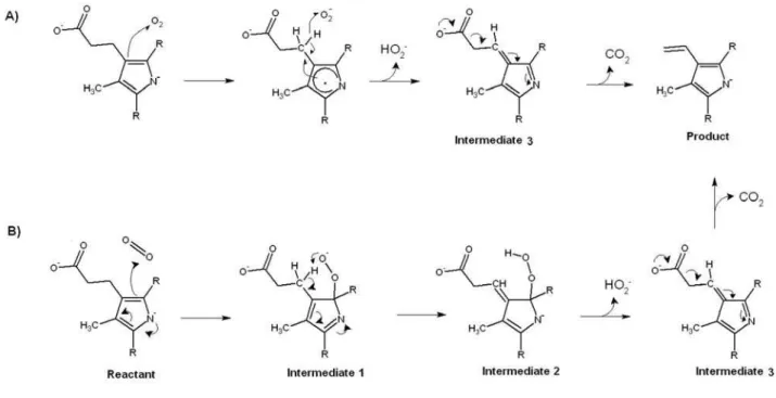
Figure 1: Proposed reaction mechanisms for coproporphyrinogen oxidase. A) Arigoni model9. B) Lash model19. The Arigoni model is energetically feasible only if the pyrrole ring nitrogen is deprotonated, whereas the Lash model may occur with either protonated or deprotonated substrate, although for simplicity only the mechanism for the deprotonated state is shown.
II. Methods
Molecular docking
the substrate and to the narrow dimensions of the binding site, docking was performed with an unsubstituted cyclic tetrapyrrole, rather than with the full coproporphyrinogen substrate. The docking poses obtained were then used as starting points for molecular dynamics simulations after adding the appropriate substitutents to each pyrrole ring.
Molecular dynamics
All molecular dynamics simulations were run with YASARA20 with the AMBER9921 forcefieldunder periodic-boundary conditions and with explicit water, using a multiple time step of 1.25 fs for intramolecular and 2.5 fs for intermolecular forces. Simulations were performed in cells 10 Å larger than the protein dimer along each axis (final cell dimensions 116 × 94 × 77 Å3) , and counter-ions were added to a final concentration of 0.9 % NaCl. A 7.86 Å cutoff was taken for Lennard-Jones forces and the direct space portion of the electrostatic forces, which were calculated using the Particle Mesh Ewald method22 with a grid spacing <1 Å, 4th order B-splines and a tolerance of 10-4 for the direct space sum. Simulated annealing minimizations started at 298 K, velocities were scaled down with 0.9 every ten steps for a total time of 5 ps. After annealing, simulations were run at 298 K. Temperature was adjusted using a Berendsen thermostat23 based on the time-averaged temperature, i.e., to minimize the impact of temperature control, velocities were rescaled only about every 100 simulation steps, whenever the average of the last 100 measured temperatures converged. Substrate parameterization was performed with the AM1BCC protocol24, which assigns atomic charges by applying simple additive bond charge corrections (BCCs) to AM1 atomic charges and has been shown to provide a fast and robust alternative to HF/6-31G* ESP-fit charges for ligand parameterization for molecular dynamics applications. Protein sidechain protonation patterns were assigned according to Krieger et al.25. All simulations were run for at least 10 ns after an initial 500 ps equilibration.
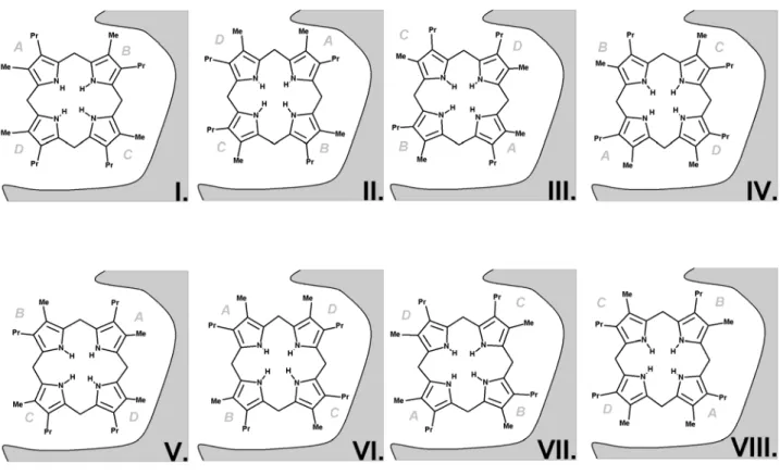
Figure 2: The eight possible substrate orientations in coporporphyrinogen III oxidase active site cavity.
The available crystal structures9-10 show that coproporphyrinogen III oxidase may exist in two main conformations, distinguishable due to the relative position of helixes H2 (Ser79-Lys87) and H8 (Arg275-Leu281), which may either move apart or towards each other, thereby opening or closing access from the solvent to a cavity which contains the putative active site10. Our preliminary docking attempts with AutoDock showed that in the “open” position a tetrapyrrole substrate may fit the solvent-accessible narrow cavity in such a way that the substituents of two of its pyrrole rings remain totally or partially solvent-exposed. However, the cramped dimensions of this putative active site does not allow the free rotation of the substrate during the docking procedure and therefore the search for the correct binding conformation had to be further refined through molecular dynamics simulations. The binding pose obtained from docking experiments with unsubstituted tetrapyrrole was used as starting point for eight different molecular dynamics simulations (after inclusion of pyrrole substituents at the appropriate positions), representing each of the putative substrate orientations. Since the correct binding mode has been experimentally shown to implicate interactions with H131, R135, D274 and R275, additional simulations were performed with an in silico mutant where these four amino acids have been changed to alanine. The comparison of the simulations performed in wild-type and mutant proteins assisted our identification of the correct binding mode and improved the characterization of the relevant interactions between the substrate and the protein, as will be shown subsequently.
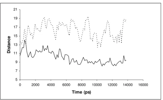
The dimer structure remained quite stable along the simulations, except in a short unstructured stretch of the N-terminal and in the protein regions involved in the movement of the H2 helix away from/towards helix H8 (Figure 3). (i.e. the opening/closing of the substrate-binding cavity): these include the H2 helix itself (Lys79-Asp89), the loop between this helix and -sheet S4 (His90-Gly108), and two sheet termini contacting this loop: the Cterminus of sheet S5 and the Nterminus of -sheet S6 (Glu138-Trp149). In the substrate-free subunit, the C-terminal of helix H2 remains ≈ 13.1 ± 1.5 Å from the N-terminal of helix H8 along the simulations, whereas its N-terminal lies ≈ 15.5 ± 1.8 Å
binding modes, whereas the H2 C-terminal to H8 N-terminal distance varies dramatically with the binding mode considered (Table 1) For all binding modes (with the exception of binding modes IV and VI), the quadruple mutation of amino acids H131, R135, R275 and D274 to alanine causes this inter-helical distance to increase dramatically (Table 2), yielding a very open conformation of the active site. As explained in the following section, this is mostly due to movements of the substrate in the active site, rather than in the intrinsic flexibility of the enzyme. In all simulations performed the Root Mean Square Deviations of the structures (after excluding the most flexible region between amino acids 77 and 110) remain low (around 2 Å) (Supporting Information). Most of the remaining variability is due to the unstructured N-terminal and to the loops (45-50 and 138-149) contacting the flexible 77-110 region (See Supporting Information for RMSF graphs).
5 7 9 11 13 15 17 19 21
0 2000 4000 6000 8000 10000 12000 14000 16000
Time (ps)
D
istan
ce
Figure 3: Distances between the termini of helixes H2 and H8 in the substrate-bearing chain of coproporphyrinogen III oxidase (Binding mode VI). Continuous line: Cα 82-Cα 282 (N-terminal H2 – C-terminal H8); Broken line: Cα 89-Cα 275 (C-terminal H2 – N-terminal H8). Additional graphs for all
Table 1: Distances (in Å) between the termini of helixes H2 and H8 of substrate-bearing (chain A) and substrate-free (chain B) wild-type coproporphyrinogen III oxidase.
Binding mode Chain A chain B
Cα 82-Cα 282 Cα 89-Cα 275 Cα 82-Cα 282 Cα 89-Cα 275
I 16.35±1.58 12.60±1.03 14.62±1.36 13.17±1.40
II 12.32±1.39 10.97±0.55 13.69±1.09 13.75±1.13
III 15.93±1.06 11.95±1.17 15.75±1.40 12.88±1.39
IV 8.77±0.78 10.02±0.37 17.05±1.54 12.37±1.20
V 14.53±0.96 11.27±0.66 15.63±1.40 14.25±1.61
VI 9.98±1.31 10.10±0.43 16.21±1.76 12.43±1.22
VII 16.23±1.00 12.32±0.62 16.22±1.64 12.99±0.85
VIII 13.12±2.12 11.08±1.15 15.46±1.45 13.06±1.59
apoprotein 14.33±1.47 12.10±1.09 16.55±1.61 12.40±0.40
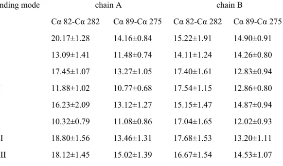
Table 2: Distances (in Å) between the termini of helixes H2 and H8 of substrate-bearing (chain A) and substrate-free (chain B) mutant coproporphyrinogen III oxidase.
Binding mode chain A chain B
Cα 82-Cα 282 Cα 89-Cα 275 Cα 82-Cα 282 Cα 89-Cα 275
I 20.17±1.28 14.16±0.84 15.22±1.91 14.90±0.91
II 13.09±1.41 11.48±0.74 14.11±1.24 14.26±0.80
III 17.45±1.07 13.27±1.05 17.40±1.61 12.83±0.94
IV 11.88±1.02 10.77±0.68 17.54±1.15 12.86±0.80
V 16.23±2.09 13.12±1.27 15.15±1.47 14.87±0.94
VI 10.32±0.79 11.08±0.86 17.04±1.65 12.02±0.93
VII 18.80±1.56 13.46±1.31 17.68±1.53 13.20±1.11
The precise shape and limits of the active site cavity vary with substrate orientation according to the interactions between the pyrrole substituents and the amino acid sidechains: a detailed description of each possible binding mode is therefore provided in the next section. The overall shape of the active site cavity as well as important aminoacids interacting with the substrate in at least one orientation are depicted in Figure 4. In every case, the narrow confines of the substrate-binding crevice prevent the substrate from assuming the “domed” conformation postulated previously12,19, and impose a chair/chaise longue conformation instead. Unless otherwise noted, the distances mentioned in these descriptions are measured between the terminal carbon of methyl (or propionate) pyrrole substituents and C (Asp residues), Cε (Arg/His residues), sidechain O (Thr/Ser residues), sidechain N (Asn
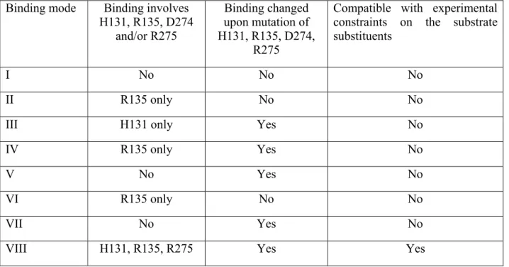
Table 3: Comparison of predicted features of each binding mode with experimentally derived information.
Binding mode Binding involves H131, R135, D274
and/or R275
Binding changed upon mutation of H131, R135, D274,
R275
Compatible with experimental constraints on the substrate substituents
I No No No
II R135 only No No
III H131 only Yes No
IV R135 only Yes No
V No Yes No
VI R135 only No No
VII No Yes No
VIII H131, R135, R275 Yes Yes
III.A Binding mode I
through short-range interactions (<4 Å away) with Ser55, Asn70, Arg287, and Ser290. No changes in binding mode are apparent upon mutation of H131, R135, R275 and D274 to alanine (Supporting information, Figure S1). The solvent-exposure of the ring B methyl group in this binding mode is not consistent with the experimentally verified requirement of a small non-polar substituent (rather than e.g. acetate13) at that position. The non-involvement of H131, R135, R275 and D274 in this substrate binding mode also argues against its mechanistical relevance.
III.B Binding mode II
This simulation afforded a very stable binding conformation, with seven different short-range contacts between polar/charged side chains and the substrate propionate groups maintained throughout the experiment: the A propionate remains bound to Arg135 (3.94 ± 0.12 Å) and His90 (4.31 ± 0.25 Å), Ser117 (3.22 ± 0.17 Å) and Asn133 (4.33 ± 0.41 Å) surround propionate B, the C propionate is kept close to Ser72 (3.40 ± 0.16 Å) and Ser 290 (3.79 ± 0.19 Å) though hydrogen bonds, and the exposed D propionate negative charge is stabilized by Arg46 (4.27 ± 0.17 Å). Both the D- and the A-ring methyl groups remain accessible to solvent, though lined with hydrophobic residues (Val74, Phe111 and Phe279 for D methyl, Met86 and Phe137 for A methyl). Apart from a marked increase in the mobility of Arg46, no changes can be seen in the binding mode upon quadruple mutation (Supporting Information, Figure S2).
III.C Binding mode III
stabilized by Arg287 (4.02 ± 0.19 Å), Ser72 (4.64 ± 0.52 Å), and Ser290 (4.47 ± 0.77 Å).In the mutated protein the B propionate more easily approaches Ser290 (3.54 ± 0.24 Å), Arg287 and Asn70 due to the removal of the stabilizing His131 that, in the wild type-protein, attracted propionate A and pulled the substrate in the opposite direction (Supporting information, Figure S3). This subtle difference in binding allows the entrance to the crevice to widen, so that in the mutated protein the D-ring is more exposed to solvent and therefore a substituent slightly larger than methyl (e.g. vinyl) may be more easily accommodated at this position than in the WT protein. In the WT protein there is enough free room around the ring B methyl to accommodate polar substituents than methyl (e.g. acetate), nn contrast to the experimental observation13.
III.D Binding mode IV
In this substrate conformation, only the substituents present on ring B are exposed to the solvent. The substrate remains bound to the enzyme through stable interactions involving all remaining propionates: propionate A interacts with Ser72 (3.30 ± 0.24 Å) and Asn70 (4.143± 0.41 Å), propionate D hydrogen-bonds Asn133 (3.43 ± 0.17 Å) and propionate C is responsible for electrostatic interactions with His90 (3.62 ± 0.15 Å) and Arg135 (4.11 ± 0.15 Å). In the quadruple mutant, the lack of Arg135 allows the substrate to change its binding mode: propionate C is now only attracted by His90 (4.11 ± 0.15 Å), propionate D approaches Asn133 (3.68 ± 0.32 Å), and propionate A slides past Asn70 (which is now left at 7.00 ± 0.46 Å), while remaining close to Ser72 (3.53 ± 0.17 Å) and approaching Arg46 (4.20 ± 0.17 Å) (Supporting Information, Figure S4). The lack of interactions with Arg275 and His131 argues against the relevance of this binding mode to the catalytic effect of the enzyme, as does the solvent exposure of the B methyl group.
Even in the wild-type protein, binding in this conformation is almost completely due to interactions with only one of the substrate carboxylate groups. In this simulation, propionates A and B remain solvent-exposed throughout, whereas propionate C remains within short range of Ser290 (3.34 ± 0.34 Å) and Arg287 (4.39 ± 0.32 Å). In the first 8 ns of the simulation Asn70 maintains a stable hydrogen-bond with propionate C (≈91% of the time). Although the stability of this bond decreases after this point (to 54% of the time), Asn70 remains quite close to propionate C (average distance in the first 10.5 ns is 3.48 ± 0.28 Å). Propionate D interacts only with Ser117 (3.47 ± 0.39 Å), whereas the A, B and D methyl groups remain quite exposed to the solvent, though surrounded by non-polar residues. In the simulation of the mutated protein, the crevice opens and helixes H2 move away from each other. The substrate partially exits the binding region, and the only surviving binding interactions are those between Arg287 and propionate C (3.83 ± 0.13 Å) and a new (less stable) interaction between this propionate and Ser72 (3.85 ± 1.03 Å). Eventually, a stable interaction between Arg46 and propionate C also develops, but this is not enough to maintain the enzyme-substrate complex tightly bound (Supporting information, Figure S5). The experimental results of enzymatic conversion of substituted porphyrinogens suggest that the active site should have enough free space around the position where the D-ring methyl is accomodated, whereas the methyl group in ring A cannot be replaced by larger substituents5,13-15. The relatively open environment around the A methyl group in this binding conformation, the paucity of electrostatic contacts between the protein and substrate, and the lack of interactions involving the experimentally determined amino acids, all argue against the mechanistic relevance of this substrate binding mode.
III.F Binding mode VI
± 0.41 Å), respectively, whereas propionate D remains strongly bound by two stable ionic interactions with His90 (3.70 ± 0.20 Å) and Arg135 (4.13 ± 0.15 Å). The methyl group on ring D is closely surrounded by Leu78, Phe279, Met86 and Phe137, which renders this binding mode (like the previous one) incompatible with the observed catalysis of several D-ring substituted porphyrinogens. In the mutated enzyme the removal of Arg135 releases propionate D, which remains attracted only by His90, the substrate C propionate moves further towards Asn133, and the propionate B approaches Asn70 and Ser72. Propionate A remains anchored to Arg46, and the binding mode maintains high similarity to that observed with the wild-type protein (Supporting Information, Figure S6).
III.G Binding mode VII
Throughout the simulation of this binding mode, the methyl groups from rings A and D remain exposed to the solvent. This behavior is not consistent with the lack of activity of the enzyme on substituted substrates bearing larger substituents than methyl (on ring A) or vinyl (on ring D). The simulation showed this putative binding mode to be the least stable of all those analyzed: only propionate A establishes stable hydrogen bonds with enzyme amino acids (Ser72, 3.43 ± 0.23 Å and Asn70, 4.36 ± 0.41 Å), whereas the interactions between propionate B and Arg135 and Asn 133 do not survive past the initial 20% of the simulation and Arg46 only sporadically interacts with propionate D. In the mutated enzyme, the cavity opens widely and this binding mode becomes even less effective, as the interaction between Asn70 and propionate A disappears, and the distance between Ser72 and propionate A becomes more variable and (on average) longer (4.39 ± 0.61 Å).(Supporting Information, Figure S7).
group in binding pose VIII is not fully occupied by this substituent and that, in agreement with experimental results, this position can accommodate non-polar substituents with two (but not three) carbon atoms.
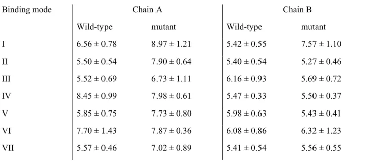
Asp274 remains close to propionate A throughout the simulation (4.70 ± 0.30 Å away from its carboxylate group, and 4.48 ± 0.30 Å away from its carbon), but its negative charge is expected to destabilize the substrate carboxylate and its role seems to be unrelated to substrate binding. Two possible roles are suggested by the molecular simulations: on the one hand, its proximity to the propionate may facilitate the decarboxylation reaction (by increasing the energy of the negatively charged reactant state); on the other hand, each carboxylate oxygen in Asp274 establishes a hydrogen bond with a different enzyme amino acid (Tyr196 on the C-terminus of helix H4 and Thr277 on helix H8) and plays therefore a role in the structural stability of the enzyme. Analysis of the Tyr196-Thr277 pair in each of the dimer subunits in the mutant and wild-type simulations (Table 4) clearly show that (except in the simulations of binding modes IV and VI) the mutation of Asp274 to alanine in the substrate-bound chain leads to an increase in the average distance and on the distance fluctuations of these residues relative to the non-mutated empty subunit.
Table 4: Distances (in Å) between the polar hydrogens in Tyr196 and Thr227 in wild-type and mutant coproporphyrinogen III oxidase.
Binding mode Chain A Chain B
Wild-type mutant Wild-type mutant
I 6.56 ± 0.78 8.97 ± 1.21 5.42 ± 0.55 7.57 ± 1.10
II 5.50 ± 0.54 7.90 ± 0.64 5.40 ± 0.54 5.27 ± 0.46
III 5.52 ± 0.69 6.73 ± 1.11 6.16 ± 0.93 5.69 ± 0.72
IV 8.45 ± 0.99 7.98 ± 0.61 5.47 ± 0.33 5.50 ± 0.37
V 5.85 ± 0.75 7.73 ± 0.80 5.98 ± 0.63 5.43 ± 0.41
VI 7.70 ± 1.43 7.87 ± 0.36 6.08 ± 0.86 6.32 ± 1.23
VIII 6.44 ± 0.62 9.03 ± 1.43 5.30 ± 0.46 6.48 ± 1.02
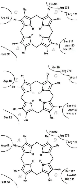
Figure 6: Schematic representation coproporphyrinogen III (top panel), harderoporphyrinogen III (center panel) and porotoporphyrinogen IX (bottom panel) to the enzyme active site
The binding pose obtained from VIII also explains why only rings A and B can be decarboxylated by the enzyme (Figure 6). It is known experimentally that decarboxylation first occurs in the active site region that holds ring A, which is shown by our simulations of binding mode VIII to be the region surrounding Asn133 and His 131. After the negative propionate in ring A is converted to a neutral vinyl group the favorable interaction with His131 disappears, and the binding affinity decreases. This partially decarboxylated intermediate (harderoporphyrinogen III) may leave the active site, and can only bind again in a new conformation (rotated ≈90º vs. the original one) with the vinyl group in ring A
occupying the original binding site of the ring D methyl (which has enough room for a two-carbon substituent) (Figure 6, center panel). Ring C can easily occupy the binding site originally belonging to ring B, due to the identical distribution of substituents in these two rings, and ring D may occupy the original position on ring C (in spite of the opposite distribution of substituents) because this site is solvent-exposed and can therefore easily accommodate a propionate group in the place of the original methyl group. After the second decarboxylation, the binding affinity again decreases due to the loss of propionate-His131 interaction, and the product (protoporphyrinogen IX) leaves the active site. Protoporphyrinogen may not bind the enzyme again, as the only binding modes possible (Figure 6, bottom panel) are now less favorable since they depend on the existence of propionate groups on rings A and B (see description of binding modes IV and VI above). Furthermore, these binding modes also place the remaining propionate groups far from the detected active site (His131, Asn133 and Ser117), which renders catalysis ineffective.
In this study, the application of molecular dynamics simulations of enzyme in explicit solvent and periodic boundary conditions to the eight possible substrate orientations in coproporphyrinogen III oxidase active site cavity enabled the determination of the putative substrate binding mode and the extensive characterization of the enzyme active site, which was quite conclusive as far as the catalytic determinants of this particular reaction are concerned. In fact, the MD simulations described here enabled the determination of a substrate binding mode fully consistent with the known selectivity of the active site towards substituted tetrapyrroles, and explained the lack of activity of the H131A, R135A, D274A and R275Amutants and the reasons behind the non-occurrence of catalysis on the C and D rings of the tetrapyrrole. The finding of a strong hydrogen-bonding role for the invariant G276 residue in the successful binding mode offers a powerful argument against the reaction mechanisms that require the deprotonation of the reacting pyrrole ring, as this interaction is only relevant with protonated pyrrole nitrogens and the absence of cationic side chains close to the substrate pyrrole nitrogens prevents the protein from lowering the high pKa of pyrrole to values compatible with its deprotonation at physiological pH. Since the Arigoni mechanism (Figure 1A) is only feasible with a deprotonated pyrrole18, these constraints leave the Lash mechanism (Figure 1B) with neutral substrate as the only mechanism compatible with the experimental and computational findings.
V. References
1. Yoshinaga, T. & Sano, S. (1980) J. Biol. Chem. 255, 4727–4731.
2. Medlock, A. E. & Dailey, H. A. (1996) J. Biol. Chem. 271, 15765–15770.
3. Elder, G. H., Evans, J. O., Jackson, J. R. & Jackson, A. H. (1978) Biochem. J., 169, 215–223. 4. Yoshinaga, T. & Sano, S. (1980) J. Biol. Chem. 255, 4722–4726.
5. Lash, T. D., Mani, U. N., Drinan, M. A., Zhen, C., Hall, T. & Jones, M. A. (1999) J. Org. Chem. 64, 464–477.
6. Akhtar, M. (2003) in The Porphyrin Handbook, eds. Kadish, K. M., Smith, K. M. & Guilard, R. (Elsevier Science, New York), Vol. 12, pp. 69–86.
7. Cavaleiro, J.A.S.; Kenner, G.W & Smith, K.M. (1974) . Chem. Soc. Perkin Trans. I, 10, 1188-1194
8. Sassa, S. & Kappas, A. (2000) J. Intern. Med. 247, 169-178
9. Lee, D.-S., Flachsová, E., Bodnárová, M., Demeler, B., Martásek, P. & Raman, C. S. (2005) Proc. Natl. Acad. Sci. USA 102, 14232–14237
10. Phillips, J. D., Whitby, F. G., Warby, C. A., Labbe, P., Yang, C., Pflugrath, J. W., Ferrara, J. D., Robinson, H., Kushner, J. P. & Hill, C. P. (2004) J. Biol. Chem. 279, 38960–38968.
11. Gitter S.J., Cooper C.L., Friesen J.A., Lash, T.D., Jones M.A. (2007) Med. Sci Monit. 13, BR1-10
12. Stephenson JR, Stacey JA, Morgenthaler JB, Friesen JA, Lash TD, Jones MA. (2007) Protein Sci. 16, 401-410.
13. Elder, G.H.; Evans, J.O. (1978) Biochem. J., 169, 205-214 14. Porra, R. J. & Falk, J. E. (1964) Biochem. J., 90, 69-75
15. Al-Hazimi, H.M.G.; Jackson, A.H.; Knight, D.W.; Lash, T.D.(1987) J. Chem. Soc. Perkin Trans. I, 265-276
17. Jones , M.A; He, J. & Lash T.D. (2002) J. Biochem., 131, 201-205
18. Silva, P.J. & Ramos, M.J. (2008) Bioorg. Med. Chem. 16, 2726-2733
19. Lash , T.D. (2005) Bioorg. Med. Chem. Lett. 15, 4506-4509
20. Krieger E, Darden T, Nabuurs S, Finkelstein A, Vriend G (2004) Proteins 57, 678-683
21. Wang J, Cieplak P, Kollman PA (2000) J. Comput. Chem. 21, 1049-1074
22. Essman U, Perera L, Berkowitz ML, Darden T, Lee H, Pedersen LG (1995) J. Chem. Phys. 103, 8577-8593.
23. Berendsen HJC, Postma JPM, van Gunsteren WF, DiNola A, Haak JR. (1984) J. Chem. Phys.
81, 3684-3690.
24. Jakalian A, Jack DB and Bayly CI (2002) J. Comput. Chem. 23,1623-1641
25. Krieger E, Nielsen JE, Spronk CA, Vriend G (2006) J. Mol. Graph. Model. 25,481-486
26. Shoolingin-Jordan P.M. (2003) The Biosynthesis of Coproporphyrinogen III. In Kadish, K.M., Smith, K.M. and guilard, M. (eds.) The Porphyrin handbook. Volume 12: The iron and Cobalt Pigments: Biosynthesis, Structure and Degradation. Elsevier Science