Comparison between conventional protective
mechanical ventilation and high-frequency
oscillatory ventilation associated with the prone
position
INTRODUCTION
Mechanical ventilation (MV) is the most important treatment for acute respiratory distress syndrome (ARDS) and is capable of modifying the evolution
of the disease.(1) Although protective conventional MV (CMV) is efective in
many patients, a signiicant number present with severe respiratory failure, in which CMV may not guarantee oxygenation and ventilation. In these cases, when pulmonary protection is required, high-frequency oscillatory ventilation
(HFOV) becomes an interesting therapeutic alternative(2) because it uses a
José Roberto Fioretto1, Susiane Oliveira Klefens1,
Rafaelle Fernandes Pires1, Cilmery Suemi
Kurokawa1, Mario Ferreira Carpi1, Rossano
César Bonatto1, Marcos Aurélio Moraes1, Carlos
Fernando Ronchi1,2
1. Department of Pediatrics, Faculdade de Medicina de Botucatu, Universidade Estadual Paulista “Júlio de Mesquita Filho” - Botucatu (SP), Brazil.
2. Faculdade de Educação Física e Fisioterapia, Universidade Federal de Uberlândia - Uberlândia (MG), Brazil.
Objective: To compare the efects of high-frequency oscillatory ventilation and conventional protective mechanical ventilation associated with the prone position on oxygenation, histology and pulmonary oxidative damage in an experimental model of acute lung injury.
Methods: Forty-ive rabbits with
tracheostomy and vascular access were underwent mechanical ventilation. Acute lung injury was induced by tracheal infusion of warm saline. hree experimental groups were formed: healthy animals + conventional protective mechanical ventilation, supine position (Control Group; n = 15); animals with acute lung injury + conventional protective mechanical ventilation, prone position (CMVG; n = 15); and animals with acute lung injury + high-frequency oscillatory ventilation, prone position (HFOG; n = 15). Ten minutes after the beginning of the speciic ventilation of each group, arterial gasometry was collected, with this timepoint being called time zero, after which the animal was placed in prone position and
Conflicts of interest: None.
Submitted on July 5, 2016 Accepted on May 11, 2017
Corresponding author: Carlos Fernando Ronchi Universidade Federal de Uberlândia Rua Benjamin Constant, 1.286
Zip code: 38.400-678 - Uberlândia (MG), Brazil E-mail: ronchi.carlos@yahoo.com
Responsible editor: Werther Brunow de Carvalho
Comparação entre ventilação mecânica convencional protetora e
ventilação oscilatória de alta frequência associada à posição prona
ABSTRACT
Keywords: Respiration, artiicial; Acute lung injury; High-frequency ventilation; Oxidative stress; Acute respiratory distress syndrome; Rabbits remained in this position for 4 hours. Oxidative stress was evaluated by the total antioxidant performance assay. Pulmonary tissue injury was determined by histopathological score. he level of signiicance was 5%.
Results: Both groups with acute lung injury showed worsening of oxygenation after induction of injury compared with the Control Group. After 4 hours, there was a signiicant improvement in oxygenation in the HFOG group compared with CMVG. Analysis of total antioxidant performance in plasma showed greater protection in HFOG. HFOG had a lower histopathological lesion score in lung tissue than CMVG.
Conclusion: High-frequency
oscillatory ventilation, associated with prone position, improves oxygenation and attenuates oxidative damage and histopathological lung injury compared with conventional protective mechanical ventilation.
tidal volume (TV) lower than the anatomical dead space volume and frequency higher than the physiological one, avoiding elevated pressures and alveolar volumes typical
of CMV.(3-5)
Due to the high mortality observed in ARDS, additional therapeutic strategies for MV have been developed,
especially for the prone position.(6) In ARDS, lung injury is
heterogeneous and varies with the position of the patient, being more signiicant in areas that depend on gravity, i.e., the dorsal lung region, when the patient is in the
supine position.(7,8) he prone position may improve gas
exchange by redistributing ventilation to better-perfused
dorsal lung areas(9,10) and by mediating homogenization
of TV distribution associated with changes in chest wall
mechanics,(11) alveolar recruitment,(12) and redirection
of compressive forces exerted by the weight of the heart
on the lungs,(13) resulting in better removal of secretions.
Recently, studies have shown that there is improved
survival in patients treated early with prone position.(14)
Considering the protective characteristics of HFOV and its capacity to redistribute ventilation to better-perfused lung areas, which results in better oxygenation in ARDS, and the potential recruitment of prone position, our hypothesis is that the sum of the beneicial efects of HFOV and prone position improves oxygenation more, makes histopathological lesions more homogeneous and of lower intensity, and attenuates oxidative damage to pulmonary tissue when compared with CMV associated with prone position.
he present study aimed to compare the efects of prone position associated with HFOV and CMV by oxygenation, histology, and pulmonary oxidative damage in an experimental model of acute lung injury induced in rabbits.
METHODS
his study was conducted at the Experimental Laboratory of the Center for Clinical and Experimental Research of the Department of Pediatrics of the
Faculdade de Medicina de Botucatu of the Universidade Estadual Paulista “Júlio de Mesquita Filho” (UNESP) and was approved by the Ethics Committee on Animal
Experimentation of the Faculdade de Medicina de Botucatu
under protocol number 795.
A prospective study in vivo conducted on laboratory
animals. White male rabbits provided by the School
of Medicine Vivarium - Botucatu Campus were used,
weighing 2.0 to 3.0kg.
he instrumentation of the animals followed a
protocol already established by the group.(15,16) Briely,
after being weighed, the animals were anesthetized and sedated with a solution of ketamine (50mg/kg) and acepromazine (2mg/kg) administered intramuscularly. Animals were placed in a surgical brace, received 100% oxygen through a nasal catheter, and underwent cervical and thoracic trichotomy for the placement of heart rate (HR)-monitoring electrodes. If the HR decreased to below 180bpm, atropine was given at a dose of 0.01mg/ kg intravenously in the auricular vein. he anterior region of the animal’s neck was anesthetized with xylocaine to perform the tracheostomy. A tracheal tube of the highest possible caliber (3.0 to 3.5mm internal diameter, Portex, Hythe, UK) was inserted through the tracheostomy and was held in position with surgical tape. MV was then
immediately started with the CMV apparatus (Inter®
7 plus, Oxy System, São Paulo (SP), Brazil). he initial parameters were as follows: pressure-regulated volume-controlled mode, with a target TV of 6mL/kg; respiratory rate (RR) of 40 cycles per minute, adjusted according to
the partial pressure of carbon dioxide (PaCO2); inspiratory
time (Ti) of 0.5 second; positive end-expiratory pressure
(PEEP) of 5cmH2O; and inspired oxygen fraction (FiO2)
of 1.0. hese parameters were maintained for a stabilization period of 10 minutes, until the moment of lung injury induction in the treated groups. After the tracheostomy, the carotid artery and the internal jugular vein were dissected. A single-lumen vascular catheter was inserted
into the common carotid (22 Gauge Jelco, Introcan®
SafetyTM, B-Braun, Melsungen, Germany), and a
double-lumen catheter (5Fr, Arrow International Inc., Reading, Philadelphia, USA) was inserted in the superior vena cava through the jugular vein. he arterial catheter was used to obtain blood gases and for continuous monitoring of mean arterial pressure (MAP) using a pressure monitoring
system (LogicCal®
from Medex, Dublin, USA) connected to a multiparameter monitor (Dixtal, Manaus, Brazil). he vena cava catheter was used for administration of continuous infusion sedatives, maintenance luids, and vasoactive drugs.
if MAP reached values below 50mmHg, continuous intravenous infusion of noradrenaline was initiated at an initial dose of 0.2μg/kg/minute; if there was no response, the dose was gradually increased to 1μg/kg/minute. he body temperature was monitored using a digital rectal thermometer and was maintained between 38°C and 40°C using heat packs, and the blood volume was maintained by continuous infusion of 4mL/kg/hour of saline solution plus 5% dextrose.
Induction of the acute lung injury model
Acute lung injury (ALI) was induced according to a
previously described technique.(15,17-19) Briely, six successive
washes of the lung were performed with warm saline (38°C) in aliquots of 30mL/kg, at a maximum pressure of
30cmH2O, through the tracheal cannula. Each washing
procedure lasted 60 seconds, 20 seconds being reserved for infusion and the remaining time for withdrawal, which was performed by gravity and external chest compression movements. After completion of the withdrawal, the procedure was repeated every 3 - 5 minutes until reaching
a PaO2/FiO2 < 100mmHg, which was conirmed after 10
minutes of stabilization. If the criterion was not reached, two more washes were performed in the sequence and, after 10 minutes, a new gasometry was obtained, and so
on, until a PaO2/FiO2 < 100mmHg was reached. After
satisfying this criterion, the animals were randomized to create the experimental groups.
Experimental groups and mechanical ventilation parameters
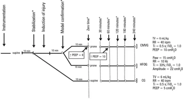
Based on previous studies performed with similar methodologies, the animals were distributed in three groups of 15 rabbits each, as follows: instrumented healthy animals (control - CG), maintained in supine position and submitted to CMV in pressure-regulated volume-controlled mode, with TV of 6mL/kg, RR of 40 cycles per
minute, a Ti of 0.5 seconds, a PEEP of 5cmH2O and an
FiO2 of 1.0; animals with ALI submitted to protective CMV
(conventional mechanical ventilation group - CMVG) in prone position, with the same initial parameters described
for CG. In this group, PEEP was increased to 8cmH2O
during the irst hour and then to 10cmH2O, and then was
maintained until the end of the experiment. Animals with ALI underwent HFOV in prone position with a mean
airway pressure of 15cmH2O, an RR of 10Hz, a Ti of
33%, a pressure range of 22cmH2O, and an FiO2 of 1.0,
in the mechanical ventilator SensorMedics 3100A (Viasys Healthcare, Yorba Linda, USA), with RR and amplitude
adjusted to maintain PaCO2 at physiological levels
(35 - 45mmHg), forming the high-frequency oscillatory ventilation (HFOG) group (Figure 1).
Figure 1 - Experimental protocol and distribution of animals according to the type of ventilation used. CMVG - conventional mechanical ventilation group; TV - tidal volume; RR - respiratory rate; Ti - inspiratory time; FiO2 - inspired oxygen fraction; PEEP - positive end-expiratory pressure; Paw - mean airway pressure; HFOG - high-frequency
Ten minutes after the beginning of the speciic ventilation of each group, new gasometry was obtained, with this timepoint being called time zero (T0), after which the animals were placed in prone position. From this moment, they were ventilated for 4 hours, and arterial blood gas measurements were collected at moments 30, 60, 120, 180, and 240 minutes. he time of 4 hours was chosen, taking into account the viability of the rabbits in this type of experiment, based on previous experiments and the studies cited above, which demonstrated early clinical
and experimental efects of the prone position.(17,18,20)
Manipulation of the lungs and determination of tissue injury. Pulmonary histology
At the end of the experiment, the animals received 1 mL of heparin and then underwent euthanasia by rapid intravenous administration of ketamine. Subsequently, the tracheal tube was occluded, and the thorax opened to exclude the presence of occult pneumothorax, to conirm the position of the vascular catheters and tracheal tube, and to collect samples for histological analysis and bronchoalveolar lavage. In animals in which bronchoalveolar lavage was performed (n = 8), the right bronchus was ligated by surgical tape, the lung/heart block was removed, the left lung was washed twice using aliquots of 15mL/kg of normal saline, and the drained luid was collected for analysis. In the animals submitted to histological analysis (n = 7), the trachea/lung/heart block was removed, the lungs and trachea were separated from the heart, and the left lung of animals not submitted to bronchoalveolar lavage was illed with 10% formalin solution. Filling was achieved by means of a column with serum equipment 30cm long, with a vial containing formalin connected to one of its ends and the trachea of the animal connected to the other end. From this system, the formaldehyde slowly dripped by gravity to ill the alveolar spaces, preserving their architecture. After a minimum of 24 hours of ixation, fragments were embedded in parain, and axial sections of the lung were then stained with hematoxylin and eosin and examined by two pathologists in a blind and independent manner. In each slide, the specimen was divided into two distinct zones, representing the dependent (dorsal) and non-dependent (ventral) regions of the lung. Ten microscopic ields were randomly selected for the examination, ive in each region, totaling 50 analyses for each animal. Pulmonary histological lesions were quantiied by a score composed of seven variables (alveolar and interstitial inlammation, alveolar and interstitial hemorrhage, edema, atelectasis,
and necrosis). he severity of the lesion was classiied for each of the seven variables as follows: zero if no lesion was observed; 1 if injured in 25% of the ield; 2 if injured in 50% of the ield; 3 if injured in 75% of the ield; and 4 if difuse injury. he maximum possible score was 28, and
the minimum score was zero.(21,22)
Concentration of malondialdehyde
Concentrations of malondialdehyde (MDA), a marker of lipid oxidative damage, were measured in pulmonary lavage luid and plasma using the method of Esterbauer et al.(23)
Pulmonary oxidative stress: total antioxidant performance assay
Lung oxidative stress was evaluated using the total antioxidant performance (TAP) assay described by
Aldini et al.(24) Briely, TAP assay, validated by Beretta
et al.,(25) determines the antioxidant capacity by measuring
oxidative stress and is the only approach that captures the antioxidant network of the lipophilic and hydrophilic
compartments and their interactions.(26) It is based on
the generation of lipophilic radical (MeO-AMVN) and an oxidizable lipophilic substrate (BODIPY), which speciically measures the oxidation of the lipid compartment related to the actions of liposoluble and water-soluble antioxidants through a mechanism of
synergism and cooperation.(27)
For each sample, 100μL of plasma and 100µL of phosphatidylcholine (PC) standard (PC1 and PC2) were pipetted separately. In plasma and in both PCs, 300μL of ice-cold phosphate-bufered saline (PBS) (pH 7.4) was added, and 100μL of BODIPY was then added to all samples; after a water-bath, 420µL of PBS and 80μL of 2,2’ - azobis (2-amidinopropane) dihydrochloride (AAPH) were pipetted into each sample. Samples were vortexed and then placed on a plate for analysis using the Wallack Victor X2 apparatus (Perkin-Elmer, Boston, USA) and the WorkOut 2.5 program (Dazdaq Solutions Ltd.). he entire procedure was performed under indirect light, and the samples were prepared in triplicate.
Statistical analysis
Table 1 - Comparison of experimental groups in relation to partial pressure of oxygen/inspired oxygen fraction, oxygenation index, pulmonary compliance, and mean arterial pressure, before and after injury
Variables
CG N = 15
HFOG N = 15
CMVG N = 15
Baseline values Before the LI After the LI Before the LI After the LI
PaO2/FiO2 444.26 ± 59.45 445.31 ± 54.17 70.23 ± 21.35* 465.86 ± 48.30 68.93 ± 11.60*
Oxygenation index (cmH2O/mmHg) 1.95 ± 0.32 2.25 ± 0.80 20.10 ± 12.92* 1.95 ± 0.46 14.23 ± 2.28*
Compliance (mL/cmH2O) 1.73 ± 0.62 2.06 ± 0.49 0.81 ± 0.19* 1.76 ± 0.41 0.74 ± 0.25*
MAP (mmHg) 60.13 ± 15.61 61.89 ± 14.47 69.55 ± 9.45* 65.2 ± 15.84 68.5 ± 15.05*
CG - control group; CMVG - conventional mechanical ventilation group; HFOG - high-frequency oscillatory ventilation group; LI - lung injury; PaO2 - partial pressure of oxygen; FiO2 - inspired
oxygen fraction; MAP - mean arterial pressure. * p < 0.05 comparing the moments before and after the induction within each group. Normal distribution: t test. Non-normal distribution:
Mann-Whitney rank.
among the diferent groups using Kruskal-Wallis ANOVA, with subsequent comparisons by Dunn’s test. he analysis of the behavior of a variable over time, in cases of normal distribution, was evaluated using repeated measures ANOVA, with comparisons between pairs using Bonferroni’s test; in cases of non-normal distribution, Friedman’s test for repeated measures was used, with
later comparisons by Dunn’s method. A t-test was used
to compare the number of lung washes between the two treated groups. Statistical signiicance was deined as p < 0.05.
RESULTS
Hemodynamics, pulmonary mechanics, and gas exchange
here were no signiicant diferences between groups regarding animals weight and number of washes required for lesion induction. Likewise, there were no signiicant
diferences among groups regarding PaO2/FiO2 ratio,
oxygenation index (OI), lung compliance, and MAP compared moments before and after lung injury induction. Comparison before and after lung injury within each group indicated that there was a signiicant worsening of oxygenation and a decrease in pulmonary compliance in both groups after induction, as shown in table 1.
In the evaluation of the hemodynamic state, MAP was not signiicantly diferent between the moments during the experiment, indicating homogenization of the groups and strict control of the variable, using, with vasoactive drugs administered when necessary. he percentages of animals requiring vasoactive drug were 20% in CG and 26% in HFOG and CMVG.
After lesion induction, the groups developed signiicant hypoxemia compared to the beginning of the experiment. After 4 hours of CMV, the HFOG showed a signiicant
improvement in oxygenation compared with the CMVG,
presenting a PaO2/FiO2 ratio similar to the moments before
injury induction and to the CG, as shown in igure 2.
Oxidative stress - malondialdehyde and total antioxidant performance
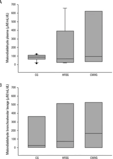
here were no signiicant diferences between the groups when MDA levels were evaluated in plasma and bronchoalveolar lavage (Figure 3).
Regarding the evaluation of TAP in plasma, HFOG presented similar antioxidant protection to CG and signiicantly higher protection than CMVG, as shown in igure 4.
Histopathology
HFOG presented a signiicantly lower histopathological lesion score than did CMVG, as shown in igure 5.
DISCUSSION
Recently, our group was the irst to publish the results of a comparison between protective CMV and HFOV regarding total antioxidant performance by TAP assay and
concluded that HFOV attenuated oxidative stress.(15)
Few studies have evaluated the association of HFOV
with prone position.(28,29) Clinical studies have concluded
that prone position associated with CMV or HFOV improves oxygenation in 12 hours, in contrast to the supine position associated with HFOV, in addition to
decreasing pulmonary inlammation. Demory et al.(29)
suggested that HFOV is able to maintain prone position-induced alveolar recruitment, and its use after the prone
position allows for the reduction of FiO2 to potentially
less toxic levels.
Figure 2 - Evolution of oxygen partial pressure/inspired oxygen fraction in the experimental period (up to 240 minutes). PaO2 - partial pressure of oxygen; FiO2 - inspired oxygen fraction; CG - control group; HFOG - high-frequency oscillatory ventilation group; CMVG - conventional
mechanical ventilation group. * p < 0.05 for the high-frequency oscillatory ventilation and conventional
mechanical ventilation groups compared with the control group; # p < 0.05 in relation to the initial moment.
Figure 3 - Concentrations of malondialdehyde in each group: (A) Plasma: High-frequency oscillatory ventilation group [control group: 87.38 (64.20 - 106.34) > high-frequency oscillatory ventilation group: 67.63 (26.40 - 327.60) < conventional mechanical ventilation group: 95.92 (34.49 - 599.06); p < 0.05). (B) Bronchoalveolar lavage: [control group: 25.75 (2.74 - 291.86) < high-frequency oscillatory ventilation group: 72.63 (0.75 - 449.64) < conventional mechanical ventilation group: 167.15 (1.85 - 462.20); p > 0.05]. CG - control group; HFOG - high-frequency oscillatory ventilation group; CMVG - conventional mechanical ventilation group. The bars above
and below the rectangles indicate the 25th and 75th percentiles, and the inner bar indicates the median.
to lesion induction, corroborating an earlier study by our
group,(15) also performed in rabbits with ALI induced by
infusion of saline in animals ventilated with HFOV in supine position. his inding conirms our hypothesis that in cases of severe hypoxemia, HFOV may be an attractive
alternative for more efective oxygenation improvement.(2)
Regarding oxidative stress, the plasma MDA concentration was lower in HFOG than in CMVG but did not reach statistical signiicance. However, when oxidative stress was evaluated by TAP, there was greater pulmonary protection in HFOG compared with CMVG animals. his result may have been due to the evaluation characteristics of the TAP assay, which is more sensitive when measuring the TAP of the two compartments (hydrophilic and lipid)
present in the biological samples.(24) Still, this result shows
that there was greater pulmonary antioxidant protection in the HFOG compared with that in the CG animals. We believe that this behavior of HFOG in relation to TAP occurred since CMV alone can damage the healthy lung by the cyclical opening and closing movements of alveolar units, whereas HFOV provides greater lung
protection by maintaining a constant lung volume.(27)
his result is in agreement with the indings of Ronchi
et al.,(15) who also used this method and obtained values
similar to those in the CG in the group ventilated with HFOV and signiicantly higher than those in the CMV group insupine position. Reinforcing our indings, in a
study conducted by Mazullo Filho et al.,(30) the authors
evaluated 12 patients admitted to the intensive care unit, comparing the irst and last days of use of CMV, and observed that patients had increased markers of oxidative stress and reduced antioxidant enzyme levels due to the use of CMV.
Histopathological indings typical of ARDS in this model include edema, polymorphonuclear iniltrate in the alveolar space, hyaline membrane formation, and capillary
congestion,(21) which were evaluated by histological scores,
including inlammation, hemorrhage, edema, atelectasis,
and necrosis.(22,31) We have demonstrated that the HFOG
Figure 4 - Total antioxidant performance in plasma for each group. CG - control group; HFOG - high-frequency oscillatory ventilation group; CMVG - conventional mechanical ventilation group. *
p < 0.05.
Figure 5 - Histopathological lesion score in lung tissue (high-frequency oscillatory ventilation group: 1.4 (1.2 - 1.8) < conventional mechanical ventilation group: 1.7 (1.4 - 3.2); * p < 0.05]. The lower edges of the rectangles indicate the 25th percentiles, the horizontal lines within the rectangles mark the medians, and the upper edges indicate the 75th percentiles.
The bars above and below the rectangles indicate the percentiles 90 and 10, respectively, and the filled
circles represent individual values. HFOG - high-frequency oscillatory ventilation group; CMVG - conventional
mechanical ventilation group.
our indings, an experimental study in pigs,(31) in which
ARDS was induced by lavage with saline, showed that HFOV associated with prone position led to a reduction in the histopathological score when compared with CMV animals. In addition, there was an improvement in
oxygenation, a signiicant reduction in pulmonary shunt
fraction, and normalization of cardiac output with lower mean airway pressures when HFOV was associated with supine position.
he present study has some limitations. First, there is no animal model capable of reproducing all of the characteristics of ALI/ARDS in humans. However, one of the most widely used ALI models in animals is alveolar lavage with heated saline, which causes surfactant depletion, resulting in lung injury very similar to that of ARDS in humans. In addition, the 4-hour experiment
under FiO2 of 1.0 may lead to lung parenchymal damage
and can interfere with the oxidative metabolism of these animals. In contrast, the use of the same oxygen concentration and the deinition of ventilatory parameters for all groups likely excluded any signiicant variations among groups due to oxygen toxicity. he choice of the number of animals was based on previous studies, and no sample calculations were performed.
CONCLUSION
High-frequency oscillatory ventilation in association with prone position improves oxygenation and leads to reduced oxidative damage, as measured by total antioxidant performance assay and attenuation of histopathological lung injury, compared with protective conventional mechanical ventilation in prone position.
ACKNOWLEDGMENTS
his study had inancial support from the Fundação
Objetivo: Comparar os efeitos da ventilação oscilatória de alta frequência e da ventilação mecânica convencional protetora associadas à posição prona quanto à oxigenação, à histologia e ao dano oxidativo pulmonar em modelo experimental de lesão pulmonar aguda.
Métodos: Foram instrumentados com traqueostomia,
acessos vasculares e ventilados mecanicamente 45 coelhos. A lesão pulmonar aguda foi induzida por infusão traqueal de salina aquecida. Foram formados três grupos experimentais: animais sadios + ventilação mecânica convencional protetora, em posição supina (Grupo Controle; n = 15); animais com lesão pulmonar aguda + ventilação mecânica convencional protetora, posição prona (GVMC; n = 15); animais com lesão pulmonar aguda + ventilação oscilatória de alta frequência, posição prona (GVAF; n = 15). Após 10 minutos do início da ventilação especíica de cada grupo, foi coletada gasometria arterial, sendo este momento denominado tempo zero, após o qual o animal foi colocado em posição prona, permanecendo assim por 4 horas.
O estresse oxidativo foi avaliado pelo método de capacidade antioxidante total. A lesão tecidual pulmonar foi determinada por escore histopatológico. O nível de signiicância adotado foi de 5%.
Resultados: Ambos os grupos com lesão pulmonar aguda apresentaram piora da oxigenação após a indução da lesão comparados ao Grupo Controle. Após 4 horas, houve melhora signiicante da oxigenação no grupo GVAF comparado ao GVMC. A análise da capacidade antioxidante total no plasma mostrou maior proteção no GVAF. O GVAF apresentou menor escore de lesão histopatológica no tecido pulmonar que o GVMC.
Conclusão: A ventilação oscilatória de alta frequência, associada à posição prona, melhora a oxigenação, e atenua o dano oxidativo e a lesão pulmonar histopatológica, comparada com ventilação mecânica convencional protetora.
RESUMO
Descritores: Respiração artiicial; Lesão pulmonar aguda; Ventilação de alta frequência; Estresse oxidativo; Síndrome do desconforto respiratório agudo; Coelhos
REFERENCES
1. ARDS Definition Task Force, Ranieri VM, Rubenfeld GD, Thompson BT, Ferguson ND, Caldwell E, Fan E, et al. Acute respiratory distress syndrome. The Berlin Definition. JAMA. 2012;307(23):2526-33.
2. Arnold JH, Hanson JH, Toro-Figuero LO, Gutiérrez J, Berens RJ, Anglin DL. Prospective, randomized comparison of high-frequency oscillatory ventilation and conventional ventilation in pediatric respiratory failure. Crit Care Med. 1994;22(10):1530-9.
3. Froese AB, McCulloch PR, Sugiura M, Vaclavik S, Possmayer F, Moller F. Optimizing alveolar expansion prolongs the effectiveness of exogenous surfactant therapy in the adult rabbit. Am Rev Respir Dis. 1993;148(3):569-77.
4. Fioretto JR, Rebello CM. High frequency oscillatory ventilation in pediatrics and neonatology. Rev Bras Ter Intensiva. 2009;21(1):96-103.
5. Rotta AT, Piva JP, Andreolio C, de Carvalho WB, Garcia PC. Progress and perspectives in pediatric acute respiratory distress syndrome. Rev Bras Ter Intensiva. 2015;27(3):266-73.
6. Casado-Flores J, Martínez de Azagra A, Ruiz-López MJ, Ruiz M, Serrano A. Pediatric ARDS: effect of supine-prone postural changes on oxygenation. Intensive Care Med. 2002;28(12):1792-6.
7. Dahlem P, van Aalderen WM, Bos AP. Pediatric acute lung injury. Paediatr Respir Rev. 2007;8(4):348-62.
8. Gattinoni L, Pesenti A, Bombino M, Baglioni S, Rivolta M, Rossi F, et al. Relationships between lung computed tomographic density, gas exchange, and PEEP in acute respiratory failure. Anesthesiology. 1988;69(6):824-32. 9. Richard JC, Janier M, Lavenne F, Berthier V, Lebars D, Annat G, et al. Effect
of position, nitric oxide, and almitrine on lung perfusion in a porcine model of acute lung injury. J Appl Physiol. 2002;93(6):2181-91.
10. Cakar N, der Kloot TV, Youngblood M, Adams A, Nahum A. Oxygenation response to a recruitment maneuver during supine and prone positions in an oleic acid-induced lung injury model. Am J Respir Crit Care Med. 2000;161(6):1949-56.
11. Pelosi P, Tubiolo D, Mascheroni D, Vicardi P, Crotti S, Valenza F, et al. Effects of the prone position on respiratory mechanics and gas exchange during acute lung injury. Am J Respir Crit Care Med. 1998;157(2):387-93.
12. Guerin C, Badet M, Rosselli S, Heyer L, Sab JM, Langevin B, et al. Effects of prone position on alveolar recruitment and oxygenation in acute lung injury. Intensive Care Med. 1999;25(11):1222-30.
13. Albert RK, Hubmayr RD. The prone position eliminates compression of the lungs by the heart. Am J Respir Crit Care Med. 2000;161(5):1660-5. 14. Guérin C, Reignier J, Richard JC, Beuret P, Gacouin A, Boulain T, Mercier
E, Badet M, Mercat A, Baudin O, Clavel M, Chatellier D, Jaber S, Rosselli S, Mancebo J, Sirodot M, Hilbert G, Bengler C, Richecoeur J, Gainnier M, Bayle F, Bourdin G, Leray V, Girard R, Baboi L, Ayzac L; PROSEVA Study Group. Prone positioning in severe acute respiratory distress syndrome. N Engl J Med. 2013;368(23):2159-68.
15. Ronchi CF, dos Anjos Ferreira AL, Campos FJ, Kurokawa CS, Carpi MF, de Moraes MA, et al. High-frequency oscillatory ventilation attenuates oxidative lung injury in a rabbit model of acute lung injury. Exp Biol Med (Maywood). 2011;236(10):1188-96.
16. Fioretto JR, Campos FJ, Ronchi CF, Ferreira AL, Kurokawa CS, Carpi MF, et al. Effects of inhaled nitric oxide on oxidative stress and histopathological and inflammatory lung injury in a saline-lavaged rabbit model of acute lung injury. Respir Care. 2012;57(2):273-81.
17. Imai Y, Nakagawa S, Ito Y, Kawano T, Slutsky AS, Miyasaka K. Comparison of lung protection strategies using conventional and high-frequency oscillatory ventilation. J Appl Physiol. 2001;91(4):1836-44.
18. Meyer J, Cox PN, Mckerlie C, Bienzle D. Protective strategies of high-frequency oscillatory ventilation in a rabbit model. Pediatr Res. 2006;60(4):401-6.
19. Lachmann B, Robertson B, Vogel J. In vivo lung lavage as an experimental model of the respiratory distress syndrome. Acta Anaesthesiol Scand. 1980;24(3):231-6.
20. Rotta AT, Gunnarsson B, Fuhrman BP, Hernan LJ, Steinhorn DM. Comparison of lung protective ventilation strategies in a rabbit model of acute lung injury. Crit Care Med. 2001;29(11):2176-84.
21. Mrozek JD, Smith KM, Bing DR, Meyers PA, Simonton SC, Connett JE, et al. Exogenous surfactant and partial liquid ventilation: physiologic and pathologic effects. Am J Respir Crit Care Med. 1997;156(4 Pt 1):1058-65. 22. Rotta AT, Gunnarsson B, Hernan LJ, Fuhrman BP, Steinhorn DM. Partial
23. Esterbauer H, Cheeseman KH. Determination of aldehydic lipid peroxidation products: malonaldehyde and 4-hydroxynonenal. Methods Enzymol. 1990;186:407-21.
24. Aldini G, Yeum KJ, Russell RM, Krinsky NI. A method to measure the oxidizability of both the aqueous and lipid compartments of plasma. Free Radic Biol Med. 2001;31(9):1043-50.
25. Beretta G, Aldini G, Facino RM, Russell RM, Krinsky NI, Yeum KJ. Total antioxidant performance: a validated fluorescence assay for the measurement of plasma oxidizability. Anal Biochem. 2006;354(2):290-8. 26. Lamb NJ, Gutteridge JM, Baker C, Evans TW, Quinlan GJ. Oxidative damage
to proteins of bronchoalveolar lavage fluid in patients with acute respiratory distress syndrome: evidence for neutrophil-mediated hydroxylation, nitration, and chlorination. Crit Care Med. 1999;27(9):1738-44.
27. Derdak S, Mehta S, Stewart TE, Smith T, Rogers M, Buchman TG, Carlin B, Lowson S, Granton J; Multicenter Oscillatory Ventilation For Acute Respiratory Distress Syndrome Trial (MOAT) Study Investigators.
High-frequency oscillatory ventilation for acute respiratory distress syndrome in adults: a randomized, controlled trial. Am J Respir Crit Care Med. 2002;166(6):801-8.
28. Papazian L, Gainnier M, Marin V, Donati S, Arnal JM, Demory D, et al. Comparison of prone positioning and high-frequency oscillatory ventilation in patients with acute respiratory distress syndrome. Crit Care Med. 2005;33(10):2162-71.
29. Demory D, Michelet P, Arnal JM, Donati S, Forel JM, Gainnier M, et al. High-frequency oscillatory ventilation following prone positioning prevents a further impairment in oxygenation. Crit Care Med. 2007;35(1):106-11. 30. Mazullo Filho JB, Bona S, Rosa DP, Silva FG, Forgiarini Junior LA, Dias AS,
et al. The effects of mechanical ventilation on oxidative stress. Rev Bras Ter Intensiva. 2012;24(1):23-9.