Risk factor paradox in the occurrence of cardiac
arrest in acute coronary syndrome patients
INTRODUCTION
Cardiovascular disease is the most important cause of premature death in western societies, and coronary heart disease the leading cause of death
worldwide, according to World Health Organization.(1)
he main cardiovascular risk factors are well validated, and include, in particular, age, hypertension, diabetes, dyslipidemia, smoking and family Silvia Aguiar Rosa1, Ana Teresa Timóteo1, Marta
Afonso Nogueira1, Adriana Belo2, Rui Cruz Ferreira1
1. Hospital de Santa Marta - Lisboa, Portugal. 2. Sociedade Portuguesa de Cardiologia -
Coimbra, Portugal. Objective: To compare patients
without previously diagnosed cardiovascular risk factors) and patients with one or more risk factors admitted with acute coronary syndrome.
Methods: his was a retrospective analysis of patients admitted with irst episode of acute coronary syndrome without previous heart disease, who were included in a national acute coronary syndrome registry. he patients were divided according to the number of risk factors, as follows: 0 risk factor (G0), 1 or 2 risk factors (G1 - 2) and 3 or more risk factors (G ≥ 3). Comparative analysis was performed between the three groups, and independent predictors of cardiac arrest and death were studied.
Results: A total of 5,518 patients were studied, of which 72.2% were male and the mean age was 64 ± 14 years. G0 had a greater incidence of ST-segment elevation myocardial infarction, with the left anterior descending artery being the most frequently involved vessel, and a lower prevalence of multivessel disease. Even though G0 had a lower Killip class
Conflicts of interest: None.
Submitted on May 21, 2016 Accepted on September 12, 2016
Corresponding author:
Silvia Aguiar Rosa Hospital de Santa Marta Rua Santa Marta 1169-1024 Lisboa, Portugal
E-mail: [email protected]
Responsable editor: Luciano César Pontes de Azevedo
Paradoxo dos fatores de risco na ocorrência de parada
cardiorrespiratória em pacientes com síndrome coronária aguda
ABSTRACT
Keywords: Cardiac arrest; Risk
factors; Acute coronary syndrome (96% in Killip I; p < 0.001) and higher ejection fraction (G0 56 ± 10% versus G1 - 2 and G ≥ 3 53 ± 12%; p = 0.024) on admission, there was a signiicant higher incidence of cardiac arrest. Multivariate analysis identiied the absence of risk factors as an independent predictor of cardiac arrest (OR 2.78; p = 0.019). Hospital mortality was slightly higher in G0, although this diference was not signiicant. By Cox regression analysis, the number of risk factors was found not to be associated with mortality. Predictors of death at 1 year follow up included age (OR 1.05; p < 0.001), ST-segment elevation myocardial infarction (OR 1.94; p = 0.003) and ejection fraction < 50% (OR 2.34; p < 0.001).
Conclusion: Even though the group without risk factors was composed of younger patients with fewer comorbidities, better left ventricular function and less extensive coronary disease, the absence of risk factors was an independent predictor of cardiac arrest.
history.(2,3) hese risk factors are incorporated in
cardiovascular risk scores, which are useful tools in clinical practices for stratifying a patient’s risk of coronary artery disease and cardiovascular death and to guide the diagnosis and treatment approach.(3-5)
However, among patients admitted with acute coronary syndrome (ACS), there is a subgroup whose pre-event stratiication classiies them as low cardiovascular risk, due to the absence of traditional risk factors.(6)
Limited data are available regarding the magnitude, clinical features and outcome of ACS in individuals without risk factors.
he aim of the present study is to analyze the baseline characteristics, clinical presentation, laboratory, echocardiographic and angiographic characteristics and outcome of patients without previously diagnosed risk factors who were admitted with a irst episode of ACS. With regards to hospital outcome, the presence of heart failure, cardiogenic shock and cardiac arrest was analyzed. In hospital and one-year follow up mortality was also evaluated, and was designated as the primary endpoint. he presence of cardiac arrest was considered as the secondary endpoint. he authors performed a comparison between groups according to the number of risk factors.
METHODS
his study was a retrospective analysis of patients admitted with irst episode of ACS without previous heart disease, who were included in the National Portuguese ACS registry (Pro ACS) in each of the 33 participant cardiology departments, between 2010 and 2014. he Portuguese Registry of ACS received the approval and authorization from the National Committee of Data Protection (authorization number 3140/2010), and is registered at ClinicalTrials.gov with the identiication number, NCT 01642329. An informed consent form was also given to all patients. Patients who presented symptoms thought to be due to ACS and electrocardiographic changes consistent with and/or elevated levels of biomarkers of myocardial necrosis were included in the registry. his study includes patients with ST-segment elevation myocardial infarction (STEMI), non-ST-segment elevation myocardial infarction (NSTEMI) and unstable angina. STEMI was deined as a persistent ST segment elevation for more than 30 minutes, and the remaining cases were considered non-ST-elevation ACS, NSTEMI, if their troponin level was
elevated above the reference limit, and unstable angina, if there were no changes in biomarkers. he diagnosis was deined by the physician at hospital admission.
he patients were divided into 3 groups, according to the number of risk factors, as follows: 0 risk factor (G0), 1 or 2 risk factors (G1 - 2) and 3 or more risk factors (G ≥ 3). he following risk factors were analyzed: age > 55 years in men and > 65 years in women, hypertension, diabetes, dyslipidemia, smoking, family history of coronary artery disease. he presence of risk factors was based on the patients’ medical history.
In each patient, baseline clinical characteristics, including demographic characteristics and comorbidities, were collected. Laboratory data on admission, electrocardiographic and echocardiographic parameters were also analyzed.
he outcome variables studied were cardiac arrest (at the prehospital level or in-hospital) and in-hospital and one-year all cause mortality.
he study protocol is in accordance with the Declaration of Helsinki.
Statistical analysis
Statistical analysis was performed using dedicated software, Statistical Package for Social Sciences (IBM SPSS, Chicago, IL), v. 19. Continuous variables were expressed as the mean ± standard deviation, and categorical variable were expressed as percentages. Study groups were compared using ANOVA for continuous variables, and Pearson’s chi-square test for categorical measures.
he predictors of death at one-year follow up were determined by Cox regression model. Once again, we considered variables that were signiicantly associated with the endpoint and had clinical relevance, and used the Stepwise Forward method considering Likelihood Ratio test to select variables. he estimated hazard ratio was considered to assess risk. he proportionality of the risks war assessed by analyzing the Schoenfeld residuals, and the functional form of a continuous variable was analyzed considering Martingale residuals.
95% conidence intervals (CI) were used, and a p-value < 0.05 was considered statistically signiicant.
RESULTS
During the study period, 5,518 patients were admitted with a irst episode of ACS and with no previous heart disease (49.7% of all patients enrolled in ProACS registry in the same period), and were included in this analysis.
he majority of patients were male (72.2%), with a mean age of 64 ± 14 years. In total, 151 patients (2.7%) were included in G0, 2,858 (51.8%) in G1 - 2 and 2,509 (45.5%) in G ≥ 3 (Figure 1).
he baseline characteristics of the three groups are presented in table 1. Patients in G0 were signiicantly younger, with lower ratio male/female when comparing with G1-2 and G ≥ 3. Patients without risk factors also presented signiicantly fewer comorbidities, particularly peripheral arterial disease, previous stroke and chronic kidney disease.
During acute events, an extensive blood analysis was performed. In G0, 7.1% of patients presented with admission blood glucose higher than 200mg/L, and 13.0% of patients had total cholesterol higher than 240mg/dL.
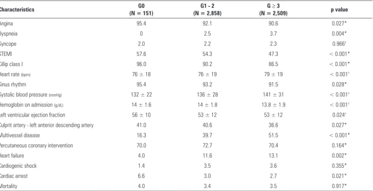
Regarding ACS clinical presentation (Table 2), G0 had a greater incidence of STEMI, but lower Killip class, heart rate and systolic blood pressure on admission.
Comparing G1 - 2 and G ≥ 3, echocardiography documented signiicantly less left ventricular systolic function impairment in G0, with a mean ejection fraction of 56 ± 10%. his fact is likely related to the lower incidence of heart failure during hospitalization in this group (Table 2).
he left anterior descending artery was the most frequently involved vessel in G0 patients, despite these individuals presenting with a lower incidence of multivessel coronary disease, compared with known risk factors patients. here was no signiicant diference in percutaneous coronary intervention between the three groups (Table 2).
During hospitalization, G0 patients presented a twofold higher incidence of cardiac arrest, when compared with the G1 - 2 and G3 groups (6.6% versus 3.0% versus 2.7%; p = 0.021). However, G0 patients did not have a signiicantly higher hospital mortality (Table 2).
A logistic regression model was built to identify the predictors of cardiac arrest, including the absence of risk factors, STEMI, systolic blood pressure, heart rate, Killip class > I, creatinine at admission, previous and in-hospital medication, culprit artery (left main and left anterior
Table 1 - Baseline clinical characteristics
Characteristics G0
(N = 151)
G1 - 2 (N = 2,858)
G ≥ 3
(N = 2,509) p value
Male 64.20 73.50 71.10 0.014*
Age (years) 49 ± 8 62 ± 15 67 ± 12 < 0.001†
Body mass index (kg/m2) 26.7 ± 3.9 26.8 ± 4.2 27.8 ± 4.3 < 0.001†
Hypertension 0 40.6 88.2 < 0.001*
Diabetes 0 6.0 44.1 < 0.001*
Dyslipidemia 0 23.7 77.9 < 0.001*
Smoker 0 34.8 34.4 < 0.001*
Family history of coronary artery disease 0 4.5 11.0 < 0.001*
Peripheral arterial disease 0.7 1.8 3.9 < 0.001*
Previous stroke 1.3 4.2 9.2 < 0.001*
Chronic kidney disease 2.7 2.5 5.4 < 0.001*
Neoplasm 3.4 4.2 4.4 0.855*
Chronic obstructive pulmonary disease 2.1 3.8 4.8 0.079*
Chronic kidney disease: creatinine > 2.0mg/dL, hemodialysis or renal transplantation. * Chi-squared test; † ANOVA. Values are expressed as (%) and mean ± standard deviation.
Table 2 - Admission characteristics and hospital outcome
Characteristics G0
(N = 151)
G1 - 2 (N = 2,858)
G ≥ 3
(N = 2,509) p value
Angina 95.4 92.1 90.6 0.027*
Dyspneia 0 2.5 3.7 0.004*
Syncope 2.0 2.2 2.3 0.966†
STEMI 57.6 54.3 47.3 < 0.001*
Killip class I 96.0 90.2 86.5 < 0.001*
Heart rate (bpm) 76 ± 18 76 ± 19 79 ± 19 < 0.001†
Sinus rhythm 95.4 93.2 91.5 0.028*
Systolic blood pressure (mmHg) 132 ± 22 136 ± 28 141 ± 31 < 0.001†
Hemoglobin on admission (g/dL) 14 ± 1.6 14 ± 1.8 13.8 ± 1.9 < 0.001†
Left ventricular ejection fraction 56 ± 10 53 ± 12 53 ± 12 0.024†
Culprit artery - left anterior descending artery 41.0 40.6 36.6 0.027*
Multivessel disease 16.3 39.7 51.5 < 0.001*
Percutaneous coronary intervention 70.0 72.7 70.4 0.164*
Heart failure 4.0 11.6 13.1 0.002*
Cardiogenic shock 1.4 3.5 3.6 0.355*
Cardiac arrest 6.6 3.0 2.7 0.021*
Mortality 4.0 3.4 3.5 0.917*
STEMI - ST segment elevation myocardial infarction; * Chi-squared test; † ANOVA. Values are expressed as (%) and mean ± standard deviation.
descending artery), percutaneous coronary intervention and left ventricular ejection fraction < 50%. his analysis identiied the absence of risk factors as an independent predictor of cardiac arrest (OR = 2.78; 95%CI 1.19 - 6.51; p = 0.019). he other independent predictors were STEMI (OR = 5.74; 95%CI 3.18 - 10.38; p < 0.001), higher heart rate (OR = 1.02; 95%CI 1.01 - 1.02; p <
Table 3 - Statistical analysis to determine the predictors of cardiac arrest
Variables Coefficient SE Multivariate analysis Univariate analysis p value* OR (95%CI) p value* OR (95%CI)
Risk factor 0† 1.022 0.434 0.019 2.78 (1.19 - 6.51) 0.007 2.57 (1.30 - 5.11)
Risk factors 1 - 2† 0.126 0.200 0.529 1.13 (0.77 - 1.68) 0.511 1.12 (0.81 - 1.54)
STEMI 1.748 0.302 < 0.001 5.74 (3.18 - 10.38) < 0.001 6.32 (4.02 - 9.94)
Heart rate 0.016 0.004 < 0.001 1.02 (1.01 - 1.02) < 0.001 1.01 (1.01 - 1.02)
SBP -0.013 0.003 < 0.001 0.99 (0.98 - 0.99) < 0.001 0.98 (0.97 - 0.98)
KK > 1 1.266 0.229 < 0.001 3.55 (2.27 - 5.56) < 0.001 4.17 (2.97 - 5.87)
Nitratesin-hospital -0.634 0.227 0.005 0.53 (0.34 - 0.83) < 0.001 0.43 (0.30 - 0.61)
SE - standard error; OR - odds ratio; 95%CI - 95% confidence intervals; STEMI - ST segment elevation myocardial infarction; SBP - systolic blood pressure; KK - Killip Kimball class. * Wald test; † comparing with 3 or more risk factors.
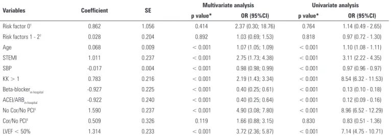
Table 4 - Statistical analysis to determine the predictors of hospital mortality
Variables Coefficient SE Multivariate analysis Univariate analysis p value* OR (95%CI) p value* OR (95%CI)
Risk factor 0† 0.862 1.056 0.414 2.37 (0.30; 18.76) 0.764 1.14 (0.49 - 2.65)
Risk factors 1 - 2† 0.028 0.204 0.892 1.03 (0.69; 1.53) 0.818 0.97 (0.72 - 1.30)
Age 0.068 0.009 < 0.001 1.07 (1.05; 1.09) < 0.001 1.10 (1.08 - 1.11)
STEMI 1.011 0.237 < 0.001 2.75 (1.73; 4.38) < 0.001 3.11 (2.22 - 4.35)
SBP -0.017 0.004 < 0.001 0.98 (0.98; 0.99) < 0.001 0.97 (0.96 - 0.97)
KK > 1 0.783 0.216 < 0.001 2.19 (1.43; 3.34) < 0.001 8.54 (6.32 - 11.53)
Beta-blockerin-hospital -0.927 0.225 < 0.001 0.40 (0.25; 0.61) < 0.001 0.13 (0.10 - 0.18)
ACEI/ARBin-hospital -0.922 0.240 < 0.001 0.40 (0.25; 0.64) < 0.001 0.12 (0.09 - 0.16)
No Cor/No PCI‡ 1.590 0.237 < 0.001 4.90 (3.08; 7.80) < 0.001 8.96 (6.52 - 12.29)
Cor/No PCI‡ 0.509 0.326 0.119 1.66 (0.88; 3.15) 0.830 0.83 (0.51 - 1.36)
LVEF < 50% 1.314 0.233 < 0.001 3.72 (2.36; 5.87) < 0.001 7.14 (4.75 - 10.71)
SE - standard error. OR - odds ratio; 95%CI - 95% confidence intervals; STEMI - ST Segment elevation myocardial infarction; SBP - systolic blood pressure; KK - Killip-Kimball class; BB - beta-blocker; ACEI/ARB - angiotensin converting enzyme inhibitors/angiotensin II receptor blockers; Cor - coronary angiography; PCI - percutaneous coronary intervention, LVEF - left ventricular ejection fraction. * Wald test; † comparing with 3 or more risk factors; ‡ comparing to coronary angiography/percutaneous coronary intervention.
Hospital all-cause mortality was slightly higher in G0, although this diference was not signiicant (Table 2). By logistic regression, we conclude that the absence of risk factors was not an independent predictor of hospital mortality (OR = 2.37; 95%CI 0.30 - 18.76; p = 0.414). Independent predictors included STEMI (OR = 2.75; 95%CI 1.73 - 4.38; p < 0.001), Killip class > I (OR = 2.19; 95%CI 1.43 - 3.34; p < 0.001), no percutaneous coronary intervention (OR = 4.90; 95%CI 3.08 - 7.80; p < 0.001) and left ventricular ejection fraction < 50% (OR = 3.72; 95%CI 2.36 - 5.87; p < 0.001). he model was well calibrated (HL: p = 0.147), and had excellent discriminant accuracy AUC = 0.92; 95%CI 0.89 - 0.94) (Table 4).
At the one-year follow up, there was no signiicant diference in survival between the three groups (Figure 2). By Cox regression analysis, the number of risk factor was not found to be associated with mortality (HR = 0.78;
95%CI 0.45 - 1.37; p = 0.393). he predictors of death at the one-year follow up were as follows: age (HR = 1.05; 95%CI 1.03 - 1.06; p < 0.001), STEMI (HR = 1.94; 95%CI 1.25 - 3.02; p = 0.003) and ejection fraction < 50% (HR = 2.34; 95%CI 1.57 - 3.47; p < 0.001) (Table 5).
DISCUSSION
In the ProACS registry, patients with no known risk factors previous to the index event represent less than 3% of the overall ACS population without previous coronary artery disease. his proportion is in line with previous published data, which also showed that about 2% of patients admitted with a irst episode of ACS had no risk factor.(6) Surprisingly, in this study, the absence of risk
Table 5 - Statistical analysis to determine predictors of death at the one-year follow up
Variables Coefficient SE Multivariate analysis Univariate analysis p value* OR (95%CI) p valor* OR (95%CI)
Risk factor 0 - 1† 0.244 0.286 0.393 0.78 (0.45; 1.37) 0.173 0.81 (0.59 - 1.10)
Age 0.046 0.009 < 0.001 1.05 (1.03; 1.06) < 0.001 1.09 (1.08 - 1.10)
STEMI 0.664 0.225 0.003 1.94 (1.25; 3.02) < 0.001 2.33 (1.80 - 3.01)
ACEI/ARBdischarge -0.598 0.227 0.008 0.55 (0.35; 0.86) < 0.001 0.18 (0.13 - 0.25)
BBdischarge -0.851 0.221 < 0.001 0.43 (0.28; 0.66) < 0.001 0.16 (0.11 - 0.22)
ASAdischarge -1.460 0.229 < 0.001 0.23 (0.15; 0.36) < 0.001 0.08 (0.06 - 0.11)
No Cor/No PCI‡ 0.784 0.248 0.002 2.19 (1.35; 3.56) < 0.001 6.95 (5.41 - 8.92)
Cor/No PCI‡ -0.251 0.323 0.439 0.78 (0.41; 1.47) 0.838 0.96 (0.67 - 1.38)
LVEF<50% 0.848 0.202 < 0.001 2.34 (1.57; 3.47) < 0.001 4.55 (3.44 - 6.02)
SE - standard error; OR - odds ratio; 95%CI - 95% confidence intervals; RF- risk factors; STEMI - ST Segment elevation myocardial infarction; ACEI/ARB - angiotensin converting enzyme inhibitors/Angiotensin II receptor blockers; BB - beta-blocker; ASA - acetylsalicylic acid; Cor - coronary angiography; PCI - percutaneous coronary intervention, LVEF - left ventricular ejection fraction. * Wald test; † comparing with 3 or more risk factors; ‡ comparing to coronary angiography/percutaneous coronary intervention.
Figure 2 - Kaplan Meier survival curves for the three study groups.
In our population, patients without known risk factors were younger, had less comorbidities and better left ventricular systolic function. Even though this group of patients had less multivessel disease, they presented more often with STEMI and more frequently had the left anterior descending artery as the culprit. his fact has been described previously in other national registries, in
which younger patients had higher STEMI incidence.(7,8)
Our indings are in accordance with previous studies that showed a higher incidence of single-vessel disease in these patients.(9-11)
In our registry, the absence of risk factors was an independent predictor of cardiac arrest on presentation and hospitalization. However, hospital mortality was not signiicantly higher in G0 patients. Previous studies showed an inverse relationship between number of risk
factors and hospital mortality. However, in a study by Canto et al., patients without risk factors were older, had more cardiogenic shock and higher Killip class, which is a diferent population from that in our registry.(12) Also, in
a CRUSADE sub-study, an inverse association between number of risk factors and mortality was reported in the non-ST-segment elevation myocardial infarction population.(13)
We can postulate that patients with more risk factors and higher frequency of multivessel disease have more collateral blood low, and this fact can limit infarct size and consequently, reduce hospital mortality and cardiac arrest. On the other hand, in the absence of risk factors, an ACS is less likely, and a lower suspicion can delay the diagnosis and efective intervention, increasing the risk of ventricular arrhythmia and mortality.
In contrast to hospital outcome, the one-year survival was higher in patients without risk factors. his fact likely relects the younger age, better left ventricular function and fewer comorbidities of these patients.
Some of the patients without known risk factors might have another less conventional RF that was not assessed, since other risk factors is not systematically collected in the ProACS registry.
also have atypical etiology, with hereditary thrombophilia and hyperhomocysteinemia being the most frequent etiologies described in previous studies.(9,16)
Little is known about the physiopathology of ACS in patients without traditional risk factors, and more studies are needed to understand these events and their correlation with poor hospital outcome.
Our study, based on a national registry with a large number of patients and recent data, accurately relects clinical practice. Since the data was drawn from a registry, this research study does not have selection bias, and the study population dimension allowed the determination of outcome predictors.
Study limitations
A registry has the advantage of representing real life clinical practice, and the indings of the study are probably applicable to a large number of tertiary hospitals. However, only traditional risk factors were reported, and as we do not have information regarding other types of risk factors, we cannot conclude which atypical factors might be associated with the worsened outcome observed. Additionally, the diagnoses were performed by diferent physicians in each department, which could generate some bias. Furthermore, a minority of patients without
Objetivo: Comparar pacientes admitidos com síndrome
coronariana aguda sem prévia identiicação de fatores de risco cardiovascular com pacientes que portavam um ou mais fatores de risco.
Métodos: Análise retrospectiva dos pacientes admitidos com o primeiro episódio de síndrome coronariana aguda sem cardio-patia prévia, incluídos em um registro nacional de síndrome co-ronariana aguda. Os pacientes foram divididos segundo o núme-ro de fatores de risco: nenhum fator de risco (G0), um ou dois fatores de risco (G1 - 2) e três ou mais fatores de risco (G ≥ 3). Realizou-se uma análise comparativa entre os três grupos e se es-tudaram os preditores independentes de parada cardíaca e óbito.
Resultados: O total apurado foi de 5.518 pacientes, 72,2% deles do sexo masculino, com média de idade de 64 ± 14 anos. O G0 teve uma incidência maior de infarto do miocárdio com elevação do segmento ST, sendo o vaso mais frequentemente envolvido a artéria descendente anterior esquerda, e menor prevalência de envolvimento de múltiplos vasos. Embora o G0
tivesse uma classe Killip mais baixa (96% Killip I; p < 0,001) e maior fração de ejeção (G0: 56 ± 10% versus G1 - 2 e G ≥ 3: 53 ± 12%; p = 0,024) na admissão, houve incidência signiicante-mente maior de parada cardíaca. A análise multivariada identi-icou ausência de fatores de risco como um fator independente para parada cardíaca (OR 2,78; p = 0,019). A mortalidade hos-pitalar foi ligeiramente maior no G0, embora sem signiicância estatística. Segundo a análise de regressão de Cox, o número de fatores de risco não se associou com mortalidade. Os preditores de óbito em 1 ano de seguimento foram infarto do miocárdio com elevação do segmento ST (OR 1,05; p < 0,001) e fração de ejeção inferior a 50% (OR 2,34; p < 0,001).
Conclusão: Embora o grupo sem fatores de risco fosse com-posto de pacientes mais jovens e com menos comorbidades, me-lhor função ventricular esquerda e coronariopatia menos exten-sa, a ausência de fatores de risco foi um preditor independente de parada cardíaca.
RESUMO
Descritores: Parada cardíaca; Fatores de risco; Síndrome coronariana aguda
known risk factors presented evidence of diabetes and dyslipidemia in blood samples collected during the acute event.
Finally, as the registry does not collect detailed information on the cause of death and thus, only the all-cause mortality data was presented.
CONCLUSION
REFERENCES
1. World Health Organization. Cardiovascular diseases [Internet]. [cited 2016 Oct 12]. Available from: http://www.who.int/mediacentre/factsheets/ fs317/en/
2. Perk J, De Backer G, Gohlke H, Graham I, Reiner Z, Verschuren WM, Albus C, Benlian P, Boysen G, Cifkova R, Deaton C, Ebrahim S, Fisher M, Germano G, Hobbs R, Hoes A, Karadeniz S, Mezzani A, Prescott E, Ryden L, Scherer M, Syvänne M, Scholte Op Reimer WJ, Vrints C, Wood D, Zamorano JL, Zannad F; Fifth Joint Task Force of the European Society of Cardiology and Other Societies on Cardiovascular Disease Prevention in Clinical Practice; European Association for Cardiovascular Prevention and Rehabilitation. European Guidelines on cardiovascular disease prevention in clinical practice (version 2012): The Fifth Joint Task Force of the European Society of Cardiology and Other Societies on Cardiovascular Disease Prevention in Clinical Practice (constituted by representatives of nine societies and by invited experts). Atherosclerosis. 2012;223(1):1-68.
3. Goff DC Jr, Lloyd-Jones DM, Bennett G, Coady S, D’Agostino RB, Gibbons R, Greenland P, Lackland DT, Levy D, O’Donnell CJ, Robinson JG, Schwartz JS, Shero ST, Smith SC Jr, Sorlie P, Stone NJ, Wilson PW, Jordan HS, Nevo L, Wnek J, Anderson JL, Halperin JL, Albert NM, Bozkurt B, Brindis RG, Curtis LH, DeMets D, Hochman JS, Kovacs RJ, Ohman EM, Pressler SJ, Sellke FW, Shen WK, Smith SC Jr, Tomaselli GF; American College of Cardiology/American Heart Association Task Force on Practice Guidelines. 2013 ACC/AHA guideline on the assessment of cardiovascular risk: a report of the American College of Cardiology/ American Heart Association Task Force on Practice Guidelines. Circulation. 2014;129(25 Suppl 2):S49-73.
4. Conroy RM, Pyörälä K, Fitzgerald AP, Sans S, Menotti A, De Backer G, De Bacquer D, Ducimetière P, Jousilahti P, Keil U, Njølstad I, Oganov RG, Thomsen T, Tunstall-Pedoe H, Tverdal A, Wedel H, Whincup P, Wilhelmsen L, Graham IM; SCORE project group. Estimation of ten-year risk of fatal cardiovascular disease in Europe: the SCORE project. Eur Heart J. 2003;24(11):987-1003.
5. Schünemann HJ, Oxman AD, Brozek J, Glasziou P, Jaeschke R, Vist GE, Williams JW Jr, Kunz R, Craig J, Montori VM, Bossuyt P, Guyatt GH; GRADE Working Group. Grading quality of evidence and strength of recommendations for diagnostic tests and strategies. BMJ. 2008;336(7653):1106-10. Erratum in BMJ. 2008;336(7654). Schünemann, A Holger J [corrected to Schünemann, Holger J].
6. Saab F, Mukherjee D, Gurm H, Motivala A, Montgomery D, Kline-Rogers E, et al. Risk factors in first presentation acute coronary syndromes (ACS): how do we move from population to individualized risk prediction? Angiology. 2009;60(6):663-7.
7. Chen TS, Incani A, Butler TC, Poon K, Fu J, Savage M, et al. The demographic profile of young patients (<45 years-old) with acute coronary syndromes in Queensland. Heart Lung Circ. 2014;23(1):49-55.
8. Prajapati J, Jain S, Virpariya K, Rawal J, Joshi H, Sharma K, et al. Novel atherosclerotic risk factors and angiographic profile of young Gujarati patients with acute coronary syndrome. J Assoc Physicians India. 2014;62(7):584-8.
9. Zimmerman FH, Cameron A, Fisher LD, Ng G. Myocardial infarction in young adults: angiographic characterization, risk factors and prognosis (Coronary Artery Surgery Study Registry). J Am Coll Cardiol. 1995;26(3):654-61. 10. Avezum A, Makdisse M, Spencer F, Gore JM, Fox KA, Montalescot G,
Eagle KA, White K, Mehta RH, Knobel E, Collet JP; GRACE Investigators. Impact of age on management and outcome of acute coronary syndrome: observations from the Global Registry of Acute Coronary Events (GRACE). Am Heart J. 2005;149(1):67-73.
11. Wolfe MW, Vacek JL. Myocardial infarction in the young. Angiographic features and risk factor analysis of patients with myocardial infarction at or before the age of 35 years. Chest. 1988;94(5):926-30.
12. Canto JG, Kiefe CI, Rogers WJ, Peterson ED, Frederick PD, French WJ, Gibson CM, Pollack CV Jr, Ornato JP, Zalenski RJ, Penney J, Tiefenbrunn AJ, Greenland P; NRMI Investigators. Number of coronary heart disease risk factors and mortality in patients with first myocardial infarction. JAMA. 2011;306(19):2120-7.
13. Roe MT, Halabi AR, Mehta RH, Chen AY, Newby LK, Harrington RA, et al. Documented traditional cardiovascular risk factors and mortality in non-ST-segment elevation myocardial infarction. Am Heart J. 2007;153(4):507-14. 14. Misteli GS, Stute P. Depression as a risk factor for acute coronary
syndrome: a review. Arch Gynecol Obstet. 2015;291(6):1213-20. 15. Choi J, Daskalopoulou SS, Thanassoulis G, Karp I, Pelletier R, Behlouli
H, Pilote L; GENESIS-PRAXY Investigators. Sex- and gender-related risk factor burden in patients with premature acute coronary syndrome. Can J Cardiol. 2014;30(1):109-17.