SUMMARY
Objective: To draw attention to complications that might arise in any Kawasaki disease (KD) stage, risk factors contributing to the onset of complications and possible transient or permanent disease sequelae. Methods: Prospective study (clinical cohort) conducted between April 2002 and April 2009 of 115 patients with KD admitted to the Pediatric Rheumatology Clinic of the General Hospital of the Federal District, Brazil. All patients were sequentially assessed with clinical and laboratory examinations, Doppler echocar-diography, imitanciometry, auditory evoked potentials, psychological evaluation, oph-thalmologic examination and, in one patient with chorea, cerebral magnetic resonance angiography. In all patients, a questionnaire assessing the possible presence of cognitive, emotional, behavioral and social disorders was applied. Results: Twenty-ive patients (21.7%) had coronary aneurisms. hirty-eight patients (33%) had a sensorineural audi-tory loss during the acute and subacute phases of the disease and 13 patients (11.3%) maintained the auditory loss six months ater the irst assessment. Other complications observed were as follows: facial palsy in one patient (0.9%), ataxia in acute and subacute phases in 11 (9.5%); 15 patients had ophthalmologic complications (13.2%), with uveitis in 13, papilledema in one patient, and conjunctival hemorrhage in another patient. One patient experienced chorea (0.9%), with a magnetic resonance angiography showing changes consistent with cerebral ischemia. In one patient, a thoracic aorta aneurism was found (0.9%) and another patient had a necrotizing vasculitis progressing to peripheral gangrene and tongue tip loss (0.9%). Behavioral changes over convalescence were ob-served in 23 children. Conclusion: KD may progress with several complications even within months of the disease acute phase, eventually resulting in permanent sequelae. he earlier the diagnosis and therapeutic intervention with IV IgG administration are, the lower will be the occurrence of complications; the presence of thrombocytosis, ane-mia and elevated and extended inlammatory activity are risk factors for complication arising.
Keywords: Mucocutaneous lymph node syndrome; coronary aneurism; aortic aneu-rism; sensorineural auditory loss; clinical course.
Prospective study of Kawasaki disease complications: review of 115
cases
NATÁLIA RIBEIRO DE M. ALVES1, CRISTINA MEDEIROS R. DE MAGALHÃES2, ROSEANE DE FÁTIMA R. ALMEIDA3, REGINA CÂNDIDO R. DOS SANTOS4,
LENORA GANDOLFI5, RICCARDO PRATESI6
1 Medical Trainee, Pediatric Rheumatology Unit, Hospital de Base de Brasília, Brasília, DF, Brazil
2 Ph.D. in Health Sciences; Attending Pediatric Rheumatologist Physician, Pediatric Rheumatology Unit, Hospital de Base de Brasília; Professor, Medical School, Escola Superior de
Ciências da Saúde, Brasília, DF, Brazil
3 M.D., Specialized in Echocardiography; Attending Cardiologist, Cardiology Service, Hospital de Base de Brasília, Brasília, DF, Brazil
4 Ph.D. in Ophthalmology; Attending Ophthalmologist, Ophthalmology Service, Hospital de Base de Brasília; Professor, Medical School, Escola Superior de Ciências da Saúde, Brasília,
DF, Brazil
5 Ph.D. in Gastroenterology; Post-doctorate in Autoimmune Diseases; Attending Physician and Professor, Medical School, Post-Graduation Programs in Medical Sciences and Sciences
of Health, Universidade de Brasília, Brasília, DF, Brazil
6 Ph.D. in Immunology and Applied Genetics; Post-doctorate in Autoimmune Diseases; Professor Emeritus, Medical School, Universidade de Brasília, Brasília, DF, Brazil
Study conducted at Pediatrics Service, Pediatric Rheumatology Unit, Hospital de Base de Brasília, Secretary of Health of the Federal District, Brasília, DF, Brazil
Submitted on: 08/30/2010
Approved on: 03/09/2011
Correspondence to:
Riccardo Patresi SQN 212 – Bloco F – Apto. 605 Asa Norte CEP: 70864-060 Brasília – DF Phone: +55 61 3307-2134 (UnB);
+55 61 3340-9192 (home); +55 61 9298-6028 [email protected] or [email protected]
INTRODUCTION
Kawasaki disease (KD), also known as mucocutaneous lymph node syndrome, is a primary vasculitis oten seen in childhood, being mediated by IgA1, afecting more fre-quently small- and medium-size vessels and it can lead to a ibrinoid necrosis in vessel walls. he disease compromis-es vcompromis-essels from the intima layer to the perivascular area, forming aneurisms in several diferent stages in a child-hood polyarteritis. his is a systemic vasculitis, but prefer-entially found in coronary arteries2.
KD diagnosis is purely clinical, based on the presence of fever persisting for at least ive days and further four out of ive diagnostic criteria: limb changes, such as hand and/ or foot erythema or swelling during the acute stage and ingertip scaling over convalescence; polymorphous exan-thema; bilateral conjunctivitis with no purulent discharge, lip erythema and cracking, raspberry tongue, throat red-ness and anterior cervical adenopathy with a 1.5 cm or larger size. As there are no speciic laboratory markers, a high level of suspicion is required, usually resulting in a rise in the number of cases diagnosed3.
he most severe complication of the disease is coronary vasculitis, leading to coronary artery changes and afect-ing 15%-20% of patients if let untreated4. hese changes include aneurisms, coronary artery ectasias and stenoses, responsible for 2% of mortality. he intravenous immuno-globulin G (IVIG) administration over the 10 irst days of the disease leads to a reduced coronary artery impairment of 3%-8% and mortality to 0.2%2,3.
Other KD complications have been described, such as aneurisms of other arteries: aortic aneurisms, with a higher number of reported cases involving the abdominal aorta5-7; axillary artery aneurism8; brachiocephalic artery aneurism9; aneurisms of iliac and femoral arteries, and re-nal artery aneurism10,11.
Gastrointestinal complications in KD are described in the literature as case reports similar to those observed in Henoch-Schönlein purpura, such as: intestinal obstruc-tion12, colon swelling13, intestinal ischemia14, intestinal pseudo-obstruction15 and acute abdomen16.
Ophthalmologic changes associated with KD have been described since the 1980’s, being found as uveitis, iridocyclitis, conjunctival hemorrhage17-19, optic neuritis20, amaurosis and ocular artery obstruction21.
KD can also be found as necrotizing vasculitis, pro-gressing into peripheral gangrene, according to a number of case reports22-24 described in the literature.
he neurological complications per central ner-vous system (CNS) lesion are increasingly found in lit-erature. he neurological complications found are me-ningoencephalitis25,26, subdural efusion27,28, cerebral hypoperfusion29, cerebral ischemia and infarct30, cerebelar infarction31, manifesting with seizures, chorea, hemiplegia, mental confusion, lethargy and coma8 or even a cerebral infarction with no neurological manifestations30,31.
Other neurological complications from cranial nerve involvement are reported as ataxia3, facial palsy32-34 and sensorineural auditory loss35-37.
he irst description for hearing loss in KD was made by Suzuki et al.35 in Japan in 1988. In 1992, Sundel et al.36 assessed 40 patients by standard audiometry and BERA (Brainstem Evoked Response Audiometry) in patients who could not undergo an audiometry. Seven patients (17.5%) had a sensorineural auditory loss and 14 patients had an inconclusive result. In 2001, Knott et al.37 reported another study where they performed an audiological as-sessment in 62 patients over the irst thirty days of KD; from these, 13 had a sensorineural auditory loss and two patients kept the sensorineural auditory deicit in a second audiological assessment 10 days later.
Behavioral changes, such as attention deicits, learn-ing deicits, emotional disorders (emotional lability, fear of night and night terrors) and internalization problems (anxious, depressive or aggressive behavior) have been described in studies in the literature38,39 conducted on the basis of the questionnaires Child Behavior Check List (CBCL4-18/1991), Strengths and Diiculties Question-naire (1997) and Parenting Stress Index (PSI/1983).
he purpose of the current study was to obtain data regarding KD complications and identify the incidence of theses complications in a sample of 115 cases followed in a clinic specialized in pediatric rheumatology in Brasília, Brazil. he objective of the study was to draw attention to the diagnosis of KD complications that are likely to oc-cur even ater the acute and subacute stages of the disease, possible transient or permanent sequelae and risk factors for these complications.
METHODS
A clinical cohort of patients with Kawasaki disease was used and followed in the Pediatric Rheumatology Clinic of Hospital de Base do Distrito Federal (HBDF). he study evaluated 115 patients having KD diagnosed in accor-dance with the American Heart Association (AHA)3 crite-ria. he patients were seen at the Pediatric Rheumatology Clinic of the HBDF from April 2002 to April 2009 (inclu-sion criteria). he incomplete KD forms and the patients having conditions that could lead to the same complications assessed were excluded. All patients provided an informed consent to participate in the study, which was approved by the Research Ethics Committee of Brasilia Health Secretary.
Clinical and laboratory examinations, as well as the echocardiograms, were performed at the admission, one month later and thereater three-month apart in the irst year ater the DK onset and six-month apart later, being performed by the same cardiologist and using the same echo-Doppler apparatus (ALOKA SSD 2200, with 2.5 and 5.0 MHz transducer made in Japan).
he ophthalmologic examination was performed dur-ing KD acute and subacute stages ater the irst examina-tion in all patients.
A questionnaire based on questionnaires Child Be-havior Check List (CBCL4-18/1991); Strengths and Dif-iculties Questionnaire (1997); Parenting Stress Index (PSI/1983), assessing cognitive, emotional, behavioral or social diiculties was applied to all patients 30 days ater the acute phase. he patients having behavioral, emotional or cognitive disorders were given periodical psychological follow-up.
he imitanciometry was initially performed in all pa-tients to exclude those with an hearing loss from conduc-tive changes. he AZ7 INTERACOUSTICS (Denmark, 2000) imitanciometer was used in auditory assessment. he auditory assessment was made through Brainstem Evoked Potentials (BERA) in all patients over the irst 30 days of disease and six months later in patients having a hearing loss initially. he equipment used for BERA was MEDELEC SAPHIRE IV PREMIERE PLUS (England, 1996) with silver surface electrodes in positions based on the international system 10-20-Fz, A1, A2.
STATISTICALANALYSIS
he Pearson’s chi-squared test was used to ind the associa-tion between KD complicaassocia-tion occurrence and the follow-ing variables: thrombocytosis (platelet level over 500,000); anemia with hemoglobin under 10 g/dL; ESR over 50 mm for more than 30 days and IVIG treatment in the acute phase and ater the acute phase. Diferences with a p < 0.05 were considered statistically signiicant. he sotware SAS 9.2 for Windows was employed for data analysis.
RESULTS
Out of 115 patients, 77 (67%) were males, and the patients’ ages ranged from two months to 11 years, with a mean of 3.2 years. Eighty-ive were Caucasian, 24 had a dark com-plexion, 1 was Afrodescendant and 5 were Japanese de-scendants. Seventy-four per cent of patients were younger than ive years old.
All patients received intravenous immunoglobulin (IVIG) 2 g/kg/dose although only 58 (%) were given IVIG within the irst 10 days of disease.
Twenty-ive patients (21.7%) had coronary aneurisms, with four patients having aneurisms in both coronary ar-teries, one only on the right artery and 20 only on the let; two of them were middle-sized and the others were small. Out of the 25 patients having a coronary aneurism, four
patients were under six months old, and two were older than 5 years; 10 patients used gamma globulin in acute phase and 15 patients in the subacute phase.
One patient (0.9%) had a thoracic aorta aneurism diag-nosed in the subacute phase through the histopathological study of the aneurism and the clinical history data.
hirty-eight patients (33%) had a sensorineural au-ditory loss during the acute and subacute phases of the disease and 13 (11.3%) were keeping the hearing loss six months ater the irst assessment. Out of these 38 patients, 20 used gamma globulin in the subacute phase.
One patient had a facial palsy in the subacute phase and 11 patients (9.5%) had ataxia, with 30% in the acute phase and the others in the subacute phase. he presence of ataxia was observed in patients with high and extended inlammatory activity.
Fiteen patients (13.2%) had ophthalmologic compli-cations: 13 had an anterior uveitis, one had a papilledema and another patient had a conjunctival hemorrhage, 40% in the acute phase, the others in the subacute phase, and these complications were not maintained in the second ophthalmologic assessment 30 days later.
A patient had a course with chorea six months ater the acute phase and it was controlled by using valproate; the brain magnetic resonance angiography showed lesions consistent with brain ischemia near the caudate nucleus. he changes were still present in a magnetic resonance an-giography performed one year ater the irst study.
One patient with a high and extended inlammatory activity associated with anemia and thrombocytosis had a necrotizing vasculitis with peripheral gangrene and conse-quent tongue tip loss.
Twenty-three (20%) patients had behavioral changes over the convalescence: all of them had irritability, 13 had a course with aggressiveness, nine with an attention dei-cit, seven with a learning deicit and two with an antisocial behavior. Six patients remained with an aggressive behav-ior, three with a learning deicit and ive with an attention deicit six months ater the acute phase.
Among the patients with a coronary aneurism, 4 kept the small let coronary artery aneurism for 9 months, two years, two and a half years and one patient has been keep-ing a medium right coronary artery aneurism six years af-ter the KD acute phase.
Five patients (4.3%) had an acute phase recurrence. hree of them used IVIG and two of them had a coronary aneurism. he other two used gamma globulin in the sub-acute phase and one of them had a course with a coronary aneurism.
40 38 36 34 32 30 28 26 24 22 20 18 16 14 12 10 8 6 4 2 0
Senso- rineural hearing loss
Coronary
aneurism Facialpalsy Ataxia ischemiaBrain Behaviorchange Peripheralgangrene Ophthal- mologic aneurismAortic changes
Figure 1 – Incidence of KD complications in a sample of 115 patients referred to the Pediatric Rheumatology Clinic of HBDF from January 2002 to January 2011.
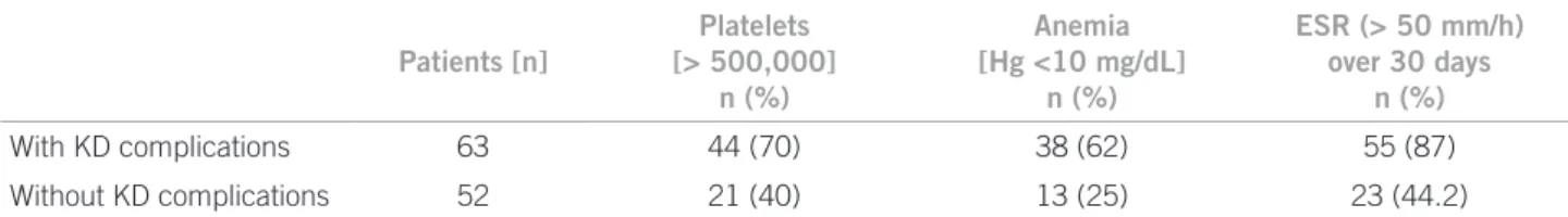
Table 1 – Relationship between KD complications and the presence of thrombocytosis (platelet count > 500,000), anemia with hemoglobin (Hb) < 10 mg/dL and ESR > 50 mm/h for over 30 days
Patients [n]
Platelets [> 500,000]
n (%)
Anemia [Hg <10 mg/dL]
n (%)
ESR (> 50 mm/h) over 30 days
n (%)
With KD complications 63 44 (70) 38 (62) 55 (87)
Without KD complications 52 21 (40) 13 (25) 23 (44.2)
Table 2 – Relationship between KD complications and IV immunoglobulin use over the acute phase and after the acute phase
Patients (n) IVIG over the acute phase, n (%) IVIG after the acute phase, n (%)
With KD complications 63 25 (40) 38 (60)
Without KD complications 52 33 (63.4) 19 (36.6)
he persistent complications were: coronary aneurism in four patients, sensorineural hearing loss in 13, behav-ioral changes in six patients.
he complications were more frequent in patients hav-ing more anemia, more thrombocytosis and higher and longer inlammatory activity (Table 1). he incidence of complications in KD was higher in patients using IVIG ater the ten days of disease progress (Table 2). By statisti-cal analysis, a signiicant association between frequency of complications and the following variables could be detect-ed: anemia with Hb < 10 g/dL (p = 0.0001); thrombocyto-sis (p = 0.0015); ESR > 50 mm/h for longer than 30 days (p = 0.0001); and IVIG treatment ater the acute phase (10 irst days of disease) (p = 0.0111).
DISCUSSION
Takahashi et al.40 observed in post-mortem studies of chil-dren having KD with no echocardiography abnormali-ties and dying years later from other causes that coronary arteries were markedly abnormal, with impressive pro-atherosclerotic changes persisting even in the absence of acute lesions in this artery. his cardiovascular damage unrecognized or unidentiied in the acute phase will mani-fest later as a cardiovascular disease in the adult, explain-ing many myocardial infarction in adolescents and young adults with no apparent cause41.
Coronary aneurisms were classiied by a new guide-line by AHA3 as small aneurisms (inner diameter from 3 to 5 mm), medium aneurisms (5 to 8 mm) and giant (> 8 mm). Other changes that can be found in KD even in the absence of aneurisms are vessel ectasias or stenoses. Giant coronary aneurisms have a higher risk for thrombo-sis, calciication and/or stenosis and consequently myocar-dial infarction. Small- and medium-size aneurisms usually regress spontaneously and rarely generate other complica-tions3.
Aortic aneurisms should also be assessed for and the aortic diameter should be measured with imaging studies because there are evidence the aorta dilation is common in patients with KD and these dilations do not regress over the irst year of the disease5.
he incidence of CNS involvement in KD ranges from 1.1% to 3.7%25,41, being found as seizures, ataxia, coma, lethargy, subdural efusion, hemiplegia, facial palsy and sensorineural hearing loss.
he facial palsy is oten unilateral and more frequent on the let, being transient and seemingly associated with coronary impairment. It resolves spontaneously and com-pletely, leaving no sequelae. Poon et al.32 published a study in 2000 of 28 patients with facial palsy as a KD complica-tion in patients with 3 to 25 months of age, preferentially in females with a 1.4:1 ratio, being transient over a range from two days to three months, more frequent on the let; 54% of the patients also had a coronary aneurism, but only one patient had used IV gamma globulin. In the current study, the only patient showing a facial palsy was diag-nosed on the 26th day of disease, experiencing extended fe-ver, high and long-lasting inlammatory activity. he facial palsy had an onset in the subacute phase on the let and improved within 30 days with physical therapy and the pa-tient also had ataxia, hearing loss and a small let coronary aneurism. It is important to have a raised suspicion level for KD in children with extended fever and facial palsy.
inner ear abnormalities because of an intense cochlea or wall vessel (vasa nervorum and perineural vessels)
inlam-mation, leading to a vasculitis neuropathy43,44.
he number of cases published on sensorineural hear-ing deicit in KD seems underestimated. Reduced hearhear-ing acuity may be slight and transient, as the condition is seen in small children and it could not be perceived by parents. he hearing loss is oten detected only by audiometry or, in small children in whom tone audiometry is diicult to be performed, by brainstem auditory evoked potential as-sessment (BERA)42. Unlike studies previously conducted, our study found a high percentage of patients with senso-rineural auditory loss both in the irst 30 days of disease (33%) and in an assessment six months later (11.3%). his diference was likely to occur because prior studies did not have BERA performed in all patients and when this oc-curred, the second assessment was done within a variable length of time36,37.
We agree with Knott et al.37 that sensorineural hearing loss is a frequent complication in KD, but we disagree with these authors that persistent sensorineural hearing loss is rare, since we found 11.3% of our patients sustaining a hearing loss in an assessment six months later. Similarly, we agree with the study by Sundel et al.36 that a high and extended inlammatory activity is a risk factor for hearing loss in KD, but we observed anemia and thrombocytosis are also predisposing factors to a hearing loss in KD42.
Ataxia occurs in the acute and subacute phases and is usually transient3. In our study, 30% of patients had ataxia in the acute phase and the others, in the subacute phase, with its disappearing over convalescence.
CNS lesions may occur and despite their severity, as shown in a number of case reports in literature29, they are mostly self-limited indings, whose clearing could take months, but could also leave sequelae, such as brain atro-phy, ischemic lesions30, including cognitive and behavioral conditions39. Meningoencephalitides, subdural efusions, hypoperfused brain, ischemia, cerebral and cerebelar in-farction are CNS changes also being frequently detected from the better KD recognition and from a more extended follow-up in these patients26-31. CNS lesions should always be considered in KD, mainly in severe cases with high and extended inlammatory activity and the presence of a coronary impairment with or without neurological mani-festations31.
Post-mortem histopathological studies in cases of KD are scarce. Amano et al.43 found ganglionitis and cranial and peripheral nerve neuritis, endoarteritis, periarteritis, cho-riomeningitis and leptomeningitis, in addition to atrophy, degenerative changes with neuron loss, marginal and sub-ependimal gliosis and glial nodes around degenerated neu-rons in post-mortem study of 30 children afected by KD.
Ophthalmologic complications, according to case re-ports found in literature, occur in the acute and subacute
phases, are usually transient and clear within months of the acute phase18. Burns et al.45 assessed 41 patients in KD acute phase and observed 27 patients with anterior uve-itis (25 bilaterally), 5 patients with punctate keratuve-itis and 3 with keratitis and uveitis. In the current study, 15 patients had ophthalmologic complications, such as anterior uve-itis, papilledema and conjunctival hemorrhage, showing transiently in the acute and subacute phases.
Histologically, KD can be a vasculitis in the polyar-teritis group with a whole-vessel wall involvement and as it progresses with vessel ibrinoid necrosis, because of the high inlammatory involvement of the vessel, it can reach the point of a necrotizing vasculitis up to a periph-eral gangrene, as seen in case reports in literature22-24. he patient having necrotizing vasculitis progressed to peripheral gangrene with a tongue tip loss. his patient was diagnosed at the subacute phase as KD and showed high and extended inlammatory activity associated with a major thrombocytosis.
KD is associated with signiicant behavioral sequelae, such as concentration diiculty, emotional lability, hyper-activity, aggressiveness, diiculty in social relationship and others. hese complications should be considered during the follow-up of KD patients and they should be referred to psychological evaluation. Studies based on Child Be-havior Check List (CBCL4-18/1991), Strengths and Dif-iculties Questionnaire (1997), Parenting Stress Index (PSI/1983), such as the study by Conway et al.38 in 2005, where anxiety, depression, conditions associated with the conduct (aggressiveness, hyperactivity, disobedience and argumentative behavior) were found in 40% of patients and few social interrelationship or school achievement changes were found. King et al.39 2000 study based on the same questionnaires, 34% of patients were found with a behavior change, such as attention deicit, learning deicit, emotional efects (fear of night and night terrors). Inter-nalization problems predominated (anxious and depres-sive behavior). In the current study, 20% of patients had behavior changes predominating in agreement with Con-way et al.’s38 indings of conduct changes (aggressiveness, hyperactivity, argumentative behavior) and with King
et al.39 indings, with attention deicit, learning deicit and
emotional efects (emotional lability) predominating for a period of six months ater KD acute phase. he most fre-quent change, occurring usually in the acute phase, is ir-ritability.
caused by brain lesions, sensorineural hearing loss and behavior changes, possibly leading to residual disabilities with learning and interpersonal relationship damage.
From the data analysis, we can conclude the earlier the diagnosis and therapeutic intervention with IVIG are, the lower the occurrence of complications will be; the pres-ence of thrombocytosis, anemia and high and extended inlammatory activity are risk factors for complications. In addition, maintaining an attentive and periodical patient follow-up is important, as many of these complications are late in onset and can become permanent.
REFERENCES
1. Rowley AH, Shulman ST, Spike BT, Mask CA, Baker SC. Oligo-clonal IgA response in vascular wall in acute Kawasaki disease. J Immunol. 2001;166:1334-43.
2. Burns JC, Glodé MP. Kawasaki disease syndrome. Lancet 2004;364:533-44.
3. Newburger JW, Takahashi M, Gerber MA, Gewitz MH, Tani LY,
Burns JC et al. Diagnosis, treatment, and long-term management
of Kawasaki disease: a statement for health professionals from the Committee on Rheumatic Fever, Endocarditis, and Kawasaki Dis-ease, Council on Cardiovascular Disease in the Young, American Heart Association. Pediatrics 2004;114:1708-33.
4. Newburger JW, Takahashi M, Burns JC, Breiser AS, Chung KJ,
Glode MP et al. The treatment of Kawasaki syndrome with
intra-venous gamma globulin. N Engl J Med. 1986;315:341-7. 5. Ravekes WJ, Colan SD, Gauvreau K, Baker AL, Sundel RD, van
der Veld ME, Aortic root dilation in Kawasaki disease. Am J Car-Aortic root dilation in Kawasaki disease. Am J Car-Am J Car-diol. 2001;87:919-22.
6. Fuyama Y, Hamada R, Uehara R, Yano I, Fujiwara M, Matoba M
et al. Long- term follow up of abdominal aortic aneurysm compli-Long- term follow up of abdominal aortic aneurysm
compli-cating Kawasaki disease: comparison of the effectiveness of dif-ferent imaging methods. Acta Paediatr Jpn. 1996;38:252-5 7. Miyake T, Yokoyama T, Shinohara T, Seto S, Oiki M. Transient
dilatation of the bdominal aorta in infant with Kawasaki dis-ease associated with thrombocytopenia. Acta Paediatr Jpn. 1995;37:521-5.
8. Tizard EJ. Complications of Kawasaki disease. Curr Paediatr. 2005;15:62-8.
9. Yang G, Thompson D, Warren A. Late-appearing brachiocephal-ic aneurysm: an atypbrachiocephal-ical vascular sequella of Kawasaki disease. Pediatr Cardiol. 2009;30:197-9.
10. Kim DS. Kawasaki disease. Yonsei Med J. 2006;47:759-72. 11. Dhillon R, Clarkson P, Donald AE, Powe AJ, Nash M, Novelli FV
et al. Endothelial dysfunction late after Kawasaki disease.
Circu-lation 1996;94:2103-6.
12. Yaniv L, Jaffe M, Shaoul R. The surgical manifestations of the in-testinal tract in Kawasaki disease. J Pediatr Surg. 2005;40:1-4. 13. Kim MY,Noh JH.A case Kawasaki disease with colonic edema. J
Korean Med Sci. 2008;23:723-6.
14. Beiler HA, Schmidt KG, Herbay A, Löffler W, Daum R. Ischemic small bowel strictures in a case of incomplete Kawasaki disease. J Pediatr Surg. 2001;36:648-50.
15. Akikusa JD, Laxer RM, Friedman JN. Intestinal pseudo obstruc-Akikusa JD, Laxer RM, Friedman JN. Intestinal pseudo obstruc-Intestinal pseudo obstruc-tion in Kawasaki disease. Pediatrics 2004;113:504-6.
16. Zulian F, Falconi F, Zancan L, Martini G. Seccieri S, LuzzatoC et
al. Acute abdomen as presenting manifestation of Kawasaki dis-Acute abdomen as presenting manifestation of Kawasaki
dis-ease, J Pediatr. 2003;142:731-5.
17. Ohno S, Miyajima T, Higuchi M, Yoshida A, Matsuda H, Nagamat-su I et al. Ocular manifestations of Kawasaki disease (mucocuta-Ocular manifestations of Kawasaki disease (mucocuta-neous lymph node syndrome). Am J Ophthalmol 1982;93:713-17. 18. Burke MJ. Eye involvement in Kawasaki disease. J Pediatr
Oph-thalmol. Strabismus 1981;18:7-11.
19. Anand S, Yang YC. Optic disc changes in Kawasaki disease. J Pediatr Ophthalmol Strabismus 2004;41:177-9.
20. Yousef N, Alhmood A, Mawri F, Kaddurah A, Diskin D, Abuham-mour W. Bilateral optic neuritis in a patient with Kawasaki dis-ease. J Pediatr Infect Dis. 2009;4:301-3.
21. Farvardin M, Kashef S, Aleyasin S, Nabavizadeh SH, Sajjadi M, Safari M. Sudden Unilateral blindness in a girl with Kawasaki dis-ease. J Pediatr Ophthalmol Strabismus 2007;44:303-4.
22. Kim YN, Choi DY, Jung MJ, Jeon I. A case of refractary Kawa-saki disease complicated by peripheral ischemia. Pediatr Cardiol. 2008;29:1110-4.
23. Dogan OF, Kara A, Devrim I, TezerH, Besbas N, Ozen S et al.
Pe-ripheral gangrene associated with Kawasaki disease and successful management using prostacycline analogue: a case report. Heart Surg Forum 2007;10:70-2.
24. Bonté Y, Mahr A, Laroche L, Guillevin L, Robeniau M. Peripheral gangrene in adult-onset Kawasaki disease. Scand J Rheumatol. 2005;34:71-3.
25. Takagi K, Umezawa T, Saji T, Morooka K, Matsuo N. Meningoen-cephalitis in Kawasaki disease. No to Hattatsu. 1990;22:429-35. 26. Tabarki B, Mahdhaoui A, Selmi H, Yacoub M, Essoussi AS. Ka-Tabarki B, Mahdhaoui A, Selmi H, Yacoub M, Essoussi AS. Ka-
Ka-wasaki disease with predominant central nervous system involve-ment. Pediatr Neurol. 2001;25:239-41.
27. Aoki N. Subdural efusion in the acute stage of Kawasaki disease (mu-cocutaneous lymph node syndrome). Surg Neurol. 1988; 29:216-7. 28. Bailie NM, Hensey OJ, Ryan S, Allcut D, King MD. Bilateral
sub-dural collections- an unusual feature of possible Kawasaki disease. Eur Paediatr Neurol. 2001;5:79-81.
29. Ichiyama T, Nishikawa M, Hayashi T, Koga M, Tashiro N, Furu-kawa S. Cerebral hypoperfusion during acute Kawasaki disease. Stroke 1998; 29:1320-1.
30. Fujiwara S, Yamano T, Hattori M, Fujiseki Y, Shimada M. Asymp-tomatic cerebral infarction in Kawasaki disease. Paediatr Neurol. 1992;8:235-6.
31. Muneuchi J, Kusuhara K, Kanaya Y, Ohna T, Furuno K, Kira R et
al. Magnetic resonance studies of brain lesions in patients with
Kawasaki disease. Brain Dev. 2006;28:30-3.
32. Poon LKH, Lun KS, Ng YM. Facial nerve palsy and Kawasaki dis-ease. HKMJ 2000;6:224-7.
33. Wright H, Waddington C, Geddes J, Newburger W, Burgner D. Facial nerve complications Kawasaki disease. Pediatrics 2008;122:783-5
34. Li ST, Chiu NC, Chen MR. Facial palsy in Kawasaki disease: report two cases. Acta Paediatr Taiwan 2008;49:24-7.
35. Suzuki H, Yanagawa T, Kihira S. Two cases of hearing loss associ-ated with Kawasaki disease. Clin Pediatr. 1988;41:167-72. 36. Sundel RP, Cleveland SS, Bener AS, Newburger JW, Mc Gill T,
Baker AL et al. Audiologic proiles of children with
Kawasakidis-ease. Am J Otol. 1992;13:512-5.
37. Knott DP, Orlof LA, Harris JP, Novak RE, Burns JC. Sensorial hearing loss and Kawasaki disease: a prospective study. Am J Oto-laryngol. 2001;22:343-8.
38. Conway CD, Ahluwalia R, Henry L, Michie C, Wood L, Tulloh R. Behaviour sequelae following acute Kawasaki disease. BMC Pedi-atrics 2005;14:1471-8.
39. King JW, Schlieper A, Birdi N, Cappelli M, Korneluk Y, Rowe PC. he efect of Kawasaki disease on cognition and behavior. Arch Pediatr Adolesc Med. 2000;154:463-8.
40. Takahashi K, Oharaseki T, Nave S. Pathological study of post coro-nary artrites in adolescents and young adults with reference to the relationship between sequelae of Kawasaki disease and atheroscle-rosis. Pediatr Cardiol. 2001;22:138-42.
41. Terasawa K, Ichinose E, Matsuishi T, Kato H. Neurological com-plications in Kawasaki disease. Brain Dev. 1983;5:371-4. 42. Magalhães CMR, Alves NRM, Oliveira KMA, Silva IMC, Gandoli
L, Pratesi R. Sensorial hearing loss: an underdiagnosed complica-tion of Kawasaki disease. J Clin Rheumatol. 2010.
43. Amano S, Hazam F. Neural involvement in Kawasaki disease. Acta Pathol Jnp. 1980;30:365-73.
44. Pagnoux C, Guillevin L. Peripheral neuropathy in systemic
vascu-litides. Curr Opin Rheumatol.2005;17:41-8.