he impact of hypertonic and normal saline in gut
reperfusion after ischemia in rats
INTRODUCTION
Gut ischemia can cause local and systemic harmful events.(1,2) Arterial
occlusion, inlammation, trauma, and all types of shock states have been
associated with intestinal ischemia.(3) he extent of the intestinal system
involved in the ischemic/reperfusion injury determines the severity of damage to distant organs. Rupture of the blood-intestinal barrier occurs precociously after ischemic/reperfusion injury, and it takes substantial time for the intestinal
mucosa to be completely repaired.(4) During this time, endotoxins and intestinal
bacteria can reach the systemic circulation and amplify the inlammatory response.(5,6)
Wilson Kohama Chimabucuro1, Bomfim Alves da Silva Junior1, Ana Iochabel Soares Moretti1, Irineu Tadeu Velasco1, Ester Correia Sarmento Rios1, Francisco Garcia Soriano1
1. Discipline of Clinical Emergency, Clinical Department, Faculdade de Medicina, Universidade de São Paulo - São Paulo (SP),
Brazil. efect of two diferent saline solutions Objective: We investigated the
on the mechanisms of injury after intestinal ischemia: oxidative stress and inlammatory responses.
Methods: Wistar rats underwent transient superior mesenteric artery occlusion and were studied for 6 hours after reperfusion. After randomization, the animals were divided into four groups: Sham; Hypertonic Saline, in which they received infusion of 4mL/kg body weight of 7.5% hypertonic saline; Saline, in which they received infusion of 33mL/kg body weight of 0.9% saline; and Non Treatment. he infusion was performed immediately prior to the reperfusion. he plasma concentrations of interleukin 6 and interleukin 10 were measured. Tissue samples (lung, liver, and intestine) were collected for malondialdehyde, myeloperoxidase, and interleukin measurements.
Conflicts of interest: None.
Submitted on March 12, 2014 Accepted on May 10, 2014
Corresponding author: Francisco Garcia Soriano
Faculdade de Medicina da Universidade de São Paulo
Avenida Dr. Arnaldo, 455, 3º andar, sala 3.134 - Cerqueira César
Zip code: 01246-000 - São Paulo (SP), Brazil E-mail: [email protected]
Responsible editor: Felipe Dal Pizzol
O impacto das soluções hipertônica e salina fisiológica na
reperfusão do trato gastrintestinal pós-isquemia em ratos
ABSTRACT
Keywords: Saline solution, hypertonic/ administration & dosage; Reperfusion injury/ drug therapy; Mesenteric artery, superior; Inlammation; Ischemia; Interleukins; Oxidative stress; Rats, Wistar
Results: he animals that received infusions (Hypertonic Saline and Saline) showed lower levels of tissue malondialdehyde, myeloperoxidase, interleukin 6, and interleukin 10 compared with the Non Treatment group. he plasma concentrations of interleukin 6 and interleukin 10 were higher in the animals treated with 7.5% hypertonic saline compared with Saline and Non Treatment groups.
Conclusion: In this model of transient intestinal ischemia, the adequate maintenance of intravascular volume decreased oxidative stress and the synthesis of inlammatory markers. Both 7.5% Hypertonic Saline and Saline attenuated the deleterious efects observed after intestinal ischemia.
Gut reperfusion leads to extensive oxidative stress in
the previously ischemic intestinal tissue.(2,7) Under these
conditions, the deleterious efects of reactive oxygen species (ROS) on cells are numerous. Adverse efects of ROS include peroxidation of the cellular membranes (both plasmatic cell membrane and intracellular organelles),
DNA breakdown, and cell death.(8)
he reperfusion of a previously ischemic organ is an intricate process, in which diferent regions of full blood low restoration coexist with other regions of partial or
no arterial blood perfusion.(9) In situations of severe
hypoperfusion, excessive ROS production can have a cumulative efect and can impair organ perfusion as a result of the formation of cellular plugs (aggregation of erythrocytes or leukocytes) at the microcirculatory level.
(10) Reperfusion of the intestine causes redistribution of
the total intravascular volume, which leads to a transient
period of systemic hypovolemia.(4)
he pioneering study by Velasco et al. showed that small amounts of 7.5% saline solution restored vital parameters and decreased mortality in dogs subjected
to severe hemorrhagic shock.(11) Since this discovery, the
7.5% hypertonic saline solution has been extensively
studied in experimental and clinical settings.(12-16) After
hemorrhagic shock, a small volume of 7.5% hypertonic saline solution improves the cardiac ejection fraction and the perfusion pressure, also with improvements
in the cardiac index.(17) Moreover, in addition to its
hemodynamic properties, 7.5% hypertonic saline solution appears to have a signiicant action as an
anti-inlammatory drug.(18,19)
Compared with the conventional treatment of hemorrhagic shock with a high volume of normal saline solution, the use of a small volume of 7.5% hypertonic
saline solution minimizes tissue edema formation.(20)
he concentration of 7.5% sodium chloride (NaCl) in colloid solutions (dextran 70 or hydroxyethyl starch) increases the pharmacological properties of these hypertonic solutions compared with the use of a
standard 7.5% saline solution.(21)
We compared the efects of a small volume of 7.5% NaCl hypertonic saline solution to those of a 0.9% NaCl isotonic saline solution in the treatment of gut ischemia and reperfusion. We analyzed the oxidative stress and its correlation with the systemic inlammatory response in an experimental model of transient intestinal ischemia in rats. he hypothesis was that compared with an isotonic saline solution, a hypertonic saline solution has more potential beneits in the treatment of intestinal ischemia.
METHODS
he experimental protocol was approved by Research
Ethics Committee’s of Hospital das Clínicas of Faculdade
de Medicina of Universidade de São Paulo and followed
the Guide for the Care and Use of Laboratory Animals (USA National Academy of Science). his study followed the National Institutes of Health (NIH) animal care guidelines. Animals that exhibited poor clinical recovery or that showed signs of severe distress after surgery were euthanized and counted as a death in their experimental groups.
Animal procedure
Adult male Wistar rats with body weight of 250 to 300g (n=101) were fasted overnight from food but had free access to water prior to the experiment. Under general anesthesia (2% xylazine hydrochloride by intraperitoneal injection - ip - in the dose of 5mg/kg of body weight prior to pentobarbital ip 5g/kg of body weight), the rats underwent tracheal intubation (polyethylene cannula, 1.7mm inner diameter) and were maintained with spontaneous ventilation. Polyethylene catheters (P10) were inserted in the right common carotid artery and the right external jugular vein. he catheters were exteriorized through the dorsal region.
A midline abdominal wall incision was performed, the abdominal viscera were gently manipulated to visualize the abdominal aorta, and the origin of the superior mesenteric artery (SMA) was identiied. In the ischemia groups, the SMA was occluded with a micro vascular clamp immediately next to the abdominal aorta. he animals were maintained under general anesthesia during the SMA occlusion. Animals that exhibited poor clinical recovery or signs of distress were sacriiced by humane euthanasia methods.
Experimental design
he dose of hypertonic solution was 4mL/kg, which has previously been demonstrated to be eicacious in the literature. he normal saline dose was of 32mL/kg; this volume of normal saline has the same amount of sodium, which is the element that determines the extracellular expansion.
Sequential blood samples were collected immediately after SMA reperfusion and at 2 and 4 hours post SMA. A blood sample (0.5mL) was collected from the animals in each group. he blood samples were centrifuged
(Centrifuge 5804 R®, Eppendorf, Hamburg, Germany),
and the plasma was immediately frozen and stored in a freezer (-80°C) for posterior cytokine analysis.
Tissue samples from the lungs (median portions), liver (right lobe), and small intestine (10cm proximal to the ileocecal valve) were collected, snap frozen (-80°C) in liquid nitrogen and stored for posterior analysis. he animals were euthanized at 2, 4, or 6 hours after intestinal reperfusion.
Myeloperoxidase assay
For the myeloperoxidase (MPO) assay, we
utilized a procedure previously described.(22,23) he
tissues were homogenized (50mg/mL) in 0.5% hexadecyltrimethylammonium bromide in 10mM
3-N-morpholinopropanesulfonic acid (MOPS) and
centrifuged at 15,000gfor 40 minutos. he suspension
was then sonicated three times for 30 seconds. An aliquot of supernatant was mixed with a solution of 1.6mM tetramethylbenzidine and 1mM hydrogen peroxide. he activity was measured spectrophotometrically as the change in absorbance at 650nm at 37°C using a Spectramax™ microplate reader (Molecular Devices, LLC, Sunnyvale, California, United States). he results are expressed as the milliunits of MPO activity per milligram of protein, which were determined with the Bradford assay.
Malondialdehyde assay
Thiobarbituric acid-reactive formation was used to quantify lipid peroxidation in tissues; the thiobarbituric acid-reactive substances were measured as previously
described.(23) The tissues were homogenized (100mg/
mL) in 1.15% potassium chloride (KCl) buffer and 100mL of the homogenates was then added to a reaction mixture that consisted of 750mL of 0.8% thiobarbituric acid, 100mL of 8.1% sodium dodecyl sulfate (SDS), 750mL of 20% acetic acid (pH 3.5), and 300mL of distilled water. The mixture was then
heated to 90°C for 60 minutes. After cooling at 4ºC, the samples were cleared by centrifugation (10,000g for 10 minutes), and their absorbance was measured at 532nm, using 1,1,3,3-tetramethoxypropane as the external standard. The level of lipid peroxides was expressed as the mmol malondialdehyde/mg of
protein.(23)
Interleukin 6 and interleukin 10
he concentration of interleukin 10 (IL-10) and IL-6 were measured in lung tissues using an enzyme-linked immunosorbent assay (ELISA) with a DuoSet kit (R&D Systems™, Minneapolis, MN, USA). Briely, the samples that contained 100mg of tissue (lungs, liver, or small intestine) were homogenized with a solution of 100µL 1.15% KCl. Aliquots of the supernatant were used for the measurement of IL-6 and IL-10 by the ELISA method. he samples were read by spectrophotometry at 450nm using a GENios Plus™ microplate reader (Tecan Group Ltd., Mannedorf, Switzerland). he tissue sample data are presented as pg/mg of protein (Bradford assay),
and the plasma data are presented as pg/mL.(23)
Statistical analysis
Statistical analyses were performed using SigmaStat™ 3.1 software (Systat Software Inc, San Jose, California, USA). All data are presented as the means±standard error deviation (±SD). he group diferences were evaluated by two-way analysis of variance (two-way ANOVA) with the Holm-Šídák method as a post-hoc test. he correlation between IL-6 and IL-10 was tested by Spearman’s rank method. A p value≤0.05 was considered signiicant.
RESULTS
Survival rate
Twenty animals were euthanatized. he total mortality rate for the individual groups was 12.12% for the Sham Group, 20% for the 7.5% HS Group, 12% for the NS Group, and 27% for the NT Group. he mortality occurred within the initial 4 hours after reperfusion, and most cases occurred between 2 and 4 hours after reperfusion.
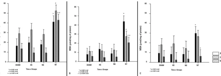
Lipid peroxidation
non-treated animals at each time point studied (2, 4, or 6 hours after reperfusion). Furthermore, it is noteworthy that the treated ischemic animals had similar MDA levels compared with the Sham Group.
Neutrophil infiltration
Similar to the results for lipid peroxidation, the reperfused groups showed basal MPO values (Figure 2) despite the increase present in the not treated animals. MPO activity was used as an index of neutrophil iniltration.
Cytokines
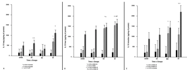
Two hours after reperfusion, there were no group diferences in the IL-6 tissue concentrations. After 4 hours, the IL-6 concentrations were signiicantly higher in the liver and intestine of the NT and NS Groups compared with the Sham and 7.5% HS Groups. After 6 hours, there were no group diferences except for the intestine, in which the IL-6 concentrations were higher in the NT Group (Figure 3).
Two hours after reperfusion, there were no group diferences in the IL-10 tissue concentrations. Four hours after reperfusion, the IL-10 concentrations were signiicantly higher in the liver and intestine in the non-treated animals and in the animals non-treated with normal saline solution compared with the sham and the animals trated with 7.5% hypertonic saline solution (similar to the IL-6 data). Six hours after reperfusion, the IL-10 levels in the liver were signiicantly higher in the saline treated group and in the non-treated animals compared with the Sham and 7.5% HS Groups. he IL-10 concentrations measured in the intestines of the non-treated animals were signiicantly higher compared with the Sham and 7.5% HS Groups 6 hours after reperfusion. Six hours after reperfusion, the IL-10 levels present in the lungs were signiicantly higher in the animals trated with 7.5% hypertonic saline solution compared with the ones treated with normal saline solution (Figure 4).
he plasma concentrations of IL-6 and 10 were studied at three intermediate time points (immediately after reperfusion and 2 and 4 hours after reperfusion) and compared among groups (Figure 5). Following the initial period of 2 hours, the plasma IL-6 concentrations were signiicantly higher in the animals treated with 7.5% hypertonic saline solution. here was also a trend towards lower IL-6 levels in tissues 4 hours after reperfusion in the animals trated with 7.5% hypertonic saline solution compared with the other groups.
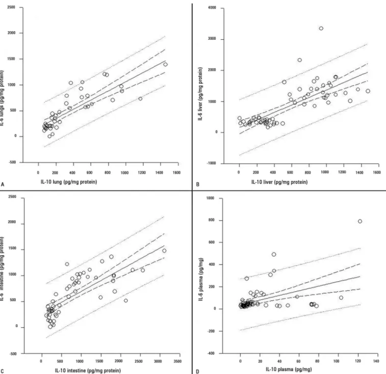
After 2 and 4 hours of reperfusion, the plasma IL-10 concentrations had a trend towards higher values in the animals treated with hypertonic saline solution compared with the other groups. hese diferences were signiicant compared with the Sham Group at each time point studied. here was a strong correlation between the IL-6 and IL-10 concentrations in tissues (R value in lungs=0.858; R value in liver=0.732; and R value in intestine=0.813) and a moderate correlation in the plasma cytokine levels (R value=0.432) using Spearman’s method (Figure 6).
DISCUSSION
Animals that underwent an SMA occlusion received intravascular luid replacement with physiological or hypertonic saline solutions immediately prior to intestinal reperfusion. he efects of both solutions were compared with the results in the sham and non-treated animals. Our results demonstrated that crystalloid luid treatment of ischemic animals caused signiicant decreases in oxidative stress markers and the inlammatory response. A hypertonic solution produced a delayed increase in gut and liver cytokines. here was an important increase in IL-10 in the lungs and plasma, as well as IL-6 in the plasma.
he arterial occlusion of large vascular territories triggers systemic mechanisms that alter the physiological
condition.(24) It has been hypothesized that the infusion
of crystalloid solutions immediately prior to SMA reperfusion can protect against hemodynamic alterations
after the intestinal blood low reestablishment.(4,24) he
pre-treatment with crystalloid solutions produces a rapid
and eicient bowel reperfusion.(25,26)
Oxidative stress, inlammation, and anti-inlammatory events that occur immediately after reperfusion contribute
to the outcome following SMA ischemia.(24) Hence, we
chose the period of 6 hours to study oxidative stress and inlammation after transient intestinal ischemia. Both crystalloid solutions decreased the oxidative stress and the inlammatory response studied in the splanchnic territory (bowel and liver) and lungs.
In this model, severe hypoperfusion occurs at the
splanchnic territory, which leads to intestinal ischemia.(15)
Treatment with 7.5% hypertonic saline solution yields hemodynamic results comparable with traditional therapeutics that use large volumes of normal saline
solution.(25) In addition, it has been demonstrated that
inlammatory responses are better attenuated with 7.5% hypertonic saline solution compared with a conventional
normal saline solution infusion,(15) and our data showed
Figure 1 - Tissue concentrations of malondialdeide (µmol/mg) in the lungs (A), liver (B) and intestine (C) in the sham group, group of animals treated with 7.5% hypertonic saline, group with normal saline solution, and not treated group. Graphics show the malondialdeide activity at 2 hours (black bar), 4 hours (light gray bar), and 6 hours (dark gray bar) after reperfusion. Gut ischemia and reperfusion increased malondialdeide in the lung, liver and gut; volume infusion (normal saline solution and hypertonic saline) protected tissues against oxidative stress. MDA - malondialdeide; HS - hypertonic saline; NS - normal saline solution; NT - not treated. *p<0.05; results as mean±standard deviation.
Figure 2 - Myeloperoxidase activity (U/mg) in the lungs (A), liver (B) and Intestine (C) in the sham group, group of animals treated with 7.5% hypertonic saline, group with normal saline solution, and not treated group. Graphics show the myeloperoxidase activity at 2 hours (black bar), 4 hours (light gray bar), and 6 hours (dark gray bar) after reperfusion. Gut ischemia and reperfusion increased neutrophil infiltration in the lung, liver and gut; volume infusion (normal saline solution and hypertonic saline) protected tissues by reducing inflammatory cell migration. MPO – myeloperoxidase; HS - hypertonic saline; NS - normal saline solution; NT - not treated. *p<0.05; results as mean±standard deviation.
Figure 4 - Tissue concentrations of interleukin 10 (pg/mg) in the lungs (A), liver (B) and intestine (C) in the sham group, group of animals treated with 7.5% hypertonic saline, group with normal saline solution, and not treated group. Graphics show the interleukin 10 concentrations at 2 hours (black bar), 4 hours (light gray bar), and 6 hours (dark gray bar) after reperfusion. Gut ischemia and reperfusion increased the interleukin 10 concentrations in the lung, liver and gut; volume infusion (normal saline solution and hypertonic saline) reduced the amount of interleukin 10 inflammation. The normal saline group presented higher interleukin 10 in the liver and gut, and the hypertonic saline presented higher levels in the lung. IL-10 - interleukin 10; HS - hypertonic saline; NS - normal saline solution; NT - not treated. *p<0.05; results as mean±standard deviation.
Figure 5 - Plasma concentrations (pmol/mg) of interleukin 6 (A) and interleukin 10 (B) in the sham group, group of animals treated with 7.5% hypertonic saline, group with normal saline solution, and not treated group. Graphics show the interleukin 6 and interleukin 10 plasma concentrations immediately prior to reperfusion (black bar) and at 2 hours (light gray bar) and 4 hours (dark gray bar) after reperfusion. Gut ischemia and reperfusion did not increase interleukin 6 or interleukin 10 concentrations in the plasma. The hypertonic solution presented higher plasma levels of interleukin 6 and interleukin 10 compared with the not treated and normal saline. IL-6 - interleukin 6; IL-10 - interleukin 10; HS - hypertonic saline; NS - normal saline solution; NT - not treated. *p<0.05; results as mean±standard deviation.
the osmotic and hyperoncotic characteristics of 7.5% hypertonic saline solution have been associated with this
particular efect in this experimental model.(27) Gonzalez
et al. studied the therapeutic efects of small volumes of hypertonic saline solutions (4mL/kg of body weight)
Figure 6 - Correlation between the levels of interleukin 6 and interleukin 10 in the lungs R=0.858 (A), liver R=0.732 (B), intestine R=0.813 (C) and plasma R=0.432 (D) in all groups by Spearman’s Method. IL-6 - interleukin 6; IL-10 - interleukin 10. p<0.05.
results obtained with these diferent hypertonic solutions were similar compared with the results achieved with higher volumes of normal saline solution (calculated as 33mL/kg of body weight). However, the authors attained
the best results with 7.5% hypertonic saline solution.(28)
It is important to highlight that the hypertonic solution delayed the inlammatory response, which is an efect that can be protective. he results obtained with crystalloid solutions were comparable with the results obtained for sham-operated animals. It is important to consider that in our study, the sham animals were subjected to moderate surgical trauma (abdominal incision, intestinal viscera manipulation, and superior mesenteric artery handling). his moderate surgical trauma was suicient to cause oxidative stress and the inlammatory response observed in our results.(29,30)
Reperfusion injury has been related to an
overproduction of ROS.(2) MDA was used as an oxidative
stress marker in this study. Our results showed that the production of MDA in diferent organs was similar in the sham and treated groups. Oxidative stress measured by MDA formation in tissues was signiicantly more intense in the non-treated animals at each time point studied. Moreover, MDA concentrations decreased over time in all experimental groups. hese data showed that oxidative stress was reduced by the adequate replacement of the total circulatory volume.
MPO activity was analyzed in diferent organs from the abdominal and extra-abdominal cavities. MPO activity has been widely used as an index of neutrophil
iniltration in tissues.(22) In this study, the MPO activity
in the 7.5% HS and NS-treated Groups was similar compared with the sham animals. Signiicantly lower levels of MPO activity were identiied in the treated animals compared with the non-treated animals. Leukocyte iniltration in tissues has been associated with
the local synthesis of chemokines and cytokines.(31) he
diferent interleukins outperform both inlammatory
and anti-inlammatory actions.(32)
he concentrations of IL-6 (pro-inlammatory) and IL-10 (anti-inlammatory) were analyzed in diferent organs, and a temporal proile of these plasmatic interleukin concentrations was studied. Many studies have proposed that the synthesis of IL-10 could be regulated by the production of IL-6, and, furthermore,
their ratio could predict the clinical outcome.(32-36) he
concentrations of IL-6 and IL-10 in tissues were very similar between the 7.5% hypertonic saline solution and sham animals throughout the time course studied. he analysis of IL-6 and IL-10 levels in tissues in the animals treated with normal saline solution peaked in the liver and small intestine in at least one measured
time point compared with the Sham and 7.5% HS Groups. Four hours after reperfusion, there was a trend towards higher IL-6 and IL-10 tissue concentrations in the NT Group. Despite the overall results for IL in tissues, the plasmatic concentrations of IL-6 and IL-10 were higher in the animals treated with 7.5% hypertonic saline solution. here are some data that demonstrated that the hypertonic solution had an anti-inlammatory action, which increased IL-10. In addition, the increased IL-10 in the plasma and lungs can explain the reduced neutrophil iniltration and oxidative stress in lungs.
We analyzed two important mechanisms of injury that occurred after transient intestinal ischemia in rats. he results of the hemodynamic parameters and biochemistry analyses were similar after treatment with crystalloid infusion. However, the clinical outcome for the animals treated with 7.5% hypertonic saline solution had a trend towards lower survival compared with the animals treated with normal saline solution, but it was not signiicant.
his study had some limitations. We could not analyze a more chronic period post-ischemia because of the diiculties in maintaining animals alive and for ethical reasons. In addition, mesenteric ischemia requires a volume infusion on a daily basis, and repeated hypertonic solution infusions present as a complication of hypernatremia. A point of strength was to compare the advantages of experimental studies in reproducing the identical scenario to ensure the therapeutic actions of a compound.
CONCLUSION
Objetivo: Investigar o papel de duas diferentes soluções sa-linas nos mecanismos de lesão após isquemia intestinal: estresse oxidativo e respostas inlamatórias.
Métodos: Ratos Wistar foram submetidos a oclusão transitória da artéria mesentérica superior e estudados du-rante as 6 horas seguintes à reperfusão. Após randomização, os animais foram divididos em quatro grupos: Falso; Solu-ção Hipertônica, os quais receberam infusão de soluSolu-ção sali-na hipertônica a 7,5% (4mL/kg de peso corpóreo); Solução Fisiológica, os quais receberam infusão de solução salina a 0,9% (33mL/kg); e Sem Tratamento. A infusão foi reali-zada imediatamente antes da reperfusão. Foram realireali-zadas dosagens sequenciais de interleucina 6 e interleucina 10 no plasma. Foram coletadas amostras de tecidos (pulmão, fíga-do e intestino) para medir malondialdeífíga-do, mieloperoxidase e interleucina.
Resultados: Em comparação ao Grupo Sem Tratamento, os ani-mais que receberam volume (Grupos Solução Hipertônica e Solução Fisiológica) mostraram níveis tissulares mais baixos de malondialde-ído, mieloperoxidase, interleucina 6 e interleucina 10. As concentra-ções plasmáticas de interleucina 6 e interleucina 10 foram mais altas nos animais tratados com solução hipertônica do que nos tratados com solução isiológica e nos sem tratamento.
Conclusão: Neste modelo de isquemia intestinal transitória, a manutenção adequada de volume intravascular diminuiu o es-tresse oxidativo e a síntese de marcadores de inlamação. Tanto a solução hipertônica quanto a isiológica atenuaram os efeitos deletérios observados após isquemia intestinal.
RESUMO
Descritores: Solução salina hipertônica/administração &
dosagem; Traumatismo por reperfusão/quimioterapia; Artéria mesentérica superior; Inlamação; Isquemia; Interleucinas; Estresse oxidativo; Ratos Wistar
REFERENCES
1. Yasuhara H. Acute mesenteric ischemia: the challenge of gastroenterology. Surg Today. 2005;35(3):185-95.
2. Cerqueira NF, Hussni CA, Yoshida WB. Pathophysiology of mesenteric ischemia/reperfusion: a review. Acta Cir Bras. 2005;20(4):336-43. Review. 3. Oldenburg WA, Lau LL, Rodenberg TJ, Edmonds HJ, Burger CD.
Acute mesenteric ischemia: a clinical review. Arch Intern Med. 2004;164(10):1054-62.
4. Khanna A, Rossman JE, Fung HL, Caty MG. Intestinal and hemodynamic impairment following mesenteric ischemia/reperfusion. J Surg Res. 2001;99(1):114-9.
5. Chang JX, Chen S, Ma LP, Jiang LY, Chen JW, Chang RM, et al. Functional and morphological changes of the gut barrier during the restitution process after hemorrhagic shock. World J Gastroenterol. 2005;11(35):5485-91. 6. Xu DZ, Lu Q, Kubicka R, Deitch EA. The effect of hypoxia/reoxygenation
on the cellular function of intestinal epithelial cells. J Trauma. 1999;46(2):280-5.
7. Lammers KM, Innocenti G, Venturi A, Rizzello F, Helwig U, Bianchi GP, et al. The effect of transient intestinal ischemia on inflammatory parameters. Int J Colorectal Dis. 2003;18(1):78-85.
8. Schoenberg MH, Beger HG. Reperfusion injury after intestinal ischemia. Crit Care Med. 1993;21(9):1376-86.
9. Stallion A, Kou TD, Latifi SQ, Miller KA, Dahms BB, Dudgeon DL, et al. Ischemia/reperfusion: a clinically relevant model of intestinal injury yielding systemic inflammation. J Pediatr Surg. 2005;40(3):470-7.
10. Esmon CT. The impact of the inflammatory response on coagulation. Thromb Res. 2004;114(5-6):321-7.
11. Velasco IT, Pontieri V, Rocha e Silva M Jr, Lopes OU. Hyperosmotic NaCl and severe hemorrhagic shock. Am J Physiol. 1980;239(5):H664-73. 12. Kolsen-Petersen JA. Immune effect of hypertonic saline: fact or fiction?
Acta Anaesthesiol Scand. 2004;48(6):667-78.
13. Rocha-e-Silva M, Poli de Figueiredo LF. Small volume hypertonic resuscitation of circulatory shock. Clinics (Sao Paulo). 2005;60(2):159-72. 14. Jonas J, Heimann A, Strecker U, Kempski O. Hypertonic/hyperoncotic
resuscitation after intestinal superior mesenteric artery occlusion: early effects on circulation and intestinal reperfusion. Shock. 2000;14(1):24-9. 15. Gonzalez EA, Kozar RA, Suliburk JW, Weisbrodt NW, Mercer DW, Moore
FA. Hypertonic saline resuscitation after mesenteric ischemia/reperfusion induces ileal apoptosis. J Trauma. 2005;59(5):1092-8.
16. Zakaria el R, Tsakadze NL, Garrison RN. Hypertonic saline resuscitation improves intestinal microcirculation in a rat model of hemorrhagic shock. Surgery. 2006;140(4):579-87; discussion 587-8.
17. Suzuki K, Aoyagi S, Koie H, Asano R. The effect of 7.2% hypertonic saline solution on m-mode echocardiographic indices in normovolemic dogs. J Vet Med Sci. 2006;68(7):749-51.
18. Attuwaybi B, Kozar RA, Gates KS, Moore-Olufemi S, Sato N, Weisbrodt NW, et al. Hypertonic saline prevents inflammation, injury, and impaired intestinal transit after gut ischemia/reperfusion by inducing heme oxygenase 1 enzyme. J Trauma. 2004;56(4):749-58; discussion 758-9. 19. Kolsen-Petersen JA. Immune effect of hypertonic saline: fact or fiction?
Acta Anaesthesiol Scand. 2004;48(6):667-78.
Authors’ contributions
Wilson Kohama Chimabucurro was responsible for all analyses and the content of the article. Bomim Alves da Silva Junior contributed to the article elaboration. Ana Iochabel Soares Moretti contributed
20. Victorino GP, Newton CR, Curran B. Effect of hypertonic saline on microvascular permeability in the activated endothelium. J Surg Res. 2003;112(1):79-83.
21. Chiara O, Pelosi P, Brazzi L, Bottino N, Taccone P, Cimbanassi S, et al. Resuscitation from hemorrhagic shock: experimental model comparing normal saline, dextran, and hypertonic saline solutions. Crit Care Med. 2003;31(7):1915-22.
22. Goldblum SE, Wu KM, Jay M. Lung myeloperoxidase as a measure of pulmonary leukostasis in rabbits. J Appl Physiol (1985).1985;59(6):1978-85. 23. Bradford MM. A rapid and sensitive method for the quantitation of
microgram quantities of protein utilizing the principle of protein-dye binding. Anal Biochem. 1976;72:248-54.
24. Hayward R, Lefer AM. Time course of endothelial-neutrophil interaction in splanchnic artery ischemia-reperfusion. Am J Physiol. 1998;275(6 Pt 2):H2080-6.
25. Choi PT, Yip G, Quinonez LG, Cook DJ. Crystalloids vs. colloids in fluid resuscitation: a systematic review. Crit Care Med. 1999;27(1):200-10. 26. Bagshawa SM, Bellomo R. The influence of volume management on
outcome. Curr Opin Crit Care. 2007;13(5):541-8.
27. Drobin D, Hahn RG. Kinetics of isotonic and hypertonic plasma volume expanders. Anesthesiology. 2002;96(6):1371-80.
28. Gonzalez EA, Kozar RA, Suliburk JW, Weisbrodt NW, Mercer DW, Moore FA. Conventional dose hypertonic saline provides optimal gut protection and limits remote organ injury after gut ischemia reperfusion. J Trauma. 2006;61(1):66-73; discussion 73-4.
29. Thomas S, Karnik S, Balasubramanian KA. Surgical manipulation of the small intestine and its effect on the lung. J Surg Res. 2002;106(1):145-56. 30. Grotz MR, Ding J, Guo W, Huang Q, Deitch EA. Comparison of plasma
cytokine levels in rats subjected to superior mesenteric artery occlusion or hemorrhagic shock. Shock. 1995;3(5):362-8.
31. Salmi M, Jalkanen S. Molecules controlling lymphocyte migration to the gut. Gut. 1999;45(1):148-53. Review.
32. Taniguchi T, Koido Y, Aiboshi J, Yamashita T, Suzaki S, Kurokawa A. Change in the ratio of interleukin-6 to interleukin-10 predicts a poor outcome in patients with systemic inflammatory response syndrome. Crit Care Med. 1999;27(7):1262-4.
33. Rajappa M, Sen SK, Sharma A. Role of pro/anti-inflammatory cytokines and their correlation with established risk factors in South Indians with coronary artery disease. Angiology. 2009;60(4):419-26.
34. Zingarelli B, Yang Z, Hake PW, Denenberg A, Wong HR. Absence of endogenous interleukin 10 enhances early stress response during post-ischaemic injury in mice intestine. Gut. 2001;48(5):610-22.
35. Dugernier TL, Laterre PF, Wittebole X, Roeseler J, Latinne D, Reynaert MS, et al. Compartmentalization of the inflammatory response during acute pancreatitis: correlation with local and systemic complications. Am J Respir Crit Care Med. 2003;168(2):148-57.