Vascular access through the intraosseous route in
pediatric emergencies
Acesso vascular por via intraóssea em emergências pediátricas
INTRODUCTION
Venous puncture is an essential procedure for administering luids and medications during pediatric emergencies. his procedure is technically more diicult in children because of their anatomy and hemodynamic responses to severe pathological processes.(1,2)
For critically ill children, the intraosseous route is quick and safe because it involves non-collapsible vascular access - unlike the peripheral veins, which undergo vasoconstriction during clinical and traumatic situations, leading to shock that prevents puncture and maintenance of an appropriate route for treatment. It is possible through the intraosseous route to infuse medications, electrolyte solutions, and blood products directly into the venous plexus bone, in which the absorption and duration of action are the
Ricardo Américo Ribeiro de Sá1,Clayton Lima Melo2,3,4, Raquel Batista Dantas2,4,5, Luciana Valverde Vieira Delfim6
1. Hospital Universitário São José - Belo Horizonte (MG), Brazil.
2. Centro Universitário Una - Belo Horizonte (MG), Brazil.
3. Pontifícia Universidade Católica de Minas Gerais - PUC-Minas - Belo Horizonte (MG), Brazil. 4. Hospital Municipal Odilon Behrens - Belo Horizonte (MG), Brazil.
5. Faculdade Estácio - Belo Horizonte (MG), Brazil.
6. Admission Unit, Hospital Belo Horizonte - Belo Horizonte (MG), Brazil.
ABSTRACT
Obtaining venous access in critically ill children is an essential procedure to restore blood volume and administer drugs during pediatric emergencies. he irst option for vascular access is through a peripheral vein puncture. If this route cannot be used or if a prolonged period of access is necessary, then the intraosseous route is an efective option for rapid and safe venous access. he present work is a descriptive and exploratory literature review. he study’s aim was to describe the techniques, professional responsibilities, and care related to obtaining venous access via the intraosseous route in pediatric emergencies. We selected 22 articles (published between 2000 and 2011) that were available in the Latin American and Caribbean Health Sciences (LILACS) and MEDLINE databases and the SciELO electronic library, in addition to the
current protocol of cardiopulmonary resuscitation from the American Heart Association (2010). After the literature search, data were pooled and grouped into the following categories of analysis: historical aspects and physiological principles; indications, beneits, and contraindications; professional assignments; technical principles; care during the access; and possible complications. he results of the present study revealed that the intraosseous route is considered the main secondary option for vascular access during the emergency response because the technique is quick and easily executed, presents several non-collapsible puncture sites, and enables the rapid and efective administration of drugs and luid replacement.
Keywords: Infusions, intraosseous/ methods; Infusions, intraosseous/ instrumentation; Emergency nursing; Critical care/methods; Child
Conflicts of interest: None. Submitted on May 7, 2012 Accepted on November 27, 2012
Corresponding author:
Ricardo Américo Ribeiro de Sá Rua Rio Pomba, 408 - Carlos Prates Zip Code: 30720-290 - Belo Horizonte (MG), Brazil
same as with peripheral or central venous access.(3,4) Some studies have revealed that intraosseous puncture is successful in over 90% of the cases for which there is a clinical indication. Furthermore, the average time to perform the puncture is less than 2 minutes when performed by an experienced professional with the suitable material. Thus, intraosseous access is increasingly becoming a good alternative to fluid infusion after the failure of peripheral venous access.(1,5,6)
Despite the well-deined indication for intraosseous access in pediatric emergencies, it is noteworthy that this subject is rarely addressed in the Brazilian scientiic literature and the ield of graduate health science. Consequently, questions regarding the technique, professional tasks, and care related to intraosseous puncture in pediatric emergencies might arise.
From this perspective, the current study is relevant due to the lack of national publications regarding intraosseous venous access in pediatric patients; the aim is to provide important scientiic information so that emergency room professionals may perform this procedure safely and efectively.
herefore, the purpose of this review is to describe the technical principles, professional assignments, and care related to using the intraosseous route for vascular access during pediatric emergencies.
METHODS
he current study is categorized as a descriptive and exploratory literature review.
To accomplish the goals of the present investigation, articles were selected from the international literature published in English, Portuguese, or Spanish. We used abstracts that were available in the Virtual Health Library (VHL) - speciically in the Latin American and Caribbean Health Sciences (LILACS) and MEDLINE databases - and the Scientiic Electronic Library Online (SciELO). A search was also conducted for books in the collection of the Centro Universitário Una in Belo Horizonte (MG-Brazil) and for documents published by boards of nursing and medicine; in addition, the current protocol of the American Heart Association (AHA) for treating cardiopulmonary arrest and cardiovascular emergencies, which was published in 2010, was used. For the article search, the following keywords were used, based on the Health Science Descriptors (DeCS/BIREME): “Intraosseous infusion,” “pediatrics,” “nursing,” “emergency nursing,” and
“intensive care.” To identify the research materials, a period of 11 years (2000 to 2011) was used to obtain the most current information on the topic.
To select the articles, the following exclusion criteria were considered: dissertations and doctoral theses (due to the time scheduled), duplicate publications in the databases, and animal research. Consequently, 283 summary articles were read, and 22 articles were selected. Twenty articles were selected from MEDLINE, 1 article was selected from SciELO, and 1 article was selected from the LILACS database.
he data were collected using the following procedure of Gil:(7) (a) exploratory reading; (b) selective reading of the title and summary to identify articles relevant to the purpose of the study; (c) analytical reading to sort the information found in the articles; and (d) interpretative reading, aimed to understand the selected material and the construction of the theoretical framework for analysis.
hus, the articles that were retrieved from the literature were analytically read, with subsequent highlighting of key points that were arranged as they appeared in the texts.
he presentation of the results and discussion of the data were descriptively categorized as follows: historical aspects and physiological principles; indications, beneits, and contraindications; professional assignments; technical principles; care during the access; and possible complications. his form of presentation was chosen to provide the reader with greater clarity in assessing the applicability of the study.
RESULTS
General aspects
he intraosseous route for vascular access is not a recently developed procedure, having been irst described in 1922. Its most widespread use occurred in the 1940s, when the technique was introduced for routine care in medical emergencies and during World War II, for treating severely injured patients.(8-11)
A decade later, in 1950, the use of the intraosseous route was reduced due to the introduction of new catheters for venous puncture (catheters on needles). However, in mid-1980, the use of intraosseous vascular access started to increase in pediatric patients because of the simplicity of the procedure, new scientiic studies, the recommendations of the AHA, and the availability of new technologies.(9-11)
became possible because the bone is richly vascularized, especially in its red bone marrow. Blood vessels enter the bone through the periosteum, passing through the compact bone via the perforating holes or Volkmann canals and Haversian canals that run longitudinally through the bone. Adjacent to this region, there are plates, which are rigid spaces that have gaps with small canaliculi. Upon reaching the spongy bone, the vessels traverse macroscopic spaces between the trabeculae, which are illed with red bone marrow, until reaching the spinal canal medullary cavity.(11,12)
The connections between the segments form a network of branches, which allows oxygen and nutrients to be transferred to the bone cells as well as products and excretions to be drained back into the bloodstream.(11-13)
hrough the intraosseous route, infused luids and medications can reach the bloodstream quickly and without loss during the absorption process.(3,11,14)
During pediatric emergency treatment, venous access is a priority. Obtaining peripheral venous access in children may be impossible or too time-consuming due to anatomical and physiological characteristics, such as increased thickness of the subcutaneous adipose tissue, the presence of small-caliber vessels, and diiculty accessing large-caliber vessels (e.g., jugular and subclavian veins). During severe pathological conditions (such as cardiopulmonary arrest, septic and hypovolemic shock, prolonged status epilepticus, and severe dehydration), reduction of the circulating blood volume, vasoconstriction, and collapse of peripheral vessels may occur. hese events, together or separately, signiicantly contribute to the diiculty of venipuncture.(2,15)
In such circumstances, other access routes for infusion of luids and medications may be used. However, these routes present greater risks and disadvantages compared with the intraosseous route because they are invasive procedures and require
greater technical skill. Central venous access, for example, consists of a time-consuming technique that is more complex than the intraosseous route, making it not feasible for cardiopulmonary resuscitation. In this case, the endotracheal route would be another possibility for drug administration. However, several studies have demonstrated that absorption through this route is incomplete and inconsistent, few drugs can be administered, several side efects may be observed, and luid replacement is not possible.(1)
Compared with central and peripheral venous access, the intraosseous route has several advantages (Table 1). he only limitation of this technique is the maximum duration of the access, which is 24 hours.(16)
For these reasons, intraosseous access is indicated as the irst alternative for luid infusion after failure to gain peripheral venous access (Table 2). In addition to providing non-collapsible access, which is minimally afected by the hemodynamic state of the patient, intraosseous access is also considered a rapidly and easily implemented technique that may be used for luid replacement and the administration
Table 1 - Characteristic of the main access routes in pediatric patients.(16)
Intraosseous access
Subclavian
vein Femoral vein
Internal jugular vein
External
jugular vein Axillary vein
Venous dissection
Access route during emergency treatment ++++ ++ +++ ++ +++ + ++
Ease of performing the technique ++++ ++ +++ ++ +++ + +
Infection + ++ ++ ++ + + ++
Thrombosis 0 + ++ + + + ++++
Other complications + ++ + + 0 + 0
Appropriate for long-term use 0 +++ ++ ++ + + 0
Appropriate for short-term use ++++ ++ +++ +++ ++ +++ ++
0 - no effect/not applicable/no risk; ++++ - higher effect/most used/high risk; + - lower effect/less used/low risk.
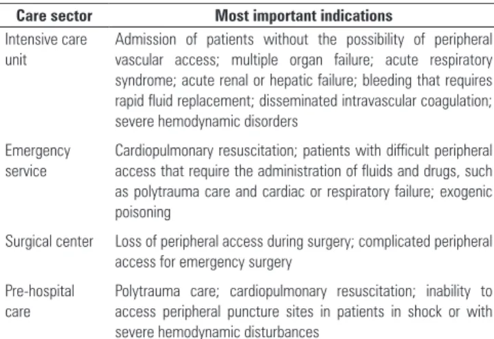
Table 2 - Most important indications for intraosseous access associated with the clinical care sectors.
Care sector Most important indications
Intensive care unit
Admission of patients without the possibility of peripheral vascular access; multiple organ failure; acute respiratory syndrome; acute renal or hepatic failure; bleeding that requires rapid fluid replacement; disseminated intravascular coagulation; severe hemodynamic disorders
Emergency service
Cardiopulmonary resuscitation; patients with difficult peripheral access that require the administration of fluids and drugs, such as polytrauma care and cardiac or respiratory failure; exogenic poisoning
Surgical center Loss of peripheral access during surgery; complicated peripheral access for emergency surgery
Pre-hospital care
Polytrauma care; cardiopulmonary resuscitation; inability to access peripheral puncture sites in patients in shock or with severe hemodynamic disturbances
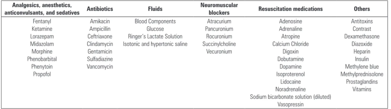
of various medications, including drugs speciied for cardiopulmonary arrest (Table 3). Furthermore, this pathway allows enables the collection of blood for laboratory tests.(1-3,13,17)
he absorption and bioavailability of several intravenous drugs are equivalent when administered through intraosseous access.(3,10,18) Pharmacokinetic
studies of morphine sulfate(4) administered
intraosseously versus intravascularly have demonstrated the similarity between the two routes.
Considering these advantages, the AHA stated in its 2010 guidelines that if it is impossible to obtain peripheral venous access (irst option), then the intraosseous route should be the second-choice technique for obtaining venous access.(19)
Intraosseous puncture for fluid and medication infusions should not be obtained (absolute contraindication) in cases with recent fractures or punctures of the bone at the site of access. In other situations, such as osteogenesis imperfecta, severe osteoporosis, osteomyelitis, cellulitis, or infection of the puncture site, physicians should consider the risk/benefit ratio (relative contraindication). Sternal puncture is contraindicated in children due to the possibility of serious complications, such as transfixation, fracture, hemothorax, and cardiac or large-caliber-vessel injury.(3,8,11,15,18)
Intraosseous access should always be performed by a trained and qualified professional. In Brazil, doctors and nurses are allowed by their respective councils to execute this procedure because these professionals are qualified to insert and manipulate intraosseous devices. The indication for the procedure is a medical decision.(20-22)
Well-deined protocols and continual training of medical and nursing staf regarding proper handling of the general apparatus and identiication of the speciic
device used in the procedures are essential for efective and safe care of patients undergoing intraosseous venous access.(3,22)
An important aspect for the success of this procedure is the necessity for continuing education for the health staf.(23) Molin et al.(24) determined that the lack of training and knowledge of the technique inluenced the rate of its use in hospitals in Denmark.
As demonstrated by Pister et al.,(17) the training of physicians and non-physicians improved the success rate in obtaining intraosseous access. In their study, the rate of success for obtaining intraosseous access with no more than 3 attempts was 100% for professionals who had received training. However, the success rate was lower, at 77%, before training. hus, the training of professionals performing this technique is crucial for efectively executing the procedure without harming the patient.
Technical principles
he preferable puncture sites in children for intraosseous access are the proximal tibia (the site may be located by placing a inger 1 cm below the tibial tuberosity and then sliding the inger 1 cm medially), the distal tibia (2 cm above the medial malleolus), and the distal femur. he humerus and calcaneus may also be used. he proximal tibia is the most commonly indicated puncture site because of the thin layer of skin that covers the anterior region of this bone and because this site would not interfere with cardiopulmonary arrest procedures, such as chest compressions and invasive respiratory access for ventilation.(15,22,25)
Several devices may be used for intraosseous puncture, and they are classiied as manual and new-generation devices (Table 4). he manual devices are inserted into the puncture site using manual pressure exerted by the operator; for this procedure, speciic
Table 3 - Most commons medications for intraosseousadministration.(3)
Analgesics, anesthetics,
anticonvulsants, and sedatives Antibiotics Fluids
Neuromuscular
blockers Resuscitation medications Others
Fentanyl Ketamine Lorazepam Midazolam Morphine Phenobarbital
Phenytoin Propofol
Amikacin Ampicillin Ceftriaxone Clindamycin Gentamicin Sulfadiazine Vancomycin
Blood Components Glucose Ringer’s Lactate Solution Isotonic and hypertonic saline
Atracurium Pancuronium
Rocuronium Succinylcholine
Vecuronium
Adenosine Adrenaline Atropine Calcium Chloride
Digoxin Dobutamine
Dopamine Isoproterenol
Lidocaine Noradrenaline Sodium bicarbonate solution (diluted)
Vasopressin
Antitoxins Contrast Dexamethasone
Diazoxide Heparin
Insulin Methylene blue Methylprednisolone
needles are available.(9,10,15,22,26) Because of the risk of needle obstruction during insertion, devices that do not have indwelling trocars should not be used.(11)
Automatic insertion devices, or new-generation devices, are placed on the puncture site and enter the spinal canal using the force of an internal spring inside the device; other devices consist of an electric bone punch, in which the needle is inserted into the medullary canal at high speeds. Both devices control the distance of insertion and use needles of various sizes.(9,10,23,26)
These new devices have advantages over the manual ones because the former provide faster access and a higher degree of safety during the puncture. In addition, the risks of fractures or of transfixing the spinal canal are minimized if the instructions are carefully followed.(1,3)
To compare the technical precision between these devices, Hartholt et al.(27) studied the results of punctures with 2 intraosseous access devices (the
Jamshidi® 15G and BIG® 15G and 18G needle),
verifying that the needle for manual insertion did not yield any adverse events during 12 punctures, while the automatic insertion device yielded 3 adverse events (1 extravasation, 1 malposition, and 1 needle displacement) during 11 punctures.
In the study conducted by Schwartz et al.,(6) the BIG® device was used, and the success rate was 87.2% (47 total punctures: 41 successful and 6 unsuccessful) in the pediatric population.
To ensure the correct use of each device, the site for which they are indicated must be examined (Table 5) to determine whether it can be used in a pediatric patient.(9)
During intraosseous access, conscious patients or patients with preservation of pain perception by the central nervous system can experience pain; in these cases, the administration of local anesthetics into the subcutaneous tissue and the intraosseous infusion of lidocaine (see the table below) are indicated before
starting luid infusion. Unconscious patients who do not respond to pain, as in cases of cardiac arrest, do not require this procedure.(11)
Care and possible complications of the access he maximum amount of time that venous access via the intraosseous route should remain in the patient is 24 hours. However, it is recommended that it remain only for the period necessary to address the emergency and obtain a more permanent vascular access (central or peripheral access) to reduce the risk of infusion-related complications caused by the intraosseous route. Within this period, several precautions should be taken to avoid complications (Table 6).(3,22)
As with all invasive procedures, the intraosseous route also can yield complications. he occurrence of adverse events during the use of this pathway is less than 1%. Recent studies have reported complication rates of approximately 0.6% and have demonstrated that the extravasation and iniltration of administered luids are the most frequent adverse events.(9,11)
he complications are related to each other, and the causes of some may be associated with unfamiliarity with the technique, as in iniltration (which occurs due to extravasation of luids) and fractures (which are related to errors during execution of the technique). Other complications, such as osteomyelitis, sepsis, cellulitis, and abscess, are related to failures regarding aseptic techniques for puncture or manipulation of the
Table 4 - Devices for intraosseous puncture.(9)
Devices Description Method of insertion Products
Manual Steel needle with a removable trocar to prevent
plugging of the needle by bone fragments
Manual insertion in the medullary space, controlled by the operator
IO Jamshidi® (Care Fusion) Needle; IO Cook® Needle (Cook Medical)
Automatic Impact Steel trocar needle, operated by a spring Upon being triggered, the device automatically inserts the trocar needle into the spinal canal via spring tension.
Bone Injection Gun (BIG)® (Wais Med LTD)
Electric Steel trocar needle, battery-operated power driver Upon being triggered, the device is inserted into the medullary canal via spinning (the device resembles an orthopedic drill bit)
EZ-IO® (Vidacare Corporation)
Table 5 - Insertion sites and intraosseous access devices.(9)
Site Adult Children Devices
Sternum ü 0 Manual, FAST 1®
Humeral head ü 0 Manual, BIG®, EZ-IO®
Distal radius ü 0 Manual
Distal ulna ü 0 Manual
Iliac crest ü 0 Manual
Distal femur ü ü Manual, BIG®, EZ-IO®
Proximal tibia ü ü Manual, BIG®, EZ-IO®
devices. Compartment syndrome and tissue necrosis are related to extravasation of luids. Fat embolism might also occur, although no cases have been reported in the literature because the bone marrow in children has little or no fat.(2,3,11,14,15,23)
According to Coronel Carvajal(2) and Lane and
Guimarães,(15) injury to the growth plate may occur. However, DeBoer et al.(3) state that this is only a theoretical concern, without any clinical evidence.
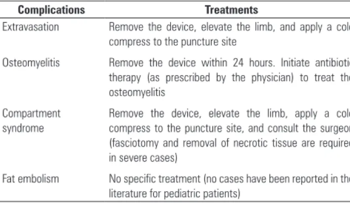
Early detection and rapid treatment are key factors for preventing these complications from generating larger lesions in the patient (Table 7). To prevent such adverse events, it is essential to follow the protocol carefully, with adequate antisepsis and proper handling of the apparatus being critical, both at the time of insertion and the withdrawal of the needle. Splinting and immobilizing the punctured limb may reduce the risks of fracture, needle dislocation, and extravasation.(2,3,15)
CLOSING REMARKS
Currently, the intraosseous route is accepted as a safe alternative for venous access because it provides several sites for non-collapsible puncture. Consequently, intraosseous access is indicated for the most severely ill children, except in clinical situations where the technique is not feasible (as previously described) or where the risk/beneit ratio is disadvantageous.
Intraosseous access is a rapidly and easily executed technique that saves time during emergency care; consequently, it enables the staf to pay closer attention to other procedures that are equally important for severely ill patients. Ultimately, use of the intraosseous route may increase the chances for a faster and more complete recovery of the child.
Despite the advantages and beneits associated with intraosseous access, its use remains underexplored in clinical practice. his fact might be explained by emergency health professionals’ lack of knowledge and appropriate training on the subject.
he results of the present study show that the available published information predominantly addresses the indications, contraindications, and complications of the procedure, as well as the technique for its execution. Most of the articles discuss the technical aspects related to the moment of the puncture access. By contrast, few studies discuss the management of infusion through this route and the maintenance of access, which may be because most of these articles were published by physicians (20 articles published by physicians and 6 by nurses), although the technique is generally the
Table 6 - Care during intraosseous access, along with its justifications.(3,21)
Care Justification
Define the injection site and the appropriate apparatus. There are devices specific for different puncture sites.
Use aseptic techniques for insertion and removal of the needle and handling apparatus. To prevent puncture-site infection, osteomyelitis, and sepsis
Fix the needle, as well as the stents and catheters. To avoid the needle being pulled out, preventing loss of access, leakage, and damage to the tissue and bone
Use a continuous infusion pump for fluids, drugs, and blood components.
To ensure the continuity and rate of infusion, which are not maintained by gravity. Furthermore, as in the intravenous route, the alarms of infusion pumps may indicate obstruction of the apparatus, which may suggest infiltration.
Inject a 10 ml bolus of physiological saline (0.9%) every 4 hours. To prevent clogging of the device, the discontinuity of infusion, and loss of the access.
Verify the operation and permeability of the apparatus. To avoid clogging of the apparatus and its consequences, such as loss of access and disruption of the infusion, which would compromise the patient’s health.
Evaluate the presence of edema, erythema, and hypersensitivity at the puncture site after removing the needle.
To enable the early detection and treatment of complications, such as bleeding and infiltration
Apply an occlusive, sterile-gauze dressing to the puncture site using aseptic technique. To prevent contamination and reduce the risk of infection at the puncture site
Support professional training and establish protocols for the procedure.
To increase the chances of success of the procedure, to instruct professionals regarding their responsibilities in clinically managing the access, and to provide safe and effective patient care
Table 7 - Managing the major complications related to intraosseous access.(11)
Complications Treatments
Extravasation Remove the device, elevate the limb, and apply a cold compress to the puncture site
Osteomyelitis Remove the device within 24 hours. Initiate antibiotic therapy (as prescribed by the physician) to treat the osteomyelitis
Compartment syndrome
Remove the device, elevate the limb, apply a cold compress to the puncture site, and consult the surgeon (fasciotomy and removal of necrotic tissue are required in severe cases)
responsibility of the nurses and their teams.
he majority of the articles analyzed in the literature are reviews. here were no experimental studies that addressed the major complications and their incidence rates, the age and morbidity of children undergoing this procedure, and the diiculties encountered in performing this technique or establishing protocols and training on the subject.
his study met its goal of describing the technical principles, responsibilities, and professional care in obtaining venous access via the intraosseous route in pediatric emergencies, in addition to contributing to the dissemination of the procedure and the provision of support to professionals working in emergency rooms.
he necessity for additional studies on this subject is emphasized, and they would enable the procedure to gain popularity. Such studies are also necessary to produce contribute concrete scientiic support for the establishment of guidelines and protocols regarding the relevant indications, techniques, and professional responsibilities, in addition to deining the appropriate materials or devices based on each type of clinical situation encountered.
RESUMO
A obtenção do acesso venoso em crianças gravemente en-fermas é um procedimento essencial para o restabelecimento
da volemia e a administração de fármacos nas emergências pediátricas. A primeira opção para obtenção de acesso vas-cular é pela punção de uma veia periférica. Quando essa via não pode ser utilizada ou sua obtenção se torna demorada, a via intraóssea consiste em efetiva opção para obtenção de um acesso venoso rápido e seguro. O presente estudo possui caráter descritivo e exploratório, realizado por meio de pes-quisa bibliográica, com o objetivo de descrever os princípios técnicos, as atribuições proissionais e os cuidados relacio-nados à obtenção do acesso venoso pela via intraóssea em emergências pediátricas. Foram selecionados 22 artigos dis-ponibilizados nas bases de dados LILACS e MEDLINE e na biblioteca eletrônica SciELO, publicados entre o período de 2000 a 2011, além do protocolo vigente de ressuscitação
car-diopulmonar da American Heart Association, de 2010. Após
a leitura das publicações, os dados foram agrupados, possibi-litando a construção de cinco categorias de análise: aspectos históricos e princípios isiológicos; indicações, vantagens e contraindicações; atribuições dos proissionais; princípios técnicos; cuidados com o acesso; e possíveis complicações. Os resultados desse estudo mostraram que a via intraóssea consolida-se, hoje, como uma segunda opção de acesso vas-cular no atendimento a emergências, por ser uma técnica de fácil e rápida execução, apresentar vários sítios de punção não colapsáveis e permitir que a administração de fármacos e a reposição volêmica sejam rápidas e eicazes.
Descritores: Infusões intraósseas/métodos; Infusões intraósseas/instrumentação Enfermagem em emergência; Cuidados críticos/métodos; Criança
REFERENCES
1. de Caen A. Venous access in the critically ill child: when the peripheral intravenous fails! Pediatr Emerg Care. 2007;23(6):422-4; quiz 425-6. 2. Coronel Carvajal C. Vía intraósea en pediatría. Rev Cuba Pediatr
[Internet]. 2003;75(3). [citado 2012 Nov 28]. Disponível em: URL: http://scielo.sld.cu/scielo.php?script=sci_arttext&pid=S0034-75312003000300011&lng=es
3. DeBoer S, Russell T, Seaver M, Vardi A. Infant intraosseous infusion. Neonatal Netw. 2008;27(1):25-32. Review.
4. Von Hoff DD, Kuhn JG, Burris HA 3rd, Miller LJ. Does intraosseous equal intravenous? A pharmacokinetic study. Am J Emerg Med. 2008;26(1):31-8. 5. Horton MA, Beamer C. Powered intraosseous insertion provides safe and
effective vascular access for pediatric emergency patients. Pediatr Emerg Care. 2008;24(6):347-50.
6. Schwartz D, Amir L, Dichter R, Figenberg Z. The use of a powered device for intraosseous drug and fluid administration in a national EMS: a 4-year experience. J Trauma. 2008;64(3):650-4; discussion 654-5.
7. Gil AC. Como elaborar projetos de pesquisa. 5a ed. São Paulo: Atlas; 2010. 8. LaRocco BG, Wang HE. Intraosseous infusion. Prehosp Emerg Care.
2003;7(2):280-5.
9. Luck RP, Haines C, Mull CC. Intraosseous access. J Emerg Med. 2010;39(4):468-75. Review.
10. Blumberg SM, Gorn M, Crain EF. Intraosseous infusion: a review of methods and novel devices. Pediatr Emerg Care. 2008;24(1):50-6; quiz 57-8.
11. Vizcarra C, Clum S. Intraosseous route as alternative access for infusion therapy. J Infus Nurs. 2010;33(3):162-74. Erratum in J Infus Nurs. 2011;34(2):123. 12. Tortora GJ, Grabowski SR. Princípios de anatomia e fisiologia. 9a ed. Rio de
Janeiro: Guanabara Koogan; 2002.
13. Engle WA. Intraosseous access for administration of medications in neonates. Clin Perinatol. 2006;33(1):161-8, ix.
14. Smith R, Davis N, Bouamra O, Lecky F. The utilisation of intraosseous infusion in the resuscitation of paediatric major trauma patients. Injury. 2005;36(9):1034-8; discussion 1039.
15. Lane JC, Guimarães HP. Acesso venoso pela via intra-óssea em urgências médicas. Rev Bras Ter intensiva. 2008;20(1):63-7.
16. Hass NA. Clinical review: vascular access for fluid infusion in children. Crit Care. 2004;8(6):478-84.
17. Pfister CA, Egger L, Wirthmüller B, Greif R. Structured training in intraosseous infusion to improve potentially life saving skills in pediatric emergencies - Results of an open prospective national quality development project over 3 years. Paediatr Anaesth. 2008;18(3):223-9.
18. Fowler R, Gallangher JV, Isaacs SM, Ossman E, Pepe P, Wayne M. The role of intraosseous vascular access in the out-of-hospital environment (resource document to NAEMSP posicion statement). Prehosp Emerg Care. 2007;11(1):63-6.
20. Conselho Federal de Medicina. Parecer Técnico nº 26/2003. Consulta acerca da solicitação de autorização de treinamento em cursos de ACLS e PALS para profissionais não - médicos. Brasília (DF): CFM; 2004. p. 1-3.
21. Conselho Regional de Enfermagem de Minas Gerais. Parecer Técnico nº 154/10. Consulta acerca da autorização legal para realização dos procedimentos de punção venosa femoral e intra-óssea pelo enfermeiro. Belo Horizonte: CFE; 2010. p. 1-4.
22. Pedreira ML. Realização de punção intra-óssea por enfermeiros [Internet]. São Paulo: COREN – SP; 2009. [citado 2011Set 27]. Disponível em: URL: http://inter.coren-sp.gov.br/sites/default/files/ Realiza%C3%A7%C3%A3o%20de%20Pun%C3%A7%C3%A3o%20Intra-%C3%B3ssea%20por%20Enfermeiros.pdf
23. Phillips L, Brown L, Campbell T, Miller J, Proehl J, Youngberg B.
Recommendations for the use of intraosseous vascular access for emergent and nonemergent situations in various health care settings: a consensus paper. Crit Care Nurse. 2010;30(6):e1-7.
24. Molin R, Hallas P, Brabrand M, Schmidt TA. Current use of intraosseous infusion in Danish emergency departments: a cross-sectional study. Scand J Trauma Resusc Emerg Med. 2010;18:37.
25. Sunde GA, Heradstveit BE, Vikenes BH, Heltne JK. Emergency intraosseous access in a helicopter emergency medical service: a retrospective study. Scand J Trauma Resusc Emerg Med. 2010;18:52.
26. Infusion Nurses Society. The role of the registered nurse in the insertion of intraosseous access devices. J Infus Nurs. 2009;32(4):187-8.