Volume replacement with saline solutions during
pancreatitis in rats and the hepatic proiles of
apoptotic proteins and heat-shock proteins
Reposição volêmica com soluções salinas em pancreatite e perfil
hepático de proteínas apoptóticas e de choque térmico
INTRODUCTION
Liver failure can occur as a consequence of the systemic inlammatory response syndrome that occurs in acute pancreatitis. In our previous studies, we have demonstrated increased lipid peroxidation levels and extracellular matrix degradation in the liver after pancreatitis.(1) In addition, we observed increased blood levels of hepatic enzymes, indicating liver cell damage. Acute pancreatitis–associated liver injury is mediated by inlammatory cytokines that are produced within tissue-resident macrophages, which are activated by the inlammatory mediators that are systemically released by the pancreas.(2) he liver, in turn, participates in systemic inlammation, releasing several Ester Correia Sarmento Rios1, Ana Iochabel
Soares Moretti1, Heraldo Possolo de Souza1,
Irineu Tadeu Velasco1, Francisco Garcia Soriano1
1. Laboratory of Medical Investigation (LIM-51), Department of Emergency Medicine, Faculdade de Medicina, Universidade de São Paulo - USP - São Paulo (SP), Brazil.
ABSTRACT
Objective: Liver failure can occur
as a consequence of the systemic inlammation after acute pancreatitis. We assessed the efect of volume repositioning with hypertonic saline solution or normal saline on hepatic cytokine production and the expression of heat-shock proteins and apoptotic proteins after acute pancreatitis.
Methods: Wistar rats were divided
in four groups: C - control animals that were not subjected to insult or treatment; NT - animals that were subjected to acute pancreatitis and received no treatment; normal saline - animals that were subjected to acute pancreatitis and received normal saline (NaCl 0.9%); and HS - animals that were subjected to acute pancreatitis and received hypertonic saline solution (NaCl 7.5%). Acute pancreatitis was induced by retrograde transduodenal infusion of 2.5% sodium taurocholate into the pancreatic duct. At 4, 12 and 24 h following acute pancreatitis induction, TNF-alpha, 1-beta, 6 and
IL-10, caspase-2 and -7, Apaf-1, AIF and HSP60 and 90 were analyzed in the liver.
Results: Casp2 decreased in the
normal saline and hypertonic saline groups (p<0.05 versus. C) at 12 h. Apaf-1, AIF and HSP90 remained unchanged. At 4 h, Casp7 increased in the NT group (p<0.01 versus C), although it remained at the baseline levels in the reperfused groups. HSP60 increased in all of the groups at 4 h (p< 0.001 vs. C). However, the hypertonic saline group showed lower expression of HSP60 than the normal saline group (p<0.05). Hypertonic saline solution maintained the production of cytokines at normal levels. Volume reperfusion with normal or hypertonic saline signiicantly modulated the expression of Casp7.
Conclusion: Volume replacement
with hypertonic or normal saline was efective in reducing caspase 7. However, only hypertonic solution was capable of regulating cytokine production and HSP60 expression at all time points.
Keywords: Heat-shock proteins;
Apoptosis; Pancreatitis; Liver This study was conducted at the Laboratory of
Medical Investigation, Department of Emergency Medicine, Faculdade de Medicina, Universidade de São Paulo - USP - São Paulo (SP), Brazil.
Conflicts of interest: None.
Submitted on September 3, 2012 Accepted on October 31, 2012
Corresponding author: Francisco Garcia Soriano
LIM-51, Faculdade de Medicina, Universidade de São Paulo
Avenida Dr. Arnaldo, 455, 3º andar, sala 3.189 - Cerqueira César
inlammatory mediators, leading to injury of other organs.(3,4) Substances that are systemically released during pancreatitis, such as nitric oxide (NO) and free radicals, can interfere with the respiration of hepatic mitochondria and can induce apoptosis.(5,6) Apoptotic cell death might play a considerable role in afecting mortality and morbidity in severe acute pancreatitis.(7) he apoptosis pathway, by death receptors or via the mitochondrial pathway, activates the inal caspase cascade for cell death.(8) Death receptor signaling has been associated with apoptosis in several hepatic diseases, such as ethanol-induced liver injury and cholestatic liver disease.(9) Apoptosis related to severe acute pancreatitis injury is known to be triggered via the mitochondrial pathway.(3)
Cell death has been observed in both apoptotic and necrotic forms in both clinical and experimental acute pancreatitis.(10) Current evidence suggests that the amounts of and balance between apoptosis and necrosis inluence the severity of acute pancreatitis.(11) Recently, heat-shock proteins and their cofactors have been revealed to be associated with apoptotic and necrotic pathways.(12) Heat-shock proteins are molecular chaperones that stabilize and refold damaged intercellular proteins, preventing intracellular protein aggregation and rendering cells resistant to stress-induced cell damage.(13)
Volume replacement, mainly with hypertonic saline alone, has shown beneits in various aspects of the pathophysiology of several diseases due to improvement of tissue hypoperfusion and decreases in oxygen consumption, endothelial dysfunction and cardiac depression, as well as reductions in a broad array of pro-inlammatory cytokines and various oxidant species.(14,15) We previously reported that hypertonic saline treatment reduces oxidative stress and tissue degeneration in the liver after pancreatitis.(1) Additionally, our group showed the efects of hypertonic saline in the expression and activity of several proteins, including heat-shock proteins (HSPs), in the lungs(16,17) and the liver.(1) However, there are no data in the literature regarding the efect of hypertonic solution on hepatic apoptosis during pancreatitis. In the present study, we assessed the efects of normal (NaCl 0.9%) and hypertonic saline (NaCl 7.5%) on the expression of apoptotic proteins and HSPs, as well as the correlation of these factors with inlammation during pancreatitis.
METHODS
Pancreatitis induction
All of the experiments were conducted in accordance with the guidelines established by the Research Ethics Committee of the Faculdade de Medicina of the
Universidade de Sao Paulo. Male Wistar rats, weighing 270-320 g, were anesthetized subcutaneously with ketamine (10 mg/kg) and xylazine (8 mg/kg). Acute pancreatitis was induced by a well-established method of retrograde infusion of 2.5% sodium taurocholate (1.0 mL/ kg; Sigma, St. Louis, MO, USA) transduodenally into the pancreatic duct via a 24-gauge angiocatheter at aconstant infusion rate of 1 mL/min. he bile duct was clamped with a microsurgical “bulldog” clamp at the hepatic hilum to prevent leakage of taurocholate solution into the liver. he hepatic hilar clamp was released after the injection. It has been reported in the literature that this model of pancreatitis causes hepatic injury and reproduces the mortality and pathophysiological changes of human pancreatitis.(18) In the present study, analysis was performed on four groups: the control group, consisting of animals that sufered neither insult nor treatment (C); the no treatment (NT) group, consisting of animals in which pancreatitis was induced, but no treatment was given; the normal saline (NS) group, consisting of animals in which pancreatitis was induced, and an intravenous bolus of normal saline (0.9% NaCl, 34 mL/kg) was administered; and the hypertonic saline (HS) group, consisting of animals in which pancreatitis was induced, and hypertonic saline (7.5% NaCl, 4 mL/ kg) was administered via the internal jugular vein over a period of 5 min at 1 h after pancreatitis induction. he volume of normal saline infused was equivalent in sodium content to 4 mL/kg of hypertonic saline. he animals were sacriiced and their livers collected at 4, 12, or 24 h after pancreatitis induction.
Semiquantitative reverse transcription-polymerase chain reaction (RT-PCR)
RT-PCR was used to determine the mRNA levels in liver tissue. Total RNA was extracted from frozen rat livers with TRIzol reagent (Invitrogen, Carlsbad, CA, USA), following the manufacturer’s instruction. RNA was dissolved in diethyl pyrocarbonate (DEPC)-treated water and was quantiied spectrophotometrically at 260 nm. First-strand c-DNA was generated by adding RNA (1
mg) to a mixture containing 1 mL of ImProm-II™ reverse transcriptase (Promega, Madison, WI, USA), 1 mL (0.5
mg/mL) of oligo (dT), 20 U/mL of Recombinant RNAsin RNAse inhibitor, 3 mM MgCl2, 6 mL of ImProm-II™ 5X reaction bufer (Promega, Madison, WI, USA) and 1
hermal Controller (MJ Research PTC-200, Watertown,
MA, USA). he PCR solution contained 1 mL of
irst-strand cDNA, 2.5 mL of 10X PCR bufer, 2 mM MgCl2, 0.5 mM dNTP mix, 1 pmol/mL of each speciic primer
and 2.5 U/mL of TaqDNA polymerase (Invitrogen,
Carlsbad, CA, USA) in a inal volume of 25 mL. To
evaluate the relative abundance of a transcript between samples, the relative RT-PCR was performed with 18S ribosomal RNA primer as an internal control. he PCR products were resolved by electrophoresis on 1% agarose gel, stained with ethidium bromide (EtBr; Horizon, Life-Technologies, USA) and visualized under ultraviolet light with a video imaging system (Pharmacia). Densitometric analyses of EtBr-stained gel bands were performed using Gene Tools software (Syngene, Cambridge, MA, USA). he data were plotted as a function of the log OD of the gene target product against the log OD of 18S rRNA. he sequences of the speciic primers (Invitrogen, Carlsbad, CA, USA) were as follows: rRNA (320bp) sense: GAAAGATGGTGAACTATGCC; and antisense: TTACCAAAAGTGGCCCACTA; HSP60 (213pb) sense: TGACACCCTTTCTTCCAACC; and antisense: AGCAAAGGGGCTAATCCAGT; and HSP90 (247pb) sense: GATTGACATCATCCCCAACC; and antisense: CTAGCCAACACCCTGAGAGC.
Western blot
Frozen tissue samples (100 mg) were pulverized in liquid nitrogen. he samples were then homogenized in a bufer containing 1% TX-100, 20 mM Tris (pH 8,0), 10% glycerol, 135 mM NaCl and proteolytic enzyme inhibitors (40 µg/mL of phenylmethylsufonylluoride and 10 µg/mL of pepstatin; Sigma, St. Louis, MO). After separation of debris by centrifugation for 45 minutes at 14,000 g, the supernatants were collected, and the protein concentration was determined by the Bradford method (Bio Rad, Hercules, CA). he samples were stored at -80ºC until assayed. Protein expression was assessed using SDS-polyacrylamide gel electrophoresis under reducing conditions. Liver tissue extracts (25-100 µg/mL) were boiled in equal volumes of loading bufer (150 mM Tris-HCl– pH 6.8, 4% SDS, 20% glycerol, 15% b-mercaptoethanol and 0.01% bromophenol blue) and were subjected to electrophoresis on 10% polyacrylamide gels. Following electrophoretic separation, the proteins were transferred to Hybond-P membranes (Amersham Pharmacia Biotech, Buckinghamshire, UK). he membranes were blocked with 5% non-fat dry milk in Tris-bufered saline and 0.5% Tween 20 (TBST) for 1 hour. Primary antibodies against the following were employed: caspase-2 (rabbit, 1:1000,
Santa Cruz Biotechnology 623), caspase-7 (rabbit, 1:1000, sc-337773), apoptotic protease activating factor 1 ( APAF-1, goat, 1:1000, sc-26685), apoptosis-inducing factor (AIF, rabbit, 1:1000, ab32516, Abcam), HSP60 (goat, 1:1000, sc1052), HSP90 a/b (goat,1:1000, sc1055) and β-actin (1:10000, Sigma, A5441); they were incubated at 4ºC overnight. After washing twice with TBST, secondary horseradish peroxidase conjugate Ab (goat anti-rabbit polyclonal sc2004 or rabbit anti-goat sc2768, Santa Cruz Biotechnology) was applied at a dilution of 1:5000 for 2 hours. he blots were washed in TBST twice over 30 min, incubated in enhanced Super Signal Detection Kit chemiluminescence reagents (Pierce, Rockford, IL, USA) and exposed to Kodak O-OMAT-AR photographic ilm (Kodak, Rochester, NY, USA). he band intensity of the original blots was quantiied using Image J software (Research Services Branch, National Institutes of Health, Bethesda, MD, USA) and was normalized to control levels (control = 1).(17)
Cytokine measurement
Plasma samples were collected from the animals just before sacriice. he cytokines TNF-α, IL-6, IL-1β and IL-10 were measured by ELISA, according to the manufacturer’s instructions (R&D Technologies, USA).
Histology analysis
After ixation in 10% formalin, the liver tissue was embedded in parain and cut into 4- to 6-µm sections. he sections were stained with hematoxylin and eosin and were analyzed qualitatively by light microscopy. he occurrence of hepatic damage, such as areas of necrosis, hemorrhage, inlammatory iniltrates and vacuolization of the cytoplasm, was assessed. he images were generated by a microscope (Leica) connected to a camera (Sony Trinitron CCD, Sony, Japan) and were input into a computer.
Statistical analysis
130kDa 42kDa A B
3
2
1
0
Ap
a
f-1
(t
o
ta
l
o
f
co
n
tro
l)
C NT NS HS C NT NS HS C NT NS HS
4h 12h 24h
350 300 250 200 150 100 50 0
300
250 200
150 100
50 0
IL
-1
b
(p
g
/mg
)
IL
-1
0
(p
g
/mg
)
A
B C NT NS HS
C NT NS HS
NT NS HS
NT NS HS
NT NS HS
NT NS HS 4h
4h
12h
12h
24h
24h
250
200
150
100
50
0
30
20
10
0
IL
-1
b
(p
g
/ml
)
IL
-1
0
(p
g
/ml
)
A
B C NT NS HS
C NT NS HS
NT NS HS
NT NS HS
NT NS HS
NT NS HS 4h
4h
12h
12h
24h
24h RESULTS
Cytokine production
Four hours after the induction of pancreatitis, increased plasma levels of IL-1β (Figure 1A) were observed in the NT and HS groups (p<0.05 versus C). After 12 hours, the plasma IL-1β levels remained increased in the NT group (p< 0.001 versus C and HS; p< 0.01 versus NS). Treatment with normal saline or hypertonic solution maintained the normal levels.
Plasma IL-10 levels (Figure 1B) increased in the NT group 4 (p< 0.05 versus C; p<0.01 versus NS and HS) and 24 hours (p< 0.05 versus C) after the induction of pancreatitis. he levels of this cytokine increased in the NS group at 12 h (p< 0.05 versus C).
he hepatic levels of IL-1b (Figure 2A) increased signiicantly in the NS group (p< 0.05 versus C) at 4 h. After 12 and 24 h, we could not observe any statistically signiicant diference in hepatic IL-1b release among the groups. he hepatic production of IL-10 (Figure 2B) did not change in the liver for the irst 4 hours after the induction of pancreatitis. However, the IL-10 levels increased in the NT group at 12 h (p< 0.05 versus C, NS, HS) and in the NS group at 24 h (p< 0.05 versus C).
he plasmatic and hepatic levels of TNF-α and IL-6 did not change in any group over 24 hours (data not shown).
Figure 1 - Plasma cytokine levels. (A) After the induction of pancreatitis, increased IL-1b was observed in the NT and HS groups. After 12 hours, the IL-1β levels remained increased in the NT group. Treatment with normal saline or hypertonic solution maintained the normal levels. (B) Pancreatitis induced an increase in IL-10 release initially. At 12 h, the levels of this cytokine increased in the NS group. Data are reported as the means ± SEMs. N = 8 rats in each group. *p<0.05 versus C; **p<0.01 versus C; ***p<0.001 versus C; #p<0.01 versus
NT; $p<0.001 versus NT.
Apoptotic protein expression
To study the cell death process, we investigated the expression of the following apoptotic proteins: Apaf-1, AIF and caspases 2 and 7.
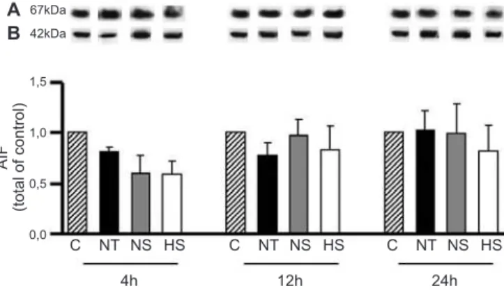
he expression of Apaf-1 (Figure 3) and AIF (Figure 4) remained at baseline levels throughout the 24 hour experimental period.
Precursors of both 2 (51 kDa) and 2L were expressed in the liver. he expression of caspase-2L (Figure 5) was unchanged for the irst 4 hours. Twelve
Figure 2 - Hepatic cytokine levels. Four, 12 and 24 hours after the induction of pancreatitis, liver homogenate was collected from control rats (C) or from rats subjected to pancreatitis without treatment (NT), rats treated with normal saline (NS) or rats treated with hypertonic solution (HS) to analyze hepatic IL-1β (A) and IL-10 (B). Data are the means ± SEMs (N = 8). *p<0.05 versus C; #p<0.05 versus NT.
Figure 3 - Expression of apoptotic protease activating factor 1 (APAF-1) estimated by western blot in the liver homogenates of control rats (C) or rats subjected to pancreatitis and treated with normal saline (NS), treated with hypertonic solution
(HS) or without treatment (NT). (A) Representative western blot of APAF-1 expression.
20kDa 42kDa A B 2,5 2,0 1,5 1,0 0,5 0,0 C a sp a se -7 (t o ta l o f co n tro l)
C NT NS HS C NT NS HS C NT NS HS
4h 12h 24h
51kDa 42kDa A B 3 2 1 0 C a sp a se -2 (t o ta l d e co n tro le s)
C NT NS HS C NT NS HS C NT NS HS
4h 12h 24h
67kDa 42kDa A B 1,5 1,0 0,5 0,0 AI F (t o ta l o f co n tro l)
C NT NS HS C NT NS HS C NT NS HS
4h 12h 24h
Figure 4 - Apoptosis-inducing factor (AIF) protein expression. AIF was quantified in the liver by western blot.C - control group; NT - pancreatitis without treatment; NS - pancreatitis with normal saline treatment; HS - pancreatitis with hypertonic saline treatment. (A) Representative western blot of AIF expression; (B) β-actin was used as a loading control (100 µg of protein/lane). Data are the means ± SEMs (N = 4 rats per group).
Figure 5 - Densitometric analysis of the hepatic protein expression of caspase-2L.
Pancreatitis was induced by retrograde infusion of 2.5% sodium taurocholate in the biliopancreatic duct of rats that were sacrificed after 4, 12 and 24 h. C - control group; NT - pancreatitis without treatment; NS - pancreatitis with normal saline treatment; HS - pancreatitis with hypertonic saline treatment. (A) Representative western blot of caspase-2L expression; (B) β-actin was used as a loading control (100 µg of protein/lane). Data are the means ± SEMs (N = 4 rats per group). *p<0.05 versus C; # p<0.01 versus C.
Figure 6 - Caspase-7 protein expression in liver homogenates. Pancreatitis was induced by retrograde infusion of 2.5% sodium taurocholate, and the rats were sacrificed after 4, 12 or 24 h. C - control group; NT - pancreatitis without treatment; NS - pancreatitis with normal saline treatment; HS - pancreatitis with hypertonic saline treatment. (A) Representative western blot of caspase-7 expression. (B) β-actin was used as a loading control (100 µg of protein/lane). Data are the means ± SEMs (N = 5 rats per group). * p<0.01 versus C; # p<0.05 versus NS.
hours after the induction of pancreatitis, the groups treated with hypertonic solution or normal saline showed decreases in caspase-2 expression compared to the control group (p<0.05). After 24 hours, caspase-2 expression decreased in all of the groups subjected to pancreatitis (p<0.01 versus C). here was an increase in caspase-7 (Figure 6) expression in the NT group at 4 hours (p<0.01 versus C). However, normal saline and hypertonic treatments maintained the baseline expression of this protein. After 12 and 24 hours, caspase-7 expression normalized in all of the groups.
Gene and protein expression of HSP60 and HSP90
We did not observe any alterations in the gene expression of HSP60 (Figure 7A) throughout the 24 hour experimental period. However, 4 hours after the induction of pancreatitis, the protein expression of HSP60 (Figure 7B) increased in all of the groups (p<0.001 versus C). At
Figure 7 - The effect of hypertonic solution on the hepatic expression of
HSP60. (A2) Densitometry of HSP60 gene expression was assessed by PCR. (B2)
Protein expression of HSP60 was analyzed by western blot. Animals were subjected to pancreatitis by retrograde infusion of 2.5% sodium taurocholate and sacrificed after 4, 12 or 24 h. (A1) Representative mRNA HSP60 (213 bp) and (C1) 18S rRNA (320 bp). (B1) Representative film of HSP60 expression. (C2) β-actin was used as the loading control (100 µg of protein/lane). Data are the means ± SEMs from 4 animals per group for protein expression and 6 animals for gene expression. *p<0.001 versus
C; #p<0.05 versus NS. 213 pb 320 pb 60kDa 42kDa A1 C1 A2 B1 C2 B2 2,0 1,5 1,0 0,5 0 3 2 1 0 lo
g10
mR N AH SP6 0 / lo
g10
rR N A H SP6 0 (t o ta l o f co n tro l)
C NT NS HS
C NT NS HS C NT NS HS C NT NS HS NT NS HS NT NS HS
274 pb
320 pb
90kDa 42kDa A1
C1
A2
B1
C2
B2 2,0
1,5
1,0
0,5
0
3,0
2,0
1,0
0
lo
g10
mR
N
AH
SP6
0
/
lo
g10
rR
N
A
H
SP6
0
(t
o
ta
l
d
e
co
n
tro
le
s)
C NT SN SH
C NT SN SH C NT SN SH C NT SN SH NT SN SH NT SN SH
4h
4h
12h
12h
24h
24h
this time, HSP60 expression was lower in the HS group than in the NS group (p<0.05). HSP90 expression (Figure 8) was not altered in any of the groups studied.
Histology analysis
he qualitative histological analysis (Figure 9) showed areas of necrosis, as well as the occurrence of hemorrhage, inlammatory iniltration and vacuolization of the cytoplasm in the livers of animals sacriiced 12 hours after the induction of pancreatitis that did not receive treatment.
We did not observe any diferences among the rats treated with isotonic or hypertonic solutions.
DISCUSSION
We have demonstrated the occurrence of hepatic injury during pancreatitis and the beneits of saline solution administration.(1,16) he present study showed the cytokine proiles and the expression of apoptotic and heat-shock proteins in the liver after the induction of acute pancreatitis. he current data support the proposal of hypertonic saline solution as an immune modulator by demonstrating the efect of sodium tonicity alone, as in our previous research.
Although it is known that the primary alterations in cytokine production occur early, we observed changes in the systemic and local production of two cytokines after the establishment of pancreatitis during the period studied. he group subjected to pancreatitis without volume treatment showed increases in plasma IL-1β and IL-10 levels at 4
Figure 8 - Densitometric analysis of gene (A2) and protein (B2) expression of HSP90 quantified in liver homogenates by PCR and western Blot, respectively.
(A1) Representative mRNA HSP90 (274 bp) and (C1) 18S rRNA (320 bp). (B1) Representative film of HSP90 expression. (C2) β-actin was used as the loading control (100 µg of protein/ lane). Data are the means ± SEMs. N = 4 animals for protein expression and 6 animals for gene expression. C - control group; NT - pancreatitis without treatment; NS - pancreatitis with normal saline treatment; HS - pancreatitis with hypertonic saline treatment.
Figure 9 - Liver histology.Panel C depicts the histology of a normal liver, showing the central vein and a portal space with the hepatic portal vein, artery and lymphatics. Panel NT shows the livers of animals subjected to pancreatitis without treatment, showing areas of hemorrhage (A) and necrosis (B), large neutrophilic infiltration (C) and vacuolization of the cytoplasm (D). The liver structure was similar in the treated groups, NS and HS. Original magnification 100x. HS - pancreatitis with hypertonic saline treatment; NS - pancreatitis with normal saline treatment; NT - pancreatitis without treatment.
HS NS NT
NT CONTROL
A
C
B
hours. After 12 hours, this group showed increased IL-1b and IL-10 levels in the plasma and tissue, respectively. Another study demonstrated increased levels of plasma cytokines after pancreatitis; however, the hepatic contents of cytokines were not studied.(18)
Our analysis of hepatic cytokine proiles demonstrated that hypertonic solution maintains normal levels for 24 hours. A slight increase in plasma IL-1b levels occurred during the irst 4 hours after pancreatitis induction. IL-1b is known to be one of the main cytokine mediators of the acute inlammatory response. he controlled release of this cytokine might induce NO production, which is important to hepatic perfusion and to the prevention of apoptosis in the liver.(19) It is interesting to note that animals treated with normal saline showed an elevated level of at least one of the cytokines studied at some time point during the analyzed period.
Increases in proinlammatory cytokines have been reported to be related to cell death.(20) In this context, we measured several elements that participate in apoptotic events. Pancreatitis induces liver damage and causes an increase in the expression of caspase-7. However, in our experiments, the same efect did not occur with caspase-2, AIF or Apaf-1, which are proteins that are related to the intrinsic pathway of apoptosis. We must consider that the signaling pathway that culminates in apoptosis can be regulated and reversed at several points.(8) Moreover, the regulation of apoptosis is more closely correlated with the activation of caspases than with intracellular protein content,(21) such as the activity of caspases and their relationship with NO. Caspase nitrosylation modulates the activity of these proteins and, in contrast, could interfere in the inal events of the apoptosis pathway.(22) Both treatments, normal saline and hypertonic solution, were eicient in maintaining the expression of caspase-7 at baseline levels. his modulation of caspases might interfere with the apoptotic potential.(23,24)
Additionally, we must consider necrosis as an important event in liver injury. Indeed, we observed necrosis in the histological analysis. In addition, in our previous studies, the induction of pancreatitis caused hepatic cell death, with the release of alanine aminotransferase (ALT) in the plasma.(25) Increases in hepatic enzymes in the blood are correlated with hepatic injury. In the liver, necrosis is usually the consequence of acute metabolic perturbation due to ATP depletion, as occurs in ischemia/reperfusion and acute drug-induced hepatotoxicity.(26) he improvement in hepatic perfusion with volume administration restores oxygen delivery and consequent ATP production,(27) thus avoiding cell necrosis.
Pancreatitis induced by cerulein increased the gene expression of heat-shock proteins and, concomitantly,
decreased the expression of these proteins.(28) In our model of experimental pancreatitis, the gene proiles of HSP60 and HSP90 remained unchanged. Conversely, HSP60 protein expression increased 4 hours after pancreatitis induction. HSP60 is involved in the regulation of the immune system, and it is capable of activating the Toll-like receptors, causing NO release. Previously, we reported that animals subjected to pancreatitis and treated with normal saline presented an increase in NO products concomitantly with increases in several inlammatory mediators.(25) In addition to the increase in HSP60 in all of the groups, the group treated with hypertonic solution presented lower protein expression than the normal saline group. It is known that HSPs are produced in response to stress and are regulated by a heat-shock factor, which is inactive under conditions of no stress.(29) Indeed, we showed the efect of hypertonic solution in reducing liver injury and inlammation during pancreatitis and its correlation with the reduction of HSP70.
he beneicial efects of hypertonic luid administration were recently demonstrated in patients with septic shock.(30) Our present study corroborates and elucidates the role of saline solutions in the regulation of the immune system beyond hemodynamic efects.
CONCLUSION
Volume replacement with hypertonic or normal saline was efective in reducing caspase 7. However, only hypertonic solution was capable of regulating cytokine production and HSP60 expression at all time points.
ACKNOWLEDGEMENTS
he authors are grateful for the inancial support provided by the Fundação de Amparo à Pesquisa do Estado de São Paulo (FAPESP, Foundation for the Support of Research in the State of São Paulo).
RESUMO
Objetivo: A falência hepática é uma consequência da
inla-mação sistêmica após pancreatite aguda. Avaliou-se o efeito da reposição volêmica com soluções salinas isiológicas ou hipertô-nica na produção hepática de citocinas e na expressão de prote-ínas ativadas por choque térmico e proteprote-ínas ligadas à apoptose durante a pancreatite aguda.
Métodos: Ratos Wistar foram divididos em quatro
aguda e não tratados; SN - animais submetidos à indução de pancreatite aguda e tratados com solução salina normal (NaCl 0,9%); SH - animais submetidos à pancreatite aguda e trata-dos com solução salina hipertônica (NaCl 7,5%). A pancreatite aguda foi induzida por infusão retrógrada transduodenal de tau-rocolato de sódio 2,5% no ducto pancreático. Após 4, 12 e 24 horas da indução da pancreatite aguda, analisaram-se, no fíga-do, TNF-α, IL-1β, IL-6 e IL-10, caspase-2, caspase-7, APAF-1, AIF, HSP60 e HSP90.
Resultados: A caspase-2 diminuiu nos grupos SN e SH
(p<0,05 versus C) após 12 horas. APAF-1, AIF e HSP90 per-maneceram inalterados. Após 4 horas da indução, a capsase-7 aumentou no grupo NT (p<0,01 versus C), embora se man-tendo em níveis basais nos grupos reperfundidos. A HSP60
au-mentou em todos os grupos após 4 horas (p<0,001 versus C). No entanto, o grupo SH mostrou menor expressão de HSP60 que o grupo SN (p<0,05). A solução salina hipertônica manteve a produção de citocinas em níveis normais. A reperfusão com volume com solução salina normal ou hipertônica, modulou signiicativamente a expressão de caspase-7.
Conclusão: A reposição volêmica com solução salina
nor-mal ou hipertônica foi efetiva em reduzir a caspase-7. Entre-tanto, somente a solução salina hipertônica foi capaz de regular a produção de citocinas e a expressão de HSP60 em todos os momentos analisados.
Descritores: Proteínas de choque térmico; Apoptose;
Pancreatite; Fígado
REFERENCES
1. Rios EC, Moretti AI, Souza HP, Velasco IT, Soriano FG. Hypertonic saline reduces metalloproteinase expression in liver during pancreatitis. Clin Exp Pharmacol Physiol. 2010;37(1):35-9.
2. Murr MM, Yang J, Fier A, Kaylor P, Mastorides S, Norman JG. Pancreatic elastase induces liver injury by activating cytokine production within Kupffer cells via nuclear factor-Kappa B. J Gastrointest Surg. 2002;6(3):474-80. 3. Sha H, Ma Q, Jha RK, Xu F, Wang L, Wang Z, et al. Resveratrol ameliorates
hepatic injury via the mitochondrial pathway in rats with severe acute pancreatitis. Eur J Pharmacol. 2008;601(1-3):136-42.
4. Tsung A, Sahai R, Tanaka H, Nakao A, Fink MP, Lotze MT, et al. The nuclear factor HMGB1 mediates hepatic injury after murine liver ischemia-reperfusion. J Exp Med. 2005;201(7):1135-43.
5. Hori Y, Takeyama Y, Ueda T, Shinkai M, Takase K, Kuroda Y. Macrophage-derived transforming growth factor-beta1 induces hepatocellular injury via apoptosis in rat severe acute pancreatitis. Surgery. 2000;127(6):641-9.
6. Takeyama Y. Significance of apoptotic cell death in systemic complications with severe acute pancreatitis. J Gastroenterol. 2005;40(1):1-10. 7. Takeyama Y, Hori Y, Takase K, Ueda T, Yamamoto M, Kuroda Y. Apoptotic
cell death of hepatocytes in rat experimental severe acute pancreatitis. Surgery. 2000;127(1):55-64.
8. Hotchkiss RS, Nicholson DW. Apoptosis and caspases regulate death and inflammation in sepsis. Nat Rev Immunol. 2006;6(11):813-22.
9. Yoon JH, Gores GJ. Death receptor-mediated apoptosis and the liver. J Hepatol. 2002;37(3):400-10.
10. Bhatia M. Apoptosis versus necrosis in acute pancreatitis. Am J Physiol Gastrointest Liver Physiol. 2004;286(2):G189-96.
11. Criddle DN, Gerasimenko JV, Baumgartner HK, Jaffar M, Voronina S, Sutton R, et al. Calcium signalling and pancreatic cell death: apoptosis or necrosis? Cell Death Differ. 2007;14(7):1285-94.
12. Takayama S, Reed JC, Homma S. Heat-shock proteins as regulators of apoptosis. Oncogene. 2003;22(56):9041-7.
13. Tsan MF, Gao B. Cytokine function of heat shock proteins. Am J Physiol Cell Physiol. 2004;286(4):C739-44.
14. Velasco IT, Pontieri V, Rocha e Silva M Jr, Lopes OU. Hyperosmotic NaCl and severe hemorrhagic shock. Am J Physiol. 1980;239(5):H664-73. 15. Friedman G, Soriano FG, Rios EC. Sepsis volume reposition with hypertonic
saline solution. Rev Bras Ter Intensiva. 2008;20(3):267-77.
16. Moretti AI, Rios EC, Soriano FG, de Souza HP, Abatepaulo F, Barbeiro DF, et al. Acute pancreatitis: hypertonic saline increases heat shock proteins
70 and 90 and reduces neutrophil infiltration in lung injury. Pancreas. 2009;38(5):507-14.
17. Yoshioka S, Mukae H, Ishii H, Kakugawa T, Ishimoto H, Sakamoto N, et al. Alpha-defensin enhances expression of HSP47 and collagen-1 in human lung fibroblasts. Life Sci. 2007;80(20):1839-45.
18. Machado MC, Coelho AM, Pontieri V, Sampietre SN, Molan NA, Soriano F, et al. Local and systemic effects of hypertonic solution (NaCl 7.5%) in experimental acute pancreatitis. Pancreas. 2006;32(1):80-6.
19. Liu L, Stamler JS. NO: an inhibitor of cell death. Cell Death Differ. 1999;6(10):937-42.
20. Mokhtari D, Kerblom B, Mehmeti I, Wang X, Funa NS, Olerud J, et al. Increased Hsp70 expression attenuates cytokine-induced cell death in islets of Langerhans from Shb knockout mice. Biochem Biophys Res Commun. 2009;387(3):553-7.
21. Chen T, Yang I, Irby R, Shain KH, Wang HG, Quackenbush J, et al. Regulation of caspase expression and apoptosis by adenomatous polyposis coli. Cancer Res. 2003;63(15):4368-74.
22. Li J, Billiar TR, Talanian RV, Kim YM. Nitric oxide reversibly inhibits seven members of the caspase family via S-nitrosylation. Biochem Biophys Res Commun. 1997;240(2):419-24.
23. Zheng TS, Flavell RA. Divinations and surprises: genetic analysis of caspase function in mice. Exp Cell Res. 2000;256(1):67-73.
24. Matikainen T, Perez GI, Zheng TS, Kluzak TR, Rueda BR, Flavell RA, et al. Caspase-3 gene knockout defines cell lineage specificity for programmed cell death signaling in the ovary. Endocrinology. 2001;142(6):2468-80. 25. Rios EC, Moretti AS, Velasco IT, Souza HP, Abatepaulo F, Soriano FG.
Hypertonic saline and reduced peroxynitrite formation in experimental pancreatitis. Clinics (Sao Paulo) 2011;66(3):469-76.
26. Malhi H, Gores GJ, Lemasters JJ. Apoptosis and necrosis in the liver: a tale of two deaths? Hepatology. 2006;43(2 Suppl 1):S31-44.
27. Angelos MG, Murray HN, Gorsline RT, Klawitter PF. Glucose, insulin and potassium (GIK) during reperfusion mediates improved myocardial bioenergetics. Resuscitation. 2002;55(3):329-36.
28. Strowski MZ, Sparmann G, Weber H, Fiedler F, Printz H, Jonas L, et al. Caerulein pancreatitis increases mRNA but reduces protein levels of rat pancreatic heat shock proteins. Am J Physiol. 1997;273(4 Pt 1):G937-45. 29. Kohn G, Wong HR, Bshesh K, Zhao B, Vasi N, Denenberg A, et al. Heat
shock inhibits tnf-induced ICAM-1 expression in human endothelial cells via I kappa kinase inhibition. Shock. 2002;17(2):91-7.