TRANSIENT EVOKED OTOACOUSTIC EMISSIONS
IN FULL-TERM AND PRETERM NEWBORNS
Emissões otoacústicas evocadas transientes
em recém-nascidos a termo e pré-termo
Helena Cristina Campos Siano(1), Silvana Frota(2)
(1) Basic Technical and Technological Education at the
Natio-nal Institute of Education for the Deaf – INES, Rio de Janeiro, RJ, Brazil.
(2) College of Speech-Language Pathology and Audiology of
the Federal University of Rio de Janeiro – UFRJ, Rio de Janeiro, RJ, Brazil.
Conlict of interest: non-existent
rigid in order to maintain their structure and lexible so as to enable stretching and shortening in a rapid contraction. This contraction elicits wave amplii
-cation and a mechanical force towards the external auditory canal, where they may be captured in the
form of OAE2 which may be spontaneous or evoked.
A study suggests that the outer hair cells (OHC)
become capable of synapsing with the efferent
system only after the 22nd week of pregnancy.
Thus, the authors believe that the cochlea has not yet reached its functional maturity before the 22nd
week and that, the end of this maturation process should probably occur during the pregnancy’s last
trimester3, or, from the 28th week onwards. Another
study found the presence of otoacoustic emissions
beginning at 27 weeks gestational age4.
Evoked otoacoustic emissions (EOAE) are regis
-tered after sound stimulation that may be transient (TEOAE) produced by click signals, that are short
lasting with a very wide frequency range and the
INTRODUCTION
Over the last few decades, audiological
inves-tigation in newborns has been made possible using objective clinical procedures that enable the diagnosis of auditory disorders in the irst days of
life. The discovery of the phenomenon of otoacoustic
emissions made by Kemp1and the possibility to
register them through the mechanical activity of the
outer hair cells (OHC) has enabled an advancement in the ield of cochlear physiology. The OHCs are innervated by efferent ibers of the medial olivoco -chlear system (MOCS) and are, at the same time,
ABSTRACT
Purpose: to evaluate and compare the amplitude of transient evoked otoacoustic emissions, observing
the variables gender and ear in preterm and term newborns with and without hearing impairment risk. Methods: the group studied consisted of 156 newborns of both genders, aged up to 54
post-conceptional weeks, allocated into three groups according to their gestational age. Group G1 was composed of 83 term newborns and G2 of 73 preterm infants. The latter was subdivided into G2A, composed of 42 newborns without hearing loss risk and G2B of 31 newborns at risk. The transient evoked otoacoustic emissions were obtained by nonlinear click stimulus presented at 84 dB SPL with the Echocheck ILO OAE Screener, Otodynamics. For data analysis, the following statistical tests were used: Mann-Whitney, chi-square or Fisher exact test, Kruskal-Wallis ANOVA and Dunn multiple, post marked Wilcoxon with p< 0, 05 was considered signiicant. Results: the amplitude of the transient
evoked otoacoustic emissions was greater in G1 (p= 0.017) than in G2 (p= 0.048) in the right and left ear and these difference was signiicant. Group G1 (p = 0,009) presented statistically greater amplitude in otoacoustic emissions than G2B in the right ear. Conclusion: the term group presented greater amplitude in otoacoustic emissions than the pre-term group. No difference in otoacoustic
emissions was observed in the variables gender or ear.
Rev. CEFAC. 2014 Jul-Ago; 16(4):1088-1096
emissions, and is thus considered by these authors
as an indicator of maturity of the peripheral auditory
system in newborns. Similar indings were observed
in another study8where 50 full-term and 50 preterm
newborns with postconceptional ages varying in between 24 hours and 11 weeks of life were evaluated. It was observed that the amplitudes of
the right ear were greater than those of the left ear and the greater the conceptional age, the greater the amplitude of the emissions.
Given the diversity of the samples and the results found in the studies conducted by several
authors, there is a need for further investigations
on the subject of time of functional maturity of the outer hair cells, thus contributing to the production of knowledge in the ield of clinical audiology.
The present study was conducted with the purpose to compare the amplitude of the transiente
evoked otoacoustic emissions, observing the variables of gender and ear, as well as the risk
factors for hearing disorder in full-term and preterm
newborns, with and without risk for hearing disorders.
METHODS
This study was submitted to the Research Ethics Committee at the Veiga de Almeida University (UVA) and was approved under protocol number 258/10.
This is a prospective, cross-sectional, experi -mental study. Data collection was conducted at the Audiology Department of the National Institute
of Education for the Deaf, INES. The newborns’
caregivers were invited to participate in the study,
after having been informed of its volunteer charac -teristic and of its methodological procedures.
The study’s sample was composed of 156 newborns, of both genders – 62 females and 94 males -, of which 83 were full-term and 73 preterm with a minimum of 8 days of life and a maximum of 54 postconceptional weeks. In order to select the sample, information about pregnancy time were collected and questions based on the questionnaire by the Joint Committee on Infant Hearing (2007)17,
concerning the main risk factors for hearing disorders were asked.
The main risk factors were: Family history of
deafness, history of congenital infections (syphilis,
toxoplasmosis, herpes, rubella, and cytomega
-lovirus); bacterial meningitis, syndrome charac -teristics, craniofacial anomalies with ear canal
and earlobe disorders; mechanical ventilation assistance >5 days; prematurity; low birth weight (<1500 kg); maternal/fetal blood incompatibility; hyperbilirubinemia with transfusion of blood-based products BL > 15 ME / 100 ml; ototoxic medication
administration for longer than 5 days, including distortion product otoacoustic emissions (DPOAE),
generated by two simultaneous pure tones in speciic frequencies2. The most widely
recom-mended technique for use in neonatal screenings
has been the transient evoked otoacoustic emissions (TEOAE) using Quickscreen mode, with the non-linear stimulus as it is of weak intensity and
is more rapid5.
Many scientiic studies in both national and inter
-national literature have been conducted using EOAE in preterm and full-term newborns, with and without risk for hearing loss4-12, however with different
methodological procedures among them. Some
authors observe greater amplitude of the EOAE in full-term newborns when compared to preterm10-12.
In one longitudinal study, when observing the
changes in response levels of the TEOAE with the increase in postconceptional age, in premature
babies – according to gestational age (30 weeks),
the authors suggested that the maturational process
is not modiied from the 38th week onwards. It has also been observed that the amplitudes of OAE in
children are much greater than in adults13. However,
another study with newborns before and after the 38 week postconceptional period showed an increase in the TEOAE after 38 weeks, indicating that the
maturation process of the active mechanisms of the cochlea and the properties of the middle ear occurs after the 38th week14.
In a study of 96 children younger than 12 months
of age, with and without auditory risk, the authors have veriied that the amplitude of the TEOAE was lower in the auditory risk group and the presence
of emissions occurred in higher percentage in the
group without auditory risk. The researchers have concluded that the auditory risk index inluenced the
incidence and the presentation of the amplitudes of
the transient evoked otoacoustic emissions15.
Other authors have studied transient otoacoustic
emissions in 44 newborns: 22 with auditory risk indicators, with postconceptional age in between 8 and 65 days of life; and 22 newborns without auditory risk indicators, with postconceptional age in between 7 and 30 days of life. The obtained results showed an expressive difference between the infants, with inferior performance of the newborns with auditory risk indicators, regarding parameters of wave repro
-ducibility, general and speciic amplitude16.
The transient evoked otoacoustic emissions were also studied by other authors7in 526 newborns,
440 of whom were born at full-term and 86 preterm.
and referred to immitanciometry and to Brainstem
Auditory Evoked Potential examination.
Following their collection, the observed data were presented as a table and the values expressed
in mean, standard deviation, median, minimum and
maximum. The following procedures were used for
statistical analysis:
• the Mann-Whitney test in order to compare the
TEOAE in between both groups of newborns;
• Kruskal-Wallis ANOVA test to compare the
TEOAE of the three subgroups of newborns and Dunn’s multiple comparisons test (non-parametric), that identiied the subgroups that were signiicantly different from one another;
• Wilcoxon signed rank test for the variation of
TEOAE between the left and right ears.
Non-parametric tests were conducted since the
variables did not have normal distribution, due to the dispersion of the data, the distribution’s lack of symmetry and rejection of the normality hypothesis, according to the Kolmogorov-Smirnov test for some subgroups.
In this study, the level of signiicance was set at
0.05 (5%) and the statistical analysis was processed using the SAS 6.11 software (SAS Institute, Inc., Cary, North Carolina). All p values considered
statis-tically signiicant according to the level of signii
-cance deined were signaled with an asterisk (*).
RESULTS
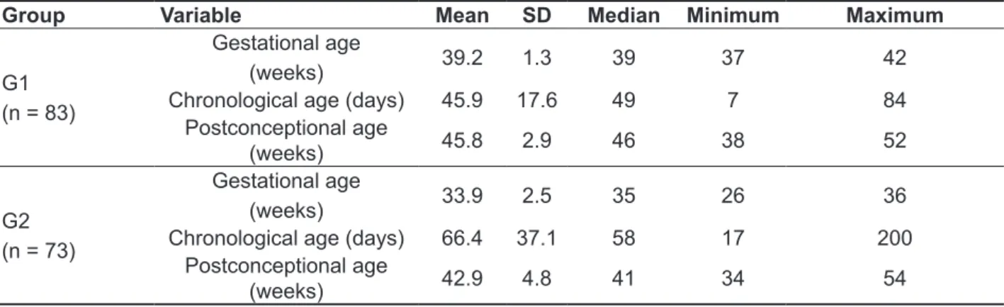
Initially, the descriptive analysis of groups G1 and G2 (Table 1) and of groups G2A and G2B (Table 2) will be shown. Gestational, chronological
and postconceptional ages were studied. It was
observed that the gestational age was signiicantly greater for the group not at risk in relation to the group at risk (G2B) (Table 2).
aminoglycosides, associated or not associated to
diuretics; and severe asphyxiation with Apgar 0-4
in the 1st minute or 0-6 in the 5th minute. Based on
the gestational age reported during the interview, two different groups were formed: the full-term
group, referred to in this study as G1 – with birth in between 37 and 42 weeks – and the preterm group, called G2 – with birth in between 26 and 37 weeks. The newborns with absence of risk indicators for
hearing disorder in their clinical history, or negative answers to the questions in the interview were
included in this study. The preterm (G2) newborns were divided into two subgroups, called G2A and G2B. G2A was composed of the preterm newborns without risk for hearing disorders; or, those who did not present any of the risk indicators for deafness. G2B was composed of the preterm newborns at risk
for hearing disorders; or, those who had at least one
risk factor for deafness according to the answers to
the interview.
The evaluation took place in a room with acoustic treatment, while the newborn was asleep in the arms of the caregiver who was seated comfortably
in a chair with arm rests.
The TEOAE were obtained using the ILO ECHOCHECK – OAE, Screener, Otodynamics equipment, non-linear click stimulus in an intensity
of 84 dB SPL, in the frequency range from 1500 to
3000 Hz. The irst ear to be tested was randomly
selected, and the one with easier access was
preferred, in order to avoid the newborn to be woken.
As an analysis criteria of the amplitude of the otoacoustic emissions, only the amplitude of the
general response was selected, and the signal/
noise ratio was studied, using a response value greater than or equal to 6 dB SPL7.
Rev. CEFAC. 2014 Jul-Ago; 16(4):1088-1096
greater than that of group G2, preterm newborns, in both ears, right (p= 0.017) and left (p = 0.048). The performance of Wilcoxon Signed Rank test showed that there is no signiicant variation in the TEOAE (in DB) in between right and left ears (Table 4).
In regards to the amplitude of the TEOAE, there
is no signiicant difference among the variables gender and ear (Table 3).
It was observed that the amplitude of the TEOAE for group G1, full-term newborns, was signiicantly
Table 1 – Descriptive analysis of groups G1 and G2 according to gestational, chronological and postconceptional ages
Group Variable Mean SD Median Minimum Maximum
G1 (n = 83)
Gestational age
(weeks) 39.2 1.3 39 37 42
Chronological age (days) 45.9 17.6 49 7 84 Postconceptional age
(weeks) 45.8 2.9 46 38 52
G2 (n = 73)
Gestational age
(weeks) 33.9 2.5 35 26 36
Chronological age (days) 66.4 37.1 58 17 200 Postconceptional age
(weeks) 42.9 4.8 41 34 54
SD: Standard Deviation
t-Student test, Mann-Whitney test
Table 2 – Analysis of gestational, chronological and postconceptional ages in the group of preterm newborns (G2), according to the presence (G2A) and absence (G2B) of risk factors
Age Group Mean SD Median Minimum Maximum p value
Gestational (weeks)
G2A 34.6 2.0 35 28 36
0.005*
G2B 32.9 2.8 33 26 36
Chronological (days)
G2A 61.8 32.9 54 17 148
0.27
G2B 72.7 41.8 62 20 200
Postconceptional
(weeks) G2AG2B 42.942.9 4.65.0 4241 3436 5454 0.95
SD: Standard Deviation t-Student test, Mann-Whitney test
Table 3 – Analysis of the transient evoked otoacoustic emissions (in dB) of the right and left ears, according to the variable gender (female and male)
Variable Gender Mean SD Median Minimum Maximum P-valuea
Right era OAE Male 18.4 5.1 19 8 30 0.60 Female 18.9 4.8 18.5 10 30
Left ear OAE Male 17.7 5.1 18 6 27 0.28 Female 18.6 4.4 19 8 30
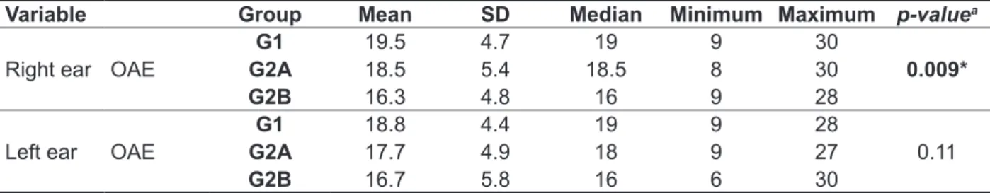
in the right ear between the subgroups of preterm not at risk, G2A and preterm at risk, G2B, and in between preterm not at risk, G2A, and full-term newborns, G1. There was no signiicant difference in the left ear, at a level of 5% between the subgroups preterm newborns not at risk G2A and preterm newborns at risk, G2B, and in between preterm newborns not at risk G2A and full-term newborns, G1 (Table 5). The Kruskal-Wallis ANOVA test showed that
there is a signiicant difference in the TEOAE of the
right ear (p = 0.009) between the subgroups G1,
G2A and G2B.
The Dunn test showed that the group of preterm
newborns at risk, G2B, had signiicantly smaller amplitude of the TEOAE in the right ear than the G1 full-term group. There was no signiicant difference
Table 4 – Analysis of the amplitude of the transient evoked otoacoustic emissions (in dB) of the right and left ears, according to the variable group
Variable Group Mean SD Median Minimum Maximum p-valuea
Right ear OAE G1 19.5 4.7 19 9 30 0.017*
G2 17.6 5.2 18 8 30
Left ear OAE G1 18.8 4.4 19 9 28 0.048*
G2 17.3 5.3 18 6 30
aMann-Whitney test.
Table 5 – Analysis of the amplitude of the transient evoked otoacoustic emissions of the right and left ears, according to the variable subgroups
Variable Group Mean SD Median Minimum Maximum p-valuea
Right ear OAE
G1 19.5 4.7 19 9 30
0.009*
G2A 18.5 5.4 18.5 8 30
G2B 16.3 4.8 16 9 28
Left ear OAE
G1 18.8 4.4 19 9 28
0.11
G2A 17.7 4.9 18 9 27
G2B 16.7 5.8 16 6 30
Rev. CEFAC. 2014 Jul-Ago; 16(4):1088-1096
the mean postconceptional age was greater than 40
weeks on the day that testing was conducted.
When analyzing age according to the presence
of risk indicators for auditory disorders in between groups G2A and G2B, group G2B had signiicantly
younger gestational age (0.005). This difference
may be due to the fact that the participants in this
group are more premature and, therefore, are more
at risk for auditory disorders. However, the statistical indings express that most newborns in both groups (G2A and G2B) were the same postconceptional age when assessed (42.9 weeks), independently of the presence of risk indicators for auditory disorder. This fact differs from studies conducted by other
authors5,8 in which the mean postconceptional age
in preterm newborns was 36.4 weeks, a younger
age than in the present study.
In the general sample, when analyzing the
amplitude of the TEOAE, according to the variables gender and ear, the mean proiles of the amplitudes show very close values, for both variables, indepen
-dently of the group, G1 or G2.
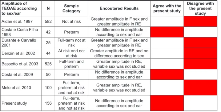
Whether or not in accordance with the present
study, other studies were selected from the available
literature, where several methodological differences
may be observed, mainly referring to gestational age and test protocols employed (types of clicks and intensity). The results of the TEOAE will be described according to the variables gender and
ear, as shown in Figure 1.
In regards to the variable ear, there was no signiicant variation of the TEOAE in each studied
group and in the sample total.
DISCUSSION
It is thought that premature infants require more
care and interventions than those born full-term, and are, therefore, more vulnerable to auditory
disorders. Authors4have stated that prematurity
constitutes a potential risk factor for the presence of auditory disorder, and may also be associated
to delay in cochlear maturity and in the myelination of the auditory pathways7. Therefore, this fact shows a need to verify cochlear function in preterm
newborns.
According to the descriptive statistical analysis, the sample characterization evidenced that the
gestational age of G1 was greater than that of G2, which was expected, considering the method
employed in this study. The mean gestational age
of G1 was 39.2 weeks and of G2 33.9 weeks, a
result that is similar to those in other studies5,9,12.
Regarding chronological age, it was observed that the subjects in G2 were evaluated later than those in G1, possibly due to the period spent in the Intensive Care Unit (ICU), where the newborns in G2 were subject to staying. Comparing the mean postconceptional ages of G1 (45.8 weeks) and of G2 (42.9 weeks), it is observed that, in both groups,
Amplitude of TEOAE according to sex/ear
N Sample
Category Encoutered Results
Agree with the present study
Disagree with the present
study
Aidan et al. 1997 582 Not at risk Greater amplitude in F sex and
greater amplitude in RE Costa e Costa Filho
1998 42 Preterm
No difference in amplitude
according to sex and ear
Durante e Carvallo
2001 25
Full-term not at
risk Greater amplitude in F sex and greater amplitude in RE
Denzin et al. 2002 44 At risk and not at risk Greater amplitude in RE and no difference according to sex
Bassetto et al. 2003 526 Full-term and preterm
Greater amplitude in RE, variable sex was not studied
Costa et al. 2009 50 Preterm No difference in amplitude according to sex and ear
Melo et al. 2010 100
Full-term,
preterm at risk and not at risk
Greater amplitude in RE, variable sex was not studied
Present study 156
Full-term,
preterm at risk and not at risk
No difference in amplitude
according to sex and ear
RE = right ear; F= Female
process of maturity is not modiied from the 38th
week onwards13.
When comparing the means of the amplitudes
of the TEOAE in the right ear, between G1, G2A and G2B, there was a statistically signiicant difference in between G2B and G1 (Table 5). G2B had TOAE amplitude in the right ear that was signii
-cantly smaller than G1. Regarding the left ear, this difference was not observed. These data strengthen the indings of other authors15,16 only in regards to
the risk indicators, but they do not report differences between ears.
It is observed that the mean of the amplitudes of the emissions of preterm newborns at risk are lower than of those not at risk, but this difference is not statistically signiicant. Similar values regarding the
mean amplitude of the emissions – around 16 dB
SPL – were observed in another study15.
There were divergences found in literature on the results of studies concerning the amplitudes of
emissions in preterm newborns, with and without risk. Some authors8found results similar to the ones
in the present study, indicating a similar functionality
of the OHC in preterm newborns at risk and not at risk, as well as the absence of auditory disorder.
Other authors15 claim that there is a difference in
emissions amplitudes between these groups.
CONCLUSION
Regarding the comparative analysis of the
amplitudes of the TEOAE, it was observed that the preterm group, G2, had smaller TEOAE amplitude than the full-term group, G1.
The preterm at risk group, G2B, had smaller
TEOAE amplitudes in the right ear than the full-term
group, G1.
There was no difference in otoacoustic emissions
regarding the variables gender and ear. The amplitudes of G1 and G2 proved similar in
both ears. Both G1 and G2 had minimum values
of 6 dB (signal-noise level) for the TEOAE. The
maximum values were the same in the right ear of both groups; they were, however, higher in the left ear in the preterm group. It was observed that some premature newborns reached amplitudes as robust as those born full-term. This inding shows that the cochlea’s functional maturity occurs in the last trimester of pregnancy, as suggested by other
authors3.
When comparing the mean TEOAE amplitude
values in between the groups, in both ears it is
seen that the mean amplitude of emissions was
signiicantly lower in G2 than in G1. Similarly,
some studies7,8 have also reported differences in the TEOAE amplitudes of full-term and preterm
newborns. However, this inding is different from the ones in the study by Goforth18 that found opposite
results, even though the analyzed groups were similar.
The parameter of amplitude of the EOAE may
be considered a signo f maturity of the peripheral auditory system of newborns, since it provides
evidence of teh presence of emissions7.
The process of maturity of the cochlea’s active
mechanisms and of the properties of the middle ear occur alongside the increase in the emissions after
38 postconceptional weeks, fact that justiies lower emissions in premature babies assessed before this
time period14.
The maturity of cochlear structures of full-term
and preterm newborns in the postnatal phase was observed in one study, due to the increase in the amplitude of the TEOAEDP in the period in between
hospital discharge and 15 to 40 days later19. Other
authors indicate that, the older the conceptional age, the greater the amplitude of the emissions8.
However, there is divergence among literature
Rev. CEFAC. 2014 Jul-Ago; 16(4):1088-1096
otoacústicas evocadas transientes em recém-nascidos a termo e pré-termo. Rev CEFAC. 2010;12(1):115- 21.
10. Morlet T, Hamburger A, Kuint J, Ari-Even RD,
Gartner M, Muchnik C et al. Assessment of medial
olicochlear system function in pre-term and full-term
newborns using a rapid test of transient otoacoustic
emissions. Clinical Otolaryngol. 2004;29:183- 92. 11. Durante AS, Carvallo RMM. Emissão Otoacústica Transitória não linear com estímulo contralateral em lactentes. Pró-Fono Rev. Atual.
Cient. 2001;13(2):271- 6.
12. Berlin CI, Hood LJ, Wen H, Szabo P, Cecola
RP, Rigby P et al. Contralateral Suppression of non-linear click-evoked otoacoustic emissions. Elsevier Science B.V. All rights reserved, Hearing Research. 1993;71(1-2):1-11.
13. Amorim AM, Lewis DR, Rodrigues GRI, Fiorini AC, Azevedo MF. Efeito de supressão das emissões otoacústicas evocadas por estímulo transiente em lactentes de risco para perda auditiva nascidos
pré-termo. Rev CEFAC. 2010;12(5):749- 55.
14. Chuang SW, Gerber SE, Thornton ARD. Evoked otoacoustic emissions in pre-term infants, Int J Pediatr Otorhinolaryngol, 1993;26:39-45.
15. Tognola G, Parazzini M, Jarger P, Brienesse P,
Ravazzani P, Grandori, F. Cochlear maturation and
otoacoustic emissions in preterm infants: a time
frequency approach. Hear Res. 2005;199:71-80.
16. Vallejo JC, Oliveira JAA, Silva MN, Gonçalves AS, Andrade MH. Análise das emissões
otoacústicas transientes em crianças com e
REFERENCES
1. Kemp T. Stimulated acoustic emissions from the human auditory system. J Acoust Soc Am.
1978;64:1386-91.
2. Lopes Filho OC, Carlos RC. Emissões Otoacústicas. In: Tratado de
3. Fonoaudiologia, São Paulo: Roca,
1997;10:221-37.
4. Lavigne-Rebillard, M.; Pujol, R. Hair cell innervation in the fetal human cochlea. Acta Otolaryngol 1988 ; 33:398-402.
5. Garcia CFD, Isaac ML, Oliveira JAA. Emissão otoacústica evocada transitória: instrumento para
detecção precoce de alterações auditivas em
recém-nascidos a termo e pré-termo. Rev Bras Otorrinolaringol. 2002;68:(3):344- 52.
6. Viveiros CM, Azevedo MF. Estudo do efeito de supressão das emissões otoacústicas evocadas transitórias em recém-nascidos a termo e
pré-termo. Fono Atual. 2004;29:(7):4-12.
7. Costa SMB, Costa Filho AO. Estudos das emissões Otoacústicas evocadas em recém-nascidos pré-termo. Pró-Fono Rev. Atual. Cient. 1998;10:(1):21- 5.
8. Basseto MCA, Chiari BM, Azevedo MF. Emissões otoacústicas evocadas transientes (EOAET): amplitude da resposta em recém-nascidos a termo e pré-termo. Rev Bras Otorrinolaringol. 2003;69(1):84-92.
9. Melo ADP, Alvarenga KF, Modolo DJ, Bevilacqua MC, Lopes AC, Agostinho-Pesse RS. Emissões
RESUMO
Objetivo: veriicar comparativamente a amplitude das emissões otoacústicas evocadas por estímulos
transientes, observando as variáveis gênero e orelha em recém-nascidos a termo e pré-termo com e sem risco para alterações auditivas. Métodos: participaram deste estudo 156 recém-nascidos, de
ambos os gêneros, com idade pós-concepcional de até 54 semanas, alocados em três grupos de acordo com a idade gestacional. O G1 foi composto de 83 recém-nascidos a termo e o G2 de 73 pré-termo. Este último, subdividido em G2A, com 42 recém-nascidos sem risco para alterações audi
-tivas e G2B com 31 recém-nascidos com risco. As emissões otoacústicas transientes foram obtidas com clique não-linear, à 84 dB NPS utilizando o Echocheck ILO EOA Screener, Otodynamics. Para análise dos resultados, foram utilizados os testes estatísticos: Mann-Whitney, qui-quadrado ou exato de Fisher, ANOVA de Kruskal-Wallis e múltiplas de Dunn, teste dos postos sinalizados de Wilcoxon; sendo considerado como signiicante o p < 0,05. Resultado: observou-se diferença signiicante nas
amplitudes das emissões otoacústicas evocadas transientes, maior em G1 (p= 0,017) do que em G2 (p=0,048) na orelha direita e esquerda. O grupo G1 (p= 0,009) apresentou amplitude das emissões otoacústicas estatisticamente maiores que G2B na orelha direita. Conclusão: o grupo a termo
apre-sentou amplitude das emissões otoacústicas maiores do que o grupo pré-termo. Não houve diferença das emissões otoacústicas entre as variáveis gênero e orelha.
2007; Disponível em: <http:// www.audiology.org/ professional/positions/jcih-early.php
19. Goforth L, Hood LJ, Berlin CI. Efferent
suppression of transient-evoked otoacoustic emissions in human infants. ARO Abstracts. 1997;20:166.
20. Marone MR. Emissões otoacústicas produto
de distorção em recém-nascidos medicados com ototóxicos. [Tese]. São Paulo: Universidade de São
Paulo; 2006. sem risco auditivo. Rev Bras Otorrinolaringol.
1999;65(4):332-6.
17. Denzin P, Carvallo RMM, Matas CG. Análises
das emissões otoacústicas transitórias em lactentes com e sem indicador de risco para
deiciência auditiva. Rev Bras Otorrinolaringol. 2002;68:874-81.
18. Joint Committee on Infant Hearing (JCIH). Position statement: principles and guidelines for early hearing detection and intervention programs
Received on: July 31, 2012 Accepted on: June 30, 2013 Mailing Address:
Helena Siano
Rua das Laranjeiras 232 – INES/DIAU Laranjeiras – RJ
CEP: 22240-001