A model of hemorrhagic cystitis
induced with acrolein in mice
1Departamento de Fisiologia e Farmacologia, 2Departamento de Morfologia,
Faculdade de Medicina, Universidade Federal do Ceará, Fortaleza, CE, Brasil
3Departamento de Farmacologia, Faculdade de Medicina de Ribeirão Preto,
Universidade de São Paulo, Ribeirão Preto, SP, Brasil C.K.L.P. Batista1,
G.A.C. Brito2, M.L.P. Souza1,
B.T.A. Leitão1, F.Q. Cunha3
and R.A. Ribeiro1
Abstract
Acrolein is a urinary metabolite of cyclophosphamide and ifosfamide, which has been reported to be the causative agent of hemorrhagic cystitis induced by these compounds. A direct cytotoxic effect of acrolein, however, has not yet been demonstrated. In the present study, the effects of intravesical injection of acrolein and mesna, the classical acrolein chemical inhibitor, were evaluated. Male Swiss mice weighing 25 to 35 g (N = 6 per group) received saline or acrolein (25, 75, 225 µg) intravesically 3, 6, 12, and 24 h before sacrifice for evaluation of bladder wet weight, macroscopic and histopathological changes by Gray’s criteria, and 3 and 24 h for assessment of increase in vascular permeability. In other animals, mesna was administered intravesically (2 mg) or systemically (80 mg/kg) 1 h before acrolein. Intravesical administration of acrolein induced a dose- and time-dependent increase in vascular permeability and bladder wet weight (within 3 h: 2.2- and 21-fold increases in bladder wet weight and Evans blue dye exuded, respectively, at doses of 75 µg/bladder), as confirmed by Gray’s criteria. Pretreatment with mesna (2-mercapto-ethanesulfonic acid), which interacts with acrolein resulting in an inactive compound, inhibited all changes induced by acrolein. Our results are the first demonstration that intravesical administration of acrolein induces hemorrhagic cystitis. This model of acrolein-induced hemorrhagic cystitis in mice may be an important tool for the evalua-tion of the mechanism by which acrolein induces bladder lesion, as well as for investigation of new uroprotective drugs.
Correspondence
R.A. Ribeiro
Departamento de Fisiologia e Farmacologia
Faculdade de Medicina, UFC Rua Coronel Nunes de Melo, 1127 60430-270 Fortaleza, CE Brasil
E-mail: [email protected]
Research supported by CNPq.
Publication supported by FAPESP.
Received July 18, 2005 Accepted July 4, 2006
Key words
•Acrolein •Bladder
•Hemorrhagic cystitis •Mesna
Introduction
Ifosfamide (IFS) and cyclophosphamide (CYP) are oxazaphosphorine-alkylating agents with a broad spectrum of antineoplas-tic activity. These drugs are also used as immunosuppressors in non-neoplastic dis-eases, such as nephrotic syndrome, systemic
side effect that limits the use of IFS and CYP (3). Both alkylating agents induce HC, but the incidence of this side effect is higher with IFS treatment (4). The cause of bladder damage appears to be related to ACR. It has been proposed that urothelial damage oc-curs by direct contact with ACR. This dam-age is followed by bladder edema, ulcer-ation, neovascularizulcer-ation, hemorrhage, and necrosis (5).
Mesna (2-mercaptoethanesulfonic acid), a thiol compound, was studied in clinical trials as a systemic uroprotective agent in the late 1970’s and became the drug of choice for this purpose within a short period of time (6). Systemic administration of mesna re-sults in regional detoxification of the urinary system (7,8). The interaction between ACR and mesna results in an inactive compound, an inert thioether, which passes innocuously through urine and does not induce any dam-age to the uroepithelium (9).
We have demonstrated that cytokines such as tumor necrosis factor (TNF)-α and
interleukin (IL)-1ß and nitric oxide (NO) are crucial mediators involved in the inflamma-tory events, including urothelial damage and hemorrhage associated with IFS- and CYP-induced HC (10-12).
It is widely accepted that ACR is the metabolite that causes HC induced by ox-azaphosphorine (5). However, most of the studies that support this conclusion were performed by injecting CYP and IFS sys-temically. No systematic studies have been reported on the potential urotoxic effect of ACR administered directly into the bladder. In the present study, we investigated whether intravesical injection of ACR induces HC in mice and whether mesna, a thiol compound, prevents this process. ACR was observed to induce a dose- and time-dependent HC which is inhibited by the co-administration of mesna, a compound clinically used to pre-vent HC in patients undergoing CYP and IFS treatments. These results confirm that ACR causes HC and suggest that
ACR-in-duced HC, in mice, could be a simple and useful model for the evaluation of new uro-protective drugs.
Material and Methods
Animals
Male Swiss mice weighing 25 to 35 g were kept in appropriate cages in temperature-con-trolled rooms, receiving food ad libitum,but without access to drinking water during the final 18 h before the experiments. All experi-mental protocols were approved by the local Animal Care and Use Committee.
Drugs
Acrolein was obtained from Sigma-Al-drich (St. Louis, MO, USA) and mesna (Mitexan®) and ifosfamide were from Asta Medica (São Paulo, SP, Brazil). All other reagents were from Sigma (St. Louis, MO, USA).
Induction of cystitis
A small midline abdominal incision was made in chloral hydrate-anesthetized mice (400 mg/kg) and 0.2 mL ACR (25, 75, and 225 µg) or saline (control) were injected into the bladder lumen through a 33-gauge needle passed through the bladder wall. After the injection, the abdominal wall was sutured with 3-0 cotton thread (Figure 1). At differ-ent times thereafter, the animals were sacri-ficed by cervical dislocation, and their blad-ders were removed by careful dissection, emptied of urine, and evaluated for the pa-rameters described below.
Measurement of vesical edema
vascular permeability, quantified by Evans blue dye extravasation. Evans blue (25 mg/ kg) was administered intravenously via the retro-orbital plexus 30 min before animal sacrifice. The bladders were dissected and placed in glass tubes containing 1 mL for-mamide. The tubes were then incubated at 56ºC overnight. The absorbance of the extracted dye was measured at 600 nm and the results are reported as the mean ± SEM amount (in µg) of Evans blue per bladder, based on a standard curve of Evans blue.
Macroscopic and microscopic evaluation
Bladders were excised, freed from sur-rounding connective tissue, and examined grossly for edema and hemorrhage. A pa-thologist performed the histological examina-tion in a single-blind fashion. Edema, bleed-ing and histological changes were evaluated according to the criteria of Gray et al. (13) as follows. Edema was considered to be severe (3+) when fluid was seen externally and inter-nally on the wall of the bladder, moderate (2+) when confined to the internal mucosa, mild (1+) when normal to moderate, and absent (0). Hemorrhage was scored as follows: (3+), in-travesical clots; (2+), mucosal hematomas; (1+), telangiectasia or dilatation of the bladder vessels; (0), normal. Histopathology was scor-ed as follows: (0), normal epithelium and absence of inflammatory cell infiltration and ulceration; (1), mild changes involving reduc-tion of epithelial cells, flattening with submu-cosal edema, mild hemorrhage and few ulcer-ations; (2), severe changes including mucosal erosions, inflammatory cell infiltration, fibrin deposition, hemorrhage, and multiple ulcer-ations. The semi-quantitative macroscopic and microscopic observations are reported as me-dians and range.
Effect of mesna on acrolein-induced hemorrhagic cystitis
The animals received the classic
uropro-tector, mesna (80 mg/kg, intraperitoneal, ip) 1 h before the injection of ACR (75 µg, intravesical, ive). Their bladders were evalu-ated 3 and 24 h after the induction of cystitis, as described above. In another set of experi-ments, mesna (2 mg; 9-fold ACR molar concentration) was administered simulta-neously with ive ACR (75 µg) and cystitis was evaluated using the same parameters and at the same times.
Statistical analysis
Data were submitted to analysis of vari-ance (ANOVA) followed by the Bonferroni test for the analysis of BWW and vesical vascular permeability. The Kruskal-Wallis and Dunn tests were used for macroscopic and microscopic analysis to compare medi-ans. The level of significance was set at P < 0.05.
Results
As illustrated in Figure 2, intravesical
and Evans blue dye exuded, respectively) and 225 g/bladder (3.4- and 37-fold increases in BWW and Evans blue dye exuded, re-spectively). The administration of ACR (75 µg/bladder) induced a time-dependent in-crease of BWW which was significant as early as 3 h (139.44%) after induction of cystitis, reaching a peak at 12 h after ACR administration (353.33%) and decreasing thereafter, but which continued to be signifi-cant up to 24 h (239.77%; Figure 3). Accord-ing to Gray’s score, ACR (75 µg/bladder) induced a time-dependent increase in mac-roscopic and micmac-roscopic scores which were significant at 3 h, with a peak occurring at 24 h (Table 1).
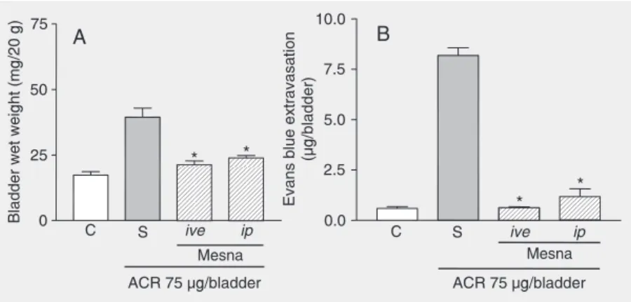
ACR-evoked increases in BWW and Evans blue extravasation, observed at 3 h after ACR injection, were significantly in-hibited by systemic or local (ive) pretreat-ment with mesna (80 mg/kg, ip, and 2 mg/ bladder, ive, respectively; Figure 4). The same was observed 24 h after ACR injection (data not shown).
Cystitis observed 3 h after ACR adminis-tration was characterized macroscopically by the presence of severe edema, receiving a score of 2 (2-3), and by extensive hemor-rhage with mucosal hematomas, receiving a score of 2 (1-3), and was significantly (P < 0.05) different from the control group which received a score of 0 (0-0) for edema and hemorrhage. The bladders evaluated after 24 h presented severe edema, receiving a score of 3 (2-3), and mucosal hematomas and intravesical clots, receiving a score of 2 (2-3). Macroscopic analysis of the bladder at 3 and 24 h after ACR injection revealed that the mesna pretreatments (ip and ive) signifi-cantly reduced (P < 0.05) the parameters analyzed (Table 2). According to Gray’s histopathological criteria, when compared with normal bladders, microscopic analysis of the bladders of mice injected 3 h before with 75 µg/bladder of ACR revealed exten-sive cystitis characterized by acute inflam-mation with vascular congestion, edema, Figure 2. Dose-dependent increase in bladder wet weight and Evans blue extravasation
induced by intravesically administered acrolein (ACR). Bladder wet weight (A) and Evans blue extravasation (B), in untreated animals (C, control) and in animals injected intravesically 3 h earlier with acrolein (25, 75, 225 µg/bladder). Data are reported as means ± SEM (N = 6). *P < 0.05 compared with control (C) (ANOVA and Bonferroni test).
Figure 3. Kinetics of the increase in bladder wet weight after injec-tion of acrolein (75 µg/bladder). The dashed line indicates the value of bladder wet weight of the control group not treated with acrolein. Data are reported as the mean ± SEM of bladder wet weight (mg/20 g) for 6 animals/ group. *P < 0.05 compared with control (ANOVA and Bonferroni test).
injection of ACR (25 to 225 g/bladder) in-duced a dose-dependent increase in both BWW and plasma leakage within 3 h, with significant differences at doses of 75 µg/ bladder (2.2- and 21-fold increases in BWW
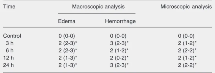
Table 1. Time course of macroscopic and microscopic changes after intravesical injection of acrolein (75 µg/bladder).
Time Macroscopic analysis Microscopic analysis
Edema Hemorrhage
Control 0 (0-0) 0 (0-0) 0 (0-0)
3 h 2 (2-3)* 3 (2-3)* 2 (1-2)*
6 h 2 (2-3)* 2 (1-2)* 2 (2-2)*
12 h 2 (1-3)* 2 (0-2)* 2 (1-2)*
24 h 2 (1-3)* 3 (2-3)* 2 (2-2)*
The results are reported as median and range within parentheses (N = 6).
hemorrhage with fibrin deposition, neutro-phil infiltration, and epithelial denudation, receiving a score of 2 (1-2). Twenty-four hours after ACR administration the micro-scopic analysis was similar, but extensive hemorrhage was observed, receiving a score of 2 (2-2). These alterations were almost abolished (P < 0.05) by treatment of the ACR-injected mice with mesna (2 mg/blad-der, ive or 80 mg/kg, ip). The scores of Gray’s parameters (median and range) are shown in Table 2.
Discussion
It is accepted that HC, a side effect ob-served in patients under CYP and IFS treat-ment, is caused by the urinary metabolite, ACR (5). Most of the studies that support this conclusion were performed by adminis-tering CYP and IFS systemically (14). How-ever, in the current study we determined whether ive administration of ACR causes HC. ACR injected into the mouse bladder induced a dose- and time-dependent HC as determined by the increase in BWW and Evans blue extravasation, parameters that have been used to quantify cystitis in rats (12) and mice (15). Both parameters were already significant 3 h after ACR adminis-tration, peaking at 12 h and decreasing there-after, with no further increase after 24 h, suggesting that a plateau had been reached. The ACR-induced HC was confirmed by macroscopic and histopathological changes of the bladder analyzed by the Gray’s crite-ria, which were used to quantify HC (13). The profile of HC observed after ACR ad-ministration was similar to that observed when CYP or IFS was administered to mice (15) or rats (12). This fact, taken together with the demonstration that ACR is found in the urine of humans (16) and of experimen-tal animals treated with CYP or IFS (17), as described in the literature (5,14,18,19), indi-cate that ACR is the urine metabolite respon-sible for the HC observed after
oxazaphos-Figure 4. Effect of different routes of administration of mesna on bladder wet weight (A) and Evans blue extravasation (B) in acrolein-induced hemorrhagic cystitis. Acrolein (ACR) (75 µg/bladder)-induced increase in bladder wet weight and Evans blue extravasation were measured 3 h after the induction of cystitis in mice treated with vehicle (saline, S). Mesna-treated animals received 2 mg/bladder mesna injected intravesically (ive) simultaneously with ACR or 80 mg/kg of mesna injected intraperitoneally (ip) 1 h before ACR injection. Data are reported as means ± SEM (N = 6). *P < 0.05 compared to saline control group (S, treated with acrolein and saline) (ANOVA and Bonferroni test).
phorine treatment.
The macroscopic and microscopic analy-sis of the bladder from ACR-injected mice, revealed intense vascular congestion and edema in the early phase (3 h), findings which were followed by neutrophil infiltration, hem-orrhage, ulceration and severe necrosis, some-times with perforation (24 h after ACR ad-ministration). Most of these findings are also observed after CYP or IFS administration to humans (3), or to experimental animals (20).
Table 2. Effect of intraperitoneally and intravesically injected mesna on the macro-scopic and micromacro-scopic effects of cystitis induced by intravesical injection of acrolein.
Group Macroscopic analysis
Edema Hemorrhage Microscopic analysis
3 h 24 h 3 h 24 h 3 h 24 h
Control 0 (0-0) 0 (0-0) 0 (0-0) 0 (0-0) 0 (0-0) 0 (0-0)
ACR 2 (2-3) 3 (2-3) 2 (1-3) 2 (2-3) 2 (1-2) 2 (2-2)
ACR + mesna ive 0 (0-1)* 0 (0-1)* 0 (0-1)* 0 (0-1)* 0 (0-1)* 0 (0-1)*
ACR + mesna ip 1 (0-1)* 0 (0-1)* 0 (0-1)* 1 (0-1)* 0 (0-2)* 0 (0-1)*
The mechanism by which ACR causes HC has not been fully elucidated. However, there is evidence that, after administration of CYP or IFS, ACR appears within the bladder, causing a reduction of endogenous glutathi-one (21,22) and inducing the generation of free radicals, such as superoxide anion and hydroxyl radical, which initiate lipid peroxi-dation and other cell damage (23,24). Re-cently, we demonstrated that the cytokines TNF-α and IL-1ß, acting by activation of the
L-arginine/iNOS/NO pathway, might also play a crucial role in the geneses of patho-physiological events of HC observed in ani-mals treated with CYP or IFS (10). In this context, previous reports have shown that ex-posure of the respiratory tract to ACR results in an increase in the production of inflamma-tory mediators such as eicosanoids and also causes a significant influx of neutrophils into the bronchoalveolar lavage (25). Moreover, evidence also exists that ACR increases the release of IL-8 by neutrophils (26).
In the present study, to validate the mo-del of ACR-induced HC, the animals were treated with mesna, a classical uroprotector agent with established clinical (8) and
ex-perimental efficacy (20,27). The increase in BWW and Evans blue extravasation was blocked by pretreatment of the mice with mesna, regardless of local or systemic ad-ministration. Furthermore, mesna also blocked the bladder’s macroscopic and microscopic changes induced by ive administration of ACR. There is evidence that ACR reacts with mesna producing an inert thioether that is eliminated in the urine (19). Thus, in this model, as is also the case for humans or animals treated with oxazaphosphorine (20), mesna presented its uroprotective effect when administered sys-temically or locally.
Our results are the first demonstration that intravesical administration of ACR in-duces HC. This model of ACR-induced HC in mice may be an important tool for the evaluation of the mechanism by which ACR induces bladder lesion, as well as, for inves-tigation of new uroprotective drugs.
Acknowledgments
The authors gratefully acknowledge M. Silvandira F. Pinheiro, J. Ivan R. Sousa and Sergio R. Rosas for technical assistance.
References
1. Calabresi P, Chabner BA. Antineoplastic agents. In: Goldman AF, Rall TW, Niss AS, Taylor P (Editors), The pharmacological basis of therapeutics. Singapore: McGraw-Hill, 1992.
2. Dechant KL, Brogden RN, Pilkington T, Faulds D. Ifosfamide/mesna. A review of its antineoplastic activity, pharmacokinetic properties and therapeutic efficacy in cancer. Drugs 1991; 42: 428-467. 3. Stillwell TJ, Benson RC Jr. Cyclophosphamide-induced hemorrhagic
cystitis. A review of 100 patients. Cancer 1988; 61: 451-457. 4. Ratliff TL, Williams RD. Hemorrhagic cystitis, chemotherapy, and
bladder toxocity. J Urol 1998; 159: 1044.
5. Cox PJ. Cyclophosphamide cystitis - identification of acrolein as the causative agent. Biochem Pharmacol 1979; 28: 2045-2049. 6. Katz A, Epelman S, Anelli A, Gorender EF, Cruz SM, Oliveira RM, et
al. A prospective randomized evaluation of three schedules of mesna administration in patients receiving an ifosfamide-containing che-motherapy regimen: sustained efficiency and simplified administra-tion. J Cancer Res Clin Oncol 1995; 121: 128-131.
7. Elias AD, Eder JP, Shea T, Begg CB, Frei E III, Antman KH. High-dose ifosfamide with mesna uroprotection: a phase I study. J Clin
Oncol 1990; 8: 170-178.
8. Scheulen ME, Niederle N, Bremer K, Schutte J, Seeber S. Efficacy of ifosfamide in refractory malignant diseases and uroprotection by mesna: results of a clinical phase II-study with 151 patients. Cancer Treat Rev 1983; 10 (Suppl A): 93-101.
9. Brock N, Pohl J. The development of mesna for regional detoxifica-tion. Cancer Treat Rev 1983; 10 (Suppl A): 33-43.
10. Ribeiro RA, Freitas HC, Campos MC, Santos CC, Figueiredo FC, Brito GA, et al. Tumor necrosis factor-alpha and interleukin-1beta mediate the production of nitric oxide involved in the pathogenesis of ifosfamide induced hemorrhagic cystitis in mice. J Urol 2002; 167: 2229-2234.
11. Gomes TN, Santos CC, Souza-Filho MV, Cunha FQ, Ribeiro RA. Participation of TNF-alpha and IL-1 in the pathogenesis of cyclo-phosphamide-induced hemorrhagic cystitis. Braz J Med Biol Res
1995; 28: 1103-1108.
247-256.
13. Gray KJ, Engelmann UH, Johnson EH, Fishman IJ. Evaluation of misoprostol cytoprotection of the bladder with cyclophosphamide (Cytoxan) therapy. J Urol 1986; 136: 497-500.
14. Brock N, Stekar J, Pohl J, Niemeyer U, Scheffler G. Acrolein, the causative factor of urotoxic side-effects of cyclophosphamide, ifos-famide, trofosfamide and sufosfamide. Arzneimittelforschung 1979; 29: 659-661.
15. Assreuy AM, Martins GJ, Moreira ME, Brito GA, Cavada BS, Ribeiro RA, et al. Prevention of cyclophosphamide-induced hemorrhagic cystitis by glucose-mannose binding plant lectins. J Urol 1999; 161: 1988-1993.
16. Al-Rawithi S, el-Yazigi A, Ernst P, Al-Fiar F, Nicholls PJ. Urinary excretion and pharmacokinetics of acrolein and its parent drug cyclophosphamide in bone marrow transplant patients. Bone Mar-row Transplant 1998; 22: 485-490.
17. Shaw IC, Graham MI, McLean AE. 2-Chloroacetaldehyde: a meta-bolite of cyclophosphamide in the rat. Cancer Treat Rev 1983; 10 (Suppl A): 17-24.
18. Kolb NS, Hunsaker LA, Vander Jagt DL. Aldose reductase-cata-lyzed reduction of acrolein: implications in cyclophosphamide toxic-ity. Mol Pharmacol 1994; 45: 797-801.
19. Wagner T. Ifosfamide clinical pharmacokinetics. Clin Pharmacokinet
1994; 26: 439-456.
20. Morais MM, Belarmino-Filho JN, Brito GA, Ribeiro RA. Pharmacolo-gical and histopatholoPharmacolo-gical study of cyclophosphamide-induced
hemorrhagic cystitis - comparison of the effects of dexamethasone and mesna. Braz J Med Biol Res 1999; 32: 1211-1215.
21. Pocernich CB, Cardin AL, Racine CL, Lauderback CM, Butterfield DA. Glutathione elevation and its protective role in acrolein-induced protein damage in synaptosomal membranes: relevance to brain lipid peroxidation in neurodegenerative disease. Neurochem Int
2001; 39: 141-149.
22. Nardini M, Finkelstein EI, Reddy S, Valacchi G, Traber M, Cross CE, et al. Acrolein-induced cytotoxicity in cultured human bronchial epi-thelial cells. Modulation by alpha-tocopherol and ascorbic acid.
Toxicology 2002; 170: 173-185.
23. Adams JD Jr, Klaidman LK. Acrolein-induced oxygen radical forma-tion. Free Radic Biol Med 1993; 15: 187-193.
24. Patel JM. Stimulation of cyclophosphamide-induced pulmonary mi-crosomal lipid peroxidation by oxygen. Toxicology 1987; 45: 79-91. 25. Leikauf GD. Mechanisms of aldehyde-induced bronchial reactivity:
role of airway epithelium. Res Rep Health Eff Inst 1992; 1-35. 26. Mio T, Romberger DJ, Thompson AB, Robbins RA, Heires A,
Rennard SI. Cigarette smoke induces interleukin-8 release from human bronchial epithelial cells. Am J Respir Crit Care Med 1997; 155: 1770-1776.