ISSN 0100-879X
BIOMEDICAL SCIENCES
AND
CLINICAL INVESTIGATION
www.bjournal.com.br
www.bjournal.com.br
Volume 43 (04) 325-408 April 2010
Braz J Med Biol Res, March 2010, Volume 43(4) 325-329
9
Effect of (-)-
∆
-tetrahydrocannabinoid on the hepatic redox state
of mice
C.E. Pinto, E. Moura, M.P. Serrão, M.J. Martins and M.A. Vieira-Coelho
Institutional Sponsors
Effect of (-)-∆
9
-tetrahydrocannabinoid on
the hepatic redox state of mice
C.E. Pinto
1, E. Moura
1,3, M.P. Serrão
1, M.J. Martins
2and M.A. Vieira-Coelho
1,31Instituto de Farmacologia e Terapêutica, 2Departamento de Bioquímica,
Faculdade de Medicina, Universidade do Porto, Porto, Portugal
3Instituto de Biologia Molecular e Celular, Universidade do Porto, Porto, Portugal
Abstract
(-)-∆9-Tetrahydrocannabinol (∆9-THC), a psychoactive component of marijuana, has been reported to induce oxidative damage
in vivo and in vitro. In this study, we administered ∆9-THC to healthy C57BL/6J mice aged 15 weeks in order to determine its ef-fect on hepatic redox state. Mice were divided into 3 groups: ∆9-THC (N = 10), treated with 10 mg/kg body weight ∆9-THC daily;
VCtrl (N = 10), treated with vehicle [1:1:18, cremophor EL® (polyoxyl 35 castor oil)/ethanol/saline]; Ctrl (N = 10), treated with
saline. Animals were injected ip twice a day with 5 mg/kg body weight for 10 days. Lipid peroxidation, protein carbonylation and DNA oxidation were used as biomarkers of oxidative stress. The endogenous antioxidant defenses analyzed were glutathione (GSH) levels as well as enzyme activities of superoxide dismutase, catalase, glutathione S-transferase, glutathione reductase, and glutathione peroxidase (GPx) in liver homogenates. The levels of mRNA of the cannabinoid receptors CB1 and CB2 were
also monitored. Treatment with ∆9-THC did not produce significant changes in oxidative stress markers or in mRNA levels of CB1 and CB2 receptors in the liver of mice, but attenuated the increase in the selenium-dependent GPx activity (Δ9-THC: 8%; VCtrl: 23% increase) and the GSH/oxidized GSH ratio (Δ9-THC: 61%; VCtrl: 96% increase), caused by treatment with the vehicle. Δ9-THC administration did not show any harmful effects on lipid peroxidation, protein carboxylation or DNA oxidation
in the healthy liver of mice but attenuated unexpected effects produced by the vehicle containing ethanol/cremophor EL®.
Key words: (-)-∆9-Tetrahydrocannabinol; Oxidative stress; Liver; Cannabinoid receptors; Cremophor EL®
Introduction
Correspondence: M.A. Vieira-Coelho, Instituto de Farmacologia e Terapêutica, Faculdade de Medicina, Universidade do Porto, Alameda Prof. Hernâni Monteiro, 4200-319 Porto, Portugal. Fax: +351-22-551-3643. E-mail: mavc@med.up.pt
Received August 12, 2009. Accepted February 25, 2010. Available online March 12, 2010. Published April 12, 2010.
The plant Cannabis sativa, also known as marijuana, is the most widely used illicit drug. Its use is common for medicinal purposes and in fact, cannabinoids, the active compounds of the plant, have been shown to possess anti-inflammatory, analgesic and antiemetic properties, among others (for a review, see Ref. 1).
In the early 1960’s, the plant’s most psychoactive cannabinoid was isolated and identified as (-)-Δ9
-tetrahydrocannabinol (Δ9-THC), a highly lipophilic
sub-stance (2). Δ9-THC is now available as a prescription
drug in some countries under the trade names Marinol®
(synthetic Δ9-THC) and Sativex® (Δ9-THC and cannabidiol
extracted from the plant). Once administered, Δ9-THC is
rapidly absorbed by tissues and metabolized, mainly in the liver, to 11-hydroxy-tetrahydrocannabinol (3).
It is known that Δ9-THC non-selectively activates the
cannabinoid receptors CB1 and CB2 (4). In healthy livers, these receptors are faintly or not expressed, respectively,
but are substantially up-regulated in experimental liver injury and in liver cirrhosis of various origins (5-7). The activation of CB1 and CB2 receptors has been described to induce hepatic profibrogenic (8) and antifibrogenic effects (9), respectively, in the liver, but no data are available regard-ing the effects of Δ9-THC administration on either healthy
or fibrotic liver.
It has been reported that Δ9-THC can induce oxidative
damage in vitro (10), and more recently in vivo (11), and it is known that, as a result of damaged cellular structures and oxidized macromolecules (DNA, lipids and enzymatic proteins) induced by increased oxidative stress, paren-chymal liver necrosis eventually occurs (12). Therefore, in the present investigation, we studied the effect of Δ9
326 C.E. Pinto et al.
defense. The levels of mRNA of each cannabinoid receptor were also monitored.
Material and Methods
Reagents
Δ9-THC (supplied in absolute ethanol) was purchased
from Lipomed AG (Switzerland). 2,4-Dinitrophenylhydrazine (DNPH) and thiobarbituric acid were purchased from Merck (Germany). Cremophor EL®, 1,2-chloro-2-dinitrobenzene,
2-vinylpyridine, 5,5’-dithio-bis(2-nitrobenzoic acid) (DTNB), 8-hydroxy-2’-deoxyguanosine (8-OHdG), cumene hy-droperoxide, glutathione reductase, nicotinamide adenine dinucleotide phosphate (NADPH), malonaldehyde, nitrotet-razolium blue chloride, nuclease P1, oxidized glutathione (GSSG), reduced glutathione (GSH), sodium pentobarbital, xanthine, and xanthine oxidase were all from Sigma (USA); alkaline phosphatase and, guanidine hydrochloride were purchased from Applichem (Germany).
Animals and treatment
C57BL/6J mice, 15 weeks of age and weighing 22-29 g, were obtained from Charles River Laboratories (Spain). The investigation conformed to the Guide for the Care and Use of Laboratory Animals published by the USA National Institutes of Health (NIH Publication #85-23, revised in 1996) and the experiments were performed according to the Portuguese law on animal welfare. Animals were kept under controlled environmental conditions (22-24°C, 12-h light/dark cycles) and provided with food and water ad libitum
before and for the duration of the experiment.
Groups of mice (N = 10) were given two daily intra-peritoneal injections, at 8 am and 8 pm, of saline (control group, Ctrl), vehicle (VCtrl, 1:1:18, cremophor EL®/ethanol/
saline solution) or Δ9-THC (a total daily dose of 10 mg/kg
body weight) for a period of 10 days and were killed on the morning after the last injection.
Sample preparation
Animals were anesthetized with sodium pentobarbital (60 mg/kg body weight) and perfused with ice-cold 0.9% NaCl. The liver was extracted, weighed and homogenized with 2 mL of a homogenization solution (25 mM KH2PO4,
30 mM Na2HPO4, 0.1% Triton X-100, pH 7.0) per gram of
liver. The resulting homogenized liver tissue was stored at -80°C.
Protein determination
The amount of protein in each liver homogenate was determined by the method of Bradford (13) using bovine serum albumin as standard.
Evaluation of oxidative stress
The amount of lipid peroxidation was determined by the thiobarbituric acid reactive substances method (14).
Briefly, each liver homogenate was centrifuged at 16,000 g for 10 min at 4°C and the supernatant solution was mixed with 10% trichloroacetic acid (1:2) and centrifuged at 6000 g for 1 min at 4°C. The resulting supernatant solu-tion was mixed with the same volume of 1% thiobarbituric acid and incubated at 95°C for 40 min. After cooling to room temperature, absorbance was read at 535 nm. The malonaldehyde equivalent nmol/mg protein was calculated using a malonaldehyde standard curve.
To determine the extent of protein carbonylation, the carbonyl content of each liver sample was measured using the supernatant obtained from the liver homogenate mixed with 10% trichloroacetic acid (1:2) (14). After centrifuging at 13,000 g for 2 min at 4°C, 0.5 mL DNPH (10 mM) in 2 M HCl solution was added to the resulting pellet. For the blank, only 0.5 mL 2 M HCl was added to the pellet. Samples were incubated for 1 h at room temperature and vortexed every 10 min, followed by another 15 min on ice after adding 0.5 mL 20% trichloroacetic acid. The pellet was then washed three times with ethanol-ethyl acetate (1:1) by centrifuging at 13,000 g for 2 min at 4°C and dissolved in 0.5 mL 6 M guanidine solution overnight. Insoluble mate-rial was removed by centrifuging at 3000 g for 15 min and absorbance was read at 340 nm. The carbonyl content was calculated using the extinction coefficient of DNPH of 22,000 M-1 cm-1 and the results are reported as carbonyl
group nmol/mg protein.
DNA damage was measured by determining the amount of 8-OHdG. DNA was extracted using the Animal Tissues Genomic DNA Mini-prep Kit from V-gene® (Bioron,
Germany), and incubated with 4 U nuclease P1, prepared in 20 mM acetate buffer, pH 5.0, for 30 min at 37ºC. This was followed by 1-h incubation at 37ºC with 1.3 U alkaline phosphatase prepared in 0.1 M Tris-HCl buffer, pH 7.5. Chloroform was added to the samples and these were vortexed for 10 s and centrifuged at 5000 g for 5 min, and the supernatant was collected for quantification of 8-OHdG (14). The samples were analyzed by high performance liquid chromatography (Gilson 302, Gilson Medical Elec-tronics, France) on a 25 x 3.2 mm ID, Superspher® RP18-4
Endcap column (HiCHROM, Germany) and monitored for electrochemical detection (Gilson 141) at 10 V. The sol-vent was 50 mM KH2PO4/K2HPO4, 10% (v/v) methanol,
pH 5.5, and the column was operated at a flow rate of 0.5 mL/min. 8-OHdG concentration (pmol 8-OHdG/µg DNA) was calculated from a calibration curve using commercial 8-OHdG as the standard.
Antioxidant enzyme activities
GSSG from the amount of total glutathione (GSX) mea-sured. GSH, GSSG and GSX data are reported as nmol/ mg protein.
The activity of the glutathione reductase (GR) enzyme, in U/mg (1 U GR defined as the amount of enzyme required to convert 1 nmol NADPH to NADP+ per min) was determined by monitoring NADPH oxidation at 340 nm for 5 min (extinc-tion coefficient of NADP+: 6.22 mM-1 cm-1) (15).
By monitoring NADPH oxidation at 340 nm for 5 min in the presence of GR, which reduces GSSG using cumene hydroperoxide as substrate, we were able to determine the glutathione peroxidase (GPx) level of activity in the liver samples (14). The selenium-dependent GPx (Se-GPx) was assayed using H2O2 as substrate instead of cumene
hydroperoxide. Both activities are reported as U/mg protein (1 U GPx and Se-GPx defined as the amount of enzyme required to convert 1 nmol NADPH to NADP+ per min).
Glutathione-S-transferase (GST) activity, in U/mg protein (1 U GST defined as the amount of enzyme required to cata -lyze the formation of 1 µmol S-2,4-dinitrophenylglutathione per min), were determined by monitoring the formation of GSH-1,2-chloro-2-dinitrobenzene conjugates for 5 min at 340 nm (extinction coefficient of S-2,4-dinitrophenylgluta -thione: 9.6 mM-1 cm-1) (14).
Catalase activity, in U/mg protein (1 U catalase defined as the amount of enzyme required to hydrolyze 1 µmol H2O2
per min), was determined by monitoring H2O2
decomposi-tion at 240 nm for 30 s (14) (extincdecomposi-tion coefficient of H2O2:
0.00394 ± 0.0002 mM-1 mm-1).
Superoxide dismutase (SOD) activity was determined by the rate of inhibition of the reduction of nitrotetrazolium blue by superoxide radicals generated by
a xanthine-xanthine oxidase system and monitored at 550 nm (14). SOD activity is reported as U/mg protein (1 unit was the amount of SOD inhibit-ing the reaction rate by 50% under the given assay conditions).
Real-time PCR
The real-time polymerase chain reaction (RT-PCR) was carried out as previously described (16). RNA from liver samples (N = 5, per group) was isolated using the PureZOL RNA isolation reagent (Bio-Rad, USA). Total RNA (1 μg per sample) was DNase treated and reverse tran-scribed according to manufacturer instructions (iScriptTM cDNA
Synthe-sis Kit, Bio-Rad). For RT-PCR, 50 μL amplification mixture (iQTM SYBR®
Green Supermix, Bio-Rad) was used containing 20 ng reverse transcribed RNA and specific primers (250 nM)
(Sigma) for cnr1 (sense: 5’-GGGCAAATTTCCTTGTAGC A-3’; anti-sense: 5’-GGCTAACGTGACTGAGAAA-3’), cnr2
(sense: 5’-CATCAGGATTTTGAACGACGA-3’; anti-sense: 5’-CTGTGCATGCCTGCTCTTAG-3’) and β-actin (sense: 5’-CTAAGGCCAACCGTGAAAAG-3’; anti-sense: 5’-AC-CAGAGGCATACAGGGACA-3’). Reactions were run in duplicate (20 μL) on an MJ Mini detector (Bio-Rad, USA). The cycling conditions were: 15 min polymerase activation at 95°C and 40 cycles at 95°C for 15 s, at 58°C for 30 s, and at 72°C for 30 s. Results were normalized to β-actin values and expressed as log2 (sample/control).
Statistical analysis
Data are reported as arithmetic means ± SEM. Sig-nificant differences between means were evaluated by ANOVA followed by the Newman-Keuls test, with the level of significance set at P < 0.05.
Results
Hepatic levels of oxidative stress biomarkers, lipid per-oxidation (0.052 ± 0.004 malonaldehyde equivalent nmol/ mg protein), protein carbonylation (0.370 ± 0.042 carbonyl group nmol/mg protein) and 8-OHdG (14.26 ± 6.00 pmol 8-OHdG/µg DNA), for controls were not significantly dif -ferent for mice treated with Δ9-THC and the vehicle (data
not shown).
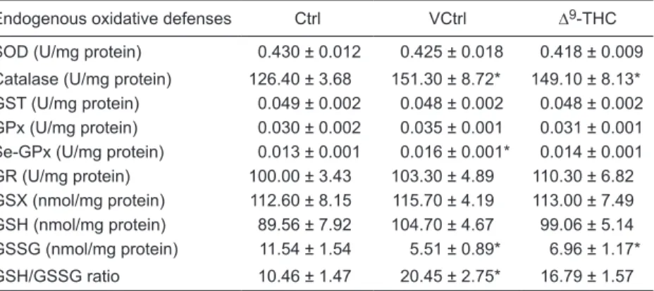
The activity levels of the enzymes involved in the en-dogenous antioxidant defense and glutathione levels are presented in Table 1. The levels of GSSG were significantly decreased in the liver of mice treated with Δ9-THC compared
Table 1. Endogenous oxidative defenses in liver homogenates of C57BL/6J mice: activ-ity levels of enzymes involved in the endogenous antioxidant defenses and glutathione levels.
Endogenous oxidative defenses Ctrl VCtrl ∆9-THC
SOD (U/mg protein) 0.430 ± 0.012 0.425 ± 0.018 0.418 ± 0.009
Catalase (U/mg protein) 126.40 ± 3.68 151.30 ± 8.72* 149.10 ± 8.13*
GST (U/mg protein) 0.049 ± 0.002 0.048 ± 0.002 0.048 ± 0.002
GPx (U/mg protein) 0.030 ± 0.002 0.035 ± 0.001 0.031 ± 0.001
Se-GPx (U/mg protein) 0.013 ± 0.001 0.016 ± 0.001* 0.014 ± 0.001
GR (U/mg protein) 100.00 ± 3.43 103.30 ± 4.89 110.30 ± 6.82
GSX (nmol/mg protein) 112.60 ± 8.15 115.70 ± 4.19 113.00 ± 7.49 GSH (nmol/mg protein) 89.56 ± 7.92 104.70 ± 4.67 99.06 ± 5.14 GSSG (nmol/mg protein) 11.54 ± 1.54 5.51 ± 0.89* 6.96 ± 1.17*
GSH/GSSG ratio 10.46 ± 1.47 20.45 ± 2.75* 16.79 ± 1.57
Data are reported as means ± SEM (N = 10). Ctrl = mice treated with saline; VCtrl = mice treated with vehicle [1:1:18, cremophor EL® (polyoxyl 35 castor oil)/ethanol/saline]; Δ9 -THC = mice treated with 10 mg (-)-Δ9-tetrahydrocannabinol/kg body weight per day for 10
days. SOD = superoxide dismutase; GST = glutathione-S-transferase; GPx = glutathione peroxidase; Se-GPx = selenium-dependent GPx; GR = glutathione reductase; GSX = total
328 C.E. Pinto et al.
to the Ctrl group. A similar decrease was also observed in the liver of mice treated with the vehicle. Neither treatment with the vehicle nor Δ9-THC induced significant changes in
the amount of GSX measured in the liver. The calculated amount of GSH for the liver of mice treated with Δ9-THC
and for the liver of mice treated with the vehicle did not differ significantly from one another or when compared with the Ctrl group (although a tendency to an increase was found for both, which was higher in the vehicle group). The GSH/ GSSG ratios determined were found to be significantly increased in the VCtrl group, but not in the Δ9-THC group,
when compared to the Ctrl group.
Treatment with Δ9-THC produced no statistically signifi
-cant changes in the enzymatic activity of the glutathione-dependent antioxidant enzymes GR and GST. Measurement of GPx, also a glutathione-dependent enzyme, tended to be increased only in the liver of mice from the VCtrl group. Se-GPx activity was only significantly increased in liver of mice from the VCtrl group.
Treatment with Δ9-THC did not produce statistically
significant changes in the activity levels of the scavenger enzyme SOD. However, the activity level of catalase, also a scavenger enzyme, was significantly increased to a similar extent in the liver of mice treated with Δ9-THC and of mice
treated with the vehicle when compared to the Ctrl group. The gene expression of the cannabinoid receptors CB1 [Ctrl: 0.000 ± 0.464; VCtrl: 0.115 ± 0.668; Δ9-THC: 0.283 ±
0.459 log2 (sample/control)] and CB2 [Ctrl: 0.000 ± 1.351;
VCtrl: 0.837 ± 0.504; Δ9-THC: 0.497 ± 0.534 log
2 (sample/
control)], as measured by RT-PCR, was not significantly changed in the liver by any of the treatments.
Discussion
In this study, we show that chronic administration of Δ9-THC did not produce any significant changes in the
hepatic redox state of healthy mice, even though there were unexpected changes induced by administration of the vehicle containing ethanol/cremophor EL® alone that
were attenuated by Δ9-THC.
Since it is known that liver injury is closely related to an increase in oxidative stress, we first assessed the level of damage to lipids, proteins and DNA in the liver of mice treated with Δ9-THC. The levels of these oxidative stress
biomarkers in the liver were not changed by Δ9-THC
treat-ment, suggesting that no damage was inflicted by either the vehicle or Δ9-THC administration.
Regardless of the lack of damage produced by Δ9-THC
treatment as measured by these parameters, we found indications that some oxidative stress was unexpectedly produced by the vehicle. Catalase activity was significantly increased by 20% in the liver of mice treated with the vehicle. Similarly, in the same group of animals, Se-GPx activity was significantly increased by 23%.
Another reliable oxidative stress indicator is the level of
glutathione, a tripeptide with antioxidant capacity. A statisti-cally significant decrease of GSSG (52%) was measured in the liver of mice treated with the vehicle. Since the GSX level measured in the liver of mice treated with the vehicle was similar to the Ctrl group, the GSH/GSSG ratio was significantly increased in the liver of mice from the VCtrl group (96%) when compared to the Ctrl group.
If the level of GSX is maintained, an increase of the GSH/GSSG ratio translates into an increased antioxidant capacity as more GSH is available (17). Although one may jump to the conclusion that the increased GSH/GSSG ratio is an indication of less oxidative stress produced by the vehicle, our results, taken together, show that some oxi-dative stress was induced by it: 1) catalase activity levels were increased; 2) the GSH levels were not significantly increased, but their tendency to increase supports the sig-nificantly increased Se-GPx activity found by us (otherwise this increased activity would lead to a GSH decrease), and 3) it has been shown that perfusing the liver with reagents that oxidize internal GSH to GSSG (by means of GPx) induces GSSG release into bile (17), which explains the reduction of GSSG observed by us.
Although some degree of oxidative stress was produced by the vehicle, the amount present or the time of exposure was not sufficient to cause any significant damage to the liver: we found no changes in the levels of the oxidative stress biomarkers measured.
It should be stated that the changes induced by the ve-hicle were unexpected since this is a very common veve-hicle used for Δ9-THC administration in vivo. In fact, the present
study shows that care should be taken in the interpretation of the results obtained with this ethanol/cremophor EL®
(polyoxyl 35 castor oil) solution, especially in oxidative stress studies.
Even though no oxidative stress was produced by Δ9
-THC administration, there is some evidence in our study that, in fact, Δ9-THC could protect the liver against
oxida-tive stress since it decreased the effects induced by the administration of the vehicle.
Δ9-THC administration increased the Se-GPx activity
by only 8%. Although much lower, this increase was not significantly different from the activity determined for the VCtrl group (a 23% increase compared to Ctrl). The total GPx activity measured was also increased (17%) in the VCtrl group, but again it was only increased by 3% in the liver of mice from the Δ9-THC group. Taken together, these
results suggest attenuation by Δ9-THC treatment of the effect
brought about by administration of the vehicle.
Similarly, when comparing the GSSG levels in the liver of Δ9-THC-treated mice with the GSSG levels in the liver
of the Ctrl group, the measured decrease (42%) was less pronounced than that induced by treatment with the vehicle (52%). The GSH level measured tended to be increased more in the VCtrl group than in the Δ9-THC group. Thus,
of mice from the VCtrl group (96%) but not in the liver of mice from the Δ9-THC group (61%) when compared with
the GSH/GSSG ratio determined for the liver of mice from the Ctrl group. However, the GSH/GSSG ratio increase in the Δ9-THC group was not significantly different from the
GSH/GSSG ratio determined for the VCtrl group, again suggesting an attenuation of the effect brought about by administration of the vehicle.
Several other studies have reported that Δ9-THC has
indeed a cannabinoid receptor-independent protective effect against increased oxidative stress conditions: 1) it prevented in vitro neuronal cell death induced by toxic reac-tive oxygen species that was unaffected by SR141716A, a CB1 receptor antagonist (18), 2) it reduced cellular oxidative stress and prevented serum-deprived cell death of human B lymphoblastoid and mouse fibroblast cells in a
nonreceptor-mediated process (19), and 3) it presented neuroprotective properties in in vitro oxidative stress tests in the brain tis-sue of CB1-deficient rats and mice (20). In fact, the gene expression of the cannabinoid receptors CB1 and CB2 measured by RT-PCR was not significantly changed, even though there were indications of some protection against oxidative stress by Δ9-THC administration. This protective
effect was suggested to be due to the high resemblance of Δ9-THC to the antioxidant vitamin E (19).
Δ9-THC administration did not show any harmful effects
on the liver of healthy mice. However, unexpected effects produced by the vehicle containing ethanol/cremophor EL® were attenuated by Δ9-THC. Further studies should
be aimed at evaluating the possible protective effect of Δ9-THC under oxidative stress conditions.
References
1. Pacher P, Batkai S, Kunos G. The endocannabinoid system as an emerging target of pharmacotherapy. Pharmacol Rev
2006; 58: 389-462.
2. Gaoni Y, Mechoulam R. Isolation, structure, and partial syn-thesis of an active constituent of hashish. J Am Chem Soc
1964; 86: 1646-1647.
3. Huestis MA. Pharmacokinetics and metabolism of the plant cannabinoids, delta9-tetrahydrocannabinol, cannabidiol and cannabinol. Handb Exp Pharmacol 2005; 168: 657-690. 4. Felder CC, Joyce KE, Briley EM, Mansouri J, Mackie K,
Blond O, et al. Comparison of the pharmacology and signal transduction of the human cannabinoid CB1 and CB2 recep-tors. Mol Pharmacol 1995; 48: 443-450.
5. Biecker E, Sagesser H, Reichen J. Vasodilator mRNA levels are increased in the livers of portal hypertensive
NO-synthase 3-deficient mice. Eur J Clin Invest 2004; 34: 283-289.
6. Moezi L, Gaskari SA, Liu H, Baik SK, Dehpour AR, Lee SS. Anandamide mediates hyperdynamic circulation in cirrhotic rats via CB(1) and VR(1) receptors. Br J Pharmacol 2006; 149: 898-908.
7. Schwabe RF, Siegmund SV. Potential role of CB2 receptors in Cannabis smokers with chronic hepatitis C. Hepatology
2005; 42: 975-976.
8. Teixeira-Clerc F, Julien B, Grenard P, Tran Van Nhieu J, De-veaux V, Li L, et al. CB1 cannabinoid receptor antagonism:
a new strategy for the treatment of liver fibrosis. Nat Med
2006; 12: 671-676.
9. Julien B, Grenard P, Teixeira-Clerc F, Van Nhieu JT, Li L,
Karsak M, et al. Antifibrogenic role of the cannabinoid recep -tor CB2 in the liver. Gastroenterology 2005; 128: 742-755.
10. Sarafian TA, Magallanes JA, Shau H, Tashkin D, Roth MD.
Oxidative stress produced by marijuana smoke. An adverse effect enhanced by cannabinoids. Am J Respir Cell Mol Biol
1999; 20: 1286-1293.
11. Mandal TK, Das NS. Effect of delta-9-tetrahydrocannabinol on altered antioxidative enzyme defense mechanisms and lipid peroxidation in mice testes. Eur J Pharmacol 2009; 607: 178-187.
12. Kaplowitz N. Mechanisms of liver cell injury. J Hepatol 2000; 32: 39-47.
13. Bradford MM. A rapid and sensitive method for the quantita-tion of microgram quantities of protein utilizing the principle of protein-dye binding. Anal Biochem 1976; 72: 248-254. 14. Faria A, Monteiro R, Azevedo I, Calhau C. Pomegranate
juice effects on cytochrome P450S expression: in vivo stud-ies. J Med Food 2007; 10: 643-649.
15. Assuncao M, Santos-Marques MJ, Monteiro R, Azevedo I, Andrade JP, Carvalho F, et al. Red wine protects against ethanol-induced oxidative stress in rat liver. J Agric Food Chem 2009; 57: 6066-6073.
16. Gilsbach R, Brede M, Beetz N, Moura E, Muthig V, Gerstner
C, et al. Heterozygous alpha 2C-adrenoceptor-deficient
mice develop heart failure after transverse aortic constric-tion. Cardiovasc Res 2007; 75: 728-737.
17. Halliwell B, Gutteridge JMC. Antioxidant defences: endog-enous and diet derived. In: Halliwell B, Gutteridge JMC (Editors), Free radicals in biology and medicine. New York: Oxford University Press; 2007. p 79-185.
18. Hampson AJ, Grimaldi M, Axelrod J, Wink D. Cannabidiol and (-)Delta9-tetrahydrocannabinol are neuroprotective an-tioxidants. Proc Natl Acad Sci U S A 1998; 95: 8268-8273. 19. Chen Y, Buck J. Cannabinoids protect cells from oxidative
cell death: a receptor-independent mechanism. J Pharmacol Exp Ther 2000; 293: 807-812.