A practical approach to assess leg muscle
oxygenation during ramp-incremental cycle ergometry
in heart failure
A.C. Barroco
1,3, P.A. Sperandio
1,2,3, M. Reis
4, D.R. Almeida
1and J.A. Neder
5 1Disciplina de Cardiologia, Universidade Federal de São Paulo, São Paulo, SP, Brasil 2
Departamento de Cardiologia, Instituto Dante Pazzanese de Cardiologia, São Paulo, SP, Brasil 3
Setor de Fisiologia Clínica do Exercício, Disciplina de Pneumologia, Universidade Federal de São Paulo, São Paulo, SP, Brasil 4
Departamento de Fisioterapia, Faculdade de Medicina, Universidade Federal do Rio de Janeiro, Rio de Janeiro, RJ, Brasil 5
Laboratory of Clinical Exercise Physiology, Division of Respiratory and Critical Care Medicine, Queen’s University, Kingston, ON, Canada
Abstract
Heart failure is characterized by the inability of the cardiovascular system to maintain oxygen (O2) delivery (i.e., muscle blood
flow in non-hypoxemic patients) to meet O2demands. The resulting increase in fractional O2extraction can be non-invasively
tracked by deoxygenated hemoglobin concentration (deoxi-Hb) as measured by near-infrared spectroscopy (NIRS). We aimed to establish a simplified approach to extract deoxi-Hb-based indices of impaired muscle O2delivery during rapidly-incrementing
exercise in heart failure. We continuously probed the right vastus lateralis muscle with continuous-wave NIRS during a ramp-incremental cardiopulmonary exercise test in 10 patients (left ventricular ejection fractiono35%) and 10 age-matched healthy
males. Deoxi-Hb is reported as % of total response (onset to peak exercise) in relation to work rate. Patients showed lower maximum exercise capacity and O2uptake-work rate than controls (Po0.05). The deoxi-Hb response profile as a function of
work rate was S-shaped in all subjects, i.e., it presented three distinct phases. Increased muscle deoxygenation in patients compared to controls was demonstrated by: i) a steeper mid-exercise deoxi-Hb-work rate slope (2.2±1.3vs1.0±0.3% peak/W,
respectively; Po0.05), and ii) late-exercise increase in deoxi-Hb, which contrasted with stable or decreasing deoxi-Hb in all
controls. Steeper deoxi-Hb-work rate slope was associated with lower peak work rate in patients (r=–0.73; P=0.01). This
simplified approach to deoxi-Hb interpretation might prove useful in clinical settings to quantify impairments in O2delivery by
NIRS during ramp-incremental exercise in individual heart failure patients.
Key words: Heart failure; Exercise; Oxygen; Muscle; Near-infrared spectroscopy
Introduction
Heart failure is a complex syndrome characterized by the inability of the cardiovascular system to maintain oxy-gen (O2) delivery [i.e., muscle bloodflow (
.
Qm)] matched
to metabolic demands (1). This is particularly true during dynamic exercise as the peripheral muscle requirements for O2 increase markedly (2,3). In fact, there is
well-established evidence that the deleterious bioenergetic con-sequences (e.g., early anaerobic metabolism) of impaired O2availability are centrally related to patients’ exercise
intolerance (4). Selected pharmacological (e.g., sildenafil intake) and non-pharmacological therapies (i.e., physical training) have been found useful in improving Q.m-O2
uptake (VO. 2) matching with important beneficial
conse-quences to patients functioning (5–7).
In this context, there is a widespread interest in non-invasive methods to detect impairments in exerciseQ.m
-. VO2
matching in heart failure patients (5). Near infrared spectro-scopy (NIRS), in particular, is an optical method that allows transcutaneous monitoring of skeletal muscle deoxygena-tion (deoxi-Hb), an index of fracdeoxygena-tional O2 extraction (8,9).
It has been postulated that muscle deoxi-Hb can reflect dynamic abnormalities inQ.m
-.
VO2coupling when the rate of
increase inVO. 2is constant, e.g., in response to a
rapidly-incremental (ramp) exercise protocol. Thus, higher values and/or faster increases in deoxi-Hb would result from insufficient Q.m relative to
.
VO2 as muscle O2 extraction
increases to compensate for insufficient O2delivery (4,10).
Despite its potential clinical usefulness, this approach has been mostly used in healthy subjects (4). Moreover, the response profile has been described by complex non-linear mathematical models (either the hyperbolic or sigmoid functions) (4,10). As pointed out by Spencer et al. (11),
Correspondence: P.A. Sperandio:<[email protected]>
fitting the whole response in a single function has little physiological rationale and it might represent a‘‘fit of conve-nience’’. Translating the deoxi-Hb signal to the clinical world using a practical and feasible approach remains an important gap to allow a wider use of NIRS for the functional assess-ment of heart failure patients.
This prospective study was designed to establish a novel, clinically-friendly approach to quantify Q.m
-. VO2
mismatch by deoxi-Hb during ramp-incremental exercise in patients with heart failure. We specifically hypothesized that impairments in peripheral muscle O2delivery would
be indicated by steeper mid-exercise deoxi-Hb-work rate slope and/or greater increases in late-exercise deoxi-Hb in patients compared to healthy controls.
Subjects and Methods
Subjects
Ten non-smoking males from the heart failure out-patient clinic of the São Paulo Hospital (New York Heart Association functional score II and III) and 10 age- and gender-matched healthy controls were assessed. Patients presented with left ventricular ejection fraction (LVEF) o35% according to 3-D transthoracic echocardiogram. They were under optimal pharmacological treatment for stage‘‘C’’patients as established by the American Heart Association and American College of Cardiology guide-lines (2). We excluded patients who had a history of recent disease decompensation (within 3 months), functional evidence of obstructive pulmonary disease (forced expira-tory volume in 1 s/forced vital capacity o0.7), anemia (hemoglobino13 g/dL), exercise-induced asthma, diabetes mellitus or other metabolic disease, significant ventricular arrhythmia, atrialfibrillation, unstable angina, acute myocar-dial infarction in the preceding year, and peripheral arterial disease associated with intermittent claudication. No patient had been previously submitted to cardiovascular rehabilita-tion to avoid the influence of physical activity on muscle oxygenation (12).
Controls were office staff and non-medical employees from the Universidade Federal de São Paulo. They were required to be sedentary as indicated by lack of regular physical activity in the preceding 5 years. No control pre-sented with a previous history of pulmonary, cardiovas-cular, autoimmune or metabolic diseases. Prior to study inclusion, the controls underwent clinical assessment, pulmonary function tests, blood analysis (including Hb) and resting electrocardiogram and echocardiogram. The study protocol was approved by the Institutional Research Ethics Board and all participants gave written informed consent (Project #0935/07).
Measurements
Cardiopulmonary exercise test. Cycle ergometer-based (Corivals400, Medical Graphics Corporation, MGC, USA) cardiopulmonary exercise test (CardiO2system, MGC) was
performed following a ramp-incremental protocol (5–10 W/min
for patients and 5–20 W/min for controls). Subjects
were asked to cycle at a frequency of 50±5 rpm. Peak
.
VO2(mL/min) was the highest value obtained at exercise
cessation: values were compared with those predicted by Neder and co-workers (13). Other measurements included: CO2output (
.
VCO2, mL/min), R (respiratory exchange ratio),
minute ventilation (VE, L/min), respiratory rate (f. ), ventila-tory equivalents for O2 and CO2 (
.
VE/VO. 2 e
.
VE/VCO. 2)
and end-tidal partial pressure of O2 and CO2 (PETO2
e PETCO2, mmHg). Heart rate (HR, bpm) was determined
using R-R distance as determined by a 12-lead electro-cardiogram (CardioPerfectt, MGC). Oxygen saturation was determined by pulse oximetry (SpO2, Onyxt, Nonim, USA).
Patients were asked about their dyspnea and leg effort every 2 min according to the 0–10 Borg scale. The
. VO2at
the lactate threshold was estimated by the gas exchange method (modifiedV-slope) and confirmed by the ventilatory method, i.e.,. VE/. VO. 2 and PETO2 increase coupled with
VE/VCO. 2and PETCO2stability.
Peripheral muscle oxygenation
Leg muscle deoxygenation was measured by the NIRO 200ssystem (Hamamatsu Photonics, Japan). The NIRS theory has been described elsewhere (9). Briefly, an optical fiber bundle carries the near-infrared light pro-duced by a laser diode to the tissue while another optical fiber bundle captures the tissue-transmitted light to a photon detector in the spectrometer. The light intensity and the transmitted light is continuously recorded and, along with the relevant extinction coefficient, used to measure changes in the hemoglobin oxygenation level (Hb) and myoglobin (Mb). The optodes (light emitting and photo-receptor sensors) were set at the vastus lateralis muscle of the left quadriceps, between the lateral epicondyle and greater trochanter of the femur, fixed with an appropriate adhesive tape and covered with a neoprene band to avoid light penetration.
The variables evaluated by NIRS were oxygenated and deoxygenated Hb concentrations (oxi-Hb and deoxi-Hb, respectively). From these primary signals, total Hb is derived, i.e., oxi-Hb+deoxi-Hb. Considering that about 70% of the Hb intramuscular signal comes from venous bed, variations in local blood volume (including venous) are expected to impact more oxi-Hb than deoxi-Hb (14–16).
Thus, many laboratories have adopted deoxi-Hb as the preferred marker for changes in the O2fractional extraction
(14,15,17), i.e., an index of Qm-. VO. 2(mis)match (18). The
device used here (continuous wave NIRS) does not mea-sure light tissue reflection and scattering (18); thus, values were recorded as a variation (D) from baseline in mMol/cm and are reported as a percent of the end-test value, i.e., 0–100%.
Statistical analysis
as means and SD. Unpairedt-test (or Mann-Whitney test, when appropriated) was used for between-group compar-isons. The slope of linear regression involving exercise deoxi-Hb as a function of work rate determined the rate of increase in the former variable. Pearson correlation was used to assess the level of linear association between continuous variables. The level of statistical significance was set ato5% (Po0.05) for all tests.
Results
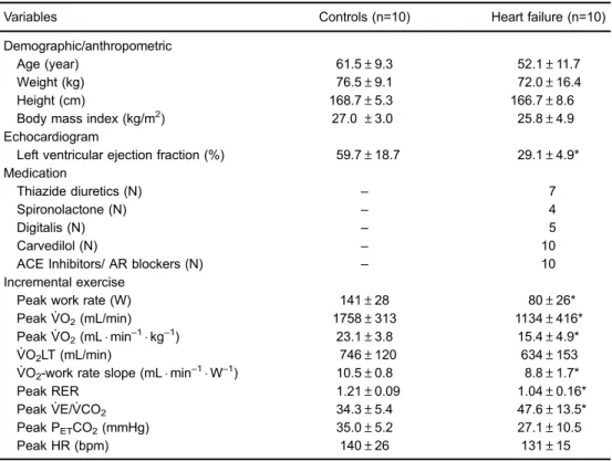
There were no significant between-group differences in anthropometric attributes (Table 1). The main etiology of heart failure was non-ischemic cardiomyopathy and, as expected by the inclusion criteria, all patients showed severe left ventricular dysfunction. Peak work rate and peakVO. 2were markedly reduced in patients; for instance,
7 patients were on Weber. ’s class C. Patients had shallower VO2-work rate slopes than controls; conversely,
. VE/VCO. 2
was higher and PETCO2 lower in patients compared to
controls (Po0.05; Table 1).
As previously described in normal subjects (4,10), deoxi-Hb response curve as a function of increasing
work rate was S-shaped i.e., it resembled a sigmoid in all subjects. From the raw signal, we initially identified two inflection points: point‘‘A’’corresponded to the work rate after exercise onset at which deoxi-Hb started to systematically increase, and from point‘‘A’’onward we applied linear regression to deoxi-Hb. Point ‘‘B’’ corre-sponded to the work rate at which there was a systematic departure from linearity.
The range of work rates before point ‘‘A’’, between points‘‘A’’and‘‘B’’and after point‘‘B’’up to peak exercise (point‘‘C’’) were named phases‘‘1’’,‘‘2’’and‘‘3’’, respec-tively. In addition to the increase in slope (S) of deoxi-Hb throughout phase 2, we calculated the deoxi-Hb difference (‘‘D’’) between points‘‘B’’and‘‘C’’(Figure 1).
As shown in Figure 2 for representative subjects and in Table 2 and Figure 2 for mean data, patients presented with significant steeper deoxi-Hb slope than controls (Po0.01). Moreover, while deoxi-Hb remained stable or even decreased during phase ‘‘3’’ in all but one control (i.e., null or negative ‘‘D’’), there were systematic increases in deoxi-Hb in all patients (Po0.05). Steeper deoxi-Hb-work rate slope was associated with lower peak work rate in patients (r=–0.73; P=0.01).
Table 1.Resting and exercise characteristics of healthy controls and patients with heart failure.
Variables Controls (n=10) Heart failure (n=10)
Demographic/anthropometric
Age (year) 61.5±9.3 52.1±11.7
Weight (kg) 76.5±9.1 72.0±16.4
Height (cm) 168.7±5.3 166.7±8.6
Body mass index (kg/m2) 27.0±3.0 25.8±4.9
Echocardiogram
Left ventricular ejection fraction (%) 59.7±18.7 29.1±4.9* Medication
Thiazide diuretics (N) – 7
Spironolactone (N) – 4
Digitalis (N) – 5
Carvedilol (N) – 10
ACE Inhibitors/ AR blockers (N) – 10
Incremental exercise
Peak work rate (W) 141±28 80±26*
PeakVO. 2(mL/min) 1758±313 1134±416*
Peak. VO. 2(mLmin–1kg–1) 23.1±3.8 15.4±4.9*
VO. 2LT (mL/min) 746±120 634±153
VO2-work rate slope (mLmin–1W–1) 10.5±0.8 8.8±1.7*
Peak RER 1.21±0.09 1.04±0.16*
PeakVE/. VCO. 2 34.3±5.4 47.6±13.5*
Peak PETCO2(mmHg) 35.0±5.2 27.1±10.5
Peak HR (bpm) 140±26 131±15
Data are reported as means ± SD or frequency (N). ACE: angiotensin-converting enzyme; AR: angiotensin receptor;VO. 2: oxygen uptake; LT: lactate threshold; RER: gas exchange ratio;
.
VE: ventilation; .
Discussion
This prospective study established a simplified approach to unravel abnormalities in peripheral muscle O2delivery
(i.e., lower bloodflow in non-hypoxemic patients) as indi-cated by changes in NIRS-based deoxi-Hb during ramp-incremental cardiopulmonary exercise test in heart failure patients. Our results indicate that, compared to controls, patients presented with steeper mid-exercise slope of deoxi-Hb as a function of work rate coupled with lack of late-exercise stability (or even decreasing deoxi-Hb). We interpret these results as evidence of faster and higher O2
extraction to compensate for impaired convective and diffu-sive O2 flow to muscle mitochondria (10). This approach
might prove useful to assess the effects of pharmacological and non-pharmacological methods aimed at improving intra-muscular microvascular hemodynamics in this patient population. The proposed approach can be easily applied in clinical settings, as it does not require data fitting with complex mathematical functions (7). Moreover, deoxi-Hb is reported as a function of work rate, andVO. 2measurements
(i.e., cardiopulmonary exercise test) are not mandatory. Importantly, the proposed parameters (slope and‘‘D’’) are largely effort-independent, being recorded during submax-imal exercise.
From a mechanistic standpoint, it has been long established that the key factors modulating O2
delivery-utilization matching in contracting appendicular muscles include: a) the muscle‘‘pump’’effect; b) local vasodilata-tion; c) parasympathetic and sympathetic tones, and d) differential patterns of musclefiber recruitment, as reviewed by other authors (4,10,19). Based on these premises, we interpreted the S-shaped pattern of muscle deoxygenation (deoxi-Hb) depicted in Figure 1 as indicating: a) an early phase (‘‘1’’) in which proportional increases in O2delivery
and O2 requirements (
.
VO2) led to a stable O2 extraction
(Bdeoxi-Hb), b) a subsequent phase (‘‘2’’) in which deficits in O2delivery relative to fast-increasing
.
VO2produced a
marked increase in O2extraction, and c) afinal phase (‘‘3’’)
in which O2delivery and O2requirements were once again
matched leading to a stable rate of O2extraction (or even
decreasing if O2 delivery becomes excessive relative to
instantaneous O2needs) (4,10). This model is consistent
with previous contentions by Spencer et al (11), who found that the deoxi-Hb response profile during ramp-incremental exercise in healthy young males consisted of three distinct phases, in which the latter two were approximately linear, i.e., phases‘‘1’’and‘‘2’’herein described.
In this context, steeper phase‘‘2’’deoxi-Hb-work rate slope in patients than controls is strongly suggestive of impaired O2 delivery-utilization matching in the former
group. It is noteworthy that these abnormalities occurred despite a shallowerVO. 2-work rate slope in patients. Thus,
even if changes in O2requirements were lower in patients,
marked deficits in Q.m likely precluded a corresponding
increase in O2delivery. In other words,
.
VO2/extraction ratio
was markedly reduced in patients, afinding consistent with impaired O2delivery. Increased O2 extraction in patients
might have also been influenced by lactacidosis-induced rightward shifts in the oxy-hemoglobin dissociation curve (Bohr effect) and/or greater recruitment of O2-costly type II
fibers. Thus, a direct quantitative (inverse) relationship between Q.mand deoxi-Hb should not be attempted.
Progressive increase in late-exercise (phase‘‘3’’) deoxi-Hb in patients, but not in controls, is another evidence of poorer muscle O2delivery-utilization matching in heart failure
conditions. In fact, there is growing evidence that despite progressive increases in work rate (andVO. 2), cardiac output
might stabilize (or even decrease) near peak exercise in these patients (20–22). Microvascular perfusion-muscle
fiber recruitment uncoupling (4,10,21) and sympathetic over-excitation (23) may also further impair Q.m near exercise
termination. Moreover, type IIfibers (with lower ATP/O2ratio)
are mostly recruited at higher compared to lower work rates (24,25), which might have contributed to muscle O2
delivery-VO. 2mismatch in phase‘‘3’’.
As a noninvasive, cross-sectional study our investiga-tion has some limitainvestiga-tions that should be highlighted. We assume, as others (5,8,26–28), that deoxi-Hb reflects
muscle fractional O2extraction (C(a-v)O2); however, we
did not measure blood gas tensions. We also assumed that deoxi-Hb at a specific site gives a rough estimate of overall muscle O2 extraction (8,14,16,28). Koga et al.
(18), however, found large heterogeneity in Q.m
-. VO2
distribution in normal subjects, a phenomenon that might be more relevant for poorly perfused muscles. There is mounting evidence that Q.m
-.
VO2 distribution
performed a cycle ergometer test as the NIRS signal quickly deteriorates during fast walking; thus, our approach is unlikely to be feasible for treadmill-based tests.
In conclusion, we presented a practical approach to interpret the deoxi-Hb signal by NIRS during ramp-incremental cycle ergometry in heart failure patients. Impairments in O2delivery, likely reflective of poor muscle
bloodflow in non-hypoxemic patients, were non-invasively uncovered by steeper mid-exercise slope of deoxi-Hb as a function of work rate and increasing (instead of stable or decreasing) deoxi-Hb near peak exercise. This novel strategy might prove useful to assess the effects of
pharmacological and non-pharmacological interventions aimed at improving skeletal muscle perfusion in this patient population.
Acknowledgements
The authors thank all colleagues from the Pulmonary Function and Clinical Exercise Physiology Unit, Federal University of São Paulo (UNIFESP), Brazil for friendly collaboration. This study was supported by the Coorde-nac¸ão de Aperfeic¸oamento de Pessoal de Nível Superior (CAPES), Brazil.
References
1. Hirai DM, Musch TI, Poole DC. Exercise training in chronic heart failure: improving skeletal muscle O2 transport and utilization. Am J Physiol Heart Circ Physiol 2015; 309: H1419–H1439, doi: 10.1152/ajpheart.00469.2015. 2. Hunt SA, Abraham WT, Chin MH, Feldman AM, Francis GS,
Ganiats TG, et al. ACC/AHA Guidelines for the diagnosis and management of heart failure in adults.J Am Coll Cardiol 2009; 53: e1–e90, doi: 10.1016/j.jacc.2008.11.013. 3. Wielenga RP, Coats AJS, Mosterd WL, Huisveld IA. The role
of exercise training in chronic heart failure.Heart1997; 78: 431–436, doi: 10.1136/hrt.78.5.431.
4. Boone J, Koppo K, Barstow TJ, Bouckaert J. Pattern of deoxy[Hb+Mb] during ramp cycle exercise: influence of aerobicfitness status.Eur J Appl Physiol2009; 105: 851–859, doi: 10.1007/s00421-008-0969-2.
5. Poole DC, Barstow TJ, McDonough P, Jones AM. Control of oxygen uptake during exercise.Med Sci Sports Exerc2008; 40: 462–474, doi: 10.1249/MSS.0b013e31815ef29b. 6. Karavidas A, Arapi SM, Pyrgakis V, Adamopoulos S.
Func-tional electrical stimulation of lower limbs in patients with chronic heart failure. Heart Fail Rev 2010; 15: 567–579, doi: 10.1007/s10741-010-9171-9.
7. Sperandio PA, Oliveira MF, Rodrigues MK, Berton DC, Treptow E, Nery LE et al. Sildenafil improves microvascular O2delivery-to-utilization matching and accelerates exercise
O2 uptake kinetics in chronic heart failure.Am J Physiol
Heart Circ Physiol2012; 303: H1474–H1480, doi: 10.1152/ ajpheart.00435.2012.
8. Ferreira LF, Townsend DK, Lutjemeier BJ, Barstow TJ. Muscle capillary blood flow kinetics estimated from pul-monary O2 uptake and near-infrared spectroscopy.J Appl Physiol2005; 98: 1820–1828, doi: 10.1152/japplphysiol. 00907.2004.
9. Ferrari M, Mottola L, Quaresima V. Principles, techniques, and limitations of near infrared spectroscopy. Can J Appl Physiol2004; 29: 463–487, doi: 10.1139/h04-031. 10. Ferreira LF, Koga S, Barstow TJ. Dynamics of noninvasively
estimated microvascular O2extraction during ramp exercise.
J Appl Physiol 2007; 103: 1999–2004, doi: 10.1152/jappl physiol.01414.2006.
11. Spencer MD, Murias JM, Paterson DH. Characterizing the profile of muscle deoxygenation during ramp incremental exercise in young men.Eur J Appl Physiol2012; 112: 3349– 3360, doi: 10.1007/s00421-012-2323-y.
12. Boone J, Bouckaert J, Barstow TJ, Bourgois J. Influence of priming exercise on muscle deoxy[Hb+Mb] during ramp cycle exercise.Eur J Appl Physiol2011; 112: 1143–1152, doi: 10.1007/s00421-011-2068-z.
13. Neder JA, Nery LE, Castelo A, Andreoni S, Lerario MC, Sachs A, et al. Prediction of metabolic and cardiopulmonary Table 2.Key variables of deoxygenated hemoglobin concentration
(deoxi-Hb)-work rate relationship in healthy controls and patients with heart failure.
Variables Controls (n=10) Heart failure (n=10)
Point‘‘A’’(W) 19±14 18±11
Point‘‘B’’(W) 111±32 53±19*
[deoxy-Hb] slope (%/W) 1.0±0.3 2.2±1.3*
D[deoxy-Hb] (%) -0.5±18.9 20.3±12.9*
responses to maximum cycle ergometry: a randomised study. Eur Respir J 1999; 14: 1304–1313, doi: 10.1183/ 09031936.99.14613049.
14. Grassi B, Pogliaghi S, Rampichini S, Quaresima V, Ferrari M, Marconi C, et al. Muscle oxygenation and pulmonary gas exchange kinetics during cycling exercise on-transitions in humans.J Appl Physiol2003; 95: 149–158, doi: 10.1152/ japplphysiol.00695.2002.
15. Boushel R, Langberg H, Olesen J, Gonzales-Alonzo J, Bulow J, Kjaer M. Monitoring tissue oxygen availability with near infrared spectroscopy (NIRS) in health and disease. Scand J Med Sci Sports2001; 11: 213–222, doi: 10.1034/ j.1600-0838.2001.110404.x.
16. Ferreira LF, Poole DC, Barstow TJ. Muscle bloodflow-O2 uptake interaction and their relation to on-exercise dynamics of O2 exchange.Respir Physiol Neurobiol2005; 147: 91– 103, doi: 10.1016/j.resp.2005.02.002.
17. Azevedo D, Medeiros WM, de Freitas FF, Ferreira Amorim C, Gimenes AC, Neder JA et al. High oxygen extraction and slow recovery of muscle deoxygenation kinetics after neuromuscular electrical stimulation in COPD patients.Eur J Appl Physiol2016; 116: 1899–1910, doi: 10.1007/s00421-016-3442-7.
18. Koga S, Poole DC, Ferreira LF, Whipp BJ, Kondo N, Saitoh T, et al. Spatial heterogeneity of quadriceps muscle deoxygena-tion kinetics during cycle exercise.J Appl Physiol2007; 103: 2049–2056, doi: 10.1152/japplphysiol.00627.2007. 19. Hirai DM, Copp SW, Holdsworth CT, Ferguson SK,
McCullough DJ, Behnke BJ, et al. Skeletal muscle micro-vascular oxygenation dynamics in heart failure: exercise training and nitric oxide-mediated function. Am J Physiol Heart Circ Physiol 2014; 306: H690–H698, doi: 10.1152/ ajpheart.00901.2013.
20. Tanabe Y, Nakagawa I, Ito E, Suzuki K. Hemodynamic basis of the reduced oxygen uptake relative to work rate during incremental exercise in patients with chronic heart failure. Int J Cardiol2002; 83: 57–62, doi: 10.1016/S0167-5273(02) 00013-X.
21. McDonough P, Behnke BJ, Padilla DJ, Musch TI, Poole DC. Control of microvascular oxygen pressures in rat muscles comprised of differentfibre types.J Physiol2005; 563: 903– 913, doi: 10.1113/jphysiol.2004.079533.
22. Rolim NP, Mattos KC, Brum PC, Baldo MV, Middlekauff HR, Negrão CE. The decreased oxygen uptake during pro-gressive exercise in ischemia-induced heart failure is due to reduced cardiac output rate.Braz J Med Biol Res2006; 39: 297–304, doi: 10.1590/S0100-879X2006000200018. 23. Ferreira LF, McDonough, Behnke BJ, Musc TI, Poole DC.
Bloodflow and O2 extraction as a function of O2 uptake in
muscles composed of differentfiber types.Respir Physiol Neurobiol 2006; 153: 237–249, doi: 10.1016/j.resp.2005. 11.004.
24. Krustrup P, Söderlund K, Mohr M, Bangsbo J. The slow component of oxygen uptake during intense, sub-maximal exercise in man is associated with additionalfibre recruitment. Eur J Physiol2004; 447: 855–866, doi: 10.1007/s00424-003-1203-z.
25. Boone J, Koppo K, Barstow TJ, Bouckaert J. Aerobic fitness, muscle efficiency, and motor unit recruitment during ramp exercise.Med Sci Sports Exerc2010; 42: 402–408, doi: 10.1249/MSS.0b013e3181b0f2e2.
26. Bauer TA, Reusch JE, Levi M, Regensteiner JG. Skeletal muscle deoxygenation after the onset of moderate exercise suggests slowed microvascular bloodflow kinetics in type 2 diabetes.Diabetes Care2007; 30: 2880–2885, doi: 10.2337/ dc07-0843.
27. Chiappa GR, Borghi-Silva A, Ferreira LF, Carrascosa C, Oliveira CC, Maia J, et al. Kinetics of muscle deoxygenation are accelerated at the onset of heavy-intensity exercise in patients with COPD: relationship to central cardiovascular dynamics.J Appl Physiol2008; 104: 1341–1350, doi: 10.1152/ japplphysiol.01364.2007.
28. Sperandio PA, Borghi-Silva A, Barroco AC, Nery LE, Almeida DR, Neder A. Microvascular oxygen delivery-to-utilization mismatch at the onset of heavy-intensity exercise in optimally treated patients with CHF.Am J Physiol Heart Circ Physiol2009; 297: 1720–1728, doi: 10.1152/ajpheart.00596. 2009.
29. Lanfranconi F, Borrelli E, Ferri A, Porcelli S, Maccherini M, Chiavarelli M, et al. Noninvasive evaluation of skeletal muscle oxidative metabolism after heart transplant.Med Sci Sports Exerc 2006; 38: 1374–1383, doi: 10.1249/01.mss. 0000228943.62776.69.
30. Jendzjowsky NG, Tomczak CR, Lawrance R, Taylor DA, Tymchak WJ, Riess KJ, et al. Impaired pulmonary oxygen uptake kinetics and reduced peak aerobic power during small muscle mass exercise in heart transplant recipients. J Appl Physiol2007; 103: 1722–1727, doi: 10.1152/japplphy siol.00725.2007.
31. Diederich ER, Behnke BJ, McDonough P, Kindig CA, Barstow TJ, Poole DC, et al. Dynamics of microvascular oxygen partial pressure in contracting skeletal muscle of rats with chronic heart failure. Cardiovasc Res2002; 56: 479–486, doi: 10.1016/S0008-6363(02)00545-X.