Effe ct o f io dide o n Fas, Fas-ligand and
Bcl-w m RNA e xpre ssio n in thyro id o f
NO D m ice pre tre ate d with m e thim azo le
Disciplina e Laboratório de Imunologia, Departamento de Clínica Médica, Faculdade de Ciências Médicas, Universidade Estadual de Campinas, Campinas, SP, Brasil
L.H.B. Boechat, C.A. Vilella and R.L. Zollner
Abstract
Nonobese diabetic (NOD) mice and a derived strain, NOD.H.2h4, have been used as a model for experimental spontaneous thyroiditis and thyroiditis induced by iodide excess after a goiter-inducing pe-riod. Some authors have proposed that iodide, given after methima-zole or propylthiouracil, is capable of inducing apoptosis in thyroid cells and that anti-thyroid drugs can modulate the expression of apoptosis components such as Fas and its ligand (Fas-L). Here we evaluated the effect of potassium iodide (20 µg/animal for 4 days, ip) given to NOD mice at the 10th week of life after exposure to methima-zole (1 mg/ml) in drinking water from the 4th to the 10th week of life. Fas, Fas-L and Bcl-w expression were analyzed semiquantitatively by RT-PCR immediately after potassium iodide administration (group MI44D) or at week 32 (MI32S). Control groups were added at 10 (C10) and 32 weeks (C32), as well as a group that received only methimazole (CM10). An increase in the expression of Fas-L and Bcl-w (P<0.01, ANOVA) Bcl-was observed in animals of group MI44D, Bcl-while Fas was expressed at higher levels (P = 0.02) in group C32 (72.89 ± 47.09 arbitrary units) when compared to group C10 (10.8 ± 8.55 arbitrary units). Thus, the analysis of Fas-L and Bcl-w expression in the MI44D group and Fas in group C32 allowed us to detect two different patterns of expression of these apoptosis components in thyroid tissue of NOD mice.
Co rre spo nde nce R.L. Zollner
Disciplina e Laboratório de Imunologia
Departamento de Clínica Médica FCM, UNICAMP
Caixa Postal 6111 13081-970 Campinas, SP Brasil
Fax: + 55-19-3289-3709 E-mail: zollner@ unicamp.br
Research supported by CNPq (No. 141759/95-0). L.H.B. Boechat was the recipient of a CNPq fellowship. Publication supported by FAPESP.
Received June 8, 2001 Accepted January 29, 2002
Ke y wo rds
·Thyroiditis
·Nonobese diabetic mice ·Apoptosis
·Methimazole
·Iodide
·Fas mRNA
·Fas-L mRNA
·Bcl-w mRNA
Intro ductio n
Over the last decade, apoptosis has be-come one of the main topics of interest in the biological sciences. This phenomenon is criti-cal to life and disease processes, playing a key role in regulatory activities, including immunology and immunopathology (1). Fas (CD95) and its natural ligand (Fas-L) are
tissues (3-5).
Nonobese diabetic (NOD) mice have been developed as a model for type 1 diabetes, but also express lymphocytic infiltrates in other organs including the thyroid (6). Many et al. (7) have shown that NOD mice submitted to an overload of iodine after a goiter-inducing phase developed thyroid infiltrates similar to those observed in human Hashimotos thy-roiditis and also expressed autoantibodies directed at thyroid cell membrane antigens. NOD.H.2h4, a strain derived from NOD, is also prone to the development of thyroid infiltrates that are clearly related to the amount of iodine intake (8,9).
Iodine-containing compounds such as amiodarone (10) and radiographic contrast media (11) have also been implicated in the induction of apoptosis in thyroid and vascu-lar epithelium, respectively. Recently, Vitale et al. (12) have shown that iodide induces apoptosis of thyroid cells in culture and Burikhanov and Matsuzaki (13) demon-strated that potassium iodide can do the same
in vivo when given to rats with
propylthio-uracil- or methimazole-induced goiter. In the present study we investigated the expression of messenger RNA specific for Fas, Fas-L and Bcl-w in goitrous NOD mice immediately after the administration of po-tassium iodide and at a later stage (32 weeks) by semiquantitative analysis.
Mate rial and Me tho ds
Animals and e xpe rime ntal gro ups
Male NOD mice used in this protocol were bred under germ-free conditions until the 4th week of life, when they were trans-ferred to a specific pathogen-free animal facility in our laboratory. Control animals (5 per group) had free access to autoclaved water and food until 10 (group C10) or 32 weeks of life (group C32) when they were sacrificed. The animals in the experimental groups received methimazole (Tapazole®
,
Eli Lilly do Brasil Ltda., São Paulo, SP, Brazil) diluted in autoclaved drinking water (1 mg/ml) from the 4th to the 10th week of life when it was discontinued and then re-ceived potassium iodide by the intraperito-neal route (20 mg animal-1
day-1
) for 4 days. Five animals were sacrificed immediately after the administration of potassium iodide (group MI44D) and five were kept alive until 32 weeks of age (group MI32S). Five ani-mals were sacrificed at the end of the methi-mazole treatment without being injected with potassium iodide (group CM10).
To tal RNA e xtractio n
Mice were anesthetized by intraperito-neal injection of 3% sodium pentobarbital (20 mg/kg) and then sacrificed by careful cervical dislocation. Thyroids were dissected and the tissue immediately placed into RNase-free plastic tubes containing 4 M guanidine thiocyanate, 0.1 M 2-mercaptoethanol, 25 mM sodium citrate, pH 7.0, and 0.5% sarcosyl, frozen in liquid nitrogen and main-tained at -80ºC until use. Total RNA extrac-tion was performed according to the method of Chomczynski and Sacchi (14) and all aliquots were analyzed by spectrophotom-etry (SpectraMax 190, Molecular Devices, Sunnyvale, CA, USA) before use. The ratio of readings at 260 nm/280 nm was deter-mined and only samples presenting a ratio between 1.6 and 1.8 were used.
Re ve rse transcriptase -po lym e rase chain
re actio n
added. The temperature was then set at 42ºC for 2 min and 1 ml Superscript II (GibcoBRL) was added to each tube. The reaction was developed at 42ºC for 50 min followed by 15 min at 70ºC and then 4ºC at the end. cDNA samples were stored at -20ºC until use.
The polymerase chain reaction (PCR) was performed in 200-ml tubes containing 2 ml cDNA, 100 ng sense primer, 100 ng anti-sense primer, and 43 ml reaction solution (5 ml PCR buffer, 5 ml 0.5 mM dNTP mix, 1.5 ml 50 mM MgCl2, and 31.5 ml ultrapure
water). Each sample was overlaid with min-eral oil (Sigma, St. Louis, MO, USA) and incubated in a thermocycler (GeneAmpl 9700, Perkin-Elmer, Foster City, CA, USA) using one cycle at 94ºC for 3 min followed by 40 cycles of 94ºC for 60 s, 58ºC for 45 s and 72ºC for 90 s. PCR fragments were visualized by agarose gel electrophoresis and ethidium bromide staining. Sample contami-nation by genomic DNA was verified sub-mitting the RNA sample to PCR amplifica-tion omitting the reverse transcriptase step. Cyclophilin (housekeeping gene) was co-amplified as an internal control. The primers used are listed in Table 1.
Se m iquantitative m e asure m e nt o f re ve rse
transcriptase -PCR pro ducts
PCR products were submitted to electro-phoretic analysis in 1.5% agarose gel with ethidium bromide (2 ml/50 ml). Samples contained 8 ml cDNA, 1.5 ml PCR loading buffer and 5.5 ml ultrapure water and mi-grated in the presence of TBE buffer con-taining 2 ml/100 ml ethidium bromide for 45 min (70 V, 150 mA). PCR products were visualized by excitation of ethidium bro-mide under ultraviolet light, digitally recorded with a Nucleovision®
system (NucleoTech, San Mateo, CA, USA) and their molecular weight and band pixel area were calculated using the Gel Expert®
Software (Nucleo Tech).
Semiquantitative measurements of ex-pression (SE) of apoptosis components were calculated for each sample by the following formula and expressed as arbitrary absor-bance units (AU): SE (AU) = pixel area of the product to be analyzed/pixel area of cyclophilin x 100.
Statistical analysis
All results are reported as means ± SD. Comparisons of semiquantitative measure-ments of expression were made with ANOVA or the Student t-test when appropriate.
Re sults
Fre que ncy o f e xpre ssio n o f PCR pro ducts
All cDNA samples used for the study expressed the product of the housekeeping gene cyclophilin (276 bp), confirming their quality. The expected products for the Fas-L (206 bp) and Bcl-w (230 bp) genes were expressed in all animals studied. Expression of the Fas product (267 bp) was observed in all animals from groups C10, MI44D and C32, but only in four among five animals from group CM10 and three among five animals from group MI32S.
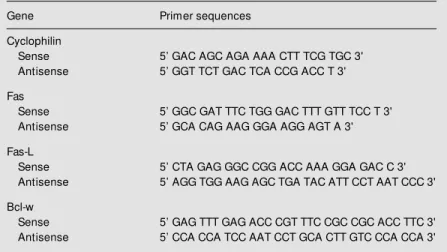
Table 1. Primers for mouse cyclophilin, Fas, Fas-ligand (Fas-L) and Bcl-w reverse transcriptase-PCR.
Gene Primer sequences
Cyclophilin
Sense 5’ GAC AGC AGA AAA CTT TCG TGC 3' Antisense 5’ GGT TCT GAC TCA CCG ACC T 3'
Fas
Sense 5’ GGC GAT TTC TGG GAC TTT GTT TCC T 3' Antisense 5’ GCA CAG AAG GGA AGG AGT A 3'
Fas-L
Sense 5’ CTA GAG GGC CGG ACC AAA GGA GAC C 3' Antisense 5’ AGG TGG AAG AGC TGA TAC ATT CCT AAT CCC 3'
Bcl-w
Se m iquantitative m e asure m e nt o f re ve rse
transcriptase -PCR pro ducts
Mean Fas expression was higher (P = 0.01) for group C32 (72.89 ± 47.09 AU) than for group C10 (10.8 ± 8.55 AU). No differ-ence was detected when group C10 was compared to group CM10. The expression of Fas-L and Bcl-w gene products was more
intense in group MI44D (P<0.01, ANOVA) (Figure 1).
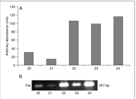
Analysis of the individual responses showed that in all groups except group MI32S some animals overexpressed Fas, suggesting a different behavior of the signaling for the synthesis of this apoptosis component among the NOD mice studied. Specifically, two animals in group C10 and one in groups CM10 and MI44D expressed Fas at clearly higher levels than the other components of the group. This fact was more prominent in group C32, where three animals had semi-quantitative levels of expression of about 100 AU, while the other two were below 30 AU (Figure 2).
The expression of Fas-L and Bcl-w was uniform, except for group MI44D, where high standard deviations (40.69 and 40.68% of the mean, respectively) reflected the ex-istence of scattered values. Mean levels of Fas-L and Bcl-w expression were higher (P<0.01, ANOVA) in group MI44D than in the others.
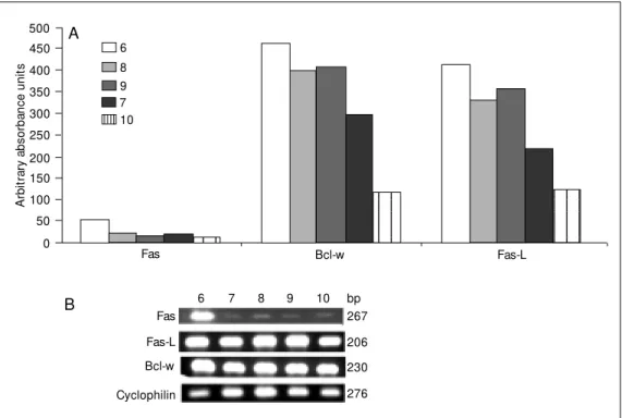
When animals from group MI44D were analyzed individually, it was possible to ob-serve two patterns of expression of Fas-L and Bcl-w (Figure 3A). Animals number 6, 8 and 9 showed higher amplification of the PCR product for both Fas-L and Bcl-w than animals number 7 and 10. Animal number 6 overexpressed Fas, as detected visually in 1.5% agarose gel (Figure 3B).
D iscussio n
Iodine has been linked to thyroid physi-ology and pathphysi-ology since the beginning of endocrinology. Some investigators have re-lated the increase in the prevalence of au-toimmune thyroid diseases (Hashimotos thyroiditis and Graves disease) to iodine supplementation in deficient areas (15), al-though these findings have recently been questioned (16). Experimental models of thy-roiditis have been classically based on ad-ministration of thyroglobulin whole molecule
A
rb
it
ra
ry
a
b
s
o
rb
a
n
c
e
u
n
it
s
140
120
100
80
60
40
20
0
20 21 22 23 24
A
B
Fas 267 bp
20 21 22 23 24
Figure 2. Individual levels of Fas expression in control NOD mice at the 32nd w eek of life. A, Results of semiquantitative measurements expressed as arbitrary absorbance units. B, Representative results of PCR analyzed in 1.5% agarose gel.
Figure 1. Semiquantitative measurement of the expression of Fas, Fas-ligand (Fas-L) and Bcl-w in control NOD mice (C10 or C32), treated w ith methimazole (CM 10) or potassium iodide after methimazole (groups M I44D and M I32S), from the 4th to the 10th w eek of life. * P<0.01 compared to the other groups (ANOVA); * * P = 0.02 compared to C10 (Student t -test).
M I44D
A
rb
it
ra
ry
a
b
s
o
rb
a
n
c
e
u
n
it
s
500
M I32S 450
400 350 300
250 200 150 100 50 0
C32 CM 10
C10
Fas Fas-L Bcl-w
* *
(17) or its related peptides (18) associated or not with adjuvants. In both cases, iodination of tyrosines is considered fundamental for the development of experimental autoim-mune thyroiditis.
Experimental thyroiditis induced by ad-ministration of supraphysiologic doses of iodine has been described in NOD mice or their derived strains such as NOD.H.2h4 (6), and for other models of thyroid autoimmu-nity such as BB/W rats (19) and obese strain chickens (20). The mechanisms involved have been related to direct toxicity as a con-sequence of high peroxide levels due to the strong expression of thyroid peroxidase in goiter-inducing conditions (7,21). Recently, many authors have related iodine to the de-velopment of apoptosis in humans (10,11) or in experimental models (22).
We submitted NOD mice to methimazole in drinking water for 6 weeks in order to promote goiter-inducing conditions and then administered high doses of potassium iodide to create the conditions usually observed in experimental models of thyroiditis in order to evaluate the changes in expression of Fas,
Fas-L and Bcl-w.
We did not detect differences in the mean levels of Fas-L expression when control 10-week-old animals were compared to those receiving methimazole without an iodine overload. This result is in contrast with those observed by Mitsiades et al. (23) who re-ported an increase in Fas-L expression in patients treated with methimazole for Graves disease.
Ours is the first study to evaluate the expression of Bcl-w in an experimental mo-del of thyroiditis. In accordance with what is described in the literature for other organs (24), Bcl-w was expressed in all animals studied, although we observed higher levels of its messenger RNA in animals sacrificed 4 days after iodine administration.
The levels of Fas gene expression mea-sured semiquantitatively were more promi-nent in the control group at 32 weeks of life, although this was not a homogeneous obser-vation. In fact, two distinct levels of expres-sion were demonstrable, with three NOD mice presenting more Fas messenger RNA than the other two, a fact that could also be
Figure 3. Individual levels of ex-pression of Fas, Fas-ligand (Fas-L) and Bcl-w in NOD m ice treated w ith potassium iodide (20 mg/animal) after receiving methimazole in drinking w ater from the 4th to the 10th w eek of life. A, Semiquantitative mea-surement for each animal (num-bers 6-10), show ing different patterns of response for Fas-L and Bcl-w . B, Representative 1.5% agarose gel eletrophore-sis.
Fas
Fas-L
Bcl-w
Cyclophilin
267
206
230
276
B 6 7 8 9 10 bp
A
rb
it
ra
ry
a
b
s
o
rb
a
n
c
e
u
n
it
s
500
123 123 123 123 123 123
123 123 123 123 123
450
400
350
300
250
200 150
100
50
0
Fas Bcl-w Fas-L
A
observed in one animal of group MI44D and in two control animals at 10 weeks.
Higher levels of Fas-L and Bcl-w expres-sion have been observed in animals sacri-ficed 4 days after iodine administration (25). Fas-L seems to be expressed constitutively in human and murine thyroid tissue and its association with Fas in neighboring cells seems to lead to apoptosis in a fratricidal way (26). Accordingly, the iodine-induced elevated expression of this apoptosis-related gene could play an important role also in the model of experimental thyroiditis in NOD mice. Our results agree with those of Bluher et al. (22), who described high expression of Fas-L in BB/W rats after iodide administra-tion. However, these authors have also de-tected an increase in Fas expression in com-parison with control animals. This differ-ence may be related to the design of the study, since these investigators administered iodide for longer periods of time. On the other hand, Burikhanov and Matsuzaki (13) identified maximum DNA fragmentation af-ter 3 h of potassium iodide doses, with a
decrease in apoptosis after 12 h. This indi-cates a very fast process, perhaps not ob-served in our study design.
The NOD mice used in the present study presented different levels of response to Fas-L and Bcl-w signaling after iodide adminis-tration. This was evident in the animals ob-served immediately after the injection of iodide. Fas is considered to be constitutively expressed in thyroid tissue as described by Giordano et al. (26). However, some ani-mals overexpress the signal for the Fas gene, as clearly observed in group C32. Concern-ing the present results, we have observed that, at least with respect to thyroid tissue, the NOD mice in our colony present two different patterns of Fas, Fas-L and Bcl-w expression. Extension of this analysis in fu-ture research must include other organs, mainly pancreas, in order to evaluate thyroid specificity. Also, future confirmation of these biological responses to iodide excess could allow us to characterize a subgroup of NOD mice susceptible to iodide-induced experi-mental thyroiditis.
Re fe re nce s
1. Granville DJ, Carthy CM , Hunt DW & M cM anus BM (1998). Apoptosis: molecu-lar aspects of cell death and disease.
Laboratory Investigation, 78: 893-913. 2. Nagata S & Golstein P (1995). The Fas
death factor. Science, 267: 1449-1456. 3. Gibson L, Holmgreen SP, Huang DC,
Ber-nard O, Copeland NG, Jenkins NA, Sutherland GR, Baker E, Adams JM & Cory S (1996). Bcl-w , a novel member of the bcl-2 family, promotes cell survival.
Oncogene, 13: 665-675.
4. Yan C, Chen J, Chen D, M inami M , Pei W, Yin XM & Simon RP (2000). Overexpres-sion of the cell death suppressor Bcl-w in ischemic brain: implications for a neuro-protective role via the mitochondrial path-w ay. Journal of Cerebral Blood Flow and M etabolism, 20: 620-630.
5. M eehan T, Loveland KL, de Kretser D, Cory S & Print CG (2001). Developmental regulation of the bcl-2 family during sper-matogenesis: Insights into the sterility of bcl-w -/- male mice. Cell Death and
Differ-entiation, 8: 225-233.
6. Dam ot t e D, Colom b E, Cailleau C, Brousse N, Charreire J & Carnaud C (1997). Analysis of susceptibility of NOD mice to spontaneous and experimentally induced thyroiditis. European Journal of Immunology, 27: 2854-2862.
7. M any M C, M anirat unga S, Varis I, Dardenne M , Drexhage HA & Denef JF (1995). Tw o-step development of Hashi-moto-like thyroiditis in genetically autoim-mune prone non-obese diabetic mice: ef-fects of iodine-induced cell necrosis. Jour-nal of Endocrinology, 147: 311-320. 8. Rasooly L, Burek CL & Rose NR (1996).
Iodine-induced autoimmune thyroiditis in NOD-H-2h4 mice. Clinical Immunology and Immunopathology, 81: 287-292. 9. Rose NR, Saboori AM , Rasooly L & Burek
CL (1997). The role of iodine in autoim-mune thyroiditis. Critical Review s in Im-munology, 17: 511-517.
10. Di M atola T, D’Ascoli F, Fenzi G, Rossi G, M artino E, Bogazzi F & Vitale M (2000).
Amiodarone induces cytochrome c re-lease and apoptosis through an iodine-independent mechanism. Journal of Clini-cal Endocrinology and M etabolism, 85: 4323-4330.
11. Zhang H, Holt CM , M alik N, Shepherd L & M orcos SK (2000). Effects of radiographic cont rast m edia on prolif erat ion and apoptosis of human vascular endothelial cells. British Journal of Radiology, 73: 1034-1041.
12. Vitale M , Di M atola T, D’Ascoli F, Salzano S, Bogazzi F, Fenzi G, M artino E & Rossi G (2000). Iodide excess induces apopto-sis in thyroid cells through a p53-indepen-dent m echanism involving oxidat ive stress. Endocrinology, 141: 598-605. 13. Burikhanov RB & M atsuzaki S (2000).
Ex-cess iodine induces apoptosis in the thy-roid of goitrogen-pretreated rats in vivo.
Thyroid, 10: 123-129.
thiocyanate-phenol-chloro-form extraction. Analytical Biochemistry, 162: 156-159.
15. Hedinger C (1981). Geographic pathology of thyroid diseases. Pathology Research and Practice, 171: 285-292.
16. M arw aha RK, Tandon N, Karak AK, Gupta N, Verma K & Kochupillai N (2000). Hashi-moto’s thyroiditis: countryw ide screening of goitrous healthy young girls in postiodi-zation phase in India. Journal of Clinical Endocrinology and M etabolism, 85: 3798-3802.
17. Stafford EA & Rose NR (2000). New er insights into the pathogenesis of experi-mental autoimmune thyroiditis. Interna-tional Review s of Immunology, 19: 501-533.
18. Wan Q, M cCormick DJ, David CS & Kong YC (1998). Thyroglobulin peptides of spe-cific primary hormonogenic sites can gen-erate cytotoxic T cells and serve as target autoantigens in experim ental autoim -mune thyroiditis. Clinical Immunology and Immunopathology, 86: 110-114. 19. Li M & Boyages SC (1994). Iodide induced
lymphocytic thyroiditis in the BB/W rat: evidence of direct toxic effects of iodide on thyroid subcellular structure. Autoim-munity, 18: 31-40.
20. Sundick RS, Bagchi N & Brow n TR (1996). The obese strain chicken as a model for human Hashimoto’s thyroiditis. Experi-mental and Clinical Endocrinology and Dia-betes, 104: 4-6.
21. M ahmoud I, Colin I, M any M C & Denef JF (1986). Direct toxic effect of iodide in ex-cess on iodine-deficient thyroid glands: epithelial necrosis and inflammation asso-ciated w ith lipofucsin accumulation. Ex-perimental and M olecular Pathology, 44: 259-271.
22. Bluher M , Krohn K, W allaschofski H, Braverman LE & Paschke R (1999). Fas and Fas ligand gene expression in autoim-mune thyroiditis in BB/W rats. European Journal of Endocrinology, 141: 506-511. 23. M itsiades N, Poulaki V, Tseleni-Balafouta
S, Chrousos GP & Koutras DA (2000). Fas ligand expression in thyroid follicular cells from patients w ith thionamide-treated
Graves’ disease. Thyroid, 10: 527-532. 24. Print CG, Loveland KL, Gibson L, M eehan
T, Stylianou A, Wreford N, de Kretser D, M etcalf D, Kontgen F, Adams JM & Cory S (1998). Apoptosis regulator bcl-w is es-sential for spermatogenesis but appears otherw ise redundant. Proceedings of the National Academy of Sciences, USA, 95: 12424-12431.
25. Sera N, Kaw akam i A, Nakashim a T, Nakamura H, Imaizumi M & Ito K (2001). Fas/FasL mediated apoptosis of thyro-cytes in Graves’ disease. Clinical and Ex-perimental Immunology, 124: 197-207. 26. Giordano C, Richiusa P, Bagnasco M ,