Involvement of Src tyrosine kinase and
protein kinase C in the expression of
macrophage migration inhibitory factor
induced by H
2
O
2
in HL-1 mouse
cardiac muscle cells
F. Rao
1,2,3, C.Y. Deng
2,3, Q.H. Zhang
1,3, Y.M. Xue
1,3, D.Z. Xiao
2,3, S.J. Kuang
2,3, Q.X. Lin
2,3,
Z.X. Shan
2,3, X.Y. Liu
2,3, J.N. Zhu
2,3, X.Y. Yu
1,2,3and S.L. Wu
1,3 1Department of Cardiology, Guangdong General Hospital, Guangdong Cardiovascular Institute, Guangdong Academy of Medical Sciences, Guangzhou, China 2Research Center of Medical Sciences, Guangdong General Hospital, Guangzhou, China 3Guangdong Academy of Medical Sciences, Guangzhou, ChinaAbstract
Macrophage migration inhibitory factor (MIF), a pleiotropic cytokine, plays an important role in the pathogenesis of atrial fibrillation; however, the upstream regulation of MIF in atrial myocytes remains unclear. In the present study, we investigated whether and how MIF is regulated in response to the renin-angiotensin system and oxidative stress in atrium myocytes (HL-1 cells). MIF protein and mRNA levels in HL-1 cells were assayed using immunofluorescence, real-time PCR, and Western blot. The result indicated that MIF was expressed in the cytoplasm of HL-1 cells. Hydrogen peroxide (H2O2), but not angiotensin II, stimulated MIF expression in HL-1 cells. H2O2-induced MIF protein and gene levels increased in a dose-dependent manner and were completely abolished in the presence of catalase. H2O2-induced MIF production was completely inhibited by tyrosine kinase inhibitors genistein and PP1, as well as by protein kinase C (PKC) inhibitor GF109203X, suggesting that redox-sensitive MIF production is mediated through tyrosine kinase and PKC-dependent mechanisms in HL-1 cells. These results suggest that MIF is upregulated by HL-1 cells in response to redox stress, probably by the activation of Src and PKC.
Key words: Macrophage migration inhibitory factor; HL-1 cells; Hydrogen peroxide; Atrial fibrillation; Protein kinases
Introduction
Recent evidence indicates that inflammation plays an important role in the pathology of atrial fibrillation (AF). In atrial tissue, inflammation is associated with activation of a variety of cells including lymphocytes, monocytes/ macrophages, fibroblasts, and cardiac myocytes, which express and secrete proinflammatory cytokines such as interleukin 6, tumor necrosis factor a, and C-reactive protein. These cytokines can modulate atrial functions and atrial remodeling, leading to development of AF (1-7). Macrophage migration inhibitory factor (MIF), a proin-flammatory cytokine, also functions as a regulator of the inflammatory response. It has been associated with inflammatory cardiovascular diseases including coronary
heart disease and myocarditis, pulmonary hypertension, and cardiac dysfunction following burn injury and sepsis (8-11). In previous research, we found that reduced calcium channel current amplitude in AF is associated with increased MIF levels (12,13). However, the regula-tion of MIF producregula-tion in atrial myocytes remains unclear. Experimental and clinical data suggest that there are interactions among angiotensin (Ang) II-related signaling, inflammation, and oxidative stress in the pathogenesis of AF-promoting structural or electrical remodeling (14,15). Reactive oxygen species (ROS) and Ang II could regulate the expression of MIF in cardiac myocytes and tubular epithelial cells (16,17). Therefore,
Correspondence: S.L. Wu and/or X.Y. Yu, Department of Cardiology, Guangdong General Hospital, Guangdong Cardiovascular Institute, Guangdong Academy of Medical Sciences, 96 Dongchuan Road, Guangzhou 510080, China. Fax: ++86-20-8376-9487. E-mail: [email protected] and/or [email protected]
in the present study, we investigated whether and how MIF is regulated in response to the renin-angiotensin system (RAS) and oxidative stress in an atrial-derived cell line (HL-1).
Material and Methods
HL-1 cardiomyocytes
HL-1 cells, a mouse cardiac cell line derived from a transplantable mouse cardiomyocyte lineage (AT-1), were obtained from the laboratory of Dr. William Claycomb (Louisiana State University Health Science Center, New Orleans, LA, USA). Cells were cultured in Claycomb medium (JRH Biosciences, USA) supplemented with 10% fetal bovine serum (JRH Biosciences), 2 mM L-glutamine (Gibco, USA), 100mM norepinephrine (Sigma, USA),
100 U/mL penicillin, and 100mg/mL streptomycin (Gibco)
in flasks precoated with fibronectin and gelatin (Sigma), then incubated at 376C in 5% CO2-95% air. The medium
was changed every 24-48 h.
Immunohistochemistry
For the immunohistochemical analyses, cultured cells were fixed in paraformaldehyde and incubated overnight with polyclonal antibodies against MIF (Santa Cruz Biotech, USA) or nonimmune immunoglobulin G (IgG) at dilutions of 1:50. The slides were then washed three times with Tris-buffered saline (TBS) for 5 min and incubated with peroxidase-labeled rabbit anti-goat antibody (Calbiochem, Germany) at a dilution of 1:2000 for 45 min. After they were washed three times for 5 min in phosphate-buffered saline (PBS), the sections were incubated in diaminobenzidine tetrahydrochloride (DAKO, Denmark) in PBS with 0.01% H2O2for 15 min.
The reaction was stopped by washing the sections in distilled water. The slides were mounted and observed under a light microscope.
Western blot analysis
Cells were lysed in 0.05 M Tris-HCl buffer, pH 8.0, containing 0.15 M sodium chloride, 0.02% sodium azide, 0.1% sodium dodecyl sulfate (SDS), 1% nonidet P (NP-40), and a Protease Inhibitor Cocktail Set (Calbiochem). Cell lysates were centrifuged at 12,000g
for 15 min at 46C. Protein concentrations were deter-mined. Samples were diluted with 4X loading buffer (Invitrogen, USA) and heated at 956C for 5 min. The proteins (30mg) were fractionated on 12% (for MIF)
SDS-polyacrylamide gels and transferred to nitrocellulose membranes (Amersham, USA) according to standard protocols. Membranes were blocked with dried skimmed milk powder in TBS Tween (TBST) for 2 h at room temperature before overnight incubation at 46C with the primary antibodies (rabbit polyclonal to MIF, 1:500). The signals were normalized to the protein levels of glycer-aldehyde 3-phosphate dehydrogenase (GAPDH, 1:1000;
Zymed, USA). After washing in TBST, the membranes were incubated for 1 h with horseradish peroxidase-conjugated anti-rabbit IgG (KPL, USA) in blocking solution. Protein bands were visualized by electrochemi-luminescence reagents (Pierce, USA), and films were evaluated densitometrically with the Gel-Pro Analyzer 4.0 software (http://gel-pro-analyzer.software.informer.com).
RNA extraction and reverse transcription polymerase chain reaction (PCR)
Total RNA extracted from cultured HL-1 cells was treated with DNase I to remove genomic DNA contamina-tion. First-strand cDNA was synthesized from 1mg total
RNA using a reverse transcription system (Promega, USA). Real-time quantitative PCRs were run in an MJ Research DNA Engine Opticon1 2 continuous
fluores-cence detection system (MJ Research, Inc., USA). cDNA (2mL) in a final volume of 25mL was amplified using
SYBR Premix Ex TaqTM(TaKaRa Biotechnology, China).
For mouse MIF, the forward primer was 59-GTG CCA GAG GGG TTT CTG T-39 and the reverse was 59-AGG CCA CAC AGC TTA CT-39; forb-actin, the forward primer was 59-TGT CCC TGT ATG CCT CTG GT-39 and the reverse was 59-GAT GTC ACG CAC GAT TTC C-39. Relative expression levels were calculated as 2-DDCT. Fold-changes were calculated according to Livak and Schmittgen (18).
Confocal microscopy
Cells were grown on glass coverslips precoated with fibronectin and gelatin, fixed in 4% paraformaldehyde for 15 min, and permeabilized with 0.25% Triton X-100 in PBS containing 1% BSA for 15 min. They were incubated overnight at 46C with a 1:100 dilution of primary antibody (mouse anti-MIF) and for 1 h at room temperature with a 1:200 dilution of secondary antibody. The coverslips were mounted onto microscope slides in Vectashield mounting medium containing DAPI for fluorescence microscopy (Vector Laboratories, USA). Fluorescent images were visualized and captured using a Leica DMI 6000 CS upright fluorescent microscope and a Leica TCS SP5 laser scanning confocal microscope (Leica, Germany).
Drugs
Genistein, 4-amino-5-(4-methylphenyl)-7-(t-butyl)py-razolo[3,4-d]pyrimidine (PP1), PD098059, SB203580, calphostin C, and GF109203X were purchased from Sigma, and U0126 was obtained from Calbiochem. These kinase inhibitors were dissolved in dimethyl sulfoxide (DMSO, Calbiochem). The concentration of DMSO in the working solutions did not exceed 1.5%.
Data analysis
Results
MIF expression and distribution in HL-1 cells
To investigate whether MIF was expressed in HL-1 cells, we used specific antibody to probe MIF in HL-1 cells. HeLa cells were used as a positive control. Immunohistochemistry assay showed that MIF was predominantly expressed in the cytoplasm of HL-1 cells, which was further confirmed by Western blot (Figure 1).
MIF production in HL-1 cells in response to redox stress
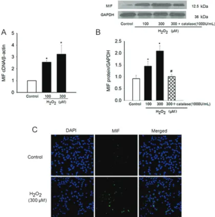
To determine whether MIF was regulated by redox stress in atrial myocytes, HL-1 cells were incubated in medium containing 100 or 300mM H2O2 for 6 h. H2O2
induced HL-1 cells to produce significantly increased levels of MIF in a concentration-dependent manner as shown by real-time PCR and Western blot (1.0vs2.6±0.1, P,0.05, and 3.2±0.8, P,0.01, gene expression for b-actin;
Figure 1. Expression of macrophage migration inhibitory factor (MIF) in HL-1 cells. A, Representative examples of immunohistochemical analysis of HL-1 cells. MIF expression in HL-1 cells with positive cytoplasmic staining (arrows). The staining specificity was tested by substituting IgG for the primary antibody.B, Western blot analysis of MIF performed on cell lysates showing a single band of 12.5 kDa.
Figure 2. H2O2-stimulated migration inhibitory factor (MIF) production in HL-1 cells.A,B, HL-1 cells were stimulated by H2O2(100 or 300mM) for 6 h or by H2O2 (300mM) for 6 h in the presence of catalase (1000 U/mL). Levels of MIF mRNA and protein were analyzed using real-time PCR or Western blotting withb-actin or GAPDH as internal controls, respectively. Mean values for each group were determined from 3 separate experiments each performed in duplicate. *P,0.05 vscontrol.#P
0.9±0.1 vs 1.5±0.2, P,0.05, and 2.1±0.2, P,0.01, protein expression for GAPDH; Figure 2A and B). This H2O2-induced MIF production was completely abolished in
the presence of catalase (1000 U/mL; from 2.1±0.2 to 1.0±0.1, P,0.05; Figure 2B). Immunofluorescence revealed similar results (Figure 2C). Morphological changes of HL-1 cells in response to H2O2treatment were
observed under an inverted microscope. Compared to controls, treated cells exhibited nuclear condensation, plasma membrane shrinkage, and a decreased beating rate, and few cells detached at 6 h after treatment; the morphological changes were H2O2-dose dependent.
MIF production in HL-1 cells in response to Ang II
We also investigated the role of Ang II in the production of MIF. In contrast to H2O2, there was no significant change
in MIF mRNA in HL-1 cells treated with three concentra-tions of Ang II (1, 10, and 100mM) for 3, 6, and 12 h,
respectively. Nevertheless, the level of MIF mRNA was significantly repressed by Ang II for the group treated with Ang II for a longer time (24 h) (1vs0.63±0.03, 0.66±0.06, and 0.66±0.07, P,0.01; Figure 3A). However, protein levels of MIF were not altered by treatment of Ang II as detected by immunoblotting (Figure 3B). Perhaps a longer treatment with Ang II would lead to the inhibitory effect of Ang II on MIF protein levels.
Effects of various kinase inhibitors on MIF production
We then examined whether protein kinases were involved in H2O2-induced MIF production in HL-1 cells
using tyrosine kinase inhibitors (genistein, a nonspecific PTK inhibitor and PP1, a specific Src antagonist), mitogen-activated protein (MAP) kinase inhibitors (PD098059 and U0126 for extracellular-regulated kinase 1/2 and SB203580 for p38-MAP kinase), and protein kinase C (PKC) inhibitors (GF109203X and calphostin C). Genistein, PP1, and GF109203X could all inhibit H2O2
-induced MIF production, whereas PD098059, U0126, SB203580, and calphostin C had no significant effect (Figure 4). These results suggest that H2O2induces MIF
production through PKC and tyrosine kinase-dependent mechanisms in HL-1 cells.
Figure 3. Effects of angiotensin II (Ang II) on the migration inhibitory factor (MIF) expression of HL-1 cells. HL-1 cells were stimulated by Ang II (1, 10, 100mM) for the indicated periods. Levels of MIF mRNA and protein were analyzed using Western blotting or real-time PCR with b-actin (A) or GAPDH (B) as internal controls, respectively. Data are reported as means±SE of MIF expression for 4 independent experiments in duplicate. *P,0.05vscontrol (one-way ANOVA).
Discussion
We demonstrated that 1) MIF was expressed in the cytoplasm of HL-1 cells; 2) levels of MIF expression were increased significantly by H2O2, but not Ang II, in a
dose-dependent manner; 3) this MIF expression was com-pletely inhibited by treatment with the tyrosine kinase inhibitors genistein and PP1, as well as the specific PKC inhibitor GF109203X.
Recent findings demonstrated the involvement of oxidative stress and inflammation in atrial tissue during AF, suggesting a potential role in the remodeling phenomenon (1,14,15). MIF, an important cytokine, is known to play a role in the pathoetiology of inflammatory cardiovascular diseases including coronary heart disease, myocarditis, pulmonary hypertension, and cardiac dys-function after burn injury and sepsis (8-10). In our previous studies (12,13), increased MIF expression was found in atrial tissue from patients with AF and contributed to the development of electrical remodeling in AF.
However, the upstream regulatory pathway of MIF in atrial myocytes is still not clear. Recent evidence has indicated that ROS could regulate the expression of MIF in cardiac myocytes (16). Ang II has been shown to upregulate MIF mRNA production and MIF protein secretion by renal tubular epithelial cells (17). These results suggest that ROS and RAS might promote oxidative stress and inflammation via induction of MIF synthesis and secretion. In the present study, we there-fore examined the concentration of MIF under the stimulation of H2O2and Ang II in HL-1 cells. We found
that H2O2was able to stimulate MIF production in HL-1
cells in a dose-dependent manner, whereas Ang II had no effect. These findings suggest that MIF may function in the myocardium as a redox-sensitive cytokine. In contrast
to H2O2, after a longer, 24-h treatment, Ang II had an
inhibitory effect on MIF mRNA levels, but not on the protein levels of MIF. Perhaps a longer treatment with Ang II would help explore the exact role of Ang II on MIF protein expression.
We also investigated the pathways of redox-sensitive intracellular signaling and found that MIF production induced by H2O2was completely inhibited by genistein,
PP1, and GF109203X, suggesting that redox-sensitive MIF production is mediated through tyrosine kinase and PKC-dependent mechanisms in HL-1 cells. However, calphostin C, another PKC inhibitor, had no effect on the MIF production induced by H2O2. This difference might
result from the different mechanisms of action of GF109203X and calphostin C. GF109203X inhibits PKC by competitive inhibition of the ATP-binding site, whereas calphostin C inhibits PKC by interaction with the protein’s regulatory domain, which is the binding site of diacylglyc-erol and phorbol esters. A study by Takahashi et al. (16) reported that H2O2-induced MIF production is mediated
through phorbol ester-insensitive PKC in cardiac myo-cytes, which could explain the different effects seen in our study. From the results of the present study, we propose that MIF is expressed by HL-1 cells in response to redox stress, probably by the activation of Src and PKC.
Acknowledgments
Research supported by the National Natural Science Foundation of China (#81000084 and#81273516), the China Postdoctoral Science Foundation (#20100470894), the Guangdong Provincial Natural Science Foundation (#10151008002000001 and#10151008002000011), and the Medical Scientific Research Foundation of Guangdong Province (#A2012006 and#A2013049).
References
1. Aviles RJ, Martin DO, Apperson-Hansen C, Houghtaling PL, Rautaharju P, Kronmal RA, et al. Inflammation as a risk factor for atrial fibrillation.Circulation2003; 108: 3006-3010, doi: 10.1161/01.CIR.0000103131.70301.4F.
2. Conway DS, Buggins P, Hughes E, Lip GY. Prognostic significance of raised plasma levels of interleukin-6 and C-reactive protein in atrial fibrillation.Am Heart J2004; 148: 462-466, doi: 10.1016/j.ahj.2004.01.026.
3. Hatzinikolaou-Kotsakou E, Tziakas D, Hotidis A, Stakos D, Floros D, Papanas N, et al. Relation of C-reactive protein to the first onset and the recurrence rate in lone atrial fibrillation. Am J Cardiol2006; 97: 659-661, doi: 10.1016/ j.amjcard.2005.09.104.
4. Smit MD, Maass AH, De Jong AM, Muller Kobold AC, van Veldhuisen DJ, Van Gelder I. Role of inflammation in early atrial fibrillation recurrence. Europace2012; 14: 810-817, doi: 10.1093/europace/eur402.
5. Pena JM, MacFadyen J, Glynn RJ, Ridker PM. High-sensitivity C-reactive protein, statin therapy, and risks of
atrial fibrillation: an exploratory analysis of the JUPITER trial.Eur Heart J2012; 33: 531-537, doi: 10.1093/eurheartj/ ehr460.
6. Qu YC, Du YM, Wu SL, Chen QX, Wu HL, Zhou SF. Activated nuclear factor-kappaB and increased tumor necrosis factor-alpha in atrial tissue of atrial fibrillation.
Scand Cardiovasc J 2009; 43: 292-297, doi: 10.1080/ 14017430802651803.
7. Deng H, Xue YM, Zhan XZ, Liao HT, Guo HM, Wu SL. Role of tumor necrosis factor-alpha in the pathogenesis of atrial fibrillation.Chin Med J2011; 124: 1976-1982.
8. Kong YZ, Yu X, Tang JJ, Ouyang X, Huang XR, Fingerle-Rowson G, et al. Macrophage migration inhibitory factor induces MMP-9 expression: implications for destabilization of human atherosclerotic plaques. Atherosclerosis 2005; 178: 207-215, doi: 10.1016/j.atherosclerosis.2004.08.030. 9. Matsui Y, Okamoto H, Jia N, Akino M, Uede T, Kitabatake
Cell Cardiol2004; 37: 557-566, doi: 10.1016/j.yjmcc.2004. 05.016.
10. Willis MS, Carlson DL, Dimaio JM, White MD, White DJ, Adams GA, et al. Macrophage migration inhibitory factor mediates late cardiac dysfunction after burn injury. Am J Physiol Heart Circ Physiol 2005; 288: H795-H804, doi: 10.1152/ajpheart.00189.2004.
11. Zhang Y, Talwar A, Tsang D, Bruchfeld A, Sadoughi A, Hu M, et al. Macrophage migration inhibitory factor mediates hypoxia-induced pulmonary hypertension. Mol Med 2012; 18: 215-223.
12. Rao F, Deng CY, Wu SL, Xiao DZ, Yu XY, Kuang SJ, et al. Involvement of Src in L-type Ca2++ channel depression induced by macrophage migration inhibitory factor in atrial myocytes. J Mol Cell Cardiol 2009; 47: 586-594, doi: 10.1016/j.yjmcc.2009.08.030.
13. Rao F, Deng CY, Wu SL, Xiao DZ, Huang W, Deng H, et al. Mechanism of macrophage migration inhibitory factor-induced decrease of T-type Ca2++ channel current in atrium-derived cells.Exp Physiol 2013; 98: 172-182, doi:
10.1113/expphysiol.2012.066761.
14. Galaris D, Korantzopoulos P. On the molecular mechanism of metmyoglobin-catalyzed reduction of hydrogen peroxide by ascorbate.Free Radic Biol Med1997; 22: 657-667, doi: 10.1016/S0891-5849(96)00382-6.
15. Engelmann MD, Svendsen JH. Inflammation in the genesis and perpetuation of atrial fibrillation.Eur Heart J2005; 26: 2083-2092, doi: 10.1093/eurheartj/ehi350.
16. Takahashi M, Nishihira J, Shimpo M, Mizue Y, Ueno S, Mano H, et al. Macrophage migration inhibitory factor as a redox-sensitive cytokine in cardiac myocytes. Cardiovasc Res
2001; 52: 438-445, doi: 10.1016/S0008-6363(01)00408-4. 17. Rice EK, Tesch GH, Cao Z, Cooper ME, Metz CN, Bucala R,
et al. Induction of MIF synthesis and secretion by tubular epithelial cells: a novel action of angiotensin II.Kidney Int