The Kinome of Pacific Oyster
Crassostrea
gigas, Its Expression during Development and
in Response to Environmental Factors
Yanouk Epelboin1, Laure Quintric2, Eric Guévélou1¤, Pierre Boudry1, Vianney Pichereau3,
Charlotte Corporeau1
*
1Ifremer, UMR 6539 CNRS/UBO/IRD/Ifremer, Laboratoire des sciences de l’Environnement Marin, Plouzané, France,2Ifremer, Service Ressources Informatiques et Communications, Plouzané, France, 3UBO, UMR 6539 CNRS/UBO/IRD/Ifremer, Laboratoire des sciences de l’Environnement Marin, Plouzané, France
¤ Current address: Aquaculture Genetics and Breeding Technology Center, Virginia Institute of Marine Science, College of William & Mary, Gloucester Point, United States of America
Abstract
Oysters play an important role in estuarine and coastal marine habitats, where the majority of humans live. In these ecosystems, environmental degradation is substantial, and oysters must cope with highly dynamic and stressful environmental constraints during their lives in the intertidal zone. The availability of the genome sequence of the Pacific oyster Crassos-trea gigasrepresents a unique opportunity for a comprehensive assessment of the signal transduction pathways that the species has developed to deal with this unique habitat. We performed anin silicoanalysis to identify, annotate and classify protein kinases inC.gigas, according to their kinase domain taxonomy classification, and compared with kinome already described in other animal species. TheC.gigaskinome consists of 371 protein kinases, making it closely related to the sea urchin kinome, which has 353 protein kinases. The absence of gene redundancy in some groups of theC.gigaskinome may simplify func-tional studies of protein kinases. Through data mining of transcriptomes inC.gigas, we identified part of the kinome which may be central during development and may play a role in response to various environmental factors. Overall, this work contributes to a better understanding of key sensing pathways that may be central for adaptation to a highly dynamic marine environment.
Introduction
The Pacific oysterCrassostrea gigasis a sessile marine invertebrate living in estuarine and inter-tidal zones and is therefore exposed to dramatic environmental fluctuations.C.gigasis one of the model species for aquaculture worldwide, but is also classified as invasive in many coun-tries, reflecting its ability to establish populations in a broad range of environmental conditions. Oysters must deploy multiple systems to cope with environmental changes, by adapting their
a11111
OPEN ACCESS
Citation:Epelboin Y, Quintric L, Guévélou E, Boudry P, Pichereau V, Corporeau C (2016) The Kinome of Pacific OysterCrassostrea gigas, Its Expression during Development and in Response to
Environmental Factors. PLoS ONE 11(5): e0155435. doi:10.1371/journal.pone.0155435
Editor:Sebastian D. Fugmann, Chang Gung University, TAIWAN
Received:January 4, 2016
Accepted:April 28, 2016
Published:May 27, 2016
Copyright:© 2016 Epelboin et al. This is an open access article distributed under the terms of the Creative Commons Attribution License, which permits unrestricted use, distribution, and reproduction in any medium, provided the original author and source are credited.
Data Availability Statement:All relevant data are within the paper and its Supporting Information files.
Funding:The authors have no support or funding to report.
metabolic activities and transmitting danger signals to their defense systems [1] [2] [3]. Research dedicated to this species has grown significantly in recent decades [4] andC.gigasis the first marine sessile bivalve for which the genome has been completely sequenced [5]. Nev-ertheless, a universal understanding of its regulatory functions and interactions is still lacking. Eukaryotes cope with their environments using a variety of mechanisms at different levels, including physiological, biochemical and molecular processes. Among these processes, post-translational modifications (PTM) have been described as one of the most important mecha-nisms for activating, modifying or suppressing protein functions and for increasing the prote-ome functional diversity [6]. PTMs change protein properties either by proteolytic cleavage or by addition of a modifying group to one or several amino acids [7]. Protein modifications include processes such as acetylation [8], methylation [9], or phosphorylation [10]. Protein phosphorylation is known to play a central role in regulating the basic functions of all eukary-otes, including DNA replication, cell cycle control, cytoskeletal rearrangement, cell movement, gene transcription, protein translation, apoptosis, differentiation and energy metabolism [11]. This process is also required to mediate defense responses and complex interactions with the external environment. The key enzymes that regulate protein phosphorylation and control cell signal transduction are protein kinases. In humans, deregulation of protein kinases is often associated with pathological states, and mutations in kinase genes are known to be involved in apoptosis, inflammation, diabetes and cancer [12]. Based on genomic data from some model species, protein kinases were identified as the largest superfamily of enzymes, representing about 2% of the whole proteome [13]. They act by phosphorylating serine, threonine or tyro-sine residues, to induce structural and functional modifications of the target proteins [14], and modifying downstream target enzymatic activities, cellular localization and/or association with regulatory proteins and factors.
The characterization of the kinome involves the identification and classification of protein kinases, and has been performed previously in some species ranging from yeast to human (results available atwww.kinase.com) [15] [16] [17] [18]. A strong positive linear correlation between kinome and proteome sizes has been described in model species, including human Homo sapiens, the nematodeCaenorhabditis elegans, the flyDrosophila melanogaster, the amoebaDictyostelium discoideum, and the yeastSaccharomyces cerevisiae[19]. Protein kinases can be divided into two superfamilies based on the 250–300 amino acid sequences of their cata-lytic domains and their kinase activity: (i) eukaryotic protein kinases (ePK) with a conserved catalytic domain, and (ii) atypical protein kinases (aPKs) which have no structural similarity with ePKs, but have been shown experimentally to display kinase activity [15].
phosphorylate serine and threonine residues [13]. Receptor protein kinases in the TK and TKL groups sense environmental stimuli and transfer signals from the cell membrane to the nucleus, through the regulation of kinases that belong to the STE group. The STE group con-tains protein kinases involved in signal transduction upstream of the MAPK family [26] [27]. Atypical protein kinases (aPKs) have been identified by biochemical methods and include proteins known to be involved in the phosphorylation-mediated regulation of a wide variety of cellular processes [28]. Many of these aPKs have been shown to bare significant structural homology to ePKs despite their lack of sequence similarity, while others are structurally distinct [13]. Some aPK families are conserved across numerous species, including prokaryotes, while others are restricted to metazoans.
To date, little is known about the classification of protein kinases in lophotrochozoa, a little-studied clade of bilaterian animals that includes marine bivalves. The recent availability of the C.gigasgenome [5] represents a unique opportunity for a comprehensive study of the kinome in a species adapted to life in the intertidal zone, a stressful and highly dynamic environment. In the present article, we describe the first genome-wide analysis ofC.gigasprotein kinases. We identified, annotated and classified protein kinases inC.gigasaccording to their kinase domain taxonomy, and compared the resulting kinome to those of other species.
Activation of protein kinases is mainly governed by post-translational modification, such as rapid (within minutes) phosphorylation/dephosphorylation processes. However, long- term (within hours) activation of kinases has been shown to induce modification of their gene expression [29]. Analyses of mRNA expression levels of protein kinases has already been done in widely-studied organism from yeast to humans, offering insight into signaling in unicellular and multicellular organisms [16]. Data mining of transcriptome data [5] allowed us to analyze specific expression of protein kinases during early development and in response to environ-mental factors. Our study sheds light on the molecular signals that might be involved in the adaptation of oysters to their environment.
Materials and Methods
Identification of ePKs and aPKs in the
C
.
gigas
genome
The identification of eukaryotic protein kinases (ePKs) and atypical protein kinases (aPKs) was based on predicted proteins ofCrassostrea gigas, as it was done similarly inCaenorhabditis ele-gans[11], microsporidia species [19] andDictyostelium[30].C.gigaspredicted proteins were retrieved from version 9 of the complete genome and downloaded from NCBI (http://www. ncbi.nlm.nih.gov/bioproject/PRJNA70283) [5]. Initially, putative protein kinases were detected using a Hidden Markov Model (HMM) profile based on known ePKs, in order to screen the 26,086 predictedC.gigasproteins. To assess the first selection, we also identified protein kinases using ePKs PFAM (PF00069) [31]. For the final catalog, we compared the annotation as protein kinase (using HMM and PFAM) with the automatic annotation of the proteome [5] using an in-house non-stringent E-value cut-off of 10−2. We carefully inspected all annotations
and were able to improve the annotation for 9 protein kinases in the proteome.
Classification
kinases with multiple conserved catalytic domains could be classified into different groups. In this case, multiple sequence alignment was performed to infer a phylogenetic tree to allow us to manually reclassify them into better fitting groups. Information about the 371 selected
sequences is provided as supplementary information (S1 Table): Genbank accession number, classification (group/family/subfamily), query definition, best hit name and percentage of iden-tity compared to other species with corresponding E-values.
Phylogenetic analysis
A phylogenetic tree was built using theC.gigasePK domains using the neighbor joining method. Sequences analysis was performed using the maximum likelihood method of PhyML [33]. For the RGC group, a tree was built from protein sequences aligned against the entire PF00069 domain with hmmalign [34], manually refined using Jalview [35], then significant blocks were selected. The oyster RGC phylogenetic tree was inferred with the maximum likeli-hood method of PhyML with 1000 bootstrap replicates, using the complete set of human, urchin, nematode, fruit fly genes encoding RGC kinases, and insulin-related peptide receptor (EKC21734.1) as outgroup. Both trees were visualized using the Figtree program (http://tree. bio.ed.ac.uk/software/figtree).
Data mining of available RNA-seq data for
C
.
gigas
Information regarding down and up-regulation of gene expression was extracted fromC.gigas RNA-seq data (49 bp single-end Illumina reads) available on the NCBI website [5]. Gene expression levels were measured by RPKM (reads per kilobase per million mapped reads) [5]. To minimize the influence of sequencing depth between samples, the total read number was normalized by multiplying a normalization factor [36]. This strategy introduces a scaling factor called Trimmed Mean of M-values (TMM), which aims at representing the“global fold-change”[36].
The RNA-seq data used were obtained from several developmental stages: egg, two cell stage, four cell stage, morula, blastula, gastrula, trochophore, D-larva, umbo larva, pediveliger, and two additional datasets consisting of spat (day 22 post fertilization) and juveniles (day 215 post fertilization) [5]. Genes encoding kinases with expression values<1 RPKM were consid-ered to be non-expressed.
Additional RNA-seq data, published by the same authors, were obtained from adult oys-ters subjected to 8 types of environmental factors [5]. Differentially expressed genes were detected using a method [37] which was constructed based on a Poisson distribution in order to avoid the influence of sequencing depth and gene length [5]. Seven oysters were exposed to different temperatures (5, 10, 15, 20, 25, 30 and 35°C) and gills were sampled after 12h and 7 days. Oysters maintained at 20°C were used as the control. Gills were collected at 7 days to evaluate the impact of salinity on 7 oysters (5, 10, 15, 20, 25, 30 and 40 ppm), with a salinity of 30 ppm as control. Fifteen oysters were exposed to air and gills and adductor muscles were collected after 1, 3, 5, 7, 9, 10 and 11 days. Control oysters were placed in aerated seawa-ter. For metal toxicity studies, gills and the digestive glands of 10 oysters exposed to zinc (1 mg/L) were sampled at 0 and 12 h, and after 5, 7, 9 and 13 days. In another experiment, gills and digestive glands were sampled in 4 oysters exposed during 12 h and 9 days to each of the five metals (zinc at 1 mg/L; cadmium at 100μg/L; copper at 100μg/L; lead at 500μg/L;
mercury at 20μg/L). Control oysters were kept in seawater at 20°C and salinity of 30 ppm
Results and Discussion
Overall description of the first marine bivalve kinome
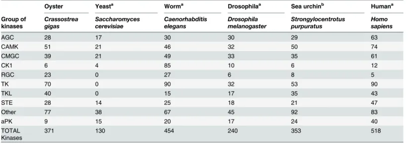
The genome ofC.gigaswas used to construct anin silicoproteome containing 26,086 single encoded proteins. Among them, we generated a non-redundant set of 371 protein kinases (S1 Table) using the HMM profile and BLAST homology searches in metazoan model organisms. The ePKs were then classified based on the known sequences of the catalytic region, together with features of the non-catalytic accessory domains, since their modular architecture is pre-dominant to define their biological roles even though it is conserved to varying degrees among the ePKs [38] (Table 1).
The resulting kinome corresponds to 1.4% of the whole proteome. This result is consistent with the correlation previously observed between kinome size and genome size [19]. Their evo-lutionary position provides important information on the evolution of kinases in Lophotrocho-zoa, a relatively distant group of species compared to those for which the kinome had already been characterized. Assignments ofC.gigasePKs and aPKs to different groups, including data from representative species, are shown inTable 1. InC.gigas(genome size: 557 Mb), we identi-fied a total of 362 ePKs, a number close to the 329 identiidenti-fied in the sea urchinS.purpuratus (genome size: 814 Mb) [17] [39], but different fromC.elegansfor which 434 ePKs (genome size: 103 Mb) have been discovered [40].
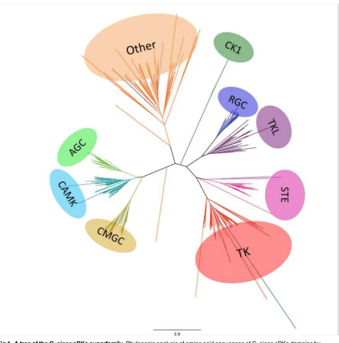
Based on our classification of ePKs and aPKs (S1 Table), we then determined which protein kinases were conserved betweenC.gigasand other species, and investigated their biological functions. Phylogenetic analyses with metazoan ePKs were performed to validate the affiliation of each oyster ePK (Fig 1). We showed that all ePKs were distributed into nine distinct classes, as described in almost all available kinomes. Indeed, some fungal species including the yeastS. cerevisiaehave a reduced kinome with the loss of one or more groups (for instance the TK group) [20] [41]. The nine ePK groups are present as a single cluster, although some protein kinases were distributed in the phylogenetic tree into unexpected groups based on the BLAST and HMM-based analyses. This may be due to the inherently imperfect nature of the heuristic methods used to generate phylogenies [17]. Overall, the nine groups of ePKs are well
Table 1. Taxonomic distribution of protein kinases (ePKs, aPKs) in various species.
Oyster Yeasta Worma Drosophilaa Sea urchinb Humana
Group of kinases
Crassostrea gigas
Saccharomyces cerevisiae
Caenorhabditis elegans
Drosophila melanogaster
Strongylocentrotus purpuratus
Homo sapiens
AGC 28 17 30 30 29 63
CAMK 51 21 46 32 50 74
CMGC 39 21 49 33 35 61
CK1 6 4 85 10 6 12
RGC 23 0 27 6 8 5
TK 70 0 90 32 53 90
TKL 40 0 15 17 35 43
STE 28 14 25 18 21 47
Other 77 38 67 45 92 83
aPK 9 15 20 17 24 40
TOTAL Kinases
371 130 454 240 353 518
a
data from [16]
b
data from [17]
represented, with a distribution similar to sea urchin, worm, drosophila or even human kinomes. The classification of 28 protein kinases remained ambiguous due to low sequence similarity and high BLAST E-values, suggesting that they might correspond to oyster-specific kinases. InS.purpuratus, 21 protein kinases with no family-level homologs in other organisms were identified as urchin-specific and belong to the Other group [17]. The function of urchin-specific protein kinases is unknown. They were shown to display weak expression during embryo development, thus suggesting that these kinases should be mainly implicated in adult-specific functions [17]. InC.gigas, another surprising feature of the oyster kinome is the large Fig 1. A tree of theC.gigasePKs superfamily.Phylogenic analysis of amino acid sequences ofC.gigasePKs domains by neighbor joining. The tree is visualized with Figtree. The color blocks represent the nine groups of ePKs shown in this tree: AGC (PKA, PKG, PKC containing), CAMK (Ca/Calmodulin-type), CMGC (CDK, MAPK, GSK, CDKL), CK1 (Casein kinases), RGC (receptor guanylate cyclase), TK (tyrosine kinases), TKL (tyrosine kinase-like), STE and Others. The aPK superfamily was excluded from this analysis.
number of protein kinases that belong to the RGC group. A detailed description of observed similarities between kinomes ofC.gigasand other species for the nine ePK groups, including RGC and the aPKs, is presented below.
AGC group
Twenty-eight AGC kinases were found inC.gigas, representing 7.5% of its kinome. This is sim-ilar to the number of AGC kinases described in the wormC.elegans(6.6%) and the sea urchin S.purpuratus(8.2%), but lower than in Drosophila and human (12.5%) (Table 1). AGC kinases have been well described in humans, with roles in signal transduction networks and an influ-ence on a large range of biological responses [21]. Specifically, they are involved in growth fac-tors, insulin and lipid signaling, and are linked to G protein coupled receptors. InC.gigas, the AGC group (28 members) contains all the families and subfamilies existing in the metazoan lineage, but has fewer members as compared to human (63 members). Interestingly, several members of the AGC group are represented by a single ortholog, in contrast to humans that have multiple redundant isoforms.C.gigashas only one protein kinase AKT (Protein kinase B) (while there are three in human and two in sea urchin) and one p70S6K (2 in human; 1 in sea urchin). In mammals, AKT was shown to be a protooncogene regulated by the lipid tyrosine kinase phosphatidylinositol 3 kinase (PI3-K) functioning as a cell survival signal to protect cells from apoptosis, and p70S6K controls the rate of protein translation during growth. Target studies of mRNA expression showed that these functions might be conserved inC.gigas, because AKT is regulated during gametogenesis [42] and p70S6K is involved in insulin signal-ing [43]. Insulin receptor, RAS, PI3-K and PDK1, all act as upstream activators of AKT, that is inducing cell survival. Each of these components of the PI3-K/AKT pathway is conserved inC. gigas, providing a powerful system for understanding cell survival signal in response to stress.
We also identified one animal-specific YANK (yet another novel kinase) (while there are four in human and one in the sea urchin) with unknown function. A detailed classification of AGC inC.gigasis provided inS1 Table.
CAMK group
Fifty-one CAMK kinases were identified inC.gigas, covering all families and subfamilies described in human (74 CAMK) and in the sea urchin (50 CAMK) (Table 1). Calcium-medi-ated signaling plays crucial roles in vertebrates during fertilization, embryonic development, signal transduction (through MAPK signaling), protein secretion, transport and memory [22] [44]. TheC.gigaskinome includes five MLCK (Myosin Light Chain Kinases), that phosphory-late the regulatory light chain of sarcomeric myosin [45], and four MARK (Microtubule Asso-ciated Kinases) that play roles in cytoskeletal organization and microtubule dynamics [46]. A similar number of TSSK (testis-specific serine/threonine-protein kinases) has been identified inC.gigas. TSSK are known to play an important role in spermatogenesis in humans [47]. In another marine bivalve, the Peruvian scallopArgopecten purpuratus, the mRNA of one TSSK was shown to be differentially expressed depending on the maturation stage, sex and tissue analyzed, suggesting a potential function of TSSK in reproductive mechanisms [48].
threonine 172 active site, as well as the binding domain for regulatoryβandγsubunits [50]. In mammals, AMPK is activated by metabolic stresses such as glucose deprivation, oxidative phosphorylation, ischemia, and hypoxia [51] [52]. InC.gigas, the activation of AMPKα con-trols metabolism during gametogenesis [42] and is activated in response to environmental-stress, for example 6h of hypoxia [53] or 14 days of exposure to low concentrations of pesti-cides [54]. In another bivalve, the freshwater musselElliptio complanata, AMPKαhas been proposed as a biomarker ofin situshort-term contamination [55].
CMGC group
Thirty nine CMGC inC.gigasare distributed between all families and subfamilies described in humans (61 members) [13] as well as in the sea urchin (35 members) [17] (Table 1). The CDK (Cyclin-Dependent Kinase) and MAPK (Mitogen-Activated Protein Kinase) families represent around 70% of this group. We identified the three major MAPK cascades inC.gigasrepresented by ERK, p38 MAPK and JNK, known to be crucial in cell signal transduction [56]. In another bivalve, the musselMytilus galloprovincialis, the p38 MAPK and JNK signaling pathways are acti-vated in response to various environmental stressors, such as temperature or heavy metals, leading to regulation of apoptosis [57] [58].C.gigasalso has five members of the DYRK (Dual specificity Tyrosine Regulated Kinase) family that play key roles in cell proliferation and apoptosis induction in response to stress such as DNA damage [59]. TheC.gigaskinome includes one casein kinase CK2 known to be involved in the response to oxidative stress inM.galloprovincialis[60] and the regulation of carbohydrate metabolism [61]. In the CMGC group, GSK-3βis present as a single ortholog and might be a key regulator of gonadal development [62], as demonstrated in the Portu-guese oyster,Crassostrea angulata[63]. InC.gigas, MAPK signaling is also mainly implied in maintaining metaphase I arrest in oocytes [64].
CK1 group
TheCrassostrea gigaskinome contains six proteins of the CK1 group, including four isoforms of casein kinase 1 and two isoforms of TTBK (Tau-tubulin kinase). CK1 and TTBK are involved in diverse cellular processes, including membrane trafficking, circadian rhythm, cell cycle progression, chromosome segregation, apoptosis, cell division, DNA repair, and cellular differentiation [65]. CK1 is the smallest group of protein kinases in most of the species for which the kinome has been identified [66], with four members in budding yeast, and six in human andC.gigas. Surprisingly, inC.elegans, 87 CK1 were characterized, and it was hypothe-sized that this huge diversification might be an adaptation allowing enhanced DNA repair in response to excessive exposure to environmental stressors and mutagens [11].
RGC group
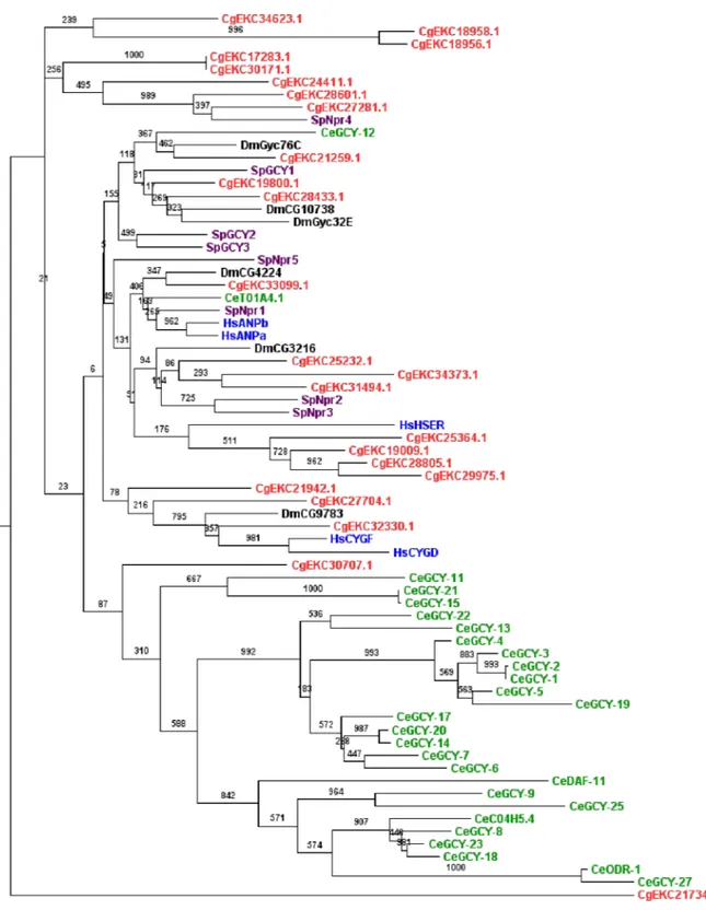
Fig 2. Phylogenetic analysis ofC.gigasRGC kinases.This tree was generated from RGC domain amino acid sequences from several species (Red:C.gigas, Purple:S.Purpuratus, Green:C.elegans, Blue:H.sapiens, Black:D.melanogaster), using the PhyML maximum likehood program and visualized with Figtree. A protein kinase from the TK group was used as outgroup (Insulin-related peptide receptor [CgEKC21734.1]). Corresponding maximum parsimony (MP) bootstrap values are shown on each branch.
abundant inC.elegans(27 members) and this particularity is not observed in other species (Fig 2). Interestingly, inC.elegans, RGC kinases have been associated with a worm-specific sensory receptor system [70] involved in alkalinity sensing [71], olfaction and odor sensing [72].C. ele-gansRGCs have also been described as salt receptor proteins [73]. InC.gigas, little is known about the function of RGCs. In the American oysterCrassostrea virginica, the peptides binding RGCs have been characterized in gills [74] [75], where their expression levels were reduced under low salinity [76]. The high number of RGC protein kinases could be an adaptation ofC. gigasliving in a highly dynamic marine environment. It would therefore be interesting to deter-mine whether the RGC group also plays a role in the oyster sensory receptor system.
TK group
InC.gigas, the TK group contains 70 ePKs representing 19% of its kinome. Evolutionarily, this group appears to be the youngest of the kinase groups, as it is absent in plants and unicellular organisms like the amoebaDictyostelium discoideumor the yeastS.cerevisiae[16] [77]. Each TK family is classified either as a membrane receptor (21 families) or as a cytoplasmic tyrosine kinase (11 families) [77]. TheC.gigaskinome contains 40 TK receptor and 30 cytoplasmic TK (S1 Table). These are key components for the relay of extracellular signals into the cell.
TK receptors are involved in various biological processes, including growth, development and immunity [78]. InC.gigas, nine TK families exist as single isoforms (Ret, insR, Ryk, Sev, CCK4, Trk, LmR, VEGFR, PDGFR), including insulin-related peptide receptors involved in growth regu-lation [79] and EGFR. The EGFR signaling module has been highly conserved throughout the course of evolution [80]. EGFR is a cell-surface receptor that plays key roles in growth and cellular proliferation, whose function is dependent on the diversity of EGFR ligands, such as epidermal growth factor or transforming growth factorα[81]. EGFR activates several signal transduction cascades that can regulate DNA synthesis, cell proliferation, differentiation, adhesion, migration and apoptosis and in mammals, EGFR contributes to the maturation of epithelial tissues, axon regeneration, wound repair and regeneration. InC.elegans, the EGFR network plays a central developmental role, determining the fate of several types of cells [81]. In Drosophila, a unique EGRF participates in gametogenesis, segmentation, wing and eye development [82]. InC.gigas, the ligand binding domain of the gene encoding EGFR exhibits a poor sequence similarity with human EGFR [83]. Based on its expression level inC.gigasand functional analysis, both inC. gigasand in a mouse cell line, it was demonstrated thatC.gigasEGFR plays a role in cell migration during wound healing of mantle and conserves the ability to activate cell proliferation [83].
Cytoplasmic TKs are involved in oxidative, temperature or osmotic regulation [84] [85] [86]. In our classification, five TK were considered as TK-unique inC.gigasbased on compari-sons with ePK domains from other species. In this study, we highlighted the fact thatC.gigas shares an expansion of the Src family with the sea urchin. InS.purpuratus, this is required for egg activation [87] and was considered to be echinoderm-specific [17]. Like the sea urchin and Drosophila,C.gigaslacks the Axl kinase receptor that is involved in hematopoiesis in mam-mals [88]. In theC.gigaskinome, the most represented TK subfamily is FERs, with eight mem-bers, whereas the sea urchin and Drosophila have just a single member and the worm, in contrast, possesses 38 FERs. InC.elegans, FRK1 is a FER that is essential for morphogenesis and differentiation of the epidermis during embryonic development [89]. The putative role of FER in oyster development remains to be investigated inC.gigas.
TKL group
domain comparisons with ePK domains from other species. The seven major families of the TKL group that have been identified in other species are present in the oyster kinome. MLK (Mixed Lineage Kinases) and RAF families are known to be sensitive to a wide range of stress-ors and are involved in MAPK signaling [90]. MLKL subfamilies inC.gigasare present as sin-gle homologs. Six members of LRRK (Leucine rich repeat kinases) were found inC.gigas, three in the sea urchin and one inC.elegans. In Drosophila andC.elegans, overexpression of LRRK induced neurodegeneration and modulated mitochondrial function [91] [92] [93]. InC.gigas, functional studies were performed on several TKL in the transforming growth factorβ
(TGF-β) pathway. Some receptors were characterized (activin-like receptors) [94] [95], including the TGF-βreceptor [96] and the Bone Morphogenetic Protein Receptor (BMPR1) [97]. The role of the TGF-βsignaling pathway was also described in germinal cell proliferation [98] [99] and immunity [100].
STE group
Twenty-nine protein kinases were classified in the STE group in theC.gigaskinome. This group contains the components of the cellular signal transduction upstream of MAPK and includes three families: STE20 (MAP4K), STE11 (MAP3K) and STE7 (MAP2K). Generally, MAP4K activates MAP3K, which activates MAP2K, which finally activates MAPK [101]. The MAPK signaling pathway is crucial in eukaryotes for response to stress and signaling into the cell [102]. The 11 subfamilies of STE20 and five subfamilies of STE11 existing in metazoans are present in theC.gigaskinome, as in the sea urchin. In contrast to humans, members of the STE11 family are present as single orthologs, except for MEKK4 which has two orthologs inC. gigas. Sea urchins and oysters have the same number of STE7 protein kinases (MEK3, MEK4, MEK5 and MEK7), known to be dual specificity protein kinases because they phosphorylate their target MAPK on both the threonine and tyrosine residues. Surprisingly, the MEK5/ERK5 signaling pathway was considered to be secondarily lost by protostomes [103]. Here, we show thatC.gigaspossesses MEK5 (STE group) and ERK5 (CMGC group), suggesting that the MEK5/ERK5 signaling pathway exists in Lophotrochozoa and is therefore not deuterostome-specific. The existence of all the components of the MAPK signaling pathway inC.gigasmay reflect its ability to be receptive and cope with environmental factors as reported inC.elegans [11]. Moreover, as suggested for the sea urchin andC.elegans[17] [18], the Pacific oyster should be a good species for investigating MAPK pathways, as the absence of redundancy would simplify functional studies.
The
“
Other
”
group
is involved in the antiviral response [106]. The NEK (NIMA-related serine/threonine Kinase) family is abundant, with 19 members in theC.gigaskinome as opposed to 11 (NEK1 to NEK11) in humans. In vertebrates, several NEK have an important role in controlling the cell cycle by contributing to the establishment of the microtubule bundle [107]. They are also important for cellular repair and recovery from DNA damage [108] [109]. This high number of NEKs might reflect an adaptation of oysters living in a highly dynamic marine environment to cope with DNA damage.
Atypical protein kinases
Regarding the atypical protein kinases, it was surprising to discover thatC.gigashad fewer members than other species studied in kinome comparisons. For example, the Pacific oyster has nine aPKs, whereas 15 are present in yeast, 17 in Drosophila, 20 in the worm, 24 in the sea urchin and 40 in humans. This group contains a number of lipid, sugar, and other small-mole-cule kinases. Indeed, the oyster atypical kinases were assigned to the alpha, RIO or PIKK (Phos-phatidyl inositol 3’kinase-related kinase) families. These proteins play a role in DNA repair and cell-cycle progression, but their functions in marine invertebrates remain unknown. The atypical protein kinases are now classified in the PKL (Protein Kinase-Like) group and share common structural features with protein kinases.
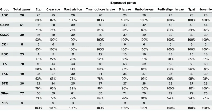
Expression of protein kinases during development
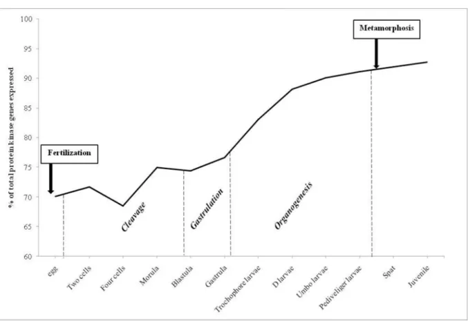
Transcriptomes from developmental stages inC.gigaswere obtained from RNA-seq (49 bp single-end Illumina reads) on a total of 250,000 zygotes maintained at 26°C and salinity around 30 ppm [5]. They provide valuable resources for studying protein kinase expression, in order to determine which kinases could be involved in the differentiation of cell types from egg to juve-nile. These transcriptomes were obtained from 38 biological samples representing three major developmental processes: cleavage, gastrulation and organogenesis. In this analysis, a threshold of 1 RPKM was used to classify genes as expressed or not (S1 Table). Our results show that more than 70% of the kinome was expressed during early development inC.gigas(Fig 3). Indeed, 70% of the kinome was expressed in eggs, 74% in embryos at the end of the cleavage (blastula stage), 77% at the end of the gastrulation, 83% in trochophore larvae, 88% in D-larvae, 90% for umbo larvae, 91% for pediveliger larvae, then 91% in spat and the maximum of 93% in juveniles (Fig 3). The lowest percentage of kinome was expressed at the 4-cell stage (68%;Fig 3) and might reflect a basal activity of weakly expressed genes, whose RPKM is close to the threshold of 1. From egg to metamorphosis, protein kinase genes belonging to all the classified groups inC.gigaswere expressed, but not from all subfamilies (S1 Table). Not surprisingly, the kinases associated with spermiogenesis, such as TSSK in the CAMK group, were weakly expressed since maturation had not occurred either in spat nor in juveniles (S1 Table).
Embryogenesis corresponds to the process by which the embryo forms and develops. It starts with the fertilization of the egg cell and is followed by mitotic divisions, known as cleav-age, leading to a late embryo called morula. From egg to juvenile, all the aPK genes are expressed, suggesting their involvement in all stages of the developing embryo. Almost all genes belonging to CMGC group, including MAPK signaling, were expressed in eggs (92%;
reflect their conserved roles in the organization of the symmetry in oyster embryos. Gastrula-tion follows the cleavage stages and is characterized by cell movements resulting in a massive reorganization of the embryo from a simple spherical ball of cells, the blastula, to a multi-lay-ered organism, the gastrula. During gastrulation, many of the cells at or near the surface of the embryo move to a more interior location. We identified several protein kinase genes belonging to different ePK groups that start to be expressed during gastrulation: PKCα, LRRK, PKG, HUNK, BRSK, DCAMKL, QIK, MEKK2 and GCN2 (S1 Table). They may be involved in con-volution and differentiation of the cells into different dermal layers. In the frogXenopus laevis, the different isoforms of protein kinase PKC are associated with Wnt signaling, leading to con-vergent extension movements [110]. LRRK2 is also involved in Wnt signaling in vertebrates [111]. PKG (c-GMP dependent protein kinase) is expressed during gastrulation of the medaka fishOryzias latipesand is necessary to maintain embryo development by phosphorylating tar-gets of SHH (Sonic Hedgehog), a crucial pathway for embryogenesis in vertebrates [112]. These pathways linked with gastrulation processes might thus be conserved between verte-brates and inverteverte-brates.
InC.gigas, after gastrulation, organogenesis starts with the development of the larva (trochophore, D-shape, umbo and pediveliger) involving cell reorganization until metamor-phosis. Some protein kinase genes start to be expressed and belong to several groups: the CAMK group, with CASK (Calcium/calmodulin-dependent Serine protein Kinase) and MLCK (Myosin Light-Chain Kinase), the STE group with YSK (Yeast Sps1/Ste20-related Kinase 4) Fig 3. Number of protein kinases genes expressed duringCrassostrea gigasdevelopment.Number of ePK and aPK genes expressed duringCrassostrea gigasdevelopment (% of total 371 protein kinases). The development stages were grouped according to the following: cleavage begins from the two cell stage to blastula, gastrulation is from blastula to gastrula stages. Genes with expression values<1 RPKM were considered to be non-expressed.
and MKC (Metastatic Kidney Cancer), and some members of NEK families in the Other group (S1 Table). Because their mRNAs are expressed from the first steps of the cleavage, MLCK and NEK might belong to the regulation of the early development inC.gigas. MLCK was shown to be crucial to larval settlement of the intertidal barnacleBalanus amphitrite, by modulating muscle contraction and motility of larvae [113]. MLCK is also involved in axon pathway for-mation in Drosophila embryos [114]. NEK proteins were identified in mammals as necessary for the development of the nervous system [107].
Metamorphosis is an important step, transforming larvae into spat with a reorganization of most larval organs to form a juvenile oyster with its definitive organs. We identified 3 protein kinase genes belonging to the TK group, Syk (Spleen tyrosine kinase), Tie (tyrosine kinase with immunoglobulin-like and EGF-like domains) and Csk (C-Src kinase) that start to be expressed between the pediveliger larval stage and spat (S1 Table). We can hypothesize that these tyrosine kinases might participate in the regulation of metamorphosis. Indeed, in the bryozoanBugula neritinaand the barnacleBalanus amphitrite, a tyrosine kinase inhibitor was shown to prevent metamorphosis [115].
Expression of protein kinases under environmental stressors
We performed anin silicoanalysis to identify down- and up-regulated kinases genes in oysters subjected to 8 different potential sources of stress: temperature, salinity, exposure to air, and to five metals (cadmium, copper, mercury, lead and zinc) [5]. Among the 371 genes encoding protein kinases, we found that 177 genes (48% of theC.gigaskinome) were differentially Table 2. Number and percentage of ePK and aPK genes expressed duringCrassostrea gigasdevelopment.
Expressed genes
Group Total genes Egg Cleavage Gastrulation Trochophore larvae D larvae Umbo larvae Pediveliger larvae Spat Juvenile
AGC 28 25 25 28 28 28 28 28 28 28
89% 89% 100% 100% 100% 100% 100% 100% 100%
CAMK 51 36 38 39 43 43 42 43 43 44
71% 75% 76% 84% 84% 82% 84% 84% 86%
CMGC 39 36 39 39 39 39 39 39 39 39
92% 100% 100% 100% 100% 100% 100% 100% 100%
CK1 6 5 6 6 6 6 6 6 6 6
83% 100% 100% 100% 100% 100% 100% 100% 100%
RGC 23 4 5 6 12 15 16 18 15 13
17% 22% 26% 52% 65% 70% 78% 65% 57%
TK 70 42 44 43 48 53 59 59 63 63
60% 63% 61% 69% 76% 84% 84% 90% 90%
TKL 40 25 27 30 31 36 37 36 39 39
63% 68% 75% 78% 90% 93% 90% 98% 98%
STE 28 22 24 25 27 27 28 28 27 28
79% 86% 89% 96% 96% 100% 100% 96% 100%
Other 77 56 59 61 65 71 70 72 72 75
73% 77% 79% 84% 92% 91% 94% 94% 97%
aPK 9 9 9 9 9 9 9 9 9 9
100% 100% 100% 100% 100% 100% 100% 100% 100%
The development stages were grouped according to the following: cleavage begins from the two cell stage to blastula, gastrulation is from blastula to gastrula stages.
expressed under at least one factor relative to the compared condition (Table 3). This corre-sponds to around 3% of the 5,844 genes identified from transcriptome datasets that were mod-ulated (up or down-regmod-ulated) by these factors [5].
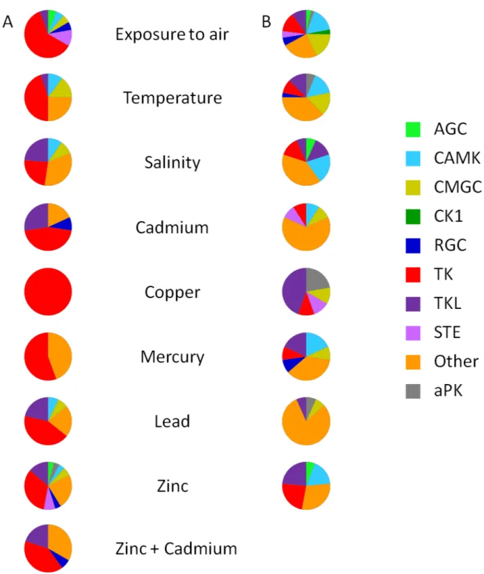
Exposure to air is a serious stressor, causing hypoxia and leading to a decrease of ATP con-centration in the hemolymph [116]. Among the different potential sources of stress, air expo-sure affected the expression of the largest number of protein kinase genes (110), corresponding to 30% of the oyster kinome (Table 3). Interestingly, three genes of the CK1 group were under-expressed following exposure to air (Table 3) without being affected by any other stressors (Fig 4). The highest numbers of differentially expressed genes under exposure to air were found in the TK (11 up-regulated, 12 down-regulated) and Other (23 down-regulated) groups (Table 3). Given the expected roles of TK and NEKs (in the Other group), the regulation of cell growth, differentiation, proliferation, cell-cycle or apoptosis might be modified by exposure to air.
Thermal stress triggered changes in the expression of 53 protein kinase genes (Table 3), cor-responding to 14% of theC.gigaskinome and 7% of the 776 differentially expressed genes identified in the transcriptome data [5]. The response ofC.gigasto thermal stress includes inhibition of apoptosis, stabilization of protein conformation and protein refolding [117]. Sev-eral genes in the TK and Other groups were up or down–regulated and we can hypothesize Table 3. Number and percentage of ePK and aPK genes up- and down-regulated under environmental stressors.
Differentially expressed genes under stressors
Group Total genes Exposure to air
Temperature Salinity Cadmium Copper Mercury Lead Zinc
Up Down Up Down Up Down Up Down Up Down Up Down Up Down Up Down
AGC 28 1 3 0 0 0 1 0 0 0 0 0 0 0 0 2 1
4% 11% - - - 4% - - - 7% 4%
CAMK 51 1 15 2 5 2 3 0 1 0 0 0 2 1 0 2 3
2% 29% 4% 10% 4% 6% - 2% - - - 4% 2% - 4% 6%
CMGC 39 1 16 3 5 2 0 0 1 0 1 0 1 1 1 3 0
3% 41% 8% 13% 5% - - 3% - 3% - 3% 3% 3% 8%
-CK1 6 0 3 0 0 0 0 0 0 0 0 0 0 0 0 0 0
- 50% - - -
-RGC 23 1 5 1 0 0 0 1 0 0 0 0 1 0 0 2 0
4% 22% 4% - - - 4% - - - - 4% - - 9%
-TK 70 11 12 9 3 5 2 5 1 2 1 5 1 6 0 17 4
16% 17% 13% 7% 7% 3% 7% 1% 3% 1% 7% 1% 9% - 24% 6%
TKL 40 1 9 1 4 5 1 3 0 0 4 0 2 3 1 7 4
2% 23% 3% 10% 13% 2% 8% - - 10% - 5% 8% 3% 18% 10%
STE 28 2 4 0 0 0 0 0 1 0 1 0 0 0 0 4 0
7% 14% - - - 4% - 4% - - - - 14%
-Other 77 0 23 5 12 7 6 2 6 0 0 4 4 3 12 12 5
- 30% 6% 16% 9% 8% 3% 8% - - 5% 5% 4% 16% 16% 6%
aPK 9 0 2 0 2 0 2 0 0 0 2 0 0 0 1 2 0
- 22% - 22% - 22% - - - 22% - - - 11% 22%
-Total 371 18 92 21 31 21 15 11 10 2 9 9 11 14 15 51 17
Transcriptome data were obtained by RNA-seq, normalized, and differentially expressed genes were detected using a referenced statistical method constructed based on a Poisson distribution [5]. Eight types of stressors are shown (exposure to air, temperature, salinity, and to 5 metals: cadmium, copper, mercury, lead and zinc).
that the functions of these kinases can be modulated by a temperature stress in oysters. The PI3K (Phosphatidylinositol-4,5-bisphosphate 3-Kinase)/AKT/mTOR (mechanistic target of rapamycin) pathway was shown to be involved in the oyster’s response to chronic thermal Fig 4. Group-level comparison of ePK and aPK genes up and down-regulated under environmental stressors.(A) Up-regulated protein kinases. (B) Down-Up-regulated protein kinases. Pie charts depict the proportion of the protein kinases group, and the total number of pie is presented inTable 3. The absence of a pie chart means no regulation was observed.
stress during three months [118]. In contrast, we observed that none of the genes from the AGC group, including AKT kinase, responded to 12 hours or 7 days of thermal stress. Simi-larly, we showed that none of the mRNA encoding STE kinases were differentially expressed after thermal stress, although MAPK signaling was previously shown to be activated for signal transduction in stress conditions [57] [58] [101]. Actually, the absence of gene regulation might not necessarily reflect the absence of function, since protein kinases are known to be mostly activated at the post-translational level.
Estuaries are characterized by variations in salinity due to rainfall and tides. Organisms like C.gigasare subjected daily to these variations and have developed mechanisms to adapt their behavior by closing their shells and ceasing to feed when exposed to low-salinity water [119]. Variations in salinity changed the expression of 35 protein kinase genes (Table 3), correspond-ing to 9.5% of theC.gigaskinome and 3% of the 1024 differentially expressed genes identified in the transcriptome data [5]. Based on our classification, we showed that protein kinase genes differentially expressed under the salinity constraint were mainly associated with the regulation of metabolism, cytoskeletal organization, and immune response (S1 Table). In the CAMK group, hyposalinity changed the expression of 3 MLRK genes (2 up-regulated; 1 down-regu-lated) (S1 Table). MLRK are known to be myosin light chain kinases associated with the passive elasticity of muscle. Our results suggest reorganization of cytoskeletal components during hyposalinity. Five kinases from TK group (Csk, Met, Eph and Ror, Fer) were up-regulated in response to hyposalinity (Table 3), indicating that variations of salinity could modulate the cell adhesion and communication, signal transduction and cytoskeleton organization inC.gigas. In TK group, hyposalinity up-regulated 1 Fer (S1 Table), a protein kinase known to be involved in signaling and regulation of cell-cell interactions [120], as already observed inC.gigasunder salinity stress [121].
In the Other group, the gene encoding PKR (protein kinase R) was up-regulated in response to hypersalinity and down-regulated in response to hyposalinity (S1 Table). InC.gigas, over-expression of the PKR gene has been associated with a protective antiviral immune response against Ostreid herpesvirus (OsHV-1μvar), induced by polyinosinic: polycytidylic acid (Poly I:
C) injection [106] [122]. In mammals, the activation of PKR is one of the mechanisms permit-ting early blocking of viral replication via inhibition of protein synthesis [123] and activation of autophagy [124]. The PKR gene inC.gigasis homologous to vertebrate ISG (interferon stim-ulated gene) and plays a role in pathogen recognition and activation of innate immunity [122]. Taken together, modulation of PKR by salinity might indicate that salinity could have an impact on the ability ofC.gigasto resist to viral infections.
Oysters living in coastal environments that suffer from human expansion may also be exposed to anthropogenic contaminations, such as heavy metals [125]. Heavy metal exposure changed the expression of 96 mRNA encoding protein kinases (S1 Table), corresponding to 26% of theC.gigaskinome and 9% of the 1024 differentially expressed genes identified in the transcriptome data [5]. With the exception of the CK1 group, genes encoding kinases in all ePK groups responded to heavy metal exposure (Fig 4). Heavy metal exposure modulated the expression of genes that mainly belong to TK, TKL and Other groups (Table 3;Fig 4). Most of these TK genes were up-regulated, and we can hypothesize that changes in TK expression could reflect that heavy metals can interfere on growth hormone regulation, as observed in fish [126].
Conclusion
transcriptome datasets. TheC.gigaskinome might have an original evolutionary position, because the number, classification and distribution of protein kinases is closer to deutero-stomes (sea urchin and human) than to protodeutero-stomes (nematode and fly). Kinase gene redun-dancy in theC.gigaskinome concerns about 30% of the genes. The lack of redundant protein kinases isoforms in several groups suggests that the Pacific oysterC.gigascould be a good spe-cies for the development of functional research dealing with protein kinases, for example, to study developmental processes, given that most protein kinases are expressed during the first stages of embryogenesis. Here we also identified the kinases that are mobilized in the Pacific oyster to deal with changes in temperature or salinity, with exposure to air, and after contami-nation by heavy metals, as well as during development. We provide new insights into the key pathways that may be crucial for adaptation to life in a highly dynamic environment.
Supporting Information
S1 Table. IdentifiedCrassostrea gigasprotein kinases.Genbank accession number, classifica-tion (group/family/subfamily), query definiclassifica-tion, best hit name, percentage of identity com-pared to other species with corresponding E-values, embryonic expression and differentially expression induced by stressors. Y: yes. U: up-regulation. D: Down-regulation.
(XLSX)
S1 Text. Zhang et al. (2012).
(PDF)
Acknowledgments
The authors are grateful to Fanny Marquer for her help in data mining of theCrassostrea gigas genome. We thank Arnaud Huvet and Fabrice Pernet for helpful discussions. We thank Helen McCombie of the Brest University Translation Bureau (BTU) for her English editing services. This works contributes to the‘Laboratoire d’Excellence’LabexMER (ANR-10-LABX-19). We additionally thank Dr. Jessica Moss Small from the Aquaculture Genetics and Breeding Tech-nology Center of the Virginia Institute of Marine Science, College of William & Mary (VA, USA) for her help with editing the English.
Author Contributions
Conceived and designed the experiments: YE LQ CC. Performed the experiments: YE LQ. Ana-lyzed the data: YE LQ VP CC. Contributed reagents/materials/analysis tools: YE LQ VP EG PB CC. Wrote the paper: YE VP EG PB CC. Designed the software used in analysis: YE LQ.
References
1. Sussarellu R, Fabioux C, Le Moullac G, Fleury E, Moraga D. Transcriptomic response of the Pacific oyster Crassostrea gigas to hypoxia. Mar Genomics. 2010; 3: 133–143. doi:10.1016/j.margen.2010. 08.005PMID:21798207
2. Timmins-Schiffman E, Nunn BL, Goodlett DR, Roberts SB. Shotgun proteomics as a viable approach for biological discovery in the Pacific oyster. Conserv Physiol. 2013; 1. doi:10.1093/conphys/cot009
3. Guo X, He Y, Zhang L, Lelong C, Jouaux A. Immune and stress responses in oysters with insights on adaptation. Fish Shellfish Immunol. 2015; 46: 107–119. doi:10.1016/j.fsi.2015.05.018PMID:
25989624
4. Guo L, Xu F, Feng Z, Zhang G. A bibliometric analysis of oyster research from 1991 to 2014. Aquac Int. 2015; 1–18. doi:10.1007/s10499-015-9928-1
6. Walsh CT, Garneau-Tsodikova S, Gatto GJ. Protein posttranslational modifications: the chemistry of proteome diversifications. Angew Chem Int Ed Engl. 2005; 44: 7342–7372. doi:10.1002/anie. 200501023PMID:16267872
7. Mann M, Jensen ON. Proteomic analysis of post-translational modifications. Nat Biotechnol. 2003; 21: 255–261. doi:10.1038/nbt0303-255PMID:12610572
8. Choudhary C, Kumar C, Gnad F, Nielsen ML, Rehman M, Walther TC, et al. Lysine acetylation targets protein complexes and co-regulates major cellular functions. Science. 2009; 325: 834–840. doi:10. 1126/science.1175371PMID:19608861
9. Erce MA, Pang CNI, Hart-Smith G, Wilkins MR. The methylproteome and the intracellular methylation network. Proteomics. 2012; 12: 564–586. doi:10.1002/pmic.201100397PMID:22246820
10. Cohen P. The regulation of protein function by multisite phosphorylation–a 25 year update. Trends Biochem Sci. 2000; 25: 596–601. doi:10.1016/S0968-0004(00)01712-6PMID:11116185
11. Plowman GD, Sudarsanam S, Bingham J, Whyte D, Hunter T. The protein kinases of Caenorhabditis elegans: A model for signal transduction in multicellular organisms. Proc Natl Acad Sci. 1999; 96: 13603–13610. doi:10.1073/pnas.96.24.13603PMID:10570119
12. Lahiry P, Torkamani A, Schork NJ, Hegele RA. Kinase mutations in human disease: interpreting geno-type–phenotype relationships. Nat Rev Genet. 2010; 11: 60–74. doi:10.1038/nrg2707PMID:
20019687
13. Manning G, Whyte DB, Martinez R, Hunter T, Sudarsanam S. The Protein Kinase Complement of the Human Genome. Science. 2002; 298: 1912–1934. doi:10.1126/science.1075762PMID:12471243
14. Theillet F-X, Smet-Nocca C, Liokatis S, Thongwichian R, Kosten J, Yoon M-K, et al. Cell signaling, post-translational protein modifications and NMR spectroscopy. J Biomol NMR. 2012; 54: 217–236. doi:10.1007/s10858-012-9674-xPMID:23011410
15. Hanks SK, Hunter T. Protein kinases 6. The eukaryotic protein kinase superfamily: kinase (catalytic) domain structure and classification. FASEB J Off Publ Fed Am Soc Exp Biol. 1995; 9: 576–596. 16. Manning G, Plowman GD, Hunter T, Sudarsanam S. Evolution of protein kinase signaling from yeast
to man. Trends Biochem Sci. 2002; 27: 514–520. PMID:12368087
17. Bradham CA, Foltz KR, Beane WS, Arnone MI, Rizzo F, Coffman JA, et al. The sea urchin kinome: A first look. Dev Biol. 2006; 300: 180–193. doi:10.1016/j.ydbio.2006.08.074PMID:17027740
18. Manning G. Genomic overview of protein kinases. WormBook. 2005; doi:10.1895/wormbook.1.60.1
19. Li Z, Hao Y, Wang L, Xiang H, Zhou Z. Genome-Wide Identification and Comprehensive Analyses of the Kinomes in Four Pathogenic Microsporidia Species. PLoS ONE. 2014; 9. doi:10.1371/journal. pone.0115890
20. Miranda-Saavedra D, Barton GJ. Classification and functional annotation of eukaryotic protein kinases. Proteins Struct Funct Bioinforma. 2007; 68: 893–914. doi:10.1002/prot.21444
21. Pearce LR, Komander D, Alessi DR. The nuts and bolts of AGC protein kinases. Nat Rev Mol Cell Biol. 2010; 11: 9–22. doi:10.1038/nrm2822PMID:20027184
22. Cohen SM, Li B, Tsien RW, Ma H. Evolutionary and functional perspectives on signaling from neuro-nal surface to nucleus. Biochem Biophys Res Commun. 2015; 460: 88–99. doi:10.1016/j.bbrc.2015. 02.146PMID:25998737
23. Schenk PW, Snaar-Jagalska BE. Signal perception and transduction: the role of protein kinases. Bio-chim Biophys Acta BBA—Mol Cell Res. 1999; 1449: 1–24. doi:10.1016/S0167-4889(98)00178-5
24. Wong SK, Garbers DL. Receptor guanylyl cyclases. J Clin Invest. 1992; 90: 299–305. PMID:
1353764
25. Knighton D, Zheng J, Teneyck L, Ashford V, Xuong N, Taylor S, et al. Crystal-Structure of the Catalytic Subunit of Cyclic Adenosine-Monophosphate Dependent Protein-Kinase. Science. 1991; 253: 407–
414. doi:10.1126/science.1862342PMID:1862342
26. Waskiewicz AJ, Cooper JA. Mitogen and stress response pathways: MAP kinase cascades and phos-phatase regulation in mammals and yeast. Curr Opin Cell Biol. 1995; 7: 798–805. doi: 10.1016/0955-0674(95)80063-8PMID:8608010
27. Bettinger BT, Amberg DC. The MEK kinases MEKK4/Ssk2p facilitate complexity in the stress signal-ing responses of diverse systems. J Cell Biochem. 2007; 101: 34–43. doi:10.1002/jcb.21289PMID:
17348032
28. LaRonde-LeBlanc N, Wlodawer A. The RIO kinases: An atypical protein kinase family required for ribosome biogenesis and cell cycle progression. Biochim Biophys Acta BBA—Proteins Proteomics. 2005; 1754: 14–24. doi:10.1016/j.bbapap.2005.07.037PMID:16182620
30. Goldberg JM, Manning G, Liu A, Fey P, Pilcher KE, Xu Y, et al. The Dictyostelium Kinome—Analysis of the Protein Kinases from a Simple Model Organism. PLoS Genet. 2006; 2: e38. doi:10.1371/ journal.pgen.0020038PMID:16596165
31. Bateman A, Coin L, Durbin R, Finn RD, Hollich V, Griffiths-Jones S, et al. The Pfam protein families database. Nucleic Acids Res. 2004; 32: D138–141. doi:10.1093/nar/gkh121PMID:14681378
32. Caenepeel S, Charydczak G, Sudarsanam S, Hunter T, Manning G. The mouse kinome: Discovery and comparative genomics of all mouse protein kinases. Proc Natl Acad Sci U S A. 2004; 101: 11707–11712. doi:10.1073/pnas.0306880101PMID:15289607
33. Guindon S, Dufayard J-F, Lefort V, Anisimova M, Hordijk W, Gascuel O. New algorithms and methods to estimate maximum-likelihood phylogenies: assessing the performance of PhyML 3.0. Syst Biol. 2010; 59: 307–321. doi:10.1093/sysbio/syq010PMID:20525638
34. Lopez A, Resnik P. Improved HMM Alignment Models for Languages with Scarce Resources. Pro-ceedings of the ACL Workshop on Building and Using Parallel Texts. Stroudsburg, PA, USA: Associ-ation for ComputAssoci-ational Linguistics; 2005. pp. 83–86. Available:http://dl.acm.org/citation.cfm?id= 1654449.1654464
35. Waterhouse AM, Procter JB, Martin DMA, Clamp M, Barton GJ. Jalview Version 2—a multiple sequence alignment editor and analysis workbench. Bioinformatics. 2009; 25: 1189–1191. doi:10. 1093/bioinformatics/btp033PMID:19151095
36. Robinson MD, Oshlack A. A scaling normalization method for differential expression analysis of RNA-seq data. Genome Biol. 2010; 11: R25. doi:10.1186/gb-2010-11-3-r25PMID:20196867
37. Chen S, Yang P, Jiang F, Wei Y, Ma Z, Kang L. De novo analysis of transcriptome dynamics in the migratory locust during the development of phase traits. PloS One. 2010; 5: e15633. doi:10.1371/ journal.pone.0015633PMID:21209894
38. Deshmukh K, Anamika K, Srinivasan N. Evolution of domain combinations in protein kinases and its implications for functional diversity. Prog Biophys Mol Biol. 2010; 102: 1–15. doi:10.1016/j. pbiomolbio.2009.12.009PMID:20026163
39. The Genome of the Sea Urchin Strongylocentrotus purpuratus. Science. 2006; 314: 941–952. doi:10. 1126/science.1133609PMID:17095691
40. C. elegans Sequencing Consortium. Genome sequence of the nematode C. elegans: a platform for investigating biology. Science. 1998; 282: 2012–2018. PMID:9851916
41. Manning G, Reiner DS, Lauwaet T, Dacre M, Smith A, Zhai Y, et al. The minimal kinome of Giardia lamblia illuminates early kinase evolution and unique parasite biology. Genome Biol. 2011; 12: R66. doi:10.1186/gb-2011-12-7-r66PMID:21787419
42. Guévélou E, Huvet A, Galindo-Sánchez CE, Milan M, Quillien V, Daniel J-Y, et al. Sex-specific regula-tion of AMP-activated protein kinase (AMPK) in the Pacific oyster Crassostrea gigas. Biol Reprod. 2013; 89: 100. doi:10.1095/biolreprod.113.109728PMID:23926284
43. Jouaux A, Franco A, Heude-Berthelin C, Sourdaine P, Blin JL, Mathieu M, et al. Identification of Ras, Pten and p70S6K homologs in the Pacific oyster Crassostrea gigas and diet control of insulin path-way. Gen Comp Endocrinol. 2012; 176: 28–38. doi:10.1016/j.ygcen.2011.12.008PMID:22202600
44. Maier LS, Bers DM. Role of Ca2+/calmodulin-dependent protein kinase (CaMK) in excitation– contrac-tion coupling in the heart. Cardiovasc Res. 2007; 73: 631–640. doi:10.1016/j.cardiores.2006.11.005
PMID:17157285
45. Stull JT, Kamm KE, Vandenboom R. Myosin light chain kinase and the role of myosin light chain phos-phorylation in skeletal muscle. Arch Biochem Biophys. 2011; 510: 120–128. doi:10.1016/j.abb.2011. 01.017PMID:21284933
46. Hayashi K, Suzuki A, Ohno S. PAR-1/MARK: a Kinase Essential for Maintaining the Dynamic State of Microtubules. Cell Struct Funct. 2012; 37: 21–25. doi:10.1247/csf.11038PMID:22139392
47. Wang X, Wei Y, Fu G, Li H, Saiyin H, Lin G, et al. Tssk4 is essential for maintaining the structural integrity of sperm flagellum. Mol Hum Reprod. 2015; 21: 136–145. doi:10.1093/molehr/gau097
PMID:25361759
48. Boutet I, Moraga D, Marinovic L, Obreque J, Chavez-Crooker P. Characterization of reproduction-specific genes in a marine bivalve mollusc: Influence of maturation stage and sex on mRNA expres-sion. Gene. 2008; 407: 130–138. doi:10.1016/j.gene.2007.10.005PMID:17976928
49. Carling D. The AMP-activated protein kinase cascade—a unifying system for energy control. Trends Biochem Sci. 2004; 29: 18–24. doi:10.1016/j.tibs.2003.11.005PMID:14729328
50. Hawley SA, Davison M, Woods A, Davies SP, Beri RK, Carling D, et al. Characterization of the AMP-activated protein kinase kinase from rat liver and identification of threonine 172 as the major site at which it phosphorylates AMP-activated protein kinase. J Biol Chem. 1996; 271: 27879–27887. PMID:
51. Hardie DG, Carling D, Carlson M. The AMP-activated/SNF1 protein kinase subfamily: metabolic sen-sors of the eukaryotic cell? Annu Rev Biochem. 1998; 67: 821–855. doi:10.1146/annurev.biochem. 67.1.821PMID:9759505
52. Choi SL, Kim SJ, Lee KT, Kim J, Mu J, Birnbaum MJ, et al. The regulation of AMP-activated protein kinase by H(2)O(2). Biochem Biophys Res Commun. 2001; 287: 92–97. doi:10.1006/bbrc.2001.5544
PMID:11549258
53. Guévélou E, Huvet A, Sussarellu R, Milan M, Guo X, Li L, et al. Regulation of a truncated isoform of AMP-activated protein kinaseα(AMPKα) in response to hypoxia in the muscle of Pacific oyster Cras-sostrea gigas. J Comp Physiol [B]. 2013; 183: 597–611. doi:10.1007/s00360-013-0743-6
54. Epelboin Y, Quéré C, Pernet F, Pichereau V, Corporeau C. Energy and Antioxidant Responses of Pacific Oyster Exposed to Trace Levels of Pesticides. Chem Res Toxicol. 2015; 28: 1831–1841. doi:
10.1021/acs.chemrestox.5b00269PMID:26313537
55. Goodchild CG, Frederich M, Zeeman SI. AMP-activated protein kinase is a biomarker of energetic sta-tus in freshwater mussels exposed to municipal effluents. Sci Total Environ. 2015; 512–513: 201–
209. doi:10.1016/j.scitotenv.2015.01.065
56. Sundaram MV. RTK/Ras/MAPK signaling. WormBook Online Rev C Elegans Biol. 2006; 1–19. doi:
10.1895/wormbook.1.80.1
57. Kefaloyianni E, Gourgou E, Ferle V, Kotsakis E, Gaitanaki C, Beis I. Acute thermal stress and various heavy metals induce tissue-specific pro- or anti-apoptotic events via the p38-MAPK signal transduc-tion pathway in Mytilus galloprovincialis (Lam.). J Exp Biol. 2005; 208: 4427–4436. doi:10.1242/jeb. 01924PMID:16339863
58. Gourgou E, Aggeli I-K, Beis I, Gaitanaki C. Hyperthermia-induced Hsp70 and MT20 transcriptional upregulation are mediated by p38-MAPK and JNKs in Mytilus galloprovincialis (Lamarck); a pro-sur-vival response. J Exp Biol. 2010; 213: 347–357. doi:10.1242/jeb.036277PMID:20038670
59. Yoshida K. Role for DYRK family kinases on regulation of apoptosis. Biochem Pharmacol. 2008; 76: 1389–1394. doi:10.1016/j.bcp.2008.05.021PMID:18599021
60. Kolaiti R-M, Baier A, Szyszka R, Kouyanou-Koutsoukou S. Isolation of a CK2αSubunit and the Holo-enzyme from the Mussel Mytilus galloprovincialis and Construction of the CK2αand CK2βcDNAs. Mar Biotechnol. 2010; 13: 505–516. doi:10.1007/s10126-010-9321-zPMID:20922551
61. Al Quobaili F, Montenarh M. CK2 and the regulation of the carbohydrate metabolism. Metabolism. 2012; 61: 1512–1517. doi:10.1016/j.metabol.2012.07.011PMID:22917893
62. Berthelin C, Kellner K, Mathieu M. Storage metabolism in the Pacific oyster (Crassostrea gigas) in relation to summer mortalities and reproductive cycle (West Coast of France). Comp Biochem Physiol B Biochem Mol Biol. 2000; 125: 359–369. doi:10.1016/S0305-0491(99)00187-XPMID:10818269
63. Zeng Z, Ni J, Ke C. Expression of glycogen synthase (GYS) and glycogen synthase kinase 3β (GSK3β) of the Fujian oyster, Crassostrea angulata, in relation to glycogen content in gonad develop-ment. Comp Biochem Physiol B Biochem Mol Biol. 2013; 166: 203–214. doi:10.1016/j.cbpb.2013.09. 003PMID:24035883
64. Portillo-López A, Gould MC, Stephano JL. MAPK is involved in metaphase I arrest in oyster and mus-sel oocytes. Biol Cell. 2003; 95: 275–282. doi:10.1016/S0248-4900(03)00054-6PMID:12941525
65. Knippschild U, Wolff S, Giamas G, Brockschmidt C, Wittau M, Würl PU, et al. The role of the casein kinase 1 (CK1) family in different signaling pathways linked to cancer development. Onkologie. 2005; 28: 508–514. doi:10.1159/000087137PMID:16186692
66. Morrison DK, Murakami MS, Cleghon V. Protein Kinases and Phosphatases in the Drosophila Genome. J Cell Biol. 2000; 150: 57–62.
67. Duda T, Yadav P, Sharma RK. Allosteric Modification, the Primary ATP Activation Mechanism of Atrial Natriuretic Factor Receptor Guanylate Cyclase. Biochemistry (Mosc). 2011; 50: 1213–1225. doi:10. 1021/bi1018978
68. Takei Y. Does the natriuretic peptide system exist throughout the animal and plant kingdom? Comp Biochem Physiol B Biochem Mol Biol. 2001; 129: 559–573. doi:10.1016/S1096-4959(01)00366-9
PMID:11399492
69. Silberbach M, Roberts CT Jr. Natriuretic peptide signalling: molecular and cellular pathways to growth regulation. Cell Signal. 2001; 13: 221–231. doi:10.1016/S0898-6568(01)00139-5PMID:11306239
70. Coburn CM, Bargmann CI. A Putative Cyclic Nucleotide–Gated Channel Is Required for Sensory Development and Function in C. elegans. Neuron. 1996; 17: 695–706. doi:10.1016/S0896-6273(00) 80201-9PMID:8893026
71. Murayama T, Takayama J, Fujiwara M, Maruyama IN. Environmental Alkalinity Sensing Mediated by the Transmembrane Guanylyl Cyclase GCY-14 in C. elegans. Curr Biol. 2013; 23: 1007–1012. doi:
72. L’Etoile ND, Bargmann CI. Olfaction and odor discrimination are mediated by the C. elegans guanylyl cyclase ODR-1. Neuron. 2000; 25: 575–586. PMID:10774726
73. Smith HK, Luo L, O’Halloran D, Guo D, Huang X-Y, Samuel ADT, et al. Defining specificity determi-nants of cGMP mediated gustatory sensory transduction in Caenorhabditis elegans. Genetics. 2013; 194: 885–901. doi:10.1534/genetics.113.152660PMID:23695300
74. Vesely DL, Gower WR, Giordano AT, Friedl FE. Atrial natriuretic peptides in the heart and hemolymph of the oyster, Crassostrea virginica: a comparison with vertebrates. Comp Biochem Physiol B. 1993; 106: 535–546. PMID:8281751
75. Poulos JE, Gower WR, Friedl FE, Vesely DL. Atrial natriuretic peptide gene expression within inverte-brate hearts. Gen Comp Endocrinol. 1995; 100: 61–68. doi:10.1006/gcen.1995.1133PMID:8575660
76. Palmer PA, Friedl FE, Giordano AT, Vesely DL. Alteration of environmental salinity modulates atrial natriuretic peptide concentrations in the gills of the oyster, Crassostrea virginica. Comp Biochem Phy-siol A PhyPhy-siol. 1995; 110: 171–178. doi:10.1016/0300-9629(94)00105-3
77. Robinson DR, Wu YM, Lin SF. The protein tyrosine kinase family of the human genome. Oncogene. 2000; 19: 5548–5557. doi:10.1038/sj.onc.1203957PMID:11114734
78. Lapraz F, Röttinger E, Duboc V, Range R, Duloquin L, Walton K, et al. RTK and TGF-beta signaling pathways genes in the sea urchin genome. Dev Biol. 2006; 300: 132–152. doi:10.1016/j.ydbio.2006. 08.048PMID:17084834
79. Gricourt L, Bonnec G, Boujard D, Mathieu M, Kellner K. Insulin-like system and growth regulation in the Pacific oyster Crassostrea gigas: hrIGF-1 effect on protein synthesis of mantle edge cells and expression of an homologous insulin receptor-related receptor. Gen Comp Endocrinol. 2003; 134: 44–56. PMID:13129502
80. Yarden Y. The EGFR family and its ligands in human cancer. signalling mechanisms and therapeutic opportunities. Eur J Cancer Oxf Engl 1990. 2001; 37 Suppl 4: S3–8.
81. Doroquez DB, Rebay I. Signal integration during development: mechanisms of EGFR and Notch path-way function and cross-talk. Crit Rev Biochem Mol Biol. 2006; 41: 339–385. doi:10.1080/
10409230600914344PMID:17092823
82. Shilo BZ. Signaling by the Drosophila epidermal growth factor receptor pathway during development. Exp Cell Res. 2003; 284: 140–149. PMID:12648473
83. Sun L, Huan P, Wang H, Liu F, Liu B. An EGFR gene of the Pacific oyster Crassostrea gigas functions in wound healing and promotes cell proliferation. Mol Biol Rep. 2014; 41: 2757–2765. doi:10.1007/ s11033-014-3130-9PMID:24469720
84. Sun X, Wu F, Datta R, Kharbanda S, Kufe D. Interaction between Protein Kinase Cδand the c-Abl Tyrosine Kinase in the Cellular Response to Oxidative Stress. J Biol Chem. 2000; 275: 7470–7473. doi:10.1074/jbc.275.11.7470PMID:10713049
85. Satoh T, Kato J, Nishida K, Kaziro Y. Tyrosine phosphorylation of ACK in response to temperature shift-down, hyperosmotic shock, and epidermal growth factor stimulation. FEBS Lett. 1996; 386: 230–
234. doi:10.1016/0014-5793(96)00449-8PMID:8647288
86. Cao H, Sanguinetti AR, Mastick CC. Oxidative stress activates both Src-kinases and their negative regulator Csk and induces phosphorylation of two targeting proteins for Csk: caveolin-1 and paxillin. Exp Cell Res. 2004; 294: 159–171. doi:10.1016/j.yexcr.2003.11.010PMID:14980511
87. Roux MM, Townley IK, Raisch M, Reade A, Bradham C, Humphreys G, et al. A functional genomic and proteomic perspective of sea urchin calcium signaling and egg activation. Dev Biol. 2006; 300: 416–433. doi:10.1016/j.ydbio.2006.09.006PMID:17054939
88. Heide I, Sokoll AC, Henz BM, Nagel S, Kreissig K, Grützkau A, et al. Regulation and possible function of axl expression in immature human mast cells. Ann Hematol. 1998; 77: 199–205. PMID:9858144
89. Putzke AP, Hikita ST, Clegg DO, Rothman JH. Essential kinase-independent role of a Fer-like non-receptor tyrosine kinase in Caenorhabditis elegans morphogenesis. Dev Camb Engl. 2005; 132: 3185–3195. doi:10.1242/dev.01900
90. Gotoh I, Adachi M, Nishida E. Identification and characterization of a novel MAP kinase kinase kinase, MLTK. J Biol Chem. 2001; 276: 4276–4286. doi:10.1074/jbc.M008595200PMID:11042189
91. Yao C, Khoury RE, Wang W, Byrd TA, Pehek EA, Thacker C, et al. LRRK2-mediated neurodegenera-tion and dysfuncneurodegenera-tion of dopaminergic neurons in a Caenorhabditis elegans model of Parkinson’s dis-ease. Neurobiol Dis. 2010; 40: 73–81. doi:10.1016/j.nbd.2010.04.002PMID:20382224
92. Imai Y, Gehrke S, Wang H-Q, Takahashi R, Hasegawa K, Oota E, et al. Phosphorylation of 4E-BP by LRRK2 affects the maintenance of dopaminergic neurons in Drosophila. EMBO J. 2008; 27: 2432–
93. Saha S, Guillily M, Ferree A, Lanceta J, Chan D, Ghosh J, et al. LRRK2 modulates vulnerability to mitochondrial dysfunction in C. elegans. J Neurosci Off J Soc Neurosci. 2009; 29: 9210–9218. doi:
10.1523/JNEUROSCI.2281-09.2009
94. Herpin A, Favrel P, Cunningham C. Gene structure and expression of cg-ALR1, a type I activin-like receptor from the bivalve mollusc Crassostrea gigas. Gene. 2002; 301: 21–30. PMID:12490320
95. Le Quéré H, Herpin A, Huvet A, Lelong C, Favrel P. Structural and functional characterizations of an Activin type II receptor orthologue from the pacific oyster Crassostrea gigas. Gene. 2009; 436: 101–
107. doi:10.1016/j.gene.2009.01.010PMID:19393178
96. Herpin A, Lelong C, Becker T, Rosa FM, Favrel P, Cunningham C. Structural and functional evidences for a type 1 TGF-beta sensu stricto receptor in the lophotrochozoan Crassostrea gigas suggest con-served molecular mechanisms controlling mesodermal patterning across bilateria. Mech Dev. 2005; 122: 695–705. doi:10.1016/j.mod.2004.12.004PMID:15817226
97. Herpin A, Lelong C, Becker T, Rosa F, Favrel P, Cunningham C. Structural and functional evidence for a singular repertoire of BMP receptor signal transducing proteins in the lophotrochozoan Crassos-trea gigas suggests a shared ancestral BMP/activin pathway. FEBS J. 2005; 272: 3424–3440. doi:
10.1111/j.1742-4658.2005.04761.xPMID:15978047
98. Fleury E, Fabioux C, Lelong C, Favrel P, Huvet A. Characterization of a gonad-specific transforming growth factor-beta superfamily member differentially expressed during the reproductive cycle of the oyster Crassostrea gigas. Gene. 2008; 410: 187–196. doi:10.1016/j.gene.2007.12.017PMID:
18234456
99. Corporeau C, Groisillier A, Jeudy A, Barbeyron T, Fleury E, Fabioux C, et al. A functional study of transforming growth factor-beta from the gonad of Pacific oyster Crassostrea gigas. Mar Biotechnol N Y N. 2011; 13: 971–980. doi:10.1007/s10126-010-9361-4
100. Lelong C, Mathieu M, Favrel P. Structure and expression of mGDF, a new member of the transforming growth factor-beta superfamily in the bivalve mollusc Crassostrea gigas. Eur J Biochem FEBS. 2000; 267: 3986–3993.
101. Qi M, Elion EA. MAP kinase pathways. J Cell Sci. 2005; 118: 3569–3572. doi:10.1242/jcs.02470
PMID:16105880
102. Plotnikov A, Zehorai E, Procaccia S, Seger R. The MAPK cascades: Signaling components, nuclear roles and mechanisms of nuclear translocation. Biochim Biophys Acta BBA—Mol Cell Res. 2011; 1813: 1619–1633. doi:10.1016/j.bbamcr.2010.12.012
103. Glatz G, Gógl G, Alexa A, Reményi A. Structural mechanism for the specific assembly and activation of the extracellular signal regulated kinase 5 (ERK5) module. J Biol Chem. 2013; 288: 8596–8609. doi:10.1074/jbc.M113.452235PMID:23382384
104. Cambray S, Pedraza N, Rafel M, Garí E, Aldea M, Gallego C. Protein Kinase KIS Localizes to RNA Granules and Enhances Local Translation. Mol Cell Biol. 2009; 29: 726–735. doi:10.1128/MCB. 01180-08PMID:19015237
105. Escoubas J-M, Briant L, Montagnani C, Hez S, Devaux C, Roch P. Oyster IKK-like protein shares structural and functional properties with its mammalian homologues. FEBS Lett. 1999; 453: 293–298. doi:10.1016/S0014-5793(99)00737-1PMID:10405163
106. Green TJ, Montagnani C. Poly I:C induces a protective antiviral immune response in the Pacific oyster (Crassostrea gigas) against subsequent challenge with Ostreid herpesvirus (OsHV-1μvar). Fish Shellfish Immunol. 2013; 35: 382–388. doi:10.1016/j.fsi.2013.04.051PMID:23685009
107. Malumbres M. Physiological relevance of cell cycle kinases. Physiol Rev. 2011; 91: 973–1007. doi:
10.1152/physrev.00025.2010PMID:21742793
108. Fry AM O’Regan L, Sabir SR, Bayliss R. Cell cycle regulation by the NEK family of protein kinases. J Cell Sci. 2012; 125: 4423–4433. doi:10.1242/jcs.111195PMID:23132929
109. Nguyen CL, Possemato R, Bauerlein EL, Xie A, Scully R, Hahn WC. Nek4 regulates entry into replica-tive senescence and the response to DNA damage in human fibroblasts. Mol Cell Biol. 2012; 32: 3963–3977. doi:10.1128/MCB.00436-12PMID:22851694
110. Kühl M, Geis K, Sheldahl LC, Pukrop T, Moon RT, Wedlich D. Antagonistic regulation of convergent extension movements in Xenopus by Wnt/β-catenin and Wnt/Ca2+ signaling. Mech Dev. 2001; 106: 61–76. doi:10.1016/S0925-4773(01)00416-6PMID:11472835
111. Berwick DC, Harvey K. LRRK2 functions as a Wnt signaling scaffold, bridging cytosolic proteins and membrane-localized LRP6. Hum Mol Genet. 2012; 21: 4966–4979. doi:10.1093/hmg/dds342PMID:
22899650
112. Yamamoto T, Suzuki N. Expression and function of cGMP-dependent protein kinase type I during medaka fish embryogenesis. J Biol Chem. 2005; 280: 16979–16986. doi:10.1074/jbc.M412433200