Research Article
Topical Antinociceptive Effect of
Vanillosmopsis arborea
Baker on Acute Corneal Pain in Mice
Laura Hévila Inocêncio Leite,
1Gerlânia de Oliveira Leite,
1Thales Silva Coutinho,
1Severino Denício Gonçalves de Sousa,
1Renata Souza Sampaio,
1José Galberto Martins da Costa,
1Irwin Rose Alencar de Menezes,
1and Adriana Rolim Campos
21Programa de P´os-Graduac¸˜ao em Bioprospecc¸˜ao Molecular, Universidade Regional do Cariri, 63105-000 Crato, CE, Brazil 2Universidade de Fortaleza, Avenida Washington Soares 1321, 60811-905 Fortaleza, CE, Brazil
Correspondence should be addressed to Adriana Rolim Campos; [email protected]
Received 25 October 2013; Revised 7 December 2013; Accepted 19 December 2013; Published 10 February 2014
Academic Editor: Olumayokun A. Olajide
Copyright © 2014 Laura H´evila Inocˆencio Leite et al. his is an open access article distributed under the Creative Commons Attribution License, which permits unrestricted use, distribution, and reproduction in any medium, provided the original work is properly cited.
his study aimed to assess the possible topical antinociceptive activity ofVanillosmopsis arborea Baker essential oil (EOVA) and to clarify the underlying mechanism, using the acute model of chemical (eye wiping) nociception in mice. EOVA (25 to 200 mg/kg; p.o. and topical) evidenced signiicant antinociception against chemogenic pain in the test model of formalin-induced neuroinlammatory pain. Local application of 5 M NaCl solution on the corneal surface of the eye produced a signiicant nociceptive behavior, characterized by eye wiping. he number of eye wipes was counted during the irst 30 s. EOVA (25, 50, 100, and 200 mg/kg; p.o. and topical) signiicantly decreased the number of eye wipes. Naloxone, yohimbine, L-NAME, theophylline, glibenclamide, and ruthenium red had no efect on the antinociceptive efect of EOVA. However, ondansetron, p-chlorophenylalanine methyl ester (PCPA), capsazepine, prazosin, and atropine prevented the antinociception induced by EOVA. hese results indicate the topical antinociceptive efect of EOVA and showed that 5-HT,�1, TRPV1, and central muscarinic receptors might be involved in the antinociceptive efect of EOVA in the acute corneal model of pain in mice.
1. Introduction
he small size of the cornea and the extensive branching of the peripheral axons of corneal neurons make this structure
the most densely innervated tissue of the body [1]. he
majority of corneal sensory ibers are polymodal nociceptors, which are activated by noxious mechanical, thermal, and
chemical stimuli [2].
Safe, long-lasting pain relief following corneal abrasions, corneal ulcers or ophthalmic surgery is diicult to achieve with current analgesics. he use of topical NSAIDs and acetominophen is constrained by their gradual onset and limited eicacy, and the adverse side efects of systemic
opioids are well known [3].
Vanillosmopsis arborea Baker is native to the Araripe National Forest, in the Northeast of Brazil in the state of Cear´a. here are few studies concerning the traditional use of
this plant. However, biological and pharmacological studies have shown that its essential oil presents antimicrobial,
antiinlammatory and gastroprotective activities [4].
Topically applied Vanillosmopsis arborea essential oil
produced antinociception in acetic acid, cyclophosphamide,
mustard oil, capsaicin or formalin-induced visceral pain [5].
he essential oil also suppressed the ear edema induced by Croton oil and phenol but no that one induced by histamine
or capsaicin [6].
Ocular pain has not been adequately controlled in many patients and there is a lack of studies that examine the
efect ofVanillosmopsis arboreaon the perception of corneal
pain. herefore, the present study was designed to investigate
the efects of acute topical administration ofVanillosmopsis
arboreaon acute corneal pain that was induced by hypertonic saline applied locally on the corneal surface in mice.
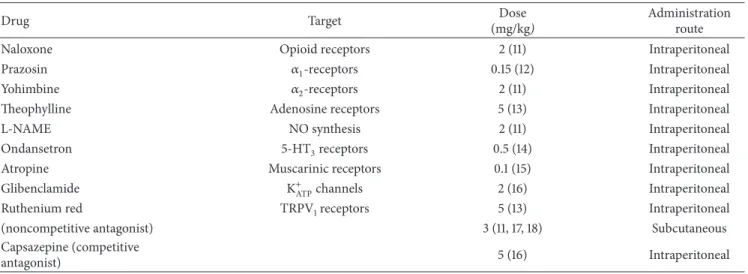
Table 1: Pharmacologic agents used for test in order to verify the possible action mechanism of EOVA antinociceptive efect.
Drug Target Dose
(mg/kg)
Administration route
Naloxone Opioid receptors 2 (11) Intraperitoneal
Prazosin �1-receptors 0.15 (12) Intraperitoneal
Yohimbine �2-receptors 2 (11) Intraperitoneal
heophylline Adenosine receptors 5 (13) Intraperitoneal
L-NAME NO synthesis 2 (11) Intraperitoneal
Ondansetron 5-HT3receptors 0.5 (14) Intraperitoneal
Atropine Muscarinic receptors 0.1 (15) Intraperitoneal
Glibenclamide K+ATPchannels 2 (16) Intraperitoneal
Ruthenium red TRPV1receptors 5 (13) Intraperitoneal
(noncompetitive antagonist) 3 (11, 17, 18) Subcutaneous
Capsazepine (competitive
antagonist) 5 (16) Intraperitoneal
2. Material and Methods
2.1. Essential Oil. he essential oil from Vanillosmopsis arborea Baker bark (EOVA) was obtained from the Nat-ural Products Research Laboratory of Regional University of Cariri. he composition (w/w) of EOVA revealed the
presence of�-bisabolol to the extent of 70%. Other identiied
compounds were �-cadinol (8.4%), elemicin (6.21%), �
-bisabolene (4.46%),�-guaiene (2.31%),�-cubebene (1.76%),
and estragole (1.08%).
2.2. Animals. Male Swiss albino mice (20–25 g) obtained from the Central Animal House of Regional University of Cariri were used. hey were housed in environmentally
controlled conditions (22∘C, 12 h light-dark cycle), with free
access to standard pellet diet (Purina, S˜ao Paulo, Brazil) and water. Animals were kept in cages with raised loors to prevent coprophagy. he experimental protocols were in accordance with the ethical guidelines of National Institute of Health, Bethesda, USA.
2.3. Formalin-Induced Paw Licking. he formalin-induced nociception was performed as described previously by
Hun-skaar and Hole [7]. Groups of mice (� = 8) were treated
with vehicle (10 mL/kg, p.o.), EOVA (25, 50, 100, or 200 mg/kg p.o. or topical), or morphine (7.5 mg/kg, s.c.) 30 or 60 min
before the administration of 20�L of 1% formalin (in 0.9%
saline) into the plantar surface of the right hind paw. he duration of paw licking (s) as an index of painful response was determined at 0–5 min (early phase, neurogenic) and 20– 25 min (late phase, inlammatory) ater formalin injection.
2.4. Eye Wiping Test. Corneal nociception was induced by a local application of hypertonic saline to the corneal surface
[8, 9]. One drop (40�L) of 5 M NaCl solution was applied
locally on the corneal surface of mice using a ine dropper
[10]. he number of eye wipes performed with the ipsilateral
forepaw was counted for a period of 30 s. Topical (20�L/eye)
or orally (10 mL/kg) EOVA (25, 50, 100 or 200 mg/kg;� =
8/group) or vehicle were given 1 h before the noxiou agent.
Morphine 7 mg/kg (s.c.;� = 8) was used as positive control.
A normal control group (� = 8) received one drop of 0.15 M
NaCl (0.9%).
In order to verify the possible involvement of opioid,
noradrenergic, nitrergic, 5-HT3, muscarinic, K+ATP,
adenosin-ergic, and TRPV1systems in the efect of EOVA, the animals
(� = 8/group) were treated with the respective antagonist (Table 1), 30 min before the topical administration of EOVA (50 mg/kg). he doses of antagonists were chosen based on previous studies.
To assess the possible contribution of endogenous
sero-tonin, animals (� = 8/group) were pretreated with�
-chlo-rophenylalanine methyl esther (PCPA, 100 mg/kg, i.p., an inhibitor of serotonin synthesis) or with vehicle, once a day for 4 consecutive days. hen, 24 hours ater the last PCPA or vehicle injection, animals received EOVA (50 mg/kg) and 1 h later they were tested in the eye wiping test.
2.5. Statistical Analysis. All data are presented as mean
± s.e.m. he data were evaluated by one-way analysis of
variance with Student-Newman-Keuls post hoc test using the GraphPad Prism 4.0 statistical program. he level of
signiicance was set at� < 0.05.
3. Results and Discussion
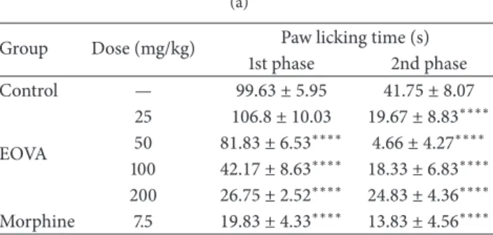
In formalin test, pretreatment with EOVA (oral and top-ical) caused signiicant diminutions of both irst phase (neurogenic) and second phase (inlammatory)
nocicep-tion responses (Tables2(a) and2(b)). Morphine (5 mg/kg),
the reference standard, also signiicantly suppressed the formalin-response at both phases.
Topically administered EOVA (25, 50, 100, or 200 mg/kg)
respectively (� < 0.001, � < 0.01, � < 0.01, and� <
0.0001, resp.) decreased the number of eye wipes induced by the local application of 5 M NaCl solution on the corneal
surface (Table 3(a)). Oral treatment with EOVA also reduced
Table 2: (a) Efect of topical EOVA on formalin test in mice. (b) Efect of oral EOVA on formalin test in mice.
(a)
Group Dose (mg/kg) Paw licking time (s)
1st phase 2nd phase
Control — 99.63 ± 5.95 41.75 ± 8.07
EOVA
25 106.8 ± 10.03 19.67 ± 8.83∗∗∗∗ 50 81.83 ± 6.53∗∗∗∗ 4.66 ± 4.27∗∗∗∗ 100 42.17 ± 8.63∗∗∗∗ 18.33 ± 6.83∗∗∗∗ 200 26.75 ± 2.52∗∗∗∗ 24.83 ± 4.36∗∗∗∗
Morphine 7.5 19.83 ± 4.33∗∗∗∗ 13.83 ± 4.56∗∗∗∗
Data are expressed as mean±s.e.m.∗∗∗∗� < 0.0001compared to control
group. ANOVA followed by Student-Newman-Keuls test. (b)
Group Dose (mg/kg) Paw licking time (s)
1st phase 2nd phase
Control — 57.67 ± 1.89 96.17 ± 6.95
EOVA
25 32.00 ± 6.27∗∗∗∗ 49.33 ± 7.96∗∗∗∗ 50 38.50 ± 4.23∗∗∗∗ 45.50 ± 9.78∗∗∗∗ 100 34.83 ± 5.81∗∗∗∗ 36.50 ± 11.43∗∗∗∗ 200 37.50 ± 4.08∗∗∗∗ 23.17 ± 10.51∗∗∗∗
Morphine 7.5 19.83 ± 4.33∗∗∗∗ 13.83 ± 4.56∗∗∗∗
Data are expressed as mean±s.e.m.∗∗∗∗� < 0.0001compared to control
group. ANOVA followed by Student-Newman-Keuls test.
EOVA was evaluated for topical antinociceptive activity in mice using experimental models of chemogenic nocicep-tion. EOVA (oral and topical) was efective in attenuating acute neurogenic and tonic inlammatory phases of the formalin response. he essential oil, given topically, elicited a dose-unrelated antinociceptive efect on the paw-licking response, just as observed in the eye wiping test.
he local application of a 5 M NaCl solution to the corneal surface produced corneal nociception. Previous work has shown that the application of hypertonic saline to the tongue and cornea transiently activates nociceptive neurons with wide dynamic range property in the trigeminal subnucleus
caudalis [11]. Moreover, infusion of hypertonic saline into the
masseter muscle produces hind paw shaking in addition to activating c-Fos positive neurons in the ipsilateral trigeminal
subnucleus caudalis [12]. he results presented here are in
agreement with previous indings [8,9].
Study of trigeminal pain and analgesic efects on trigem-inal acute pains such as headache, muscle spasms, dental problems, or postsurgery pain seems to be more problematic. he cornea is used for nociception studies in trigeminal
system [13], since corneal nociceptive receptors have a large
representation in the trigeminal ganglion through the
oph-thalmic branch of trigeminal nerve [14]. hin myelinated
ibres [15] as well as unmyelinated ibers in cornea respond
to chemical, mechanical, and thermal noxious stimuli [16].
In rat, wiping the eye with forelimb is an obvious withdrawal response to corneal chemical stimuli. Some researchers have used eye wiping test for investigating the chemical pungency
[17] or the presence of C-iber activity [18,19]. Eye wiping test
Table 3: (a) Efect of topically applied EOVA on the corneal nociception induced by a local application of a 5 M NaCl solution in mice. (b) Efect of orally administered EOVA on the corneal nociception induced by a local application of a 5 M NaCl solution in mice.
(a)
Group Dose (mg/kg) Number of eye wipes (30 s)
Control — 20.33 ± 0.66
EOVA
25 15.33 ± 0.91∗∗∗
50 16.67 ± 0.84∗∗
100 16.67 ± 0.66∗∗
200 14.33 ± 0.61∗∗∗∗
Morphine 7.5 12.17 ± 0.83∗∗∗∗
Data are expressed as mean±s.e.m.∗∗� < 0.01,∗∗∗� < 0.001, and
∗∗∗∗� < 0.0001compared to control group. ANOVA followed by
Student-Newman-Keuls test.
(b)
Group Dose (mg/kg) Number of eye wipes (30 s)
Control — 22.13 ± 1.31
EOVA
25 16.50 ± 1.26∗∗
50 14.75 ± 0.70∗∗∗
100 15.25 ± 1.09∗∗∗
200 12.63 ± 0.73∗∗∗
Morphine 7.5 11.88 ± 0.83∗∗∗
Data are expressed as mean±s.e.m.∗∗� < 0.01,∗∗∗� < 0.001, and
∗∗∗∗� < 0.0001compared to control group. ANOVA followed by
Student-Newman-Keuls test.
is a phasic analgesic test and is sensitive to centrally acting analgesics. Hypertonic saline-induced corneal pain has been introduced as a model of acute pain for study of mechanisms
of pain in the trigeminal system in rats [8].
he results of this investigation provide evidence that
the essential oil from V. arborea bark is topically active
in the attenuation of corneal pain induced by 5 M NaCl. Previous study showed the local antiinlammatory efect of
the essential oil fromVanillosmopsis arborea. It was observed
that EOVA efect can be related to release of leukotrienes, decrease the production of inlammatory eicosanoids and
inluence on the production of AA metabolites [6].
his antinociceptive efect may be related to the high�
-bisabolol content in EOVA, since that�-bisabolol possess
vis-ceral antinociceptive activity [20] and it is able to reduce the
neuronal excitability in a concentration-dependent manner
[21].
In the present study, we attempted to characterize further some of the mechanisms through which EOVA exerts its antinociceptive action in chemical model of corneal pain in mice.
he antinociceptive efect induced by the EOVA (50 mg/kg) was signiicantly inhibited by ondansetron,
PCPA, prazosin, atropine, and capsazepine (Tables 4,
5, 6, 7, and 8). On the other hand, the administration
of glibenclamide, naloxone, ruthenium red, yohimbine, LNAME or theophylline did not prevent the EOVA-induced
Table 4: Efect of pretreatment with ondansetron on the EOVA-induced corneal antinociception in mice.
Group Dose (mg/kg) Number of eye wipes (30 s)
Control — 12.50 ± 0.67
OEVA 50 8.33 ± 1.05∗∗∗
Ondansetron 0.5 5.00 ± 0.51∗∗∗∗
+OEVA 50 12.50 ± 1.91
Data are expressed as mean±s.e.m.∗∗∗� < 0.001and∗∗∗∗� < 0.0001
compared to control group. ANOVA followed by Student-Newman-Keuls test.
Table 5: Efect of pretreatment with�-chlorophenylalanine methyl esther (PCPA) on the EOVA-induced corneal antinociception in mice.
Group Dose (mg/kg) Number of eye wipes (30 s)
Control — 11.50 ± 0.84
OEVA 50 6.25 ± 0.16∗∗∗
PCPA 100 9.12 ± 0.83
+OEVA 50 14.13 ± 0.35∗∗∗
Data are expressed as mean±s.e.m.∗∗∗� < 0.001compared to control
group. ANOVA followed by Student-Newman-Keuls test.
Serotoninergic neurons also play a crucial role in the
control of pain [22] and the diversity of subtype receptors for
serotonin makes this system able to exert either facilitatory
or inhibitory function [23]. Spinal 5-HT3receptors have been
shown to mediate antinociception, possibly via GABA release
[24,25]. Concerning the mechanism through which EOVA
exerts its antinociceptive action, the present study shows that
the 5-HT3receptor is likely involved. his conclusion derives
from the fact that pretreatment of animals with the 5-HT3
antagonist, ondansetron, reversed the antinociception caused by EOVA.
In addition, the treatment of mice with tryptophan hydroxylase inhibitor (PCPA) at a dose known to decrease the cortical content of serotonin and to signiicantly reverse
morphine antinociception [26, 27] attenuated the efect of
EOVA, indicating the involvement of 5-HT in the antinoci-ceptive efect of EOVA.
In the present study, the involvement of�1-receptors in
the antinociceptive action of EOVA is suggested by the results
showing that pretreatment of animals with prazosin (an�1
-receptor antagonist) signiicantly blocked the antinociception
caused by EOVA. Prazosin, a former selective �1-receptor
antagonist, has high ainity for�2B-receptors [28]. It has been
suggested that the efects of prazosin against spinal�agonists
appear through�2B-receptors, although yohimbine interacts
with�2A-receptor site as well as�2B-receptor site [29].
he roles of acetylcholine, cholinergic agonists, and cholinesterase inhibitors, collectively termed cholinomimet-ics, have been established in the modulation of pain and
analgesia [30]. Here we show that the pretreatment of animals
with atropine prevented the antinociceptive efect induced by EOVA. his result indicates that the muscarinic receptors are involved in EOVA-induced antinociceptive efect. It is already well established that the analgesic efect of systemic morphine is mediated by a descending cholinergic pathway
Table 6: Efect of pretreatment with prazosin on the EOVA-induced corneal antinociception in mice.
Group Dose (mg/kg) Number of eye wipes (30 s)
Control — 14.17 ± 0.70
EOVA 50 8.33 ± 1.05∗∗∗
Prazosin 0.15 12.33 ± 0.80
+EOVA 50 13.33 ± 0.95
Data are expressed as mean±s.e.m.∗∗∗� < 0.001compared to control
group. ANOVA followed by Student-Newman-Keuls test.
Table 7: Efect of pretreatment with atropine or glibenclamide on the EOVA-induced corneal antinociception in mice.
Group Dose (mg/kg) Number of eye wipes (30 s)
Control — 17.00 ± 0.85
EOVA 50 11.83 ± 0.74∗∗∗
Atropine 0.1 10.83 ± 0.87∗∗∗∗
+EOVA 50 14.83 ± 0.542
Glibenclamide 2 6.66 ± 0.95∗∗∗∗
+EOVA 50 11.50 ± 0.84∗∗∗
Data are expressed as mean±s.e.m.∗∗∗� < 0.001and∗∗∗∗� < 0.0001
compared to control group. ANOVA followed by Student-Newman-Keuls test.
Table 8: Efect of pretreatment with capsazepine on the EOVA-induced corneal antinociception in mice.
Group Dose (mg/kg) Number of eye wipes (30 s)
Control — 14.32 ± 0.71
EOVA 50 11.00 ± 0.57∗
Capsazepine 5 10.67 ± 0.76∗
+EOVA 50 12.83 ± 0.47
Data are expressed as mean±s.e.m.∗� < 0.05compared to control group.
ANOVA followed by Student-Newman-Keuls test.
Table 9: Efect of pretreatment with naloxone on the EOVA-induced corneal antinociception in mice.
Group Dose (mg/kg) Number of eye wipes (30 s)
Control — 20.00 ± 0.68
EOVA 50 13.17 ± 0.79∗∗∗∗
+Naloxone 2 11.50 ± 0.88∗∗∗∗
Data are expressed as mean±s.e.m.∗∗∗∗� < 0.0001compared to control
group. ANOVA followed by Student-Newman-Keuls test.
Table 10: Efect of pretreatment with ruthenium red on the EOVA-induced corneal antinociception in mice.
Group Dose (mg/kg) Number of eye wipes (30 s)
Control — 12.50 ± 0.67
EOVA 50 8.33 ± 1.05∗
Ruthenium red 3 5.83 ± 2.33∗∗
+EOVA 50 5.16 ± 0.30∗∗
Data are expressed as mean±s.e.m.∗� < 0.05and∗∗� < 0.01compared to
control group. ANOVA followed by Student-Newman-Keuls test.
[31] as well as spinal endogenous acetylcholine acting through
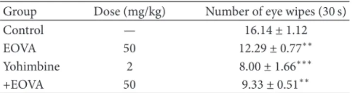
Table 11: Efect of pretreatment with yohimbine on the EOVA-induced corneal antinociception in mice.
Group Dose (mg/kg) Number of eye wipes (30 s)
Control — 16.14 ± 1.12
EOVA 50 12.29 ± 0.77∗∗
Yohimbine 2 8.00 ± 1.66∗∗∗
+EOVA 50 9.33 ± 0.51∗∗
Data are expressed as mean±s.e.m.∗∗� < 0.01and∗∗∗� < 0.001compared
to control group. ANOVA followed by Student-Newman-Keuls test.
Table 12: Efect of pretreatment with L-NAME on the EOVA-induced corneal antinociception in mice.
Group Dose (mg/kg) Number of eye wipes (30 s)
Control — 19.00 ± 0.57
EOVA 50 11.50 ± 0.67∗∗∗∗
L-NAME 10 17.50 ± 0.84
+EOVA 50 12.83 ± 0.65∗∗∗∗
Data are expressed as mean±s.e.m.∗∗∗∗� < 0.001compared to control
group. ANOVA followed by Student-Newman-Keuls test.
Table 13: Efect of pretreatment with theophylline on the EOVA-induced corneal antinociception in mice.
Group Dose (mg/kg) Number of eye wipes (30 s)
Control — 17.00 ± 0.85
EOVA 50 10.33 ± 0.55∗∗∗∗
heophylline 5 7.33 ± 2.06∗∗∗∗
+EOVA 50 9.16 ± 0.60∗∗∗∗
Data are expressed as mean±s.e.m.∗∗∗∗� < 0.001compared to control
group. ANOVA followed by Student-Newman-Keuls test.
Ruthenium red (a noncompetitive TRPV1antagonist) did
not afect the antinociceptive efect of EOVA. However,
cap-sazepine (a competitive TRPV1channel antagonist) inhibited
this response, indicating that EOVA interacts directly with
TRPV1receptors may contribute to this antinociception.
In summary, we have demonstrated that topical EOVA reduces the nociceptive behavior in models of formalin-induced paw licking and eye wiping in mice. In the corneal pain model, the observed antinociception is possibly
medi-ated by 5-HT,�1, muscarinic, and TRPV1receptors.
Conflict of Interests
here is no conlict of interests.
Acknowledgments
his research was supported by the grants and fellowships from CNPq, CAPES, and FUNCAP, Brazil.
References
[1] L. J. M¨uller, C. F. Marfurt, F. Kruse, and T. M. T. Tervo, “Corneal nerves: structure, contents and function,” Experimental Eye Research, vol. 76, no. 5, pp. 521–542, 2003.
[2] C. Belmonte, M. C. Acosta, and J. Gallar, “Neural basis of sen-sation in intact and injured corneas,”Experimental Eye Research, vol. 78, no. 3, pp. 513–525, 2004.
[3] K. Wishaw, D. Billington, D. O’Brien, and P. Davies, “he use of orbital morphine for postoperative analgesia in pterygium surgery,”Anaesthesia and Intensive Care, vol. 28, no. 1, pp. 43–45, 2000.
[4] A. V. Colares, F. Almeida-Souza, N. N. Taniwaki et al., “In vitro antileishmanial activity of essential oil ofVanillosmopsis arborea (Asteraceae) baker,” Evidence-Based Complementary and Alternative Medicine, vol. 2013, Article ID 727042, 7 pages, 2013.
[5] G. D. O. Leite, R. S. Sampaio, L. H. I. Leite et al., “Attenuation of visceral pain in mice by the essential oil fromVanillosmopsis arboreabark,”Revista Dor, vol. 12, no. 1, pp. 46–49, 2011. [6] G. D. O. Leite, L. H. I. Leite, R. D. S. Sampaio et al.,
“Mod-ulation of topical inlammation and visceral nociception by Vanillosmopsis arborea essential oil in mice,”Biomedicine and Preventive Nutrition, vol. 1, no. 3, pp. 216–222, 2011.
[7] S. Hunskaar and K. Hole, “he formalin test in mice: dissocia-tion between inlammatory and non-inlammatory pain,”Pain, vol. 30, no. 1, pp. 103–114, 1987.
[8] R. Farazifard, F. Safarpour, V. Sheibani, and M. Javan, “Eye-wiping test: a sensitive animal model for acute trigeminal pain studies,”Brain Research Protocols, vol. 16, no. 1–3, pp. 44–49, 2005.
[9] E. Tamaddonfard, E. Khalilzadeh, N. Hamzeh-Gooshchi, and S. Seiednejhad-Yamchi, “Central efect of histamine in a rat model of acute trigeminal pain,”Pharmacological Reports, vol. 60, no. 2, pp. 219–224, 2008.
[10] S. Kasture and S. Ingale, “Evaluation of analgesic activity of the leaves ofPassilora incarnataLinn,”International Journal of Green Pharmacy, vol. 6, no. 1, pp. 36–39, 2012.
[11] E. Carstens, N. Kuenzler, and H. O. Handwerker, “Activation of neurons in rat trigeminal subnucleus caudalis by diferent irritant chemicals applied to oral or ocular mucosa,”Journal of Neurophysiology, vol. 80, no. 2, pp. 465–492, 1998.
[12] J. Y. Ro, N. F. Capra, J. S. Lee, R. Masri, and Y. H. Chun, “Hypertonic saline-induced muscle nociception and c-fos acti-vation are partially mediated by peripheral NMDA receptors,”
European Journal of Pain, vol. 11, no. 4, pp. 398–405, 2007. [13] I. D. Meng, J. W. Hu, A. P. Benetti, and D. A. Bereiter, “Encoding
of corneal input in two distinct regions of the spinal trigem-inal nucleus in the rat: cutaneous receptive ield properties, responses to thermal and chemical stimulation, modulation by difuse noxious inhibitory controls, and projections to the parabrachial area,”Journal of Neurophysiology, vol. 77, no. 1, pp. 43–56, 1997.
[14] C. de Felipe, G. G. Gonzalez, J. Gallar, and C. Belmonte, “Quantiication and immunocytochemical characteristics of trigeminal ganglion neurons projecting to the cornea: efect of corneal wounding,”European Journal of Pain, vol. 3, no. 1, pp. 31–39, 1999.
[15] C. Belmonte, J. Gallar, M. A. Pozo, and I. Rebollo, “Excitation by irritant chemical substances of sensory aferent units in the cat’s cornea,”Journal of Physiology, vol. 437, pp. 709–725, 1991. [16] J. Gallar, M. A. Pozo, R. P. Tuckett, and C. Belmonte, “Response
of sensory units with unmyelinated ibres to mechanical, ther-mal and chemical stimulation of the cat’s cornea,”Journal of Physiology, vol. 468, pp. 609–622, 1993.
vanilloid receptor agonists and analgesics,” Bioorganic and Medicinal Chemistry, vol. 9, no. 1, pp. 19–32, 2001.
[18] R. Farazifard, R. Kiani, M. Noorbakhsh, and H. Esteky, “Efects of neonatal C-iber depletion on the integration of paired-whisker inputs in rat barrel cortex,”Experimental Brain Research, vol. 162, no. 1, pp. 115–121, 2005.
[19] L. Karai, D. C. Brown, A. J. Mannes et al., “Deletion of vanilloid receptor 1-expressing primary aferent neurons for pain control,”Journal of Clinical Investigation, vol. 113, no. 9, pp. 1344–1352, 2004.
[20] G. D. O. Leite, L. H. I. Leite, R. D. S. Sampaio et al., “(−)-� -bisabolol attenuates visceral nociception and inlammation in mice,”Fitoterapia, vol. 82, no. 2, pp. 208–211, 2011.
[21] M. A. Ade, J. C. R. Gonc¸alves, J. S. Cruz, and D. A. M. Ara´ujo, “Evaluation of the sesquiterpene (−)-�-bisabolol as a novel peripheral nervous blocker,”Neuroscience Letters, vol. 472, no. 1, pp. 11–15, 2010.
[22] H. L. Fields, M. M. Heinricher, and P. Mason, “Neurotrans-mitters in nociceptive modulatory circuits,”Annual Review of Neuroscience, vol. 14, pp. 219–245, 1991.
[23] L. Bardin, J. Lavarenne, and A. Eschalier, “Serotonin receptor subtypes involved in the spinal antinociceptive efect of 5-HT in rats,”Pain, vol. 86, no. 1-2, pp. 11–18, 2000.
[24] A. A. Alhaider, S. Z. Lei, and G. L. Wilcox, “Spinal 5-HT3 receptor-mediated antinociception: possible release of GABA,”
Journal of Neuroscience, vol. 11, no. 7, pp. 1881–1888, 1991. [25] M. J. Millan, “Descending control of pain,”Progress in
Neurobi-ology, vol. 66, no. 6, pp. 355–474, 2002.
[26] L. A. Pini, M. Sandrini, and G. Vitale, “he antinocicep-tive action of paracetamol is associated with changes in the serotonergic system in the rat brain,” European Journal of Pharmacology, vol. 308, no. 1, pp. 31–40, 1996.
[27] A. R. S. Santos, R. O. P. de Campos, O. G. Miguel, V. Cechinel-Filho, R. A. Yunes, and J. B. Calixto, “he involvement of K+ channels and G(i/o) protein in the antinociceptive action of the gallic acid ethyl ester,”European Journal of Pharmacology, vol. 379, no. 1, pp. 7–17, 1999.
[28] D. B. Bylund, C. Ray-Prenger, and T. J. Murphy, “Alpha-2A and alpha-2B adrenergic receptor subtypes: antagonist binding in tissues and cell lines containing only one subtype,”Journal of Pharmacology and Experimental herapeutics, vol. 245, no. 2, pp. 600–607, 1988.
[29] Y. Takano and T. L. Yaksh, “Characterization of the phar-macology of intrathecally administered alpha-2 agonists and antagonists in rats,”Journal of Pharmacology and Experimental herapeutics, vol. 261, no. 2, pp. 764–772, 1992.
[30] P. G. Jones and J. Dunlop, “Targeting the cholinergic system as a therapeutic strategy for the treatment of pain,” Neuropharma-cology, vol. 53, no. 2, pp. 197–206, 2007.
[31] C. Y. Chiang and M. Zhuo, “Evidence for the involvement of a descending cholinergic pathway in systemic morphine analgesia,”Brain Research, vol. 478, no. 2, pp. 293–300, 1989. [32] S. R. Chen and H. L. Pan, “Spinal endogenous acetylcholine
contributes to the analgesic efect of systemic morphine in rats,”
Anesthesiology, vol. 95, no. 2, pp. 525–530, 2001.
[33] R. Radhakrishnan and K. A. Sluka, “Spinal muscarinic receptors are activated during low or high frequency TENS-induced antihyperalgesia in rats,”Neuropharmacology, vol. 45, no. 8, pp. 1111–1119, 2003.
[34] K. Honda, S. Ando, K. Koga, and Y. Takano, “he spinal muscarinic receptor subtypes contribute to the morphine-induced antinociceptive efects in thermal stimulation in mice,”
Neuroscience Letters, vol. 371, no. 2-3, pp. 235–238, 2004. [35] Y. P. Chen, S. R. Chen, and H. L. Pan, “Systemic morphine
Submit your manuscripts at
http://www.hindawi.com
Stem Cells
International
Hindawi Publishing Corporationhttp://www.hindawi.com Volume 2014
Hindawi Publishing Corporation
http://www.hindawi.com Volume 2014
INFLAMMATION
Hindawi Publishing Corporation
http://www.hindawi.com Volume 2014
Behavioural
Neurology
Endocrinology
International Journal ofHindawi Publishing Corporation
http://www.hindawi.com Volume 2014
Hindawi Publishing Corporation
http://www.hindawi.com Volume 2014
Disease Markers
Hindawi Publishing Corporation
http://www.hindawi.com Volume 2014
BioMed
Research International
Oncology
Journal ofHindawi Publishing Corporation
http://www.hindawi.com Volume 2014
Hindawi Publishing Corporation
http://www.hindawi.com Volume 2014
Oxidative Medicine and Cellular Longevity
Hindawi Publishing Corporation
http://www.hindawi.com Volume 2014
PPAR Research
The Scientiic
World Journal
Hindawi Publishing Corporationhttp://www.hindawi.com Volume 2014
Immunology Research
Hindawi Publishing Corporation
http://www.hindawi.com Volume 2014
Journal of
Obesity
Journal ofHindawi Publishing Corporation
http://www.hindawi.com Volume 2014
Hindawi Publishing Corporation
http://www.hindawi.com Volume 2014
Computational and Mathematical Methods in Medicine
Ophthalmology
Journal ofHindawi Publishing Corporation
http://www.hindawi.com Volume 2014
Diabetes Research
Journal ofHindawi Publishing Corporation
http://www.hindawi.com Volume 2014
Hindawi Publishing Corporation
http://www.hindawi.com Volume 2014
Research and Treatment
AIDS
Hindawi Publishing Corporation
http://www.hindawi.com Volume 2014
Gastroenterology Research and Practice
Hindawi Publishing Corporation
http://www.hindawi.com Volume 2014
Parkinson’s
Disease
Evidence-Based Complementary and Alternative Medicine
Volume 2014 Hindawi Publishing Corporation