UNIVERSIDADE FEDERAL DO CEARÁ
FACULDADE DE FARMÁCIA, ODONTOLOGIA E ENFERMAGEM PROGRAMA DE PÓS-GRADUAÇÃO EM ODONTOLOGIA ÁREA DE CONCENTRAÇÃO: CLÍNICA ODONTOLÓGICA
SUBÁREA DE CONCENTRAÇÃO: CARIOLOGIA
PAULA VENTURA DA SILVEIRA
NOVAS TERAPIAS NO CONTROLE DE BIOFILMES ORAIS
PAULA VENTURA DA SILVEIRA
NOVAS TERAPIAS NO CONTROLE DE BIOFILMES ORAIS
Tese apresentada ao Programa de Pós-Graduação em Odontologia da Universidade Federal do Ceará, como requisito parcial à obtenção do título de Doutor em Odontologia. Área de concentração: Clínica Odontológica
Orientadora: Prof.(a). Dr.(a). Iriana Carla Junqueira Zanin dos Santos.
Coorientadora: Prof.(a). Dr.(a). Simone Duarte. Indiana University. Indiana. USA.
Dados Internacionais de Catalogação na Publicação Universidade Federal do Ceará
Biblioteca Universitária
Gerada automaticamente pelo módulo Catalog, mediante os dados fornecidos pelo(a) autor(a) S1n SILVEIRA, PAULA VENTURA DA.
NOVAS TERAPIAS NO CONTROLE DE BIOFILMES ORAIS / PAULA VENTURA DA SILVEIRA. – 2017.
106 f. : il. color.
Tese (doutorado) – Universidade Federal do Ceará, Faculdade de Farmácia, Odontologia e Enfermagem, Programa de Pós-Graduação em Odontologia, Fortaleza, 2017.
Orientação: Profa. Dra. IRIANA CARLA JUNQUEIRA ZANIN DOS SANTOS. Coorientação: Profa. Dra. SIMONE DUARTE.
1. BIOFILME DENTÁRIO. 2. GASES EM PLASMA. 3. BIOLOGIA MOLECULAR. 4. FOTOTERAPIA. I. Título.
PAULA VENTURA DA SILVEIRA
NOVAS TERAPIAS NO CONTROLE DE BIOFILMES ORAIS
Tese apresentada ao Programa de Pós-Graduação em Odontologia da Universidade Federal do Ceará, como requisito parcial à obtenção do título de Doutor em Odontologia. Área de concentração: Clínica Odontológica.
Aprovada em: ___/___/______.
BANCA EXAMINADORA
_______________________________________________ Prof.(a). Dr (a). Iriana Carla Junqueira Zanin dos Santos
Universidade Federal do Ceará (UFC)
_______________________________________________ Prof. Dr. Francisco César Barroso Barbosa
Universidade Federal do Ceará (UFC)
_______________________________________________ Prof.(a). Dr (a). Juliana Paiva Marques Lima Rolim
Centro Universitário Christus
_______________________________________________ Prof.(a). Dr (a). Lidiany Karla Azevedo Rodrigues Gerage
Universidade Federal do Ceará (UFC)
_______________________________________________ Prof.(a). Dr (a). Ramille Araújo Lima
Ao Senhor Jesus seja dada toda Honra e toda Glória. Obrigada Senhor por estar presente em todos os momentos e por ter me ajudado na conquista de mais um sonho.
À minha filha Ana Cristina, herança do Senhor Jesus para minha vida.
Ao meu esposo José Tibúrcio, o maior incentivador e companheiro nessa jornada.
AGRADECIMENTOS
À Universidade Federal do Ceará, na pessoa do seu Magnífico Reitor Prof. Dr. Herry de Holnada Campos; à Faculdade de Farmácia, Odontologia e Enfermagem, na pessoa da sua diretora Profa. Dra. Lidiany Karla Azevedo Rodrigues Gerage; ao Curso de Odontologia, na pessoa do seu coordenador Prof. Dr. Juliano Santori Mendonça; ao Programa de Pós-Graduação em Odontologia na pessoa do seu coordenador Prof. Dr. Vicente de Paulo Aragão Sabóia, pela participação desta conceituada instituição na minha formação científica, profissional e pessoal.
À CAPES, pelo apoio financeiro ao período de doutorado sanduíche em Nova York.
À NYU, pela oportunidade e por todos os colegas que me receberam tao bem. À minha querida Orientadora Profa. Dra. Iriana Carla Junqueira Zanin dos Santos, por todo o auxílio durante o curso e por ser um exemplo que nos inspira, orientando-me não só na caminhada do curso, mas de maneira mais ampla, com olhar especial sobre a vida.
À Profa. Dra. Lidiany Karla Azevedo Rodrigues Gerage, por todo o auxílio durante o curso e por ser um exemplo que nos inspira e nos motiva a seguir em frente.
À Profa. Dra. Simone Duarte, pela excelente recepção na Universidade de Nova York e por ser um exemplo, orientando-me muito além da vida profissional, assim como pelas prestimosas sugestões feitas ao trabalho.
Aos professores do curso de Pós-Graduação em Odontologia pelos esforços para o crescimento e a melhoria do doutorado.
Aos professores doutores, imprescindíveis na análise estatística do material da pesquisa, pelas suas disponibilidades e auxílio inestimáveis
Aos funcionários da pós-graduação, Lucia Ribeiro Maques Lustosa, Janaíne Marques Leal e Joana karla de Assis Pinheiro pela constante disponibilidade.
Ao Curso de Física que colaborou com o material necessário ao transporte do material biológico da pesquisa.
Aos professores participantes da banca examinadora, pelo tempo disponibilizado e pelas valiosas colaborações e sugestões.
Aos participantes do estudo, que se permitiram ajudar ao desenvolvimento da Ciência.
sempre foi muito disponível às nossas demandas e às da pesquisa.
Aos colegas do curso de Pós-Graduação em Odontologia pelas experiências e amizade compartilhada.
Em especial, às amigas Patrícia Maria Costa de Oliveira, Cecília Atem Gonçalves, Sarah Florindo de Figueiredo Guedes e Sonia Luque Peralta, que apoiaram e me incentivaram em todos os momentos do curso, desde a seleção à defesa de Tese, me estimulando, organizando minhas ideias e compartilhando os seus êxitos.
Aos amigos: Sarah Florindo de Figueiredo Guedes, Karla Shangela da Silva Alves, Roberto Haniery Ponte Alves, Shane Maule, Shawn Maule, Beatriz Helena Dias Panariello, Cecília Atem Golçalves, Sonia Luque Peralta, Beatriz Golçalves Neves, Daniela da Silva Bezerra, Daniela Nunes Pinto, Adalia Samara Gadelha de Holanda Lima e Héllen Súzany Freire Silva que se fizeram presentes nos momentos difíceis da pesquisa, apoiando as etapas que exigiam equipe extensa e empenhada. Em especial, à Daniela da Silva Bezerra e Sonia Luque Peralta, pelo apoio na reta final da pesquisa.
Aos amigos que fiz durante o doutorado sanduiche: Beatriz Helena Dias Panariello, Cecília Atem Gonçalves, Adriana da Fonte Carreiro e Aline Rogeria Freire de Castilho. Vocês foram presentes de Deus para minha vida. Sou grata por tudo que vivemos juntas naquele período tão importante da minha vida e pela oportnidade que nos foi dada pela prof. (a). Dr. (a). Simone Duarte.
Aos companheiros da Saúde Coletiva, Periodontia e Clínica Integrada: Carlos Santos de Castro Filho, Denis Bezerra Araújo, Jorge Francisco Fiamengui, Ítalo Sarto Carvalho Rodrigues, Sonia Luque Peralta, Victor Pinheiro Feitosa, José Avelino Portela Neto, Clarice Fernades Eloy da Costa, Clarice Maia Soares de Alcântara Pinto, Renata Luzia Cavalcante Costa, Gislaine Cristina Padovani, Jozely Francisca Mello Lima, kátia do Nascimento Gomes, Camilla Borges Ferreira Gomes, Jandenilson Alves Brigido Francisco Ivan Rodrigues Mendes Júnior, Felipe Coelho, Diego Martins e Paulo Andre Gonçalves de Carvalho, que são os melhores colegas de trabalho que alguém pode ter.
As empresas Vulcabras-Azaleia, FAMETRO, Faculdade Paulo Picanço e UNICATÓLICA pelo incentivo ao engrandecimento científico de seus colaboradores e parceiros.
Ao meu pai Manoel Damião da Silveira Neto por me incentivar, mostrar caminhos e principalmente nunca deixar que eu desista de alcançar meus sonhos.
Ao meu amado esposo e amigo, José Tibúrcio Neto, sem você tenho certeza que não teria conseguido.
Aos familiares por todo apoio e torcida.
À minha amiga Carla Ribeiro Cardoso pela amizade, carinho e torcida de sempre.
Aos amigos: Maria Elisa Machado Ferreira Marcelo, Claudine Kennia de Almeida Cezario, Carlos Santos de Castro Filho, Maria Daucirlene Costa Aquino, Cecília Atem Gonçalves, Beatriz Helena Dias Panariello, Adriana da Fonte Carreiro, Sarah Florindo de Figueiro Guedes, Camilla Borges Ferreira Gomes, Patrícia Maria Costa de Oliveira e Sonia Luque Peralta, continuo aprendendo muito com vocês, vocês serão sempre exemplos para mim.
À todos os amigos que colaboraram com as etapas da pesquisa, com seu conhecimento, empatia, uma palavra de apoio, uma sugestão acertada, uma orientação. Foram muitos os que se identificaram comigo e com as dificuldades da caminhada. Agradeço, imensamento a colaboração de todos, mesmo que dos que eventualmente não tenham sido citados. Sintam-se todos abraçados e saibam que serei sempre grata.
Há muito mais a quem agradecer, certamente. A todos aqueles que, embora não nomeados, me brindaram com seus inestimáveis apoios, quero agradecer a todas as pessoas que se fizeram presentes e que se preocuparam que foram solidárias e que torceram por mim. De qualquer forma, todos os que realizam um trabalho de pesquisa sabem que não o fazem sozinhos.
O meu reconhecido e carinhoso muito obrigada!
RESUMO
Os biofilmes estão relacionados a maioria das doenças orais e os agentes antimicrobianos podem ser utilizados como tratamentos alternativos à remoção mecânica desses biofilmes. Diante da emergência de resistência microbiana frente ao uso de antimicrobianos convencionais, tem aumentado o interesse em terapias que tornem o surgimento de resistência improvável. Nesse contexto, a terapia fotodinâmica antimicrobiana e o uso dos plasmas de baixa temperatura, ambos baseados na formação de espécies reativas de oxigênio, podem ser utilizados. Assim, o objetivo deste estudo foi analisar o uso de novas terapias no controle dos biofilmes orais. No primeiro capítulo foi realizado um experimento com a fototerapia sobre a formação de biofilme de Candida albicans (SN 425). O biofilme foi exposto a luz azul e vermelha (400-690 nm), duas vezes ao dia. Foram utilizados a Clorexidina 0,12% e o NaCl (0,89%) como grupos controle. As amostras desses biofilmes foram analisadas através da contagem das unidades formadoras de colônia (UFC), peso seco e polissacarídeos. A análise de peso seco dos biofilmes de C. albicans revelou uma redução significativa após o tratamento com luz vermelha e luz azul quando comparados aos grupos controle. Embora essa redução não tenha sido acompanhada pela redução na viabilidade das células, concluiu-se que o tratamento duas vezes por dia com luz azul e vermelha foi um mecanismo promissor para a inibição do desenvolvimento de biofilmes C. albicans. No segundo capítulo, foi realizado um experimento com o plasma de baixa temperatura com oito voluntários que utilizaram dispositivos intra-orais contendo blocos de esmalte bovino, tratados 10 vezes ao dia com uma solução de sacarose a 10% durante 7 dias. Decorrido o período experimental, os blocos de esmalte receberam tratamento durante 5 minutos de plasma, clorexidina 0,12%, gás argônio ou solução salina 0,89%. As amostras de biofilmes foram recolhidas e processadas para a detecção molecular das bactérias de interesse. Os resultados demonstraram que o tratamento dos biofilmes com plasma reduziu significativamente as concentrações de Streptococcus mutans, Lactobacillus acidophillus, Streptococcus mitis, Actinoomyces naeslundis e
ABSTRACT
Biofilms are related to most diseases and antimicrobial agents can be used as alternative treatments to mechanical removal of biofilms. Faced with the emergence of microbial resistance versus the use of conventional antimicrobials, interest in therapies that make the emergence of resistance unlikely has increased. In this context, antimicrobial photodynamic therapy and the use of low temperature plasmas, both based on the formation of reactive oxygen species, can be used. Thus, the objective of this study was to analyze the use of new therapies in the control of oral biofilms. In Chapter 1, an experiment was carried out with a phototherapy on the biofilm formation of Candida albicans (SN 425). The biofilm was exposed to blue or red light (400-690 nm), without the use of photosensitizers, twice a day. Chlorhexidine 0.12% and NaCl (0.89%) were used as control groups. Biofilm samples were analyzed by counting the colony forming units (CFU), dry weight and polysaccharides. A dry weight analysis of the biofilm product of C. albicans revealed a significant reduction after treatment with blue and red light when compared to the control groups. Although it has not been accompanied by the reduction in viability of the cells and considering the growth of this microorganism in the form of hyphae, it was concluded that the treatment twice a day with blue and red light it is a promise therapy for inhibition of C. albicans biofilms. In the second chapter, a low-temperature plasma experiment was performed with volunteers using intraoral devices containing blocks of bovine enamel, treated 10 times daily with a solution of 10% sucrose for 7 days. After the experimental period, the enamel blocks were treated for 5 minutes of plasma, Chlorhexidine 0.12%, argon gas or 0.89% saline solution. The biofilm samples were collected and processed for identification of the genes of the bacteria of interest. The results demonstrated that treatment of plasma biofilms significantly reduced concentrations of Streptococcus mutans, Lactobacillus acidophillus, Streptococcus mitis,
SUMÁRIO
1 INTRODUÇÃO GERAL ... 13
2 PROPOSIÇÃO ... 19
3 CAPÍTULOS ... 20
Capítulo 1: Effect of twice-daily red and blue light treatment on matrix-rich candida biofilm development ... 20
Capítulo 2: Antimicrobial effect of Low Temperature Plasma on oral biofilm formed in situ: molecular partial identification of microbial population ... 20
3.1 Capítulo 1: ... 21
Effect of twice-daily red and blue light treatment on Candida albicans biofilm development ... 21
3.2 Capítulo 2: ... 34
Antimicrobial effect of Low Temperature Plasma on oral biofilm formed in situ: molecular partial identification of microbial population ... 34
4 CONCLUSÕES GERAIS ... 60
REFERÊNCIAS GERAIS ... 61
ANEXO ... 67
ANEXO A– PARECER DE APROVAÇÃO DA PESQUISA ... 68
ANEXO B– NORMAS PARA SUBMISSÃO DE ARTIGOS REVISTA A ... 72
13
1 INTRODUÇÃO GERAL
A grande maioria dos microrganismos na natureza é encontrado ligada às superfícies, onde crescem e formam biofilmes. Os biofilmes consistem em uma ou mais comunidades de microrganismos, embebidos em uma matriz, aderidos uns aos outros e/ou a superfícies ou interfaces (COSTERTON et al., 2005, FLEMMING et al., 2016). O biofilme oral compreende estruturas tridimensionais complexas, formado por comunidades de multiespécies microbianas sobre o tecido oral (HE, et al., 2015; HOJO, et al., 2009), incorporados em uma matriz de polissacarídeo extracelular (REESE; GUGGENHEIM B., 2007).
Geralmente, a dinâmica de formação de um biofilme ocorre em etapas distintas. Inicialmente temos a adesão dos organismos denominados colonizadores primários, que se aderem à superfície, comumente contendo proteínas ou outros compostos orgânicos. As células aderidas passam a se desenvolver, originando microcolônias que sintetizam uma matriz exopolissacarídica, que passa a atuar como substrato para a aderência de microrganismos denominados colonizadores secundários. Estes colonizadores secundários podem se aderir diretamente aos primários, ou promoverem a formação de coagregados com outros microrganismos (RICKARD et al., 2013). Assim, o biofilme corresponde a uma "entidade" dinâmica, pois, de acordo com os microrganismos que o compõem, teremos condições físicas, químicas e biológicas distintas. Estas alterações fazem com que cada biofilme seja único, de acordo com os microrganismos presentes. Nesse sentido, ao longo do tempo a composição microbiana dos biofilmes geralmente sofre alterações significativas (JENKINSON; LAPPIN-SCOTT, 2011).
Um biofilme natural formado sobre a superfície dos dentes pela aderência de diferentes espécies de bactérias e de matriz extracelular com glucanos solúveis e insolúveis é chamada de placa dentária. Ela é afetada por diversos fatores externos, como a dieta, a composição da saliva e o fluxo salivar (MARSH, 2015). A placa dentária é o biofilme mais extensivamente pesquisado (KOLENBRANDER, 2010).
Os Streptococcus sanguinis, Streptococcus oralis, Streptococcus gordonii, Streptococcus mitis, Actinomyces naeslundii, Fusobacterium nucleatum, Capnocytophaga ochraceae,
Streptococcus mutans e Streptococcus sobrinus podem estabelecer quando existir disponibilidade frequente de sacarose, portanto, em função da dieta do hospedeiro (ROSANA; LAMONTB, 2000; ZIJNGE et al., 2010; RICKARD et al., 2013).
O biofilme é composto por bactérias Gram-positivas e negativas, sofrendo interferências do fluxo salivar e dos componentes de defesa do hospedeiro, tornando-se patogênico para os tecidos periodontais e dentais em decorrência da susceptibilidade do hospedeiro. O biofilme pode ser encontrado na região supragengival ou subgengival. O biofilme supragengival caracteriza-se pela presença qualitativa de microrganismos Gram-positivos que servem de substrato para a formação do biofilme subgengival em decorrência da sua permanência sobre a estrutura dental. S. mutans encontra-se no complexo microbiano relacionado ao biofilme supragengival enquanto Porphyromonas gingivalis relacionado ao biofilme subgengival (CREA, 2014).
As bactérias associadas ao biofilme oral resistem dentro do biofilme supragengival, composto basicamente com bactérias aeróbias sacarolíticas (SOCRANSKY et al, 1998). A natureza dos biofilmes subgengivais é mais complexa compreendendo bactérias anaeróbias e proteolíticas tais como Porphyromonas gingivalis, Tannerella forsythia,
Aggregatibacter actinomycetemcomitans, além de, Staphylococci (SOUTO et al, 2006). As espécies de Cândida albicans também são muito prevalentes, sendo um fungo normalmente encontrado no corpo humano sem, contudo, ocasionar um processo patológico em indivíduos saudáveis. Estes microrganismos podem facilmente ser recuperados da mucosa oral, trato gastrointestinal, vagina e pele em condições de saúde. Entretanto, sob certas circunstâncias, este microrganismo pode causar uma infecção, conhecida como candidose, que acomete principalmente pacientes imunodeficientes, podendo evoluir para infecção sistêmica (SENEVIRATNE et al, 2008).
A incidência de infecções hospitalares por fungos tem aumentado expressivamente nas ultimas décadas. Até 60% dos óbitos oriundos de infecções hospitalares são causadas por fungos (TAMURA, 2007). Neste contexto, as espécies do gênero Cândida
15
hospedeiro, o primeiro passo para o controle destes microrganismos seria a remoção mecânica do biofilme (SOCRANSKY et al, 2002).
As doenças orais relacionadas com biofilmes afetam a maioria da população mundial. Uma descoberta importante de relevância clínica em relação aos microrganismos que crescem sobre uma superfície é a sua maior resistência aos agentes antimicrobianos (CERI et al., 1999; GILBERT et al., 2012). Desde 1970 tem sido verificado um aumento significativo nessa resistência, resultante da utilização, algumas vezes indiscriminada, de agentes antimicrobianos (BRUNTON et al, 2010). Segundo Tobudic et al (2010) os microrganismos dos biofilmes podem ser de 10 a 1000 vezes mais resistentes aos antibióticos do que as bactérias geneticamente idênticas, isso ocorre devido a própria resistência mediada pela célula, como as mutações naturais evolutivas das bactérias e a transferência de genes de resistência homólogos (HOIBY et al, 2011). Ainda, segundo Diaz (2015), através da resistência mediada pela presença do biofilme que resulta na deficiente penetração e na barreira de difusão dos antibióticos na matriz polissacarídica estável, através do crescimento lento e resposta geral ao stress, da heterogenicidade e do quorum sensing, que consite em um sistema de comunicação entre as bactérias, no qual sintetizam compostos sinalizadores de baixo peso molecular, os autoindutores bacterianos, que são excretados para o meio ambiente, as bactérias detectam a presença dos mesmos e respondem ativando ou reprimindo certos genes (MAH e O’TOOLE, 2001 e VIANA, 2016).
Além disso, as células crescidas como biofilme expressam propriedades distintas das células planctônicas, uma das quais é uma resistência aumentada aos agentes antimicrobianos. Trabalhos indicam que a estrutura física e/ou química dos exopolissacarídeos ou outros aspectos da arquitetura do biofilme também podem conferir resistência da comunidade bacteriana. Os microrganismos cultivados em biofilme podem desenvolver um fenótipo resistente a biocidas específico. Devido à natureza heterogênea do biofilme, é provável que existam múltiplos mecanismos de resistência atuando em uma única comunidade. Pesquisas esclarecem sobre como e por que as comunidades microbianas ligadas à superfície desenvolvem resistência aos agentes antimicrobianos (MAH e TOOLE, 2001).
Fatores que adicionalmente influenciam nessa resistência seriam a idade e a estrutura do biofilme, que podem restringir a penetração do agente antimicrobiano e a modificação do fenótipo, diferindo do seu estado planctônico, após formarem uma matriz de polissacarídeo extracelular, o que os tornam mais tolerantes aos antibióticos e às forças de atrito (MARSH, 2004).
totalmente eficaz contra biofilmes maduros, tornando necessário a adoção de métodos alternativos para o tratamento da placa dentária sem prejudicar os tecidos saudáveis, bem como para o tratamento das doenças bucais (WILLIANS, 2011).
Os métodos alternativos desenvolvidos para erradicar bactérias indesejáveis incluem a terapia fotodinâmica antimicrobiana (TFA), fototerapia, antimicrobianos nanoparticulados, peptideos antibacterianos e de Plasma de Baixa Temperatura (PBT) (LINS
et al., 2015; BROGDEN; BROGDEN, 2011; CARJA et al., 2009; HASAN; CRAWFORD; IVANOVA, 2013; REN et al., 2009).
Nesse contexto, a fototerapia e o uso do plasma de baixa temperatura, ambos baseados na formação de espécies reativas de oxigênio, podem ser utilizados. A Terapia fotodinâmica antimicrobiana é uma fototerapia baseada na utilização de substâncias de origem endógena ou exógenas que são ativadas na presença da luz gerando espécies reativas de oxigênio (ROS) podendo sensibilizar sistemas biológicos (DONNELY, 2008).
A utilização dessa terapia é conhecida em diversas áreas e de modo geral, atua promovendo uma desinfecção local pela associação de luzes com fotossenssibilizador (FS) endógeno ou exógeno de comprimento de onda complementar. A terapia fotodinâmica foi inicialmente idealizada para o tratamento do câncer e sua ação antimicrobiana só começou a ser efetivamente estudada nas últimas duas décadas, quando no casoda odontologia, começou a ser testada tendo como alvo as células bacterianas envolvidas no desenvolvimento das doenças bucais (FEUERSTEIN, 2015).
17
pode levar à produção de ROS (FEUERSTEIN, 2015).
Outra terapia emergente no controle das infecções orais e baseada na produção de espécies reativas de oxigênio é o uso do Plasma de Baixa Temperatura, uma recente tecnologia para o tratamento antimicrobiano, sendo uma alternativa de grande potencial aos tratamentos tradicionais, tais como antibióticos e atua também como um promotor de cicatrização de feridas, tornando-se uma ferramenta promissora em uma variedade de aplicações biomédicas, com particular importância ao combate de infecções (MAI-PROCHNOWA et al., 2014). O Plasma é o quarto estado da matéria, é um gás ionizado neutro e pode ser gerado utilizando uma variedade de gases ou misturas de gases como: argônio, hélio, ozônio ou gás oxigênio. É constituído por partículas em interação permanente, como fótons, elétrons, íons positivos e negativos, átomos, radicais livres e moléculas excitadas e não excitadas (MOREAU; ORANGE; FEUILLOLEY, 2008), as quais podem contribuir para as suas propriedades antibacterianas.
Em geral, existem dois tipos de plasma gasoso, que são classificados de acordo com as condições em que são criados, os plasmas térmicos são obtidos a alta pressão (≥105 Pa) e precisam de um maior poder substancial (até 50 MW), a temperatura do gás é quase a mesma para todos os componentes do plasma e pode ser muito elevada (5 a 20 × 103 K). Os plasmas não térmicos ou de baixa temperatura são obtidos a pressões mais baixas e utilizam um menor poder substancial. Estes últimos são caracterizados por uma temperatura muito elevada dos elétrons mais do que a do gás (temperatura macroscópica) e consequentemente não apresentam um equilíbrio termodinâmico local (MAI-PROCHNOWA et al., 2014; SCHOLTZ et al., 2015). Pesquisas têm demonstrado que o tratamento com o Plasma à Baixa Temperatura (PBT) pode inibir completamente a formação de um biofilme rico em matriz (DUARTE et al., 2011). O Plasma surge como um tratamento físico eficaz com efeito antimicrobiano, para bactérias, parasitas, fungos, esporos e vírus (WU, 2013).
As vantagens do PBT e da fototerapia, sobre terapias antibióticas são que eles podem ser utilizados para um tratamento localizado, fornecendo uma resposta bactericida rápida, o que torna a probabilidade do desenvolvimento de resistência bacteriana improvável, e essas terapias demonstram efeitos colaterais mínimos. Além disso, a temperatura é compatível com os tecidos mamíferos, o que incentiva a utilização in vivo (FLUHR et al., 2012; PATTERCKE et al., 2012). Assim, essas tecnologias têm recebido crescente atenção como possíveis terapias para o tratamento e/ou prevenção de biofilmes.
19
2 PROPOSIÇÃO GERAL
Analisar o uso de novas terapias aplicadas ao controle do biofilme oral. ESPECÍFICOS
• Descrever os efeitos da fototerapia com a luz azul ou vermelha no desenvolvimento do biofilme de C. albicans.
3 CAPÍTULOS
REGIMENTO INTERNO
Esta tese está baseada no Artigo 46 do Registro Interno do Programa de Pós-Graduação em Odontologia da Universidade Federal do Ceará, que regulamenta o formato alternativo para dissertação de mestrado e tese de doutorado e permite a inserção de artigos científicos de autoria e co-autoria do candidato. Por se tratar de pesquisa envolvendo seres humanos, ou parte deles, o projeto de pesquisa deste trabalho foi submetido à apreciação do Comitê de Ética em Pesquisa da Faculdade de Medicina da Universidade Federal do Ceará via Plataforma Brasil, tendo sido aprovado sob Caae - 40975514.0.0000.5054 (ANEXO A). Assim sendo, essa tese de doutorado é composta por dois capitulos que contém dois artigos que serão submetidos para publicação nos periódicos “Laser in Medical Science” (ANEXO B) and “Clinical Oral Investigation” (ANEXO C).
Capítulo 1: Effect of twice-daily red and blue light treatment on Candida albicans biofilm development
“Laser in Medical Science” – qualis capes A2
Capítulo 2: Antimicrobial effect of Low Temperature Plasma on oral biofilm formed in situ: molecular partial identification of microbial population
21
3.1 Capítulo 1:
Effect of twice-daily red and blue light treatment on Candida albicans biofilm development Paula Ventura da Silveira, Beatriz Helena Dias Panariello, Cecília Atem Goncalves de Araújo Costa, Shawn Maule, Shane Maule, Malvin Janal, Iriana Carla Junqueira Zanin, Simone Duarte
Abstract
Introduction: The use of blue light or red light has been proposed as a direct means of affecting local bacterial infections; however, the use of light to prevent the biofilm development of Candida albicans has received less attention. The aim of this study was to determine how the treatment with red and blue lights affects the development and composition of a matrix-rich Candida biofilm.
Methods: Red and blue light treatment were applied to Candida albicans (SN 425) biofilms twice-daily for 48 h. All the experiments were repeated on five separate occasion with two replicaties. After 18 h biofilm formation, the biofilm was exposed to non-coherent blue light and red light (Luma- Care; 420 nm and 635 nm). The distance between the light source tip and the exposed sample was 1.0 cm and the parameters adopted were energy density of 72 J cm-2 and time exposure of 12. 56 min for blue light and 18 J cm−2 for 27.3 s, 35 J cm−2 for 1 min, and 53 J cm−2 for 2 min for red light. Positive and negative control groups were treated twice-daily with 0.12% chlorhexidine (CHX) (1 min) and 0.89% NaCl (1 min), respectively. Biofilms were analyzed for colony forming units (CFU), dry-weight, and exopolysaccharides (EPS-insoluble and EPS-soluble).
Results: C. albicans biofilms dry-weight were significantly reduced by the treatment with red and blue light. The EPS-soluble content was mainly reduced by twice-daily exposure to blue light while EPS-insoluble exhibited major reduction via twice-daily treatment with red light for 1 min.
Conclusion: Twice-daily treatment with blue and red light is a promising mechanism for the reduction of matrix-rich C. albicans biofilm development.
Introduction
Candida albicans is the main specie associated with oral candidiasis and has been increasingly observed in both in immune-compromised and non-compromised individuals 1.In recent years, there has been a significant increase in the incidence of oral candidiasis. Several factors are thought to be responsible for this increase, including a growing incidence of diabetes, increased age life expectancies, the growth in HIV-infection and the AIDS epidemic, a widespread use of immunosuppressive therapy, the use of broad spectrum antibiotics and invasive clinical procedures such as solid organ or bone marrow transplantation. In this context, C. albicans is a frequent fungal biofilm-forming pathogen that can cause life-threatening infections by colonizing medical and dental devices (i.e. prostheses, implants and catheters) 2. Dimorphism is an important characteristic experienced by C. albicans in response to adverse environmental conditions, which increases its virulence. C. albicans can undergo from around yeast cells to long filamentous cells named hyphae 3. The yeast form facilitates the colonization of different sites while the hyphal form has an important role in causing disease by invading epithelial cells and causing tissue damage 3.
C. albicans attachment to mucosal tissues and to abiotic surfaces and the formation of biofilms are crucial steps for its survival and proliferation in the oral cavity4. It is estimated that most microorganisms in nature occur in biofilms 5. Biofilm growth starts when planktonic cells adhere to a surface and the proliferation of the yeast cells across the substrate surface starts, as well as the beginning of hyphal development. The final step of biofilm development is the maturation stage, in which yeast-like growth is repressed, hyphal growth is augmented, and extracellular matrix encases the biofilm 6. Exopolysaccharides (EPS), proteins, lipids, nucleic acids, lipoteichoic acids (LTA), and even lipopolysaccharides have been identified in the matrices of bacterial biofilms 6. The analyses of EPS matrix formation could advance the current understanding of the development process and structural organization of oral biofilms, which would be essential for designing novel and effective antibiofilm therapies. Furthermore, biofilm formation is one of the most important attributes for virulence in
23
complex (MGCx) 8;9. This exopolysaccharide interaction of C. albicans is essential for protection of the biofilm from drug treatment 10.
The photodynamic antimicrobial chemotherapy (PACT) has been indicated as an alternative to conventional antimicrobial therapy to kill oral bacteria. It is based on the use of extrinsic photosensitizers, light-absorbing molecules that initiate a photochemical reaction when exposed to light of a specific wavelength 11. A previous study investigated the antimicrobial effect of blue (wavelength range, 400–440 nm) and red (wavelength range, 570–690 nm) light-emitting diode (LED) into different exposure times to active different concentrations of curcumin and toluidine blue on planktonic suspensions of Streptococcus mutans. It was observed that the LED device in combination with curcumin and toluidine blue promoted an effective photoinactivation of S. mutans suspensions at ultrashort light illumination times. However, the greatest PACT limitation is the challenge for the photosensitizer to penetrate through the depths of the biofilm35
The phototherapy seems to be a promising alternative for PACT since it exceeds this challenge. Its antimicrobial mechanism is like PACT, however, the bacterial killing seems to involve the activation of endogenous photosensitizers in bacteria, such as flavins and cytochromes, that may lead to production of ROS (Reactive Oxygen Species). The antimicrobial effect of blue light alone has been demonstrated in S. mutans biofilms12; however, the effect of blue and red light in C. albicans biofilms has never been investigated. Therefore, the aim of this study was to determine how the treatment with blue light and red light affects the development and composition of a matrix-rich Candida biofilm.
Methods
Light Sources
72 J.cm−2. Thus, the biofilms were irradiated for 12.5 min. The red light (wavelength range, 570–690 nm) provided an absorption spectrum with a central wavelength at 635±10 nm with a power density of 1,460 mW cm−2 (set power =1,650 mW) and 18, 35, and 53 J cm−2 of radiant exposures. The exposure time corresponding to each tested radiant were 2 min, 1 min and 27.3 s, respectively. A work distance of 5 mm between the light source and biofilms surface was applied as it is a safe distance to avoid heating sample36.
Inoculum
The biofilm was obtained from the strain C. albicans SN 425. The microorganism stored at -80ºC were seeded onto Petri dishes with SDA (Sabourand dextrose agar) culture and incubated at 37°C for 48h. Then, around 5 colonies of the microorganism were taken with a loop and added in YNB medium (Yeast Nitrogen Base- DIFCO, Detroit, Michigan, USA) supplemented with 100 mM of glucose. The pre-inoculum was incubated at 37°C. After 16h of incubation, the pre-inoculum was diluted with fresh YNB medium supplemented with 100 mM glucose (1:10 dilution). These inoculum cultures were incubated at 37°C until the strain reached the mid-log growth phase (8 hours, OD540nm͔ =̃ 0.400 nm). Then, the inoculums were adjusted to
reach 107 cells/mL.
Biofilm formation and phototherapy
One milliliter of the inoculum of each strain was added to the wells of a 24-well polystyrene plate. The culture plate was incubated at 37°C for 90 min (adhesion phase). After this period, the wells were washed twice with sterile 0.89% NaCl solution to remove non-adhered cells. Afterwards, one (1) mL of RPMI 1640 buffered with morpholinepropanesulfonic acid (MOPS) (Sigma-Aldrich, St. Louis, Missouri, USA) at pH 7 was added to each well. After 18 hours of initial biofilm formation, the biofilm was exposed to red and blue light twice daily (9 am and 3pm), until 48 hours of biofilm formation. Positive and negative control groups were treated twice-daily with 0.12% chlorhexidine-CHX (1 min) and 0.89% NaCl (1 min), respectively.
Biofilm analysis
25
solution were added to the plates and the wells surfaces were gently scraped with a sterile spatula. The removed biofilms were added to sterile tubes. The tubes containing the removed biofilms were vortexed and an aliquot of 100 ml was separated for the dry-weight13 and another aliquot of 100 µl was separated for colony forming units (cfu/mL). The remaining content was centrifuged (10000 rpm, 10 min). The supernatant had 1 mL collected for EPS-soluble analysis by phenol: chloroform method 14 and the biofilm pellet was resuspended and washed with milli-Q water; this procedure was repeated three times. Then, the biofilm pellet was resuspended with 1 mL of milli-Q water and this aliquot was used for the EPS-insoluble analysis by phenol: chloroform method 14.
Statistical analyses
All the experiments were repeated on five separate occasion with two replicates. The polysaccharide content was normalized by the dry-weight. Colony forming units was transformed to Log10. Data was analyzed by two-way ANOVA and Tukey’s test.
The p value was <0.05 for statistical significance.
Results
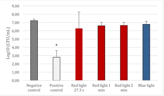
Figure 1 shows the results of Log10 CFU/mL of C. albicans. The First bar
represents the negative control. Each group was compared with the negative and positive control. There was significant difference in Log10 CFU/ml between the
negative and positive control samples (p<0.05). However, there was no significant difference between light groups and negative control (p>0.05).
0,00 1,00 2,00 3,00 4,00 5,00 6,00 7,00 8,00 9,00 Negative control Positive control Red light 27.3 s
Red light 1 min
Fig 1. Mean and standard deviations of Log10 CFU/mL of C. albicans. Comparison was
made between the twice-daily light treatment and the controls-0.12% CHX (positive control) and 0.89% NaCl (negative control). The * points to significant differences (p<0.05) in comparison to the other groups.
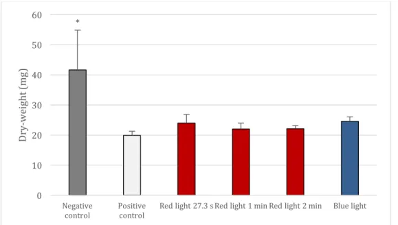
The figure 2 shows the results of the dry-weight (mg) of C. albicans biofilms after periodic light treatment. A significant reduction of the dry-weight in all light-treated samples in comparison to the negative control was observed and the reduction values observed in the light-treated groups was statistically similar to the positive control.
Fig 2. Mean and standard deviations of dry-Weight (mg) of C. albicans biofilm after the twice-daily treatment with red and blue light and with 0.12% CHX (positive control) and treatment with 0.89% NaCl (negative control). The * points to significant differences (p<0.05) in comparison to the other groups.
Figure 3 shows the results of C. albicans EPS-soluble and -insoluble contents after the twice-daily treatment with red and blue light and with 0.12% CHX (positive control) and treatment with 0.89% NaCl (negative control). It was observed that twice-daily light exposure to red light for 1 and 2 min and to blue light for 12 min 56 s numerically reduced the EPS- soluble in comparison to the negative control, mainly the blue light treatment. On the other hand, the EPS-insoluble content was numerically reduced by twice-daily exposure to red light for 1 min in comparison to the negative control, to the blue light and to the other periods of exposure to red light. Thus, there is a tendency of EPS-soluble reduction by twice-daily treatment with the blue light and a tendency of reduction of EPS-insoluble by twice-daily treatment with red light for 1
* 0 10 20 30 40 50 60 Negative control Positive control
Red light 27.3 s Red light 1 min Red light 2 min Blue light
27
Fig 3. Mean values and standard deviations of EPS-soluble and insoluble content in C. albicans biofilm (µg/mg of dry-weight) after twice-daily light treatment compared to twice-daily treatment with 0.12% Chlorhexidine (positive control) and twice-daily treatment with 0.89% NaCl (negative control). The * points to significant differences (p<0.05) in comparison to the other groups.
0 1 2 3 Negative control Positive control
Red light 27.3 s
Red light 1 min
Red light 2 min Blue light μ g/ m g o f d ry -we ig h t
EPS SOLUBLE
* 0 2 4 6 8 10 12 14 Negative control Positive control Red light 27.3 sRed light 1 min
Discussion
C. albicans is the most frequent specie isolated from superficial and systemic fungal infections and is associated with high rates of mortality37. There is an increasing number of strains of this microorganism that are resistant to antifungal agents38. Treatments of oral infections caused by Candida use topical antifungal medication, such as Nystatin15; and systemic antifungal medication, such as Fluconazole16. Due to the antifungal resistance and difficulties associated with the use of conventional medications, antimicrobial photodynamic chemotherapy (PACT) has been indicated for inactivating Candida and for the treatment of superficial fungal infections17;18;19. Studies have demonstrated that species of Candida present susceptibility to PACT 18. However, this method has limitations, such as non-selective antimicrobial characteristics and difficulty to penetrate to the depths of the biofilm, resulting in less effectiveness in biofilms39. As an alternative to PACT, a previous study described the use of blue light to prevent the biofilm development of
Streptoccocus mutans in vitro12. The study found that that twice-daily treatment prevented in vitro
S. mutans biofilm matrix development, being more effective in reducing the production of EPS-insoluble than the ‘gold-standard’ anti-plaque 0.12% chlorhexidine 12. However, the effect of blue and red light in C. albicans biofilms has never been investigated. Therefore, the aim of this study was to determine how the treatment with blue light or red light affects the development and composition of a matrix-rich Candida biofilm.
The light is an essential environmental cue for various organisms 20. Light is the major source of energy in the biosphere, and an essential signal that controls growth, development, and behavior of many different physiological mechanisms in most organisms. Long term experience with phototherapy for the treatment of jaundice, cancer and dermatological conditions has demonstrated its safety as well as its effectiveness 24. LumaCare™ device is a source of light that produces the whole spectrum of visible light by changing different probes at specific wavelenghts
37
. The light source presents a high potency (1,460 mW cm−2) in the red wavelength, thus, the exposure of the biofilms to this light was short (2 min, 1 min and 27.3 s) 37. On the other hand, the blue LED (95.5 mW cm−2) has a lower penetration depth into the tissue compared to red light due to its low potency associated with scattering and absorption by biomolecules, resulting in longer illumination time (12.56 min) 37.
components. Results on the photodynamic degradation of individual components of Candida
biofilm matrix are scarce.
The present study observed a significant reduction of the total biomasses (dry-weight) of the biofilms after the treatments with red and blue lights (Figure 2). The total biomass is defined as the total weight of the biofilm after the treatments and the washes of the biofilm. By reducing the biomasses, the lights prevented biofilm development. On the other hand, the CFU count was not affect. Thus, the reduction of the biomasses happened because of the reduction of the matrix components. We observed a numerical reduction of EPS-soluble that might have influenced in the significant reduction of the biomasses. However, Candida matrix have other important components, such as eDNA and proteins40 that might have been affected by the photherapy but were not evaluated in this study. Thus, the limitation of our study is that we only evaluated EPSsoluble and -insoluble from the matrix of C. albicans. Further studies should evaluate the effect of red and blue light in eDNA and proteins.
In the present study, the twice-daily treatment with blue light resulted in a numerical reduction in the EPS-soluble content of C. albicans biofilms (Figure 3a). Moreover, the EPS-soluble was numerically reduced by the application of red light for 1 min twice a day (Figure 3a). The decrease in the concentration of polysaccharides with the twice-daily treatment with blue light and red light for 1 min indicates that the light interactions with matrix components may affect the cohesiveness and stability of the EPS and the leakage of polysaccharides from the biofilm, without causing a significant effect of cell viability (Figure 1). The reduction of polysaccharides from the matrix of C. albicans biofilms is important since these components are related to the protection of the biofilm from antifungals8. Thus, the twice-daily treatment of oral candidiasis with blue light for 12 min 56 and with red light for 1 min might function as an adjuvant to topic antifungal application, such as Nystatin.
Our results demonstrated the effect of blue and red light on C. albicans biofilms. To our knowledge, this is the first report of twice-daily use of blue and red light to prevent C. albicans
31 Therefore, we conclude that C. albicans biofilms biomasses (dry-weight) were significantly reduced by the treatment with red and blue light. Moreover, the EPS-soluble content was mainly reduced by twice-daily exposure to blue light while EPS-insoluble exhibited major reduction with twice-daily treatment with red light for 1 min. This leads us to believe that light is a promising therapeutic approach for biofilm-related C. albicans diseases, such as oral candidiasis, indicating that twice-daily treatment of Candida biofilms with either blue light and red light can function as an adjuvant to topic antifungal application.
Aknowledgment
This research was supported by CAPES Foundation from whom the first author received a scholarship (CAPES 88881.062159̷ 2014-01 PVE̷ CAPES).
Conflict of Interests
None.
Ethical Considerations
This article does not contain any studies with human or animal subjects performed by any of the authors.
References
1. Lefebvre CA, Wataha JC, Cibirka RM, Schuster GS, Parr GR. Effects of triclosan on the cytotoxicity and fungal growth on a soft denture liner. J Prosthet Dent. 2001; 85:352–356
2. Fricke K, Koban I, Tresp H, Jablonowski L, Schröder K, Kramer A, Weltmann K-D, von Woedtke T, Kocher T: Atmospheric pressure plasma: a high- performance tool for the efficient removal of biofilms. PLOS ONE 2012; 7(8): e42539
3. Sudbery PE. Growth of Candida albicans hyphae. Nature Reviews Microbiology. 2011; 9: 737-748
4. Sanchez-Vargas LO, Estrada-Barraza D, Pozos-Guillen AJ, Rivas-Caceres R. Biofilm formation by oral clinical isolates of Candida species. Arch Oral Biol. 2013; 58(10):1318–26
5. Sutherland IW. The biofilm matrix an immobilized but dynamic microbial environment. Trends Microbiol. 2001; 9: 222–227
6. Flemming HC, Wingender J. The biofilm matrix.Nat.Rev. Micro. 2016; 8, 623–633
7. Williams DW, Kuriyama T, Silva S, Malic S, Lewis MA. Candida biofilms and oral candidosis: treatment and prevention. Periodontol 2000. 2011;55(1):250–65
8. Zarnowski R, Westler WM, Lacmbouh GA, Marita JM, Bothe JR, Bernhardt J, Lounes Hadj Sahraoui A, Fontaine J, Sanchez H, Hatfield RD, Ntambi JM, Nett JE, Mitchell AP, Andes DR. Novel entries in a fungal biofilm matrix encyclopedia. MBio. 2014; 5: e01333-14.
9. Mitchell KF, Zarnowski R, Sanchez H, Edward JA, Reinicke EL, Nett JE, Mitchell AP, Andes DR. Community participationin biofilm matrix assembly and function. Proc Natl Acad Sci U S A. 2015; 112(13):4092–7
10. Mitchell KF, Zarnowski R, Andes DR. Fungal super glue: The biofilm matrix and its composition, assembly, and functions. PLoS Pathog. 2016; 12: e1005828
microcolonies formation by Streptococcus mutans in biofilms. J Appl Microbiol. 2010; 108: 2103– 2113
12. Lins de Sousa D, Araujo RL, Zanin IC, Klein MI, Janal MN, Duarte S. Effect of twice-daily blue light treatment on matrix-rich biofilm development. Plos One. 2015; 10(7): e0131941
13. Koo H, Xiao J, Klein MI. Extracellular polysaccharides matrix – an often forgotten virulence factor in oral biofilm research. Int J Oral Sci. 2009; 1:229–234.
14. Dubois M, Gilles KA, Hamilton JK, Rebers PA, Smith F. Colorimetric method for determination of sugars and related substances. Anal Chem. 1956; 28: 350–356
15. Banting DW, Greenhorn PA, McMinn JG. Effectiveness of a topical antifungal regimen for the treatment of oral candidiasis in older, chronically ill, institutionalized, adults. J Can Dent Assoc. 1995; 61:199-200, 203-205
16. Samaranayake LP, Keung Leung W, Jin L. Oral mucosal fungal infections. Periodontol 2000. 2009; 49: 39-59
17. Bliss JM, Bigelow CE, Foster TH, Haidaris CG. Susceptibility of Candida species to photodynamic effects of Photofrin. Antimicrob Agents Chemother. 2004; 48:2000–2006
18. Dovigo LN, Pavarina AC, Mima EGO, Giampaolo ET, Vergani CE, Bagnato VS. Fungicidal effect of photodynamic therapy against fluconazole-resistant Candida albicans and Candida glabrata. Mycoses. 2011; 54:123–130
19. Mima EG, Pavarina AC, Dovigo LN, Vergani CE, Costa CAS, Kurashi C, Bagnato VS. Susceptibility of Candida albicans to photodynamic therapy in a murine modelo of oral candidosis. Oral Surg Oral Med Oral Pathol Oral Radiol Endod. 2012; 109:392–401
20. Dai T, Gupta A, Huang YY, Yin R, Murray CK, Vrahas MA. Blue light rescues mice from potentially fatal Pseudomonas aeruginosa burn infection: efficacy, safety, and mechanism of action. Antimicrob agentes chemother. 2013; 57: 1238-1245
21. Al-Fattani MA, Douglas LJ. Biofilm matrix of Candida albicans and Candida tropicalis: chemical composition and role in drug resistance. J Med Microbiol. 2006; 55:999–1008.
22. Nett J, Lincoln L, Marchillo K, Massey R, Holoyda K, Hoff B, Van Handel, M, Andes D. Putative role of β-1,3 glucans in Candida albicans biofilm resistance. Antimicrob Agents Chemother. 2007; 51:510–20.
23. Vediyappan G, Rossignol T, d'Enfert C. Interaction of Candida albicans biofilms with antifungals: transcriptional response and binding of antifungals to beta-glucans. Antimicrob Agents Chemother. 2010; 54: 2096-111.
24. Tan KL. Phototherapy for neonatal jaundice. Clin Perinatol. 1991; 18:423-439
25. Yoshida H Matsui K, Horikoshi H, Ohmichi N, Ohgai M, Ikuhara Y. Cubic-formation and grain-growth mechanisms in tetragonal zirconia polycrystal. J Am Ceram Soc. 2013;86:1401-1408 26. Feverstein O, Morein OSP, Steinberg D. Synergic antibacterial effect between visible light and hydrogen peroxide on S. mutans. J Antimicrob. Chemother. 2006; 57: 872-876
27. Yang P, Cao Y, Lu X, et al. Efficacy of atmospheric pressure plasma as an antibacterial agent against Enterococcus faecalis in vitro. Plasma Sci Technol. 2011;13: 93–8.
28. Masuoka J. Surface glycans of Candida albicans and other pathogenic fungi: physiological roles, clinical uses, and experimental challenges. Clinical microbiology reviews, 2004; 17 (2): 281-310
29. Tronchin G, Pihet M, Lopes-Bezerra LM, Bouchara JP: Adherence mechanisms in human pathogenic fungi. Med Mycol 2008, 46(8):749-772
30. Feuerstein O. Light therapy: complementary antibacterial treatment of oral biofilm. Adv Dent Res. 2012; 24: 103–107
31. Malik R, Manocha A, Suresh DK. Photodynamic therapy- a strategic review. Indian Dent Res. 2010; 21:285-291
33 33. Nett J, Andes D. Candida albicans biofilm development, modeling a host-pathogen interaction. Curr Opin Microbiol. 2006; 9(4):340-5.
34. Dai T, Huang YY, Hamblin MR. Photodynamic therapy for localized infections state of the art. Photo Diagn Photodyn Ther. 2009; 6 (3-4): 170-188
35. Fontana CR, Abernethy AD, Som S, Ruggiero K, Doucette S, Marcantonio RC, et al. The antibacterial effect of photodynamic therapy in dental plaque-derived biofilms. J Periodontal Res. 2009; 44: 751–759. pmid:19602126.
36 Paschoal MA, Lin M, Santos-Pinto L, Duarte S. Photodynamic antimicrobial chemotherapy on
Streptococcus mutans using curcumin and toluidine blue activated by a novel LED device. Lasers Med Sci. 2015;30(2):885-90
37. Nobile CJ, Johnson AD. Candida albicans biofilms and human Diseases. Annu Rev Microbiol. 2015; 69: 71–92
38. Sardi JC, Scorzoni L, Bernardi T, Fusco-Almeida AM, Mendes Giannini MJ. Candida species: current epidemiology, pathogenicity, biofilm formation, natural antifungal products and new therapeutic options. J Med Microbiol. 2013; 62:10–24
39. Fontana CR, Abernethy AD, Som S, Ruggiero K, Doucette S, Marcantonio RC, et al. The antibacterial effect of photodynamic therapy in dental plaque-derived biofilms. J Periodontal Res. 2009; 44: 751–759
Antimicrobial effect of Low Temperature Plasma on oral biofilm formed in situ: molecular partial identification of microbial population
Silveira PV, Faculty of Pharmacy, Dentistry and Nursing, Federal University of Ceara, Fortaleza, Brazil
Guedes SF, Faculty of Pharmacy, Dentistry and Nursing, Federal University of Ceara, Fortaleza, Brazil
Alves KSS, Faculty of Pharmacy, Dentistry and Nursing, Federal University of Ceara, Fortaleza, Brazil
Rodrigues LKA, Faculty of Pharmacy, Dentistry and Nursing, Federal University of Ceara, Fortaleza, Brazil
Stipp RN, Department of oral diagnosis, Piracicaba Dental School, State University of Campinas, Piracicaba, São Paulo, Brazil
Duarte S, Department of Basic Sciences, Indiana University College of Dentistry, Indiana, USA Zanin ICJ#, Faculty of Dentistry, Federal University of Ceara, Sobral, Brazil
#
Corresponding Author:
Iriana Carla Junqueira Zanin, DDs, Ms, PhD
College of Dentistry, Federal University of Ceara, Sobral, CE, Brazil.
Coronel Estanislau Frota Street, s/n
Sobral – CE- Brazil
Zip code: 62010-560
Phone/fax: ++ 55 88 36132603 Mobile: ++ 55 88 97159496
E-mail: irianaz@yahoo.com.br, iriana.zanin@pq.cnpq.br
Abstract
Objectives To evaluate the antimicrobial effect of tissue tolerable plasma on oral biofilms formed in situ through the molecular characterization of the microbial population.
Material and methods For this in situ experiment, a single-blind split mouth design was used in two phases of 7 days each, in which 8 volunteers wore palatal devices containing six bovine enamel slabs, positioned in pair of three. At the end of the clinical phase, the device was randomly split and each half was allocated to one of the following treatments: Plasma (PLA); Argon (ARG); Chlorhexidine 0.12% (CHX) and Salina solution 0.89% (NaCl). In this way, at the end of the two clinical phases, biofilms form all volunteers were submitted to the four different treatments.
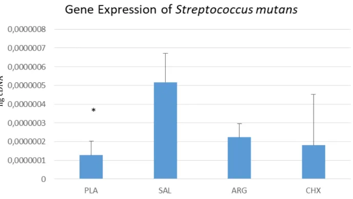
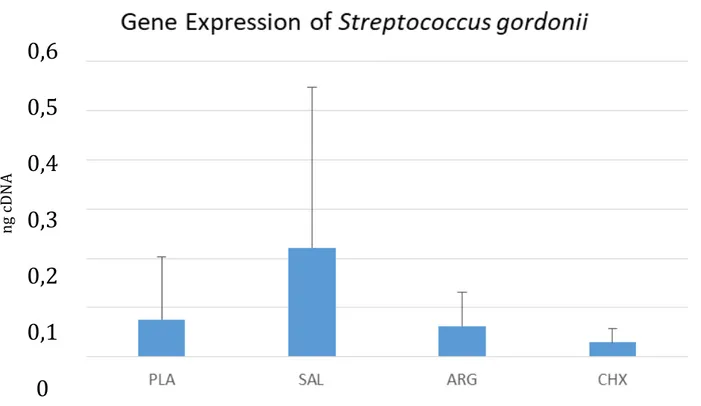
Results A total of eight samples from each group were submitted to the RT-q PCR for the bacteria detection and quantification. Data of the gene expression of each bacterium were obtained. Statistical analysis were performed using the Sigma Plot program, using Anova test, followed by the complementary tests of Student t-test and Newman Keuls, with p <0.05. In all the analyzed groups the presence of specific bacteria for each primer was observed, however some groups treated with plasma the bacterial expression of the bacteria was lower. Plasma treatment on biofilm sample presented significantly lowers concentrations of Streptococcus mutans, Lactobacillus acidophilus,
Streptococcus mitis group, Bifidobacterium group and the Actinimyces naeslundii (p < 0.05) compared to other treatment groups. Concentrations of Lactobacillus casei groups and the
Streptococcus gordonii were not significantly different before and after plasma treatment.
Clinical relevance Considering that the information about the oral microbiota related to biofilm in situ status is relevant, this study provides insights to better understand the differences in the microbiotas between different treatments.
Keywords Bacteria- biofilm- in situ- Quantitative polymerase chain reaction
Introduction
The impact of environmental factors on chronic diseases has been the focus of many studies, especially in the last couple of decades, along with an effort to understand the heterogeneity of the immunological parameters among individuals [1]. In between different environmental factors, one that stands out is the microbiota that may be defined as the sum of microbes residing in a habitat. The oral cavity is the habitat of several kinds of microorganisms, which form a complex community that can adhere to the teeth surface or to epithelial mucosa forming biofilms [2]. The microbiome is defined as the totality of genes of microbiota, in this case, of that oral microbiota [3]. In oral health, the oral cavity microbiome comprises billions of microbes.
the oral microbiota in health and disease revealed the diversity of oral biofilms, introducing new candidates for disease-associated bacterial species [13]. Molecular approaches have revealed a greater variability of the oral microbiota associated to dental caries, including Streptococcus spp
and bacteria of the genera Actinomyces, Bifidobacterium, Lactobacillus, Propionibacterium,
Veillonella, Selenomonas, and Atopobium [14].
It is well know that the accumulation of bacterial biofilms on tooth surfaces results in some of the most prevalent bacterial-induced human diseases, caries and inflammatory periodontal diseases. Current treatment of subjects with plaque-related diseases involves mechanical removal of the biofilm and the use of antiseptics and antibiotics. Thus, the increased microbial resistance against commercially available antimicrobial drugs and substances have cooperated with the search for alternative treatments for the control of pathogenic biofilms involved with diseases that affect the body, including biofilm-dependent oral diseases [15, 16]. In this context, plasma may constitute a suitable process to combat both biofilm-related resistance and antimicrobial resistance.
Plasma is a partially ionized gas generated by an electrical discharge, which creates a highly reactive environment with ions, electrons, excited atoms and molecules, vacuum ultraviolet and ultraviolet (UV) irradiation, free radicals, and chemically reactive particles [17]. It is also specific, targeting only the infected area. In addition, plasma is usually produced by low-toxicity gases and elaborates its activity by producing a mixture of products that decay within a few seconds after the treatment process [18] and the ability to achieve gas phase, without the need to reach high temperatures, allows its use in thermosensitive materials including cells and tissues [19]. The effectiveness of removing biofilms and inactivation of microorganisms with tissue tolerable plasma have been demonstrated [20].
In general, biofilms models may help us to accurately predict, in a controlled and simplified way, a clinical outcome which can lead us to preventive actions for diseases [21]. In this way, we decided to evaluate the effect of plasma under conditions more similar to those found in the mouth, using an in situ multispecies biofilm model. Method involves the use of devices that create conditions reproducing the process of biofilm formation in the oral cavity, serving as a link between the uncontrolled clinical situation and the highly controlled laboratory experiments.
37
Methods
Ethics statement
This study protocol was approved by the Research and Ethics Committee of the Federal University of Ceara, Brazil Medical School (Sisnep Caae - 40975514.0.0000.5054). All volunteers gave informed consent according to Resolution n0 196 of the National Health Council, Health Ministry, Brasilia, DF, from 10/03/1996.
Experimental design
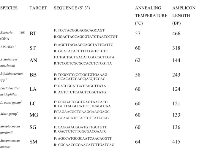
For this in situ experiment, a single-blind split mouth design was used in two phases of 7 days each, in which 8 volunteers wore palatal devices containing six bovine enamel slabs, positioned in pair of three. At the end of the clinical phase, the device was randomly split and each half was allocated to one of the following treatments: Plasma (PLA); Argon (ARG); Chlorhexidine 0.12% (CHX) and Salina solution 0.89% (SAL) as described in Fig 1. In this way, at the end of the two clinical phases, all volunteers were submitted to the four different treatments.
Figure 1. Description of the treatments in which the enamel slabs were treatds
Groups Code Treatment
Plasma PLA Plasma plume during5 min Argon ARG Argon gas flow during5 min
Chlorhexidine 0.12% CHX 50 µL on each slab during 5 min Salina solution 0.89% SAL 50 µL on each slab during 5 min
Tissue tolerable plasma
fixed holder.
Specimen preparation
Bovine teeth were used to perform this in situ study. The teeth were stored in 0.01% (v/v) thymol solution at 4°C for 30 days until used [21,22,23]. Enamel slabs with 4 x 4 x 2 mm were obtained using a water-cooled diamond saw and a cutting machine (IsoMet Low Speed Saw; Buehler, Lake Bluff, IL, USA). The adjustment of the enamel and dentin to obtain flat plates was done with the aid of a low-speed polishing machine and 320 grit paper (Carbimet Paper Discs), under water-cooling. Afterwards, the specimens were polished using three different silicon carbide waterproof papers (320, 600, and 1,200-grit) as well as polishing cloths with 1 µm diamond paste (Buehler).
In situ palatal devices
The slabs were autoclaved (121 ºC, 15 min) [24] and stored in 100% humidity until being inserted into the palatal appliances. For each subject, two acrylic palatal devices were fabricated, in which two cavities (18 x 6 x 3 mm) were prepared on the left and right sides; three slabs were attached with wax in each cavity. In order to allow biofilm accumulation, and to protect it from mechanical disturbance, a plastic mesh was positioned on the acrylic resin, leaving a 1 mm space from the slab surface [25,26].
In situ study Population
Eight healthy volunteers (5 women and 3 men), aged 19–34 years, able to comply with the experimental protocol, were selected to participate in this study. All participants received oral and written instructions about the experimental design. The inclusion criteria were normal salivary flow rate, normal buffering capacity of saliva and S. mutans colony-forming units (CFU mg-1) in biofilms of at least 105 after 36 h of oral hygiene suspension. Exclusion criteria included active caries lesions, use of antibiotics within the past 3 months prior to the study, use of fixed or removable orthodontic devices. The use of dentifrice containing any antibiotics was suspended during the experimental period.
In situ biofilm formation
39 carbonate based dentifrice, 1,450 µg fluoride (F) g-1, as monofluorophosphate (MFP); Colgate-Palmolive, São Paulo, SP, Brazil]. Also, the volunteers received oral and written instructions to wear the appliances at all times, including at night. They were allowed to remove the appliances only during meals, when consuming acid drinks, and when performing oral hygiene. When removed, the devices were kept moist in plastic boxes to keep the bacterial biofilm viable [23]. The cariogenic challenge was provided by the volunteers who dripped a 10% sucrose solution onto all the enamel slabs, 10 times a day, according to a predetermined schedule (at 08:00, 09:30, 11:00, 12:30, 14:00, 15:30, 17:00, 18:30, 20:00, and 21: 30 h) [29]. Before replacing the palatal appliance in the mouth, a 5-min waiting time was standardized to allow diffusion of the sucrose into the dental biofilm. Brushing with the dentifrice was performed three times a day, after mealtimes when the volunteers habitually carried out their oral hygiene procedures. The appliances were brushed extra-orally, except for the slab area, and volunteers were asked to brush carefully over the covering meshes, to avoid disturbing the biofilm. All volunteers consumed fluoridated water (0.70 mg F 1-1), and no restriction was made with regard to the volunteers’ dietary habits.
Plasma treatment of in situ biofilms
The distribution of treatments on the palatal device in each intra-oral phase was determined randomly by raffle. All volunteers came in fasting, removed the device from mouth and one drop of 10% sucrose was added to each slabs. Third minutes later the plastic meshes of the devices were removed with a scalpel blade (#15C), the biofilm formed in situ were exposed, and the treatments with PLA, ARG, CHX or SAL were performed. Biofilms were then scraped carefully, were and weighed and were suspended in RNAlater solution adding 5–10 volume. Samples were stored at room temperature overnigh and stored at -80 oC after that.
Extraction of RNA
After stirring at Beadbeater, 850 µl of a mixture of RLT (RNeasy Minikit ®) and 1% β -mercaptoethanol was added to the biofilm sample, vortexed for 30 seconds and centrifuged for 2 minutes (11000g / 4 ° C). Thereafter, 350 µl of the supernatant was removed for the continuation of the extraction procedure, while the other part was stored if further extraction was required. To the 350 µl initially removed, 250 µl of pure ethanol was added and vortexed. Then, the contents were transferred to a RNeasy MiniKit column (Qiagen, Valencia, CA, USA) and centrifuged. After centrifugation, the contents that had passed through the column were discarded. 700 µl of RW (RNeasy Minikit ®) were added to the columm and again centrifugation (11000g / 20 ° C) for 30 seconds was performed. The content that had passed through the column was discarded again. Thereafter, 500 µl of RPE (RNeasy Minikit ®) was added to the column and centrifuged again (11000g / 20 ° C) for 30 seconds. This step was performed twice. A centrifugation without addition of reagents (11000g / 20oC) for 2 minutes to remove all alcohol present on the column was performed. Subsequently, the column was placed in a capped eppendorf and 40 µl of RNAse free water was added and centrifuged again (11000g / 20 ° C) for 1 minute to elute. The eluate was pipetted and stored in a new labeled eppendorf.
Treatment with DNase
For each RNA sample obtained, 5 µl of the buffer and 5 µl of the Turbo DNase enzyme (Applied Biosystems, Ambiom, Austin, TX, USA) were added. After vortexing, the sample was allowed to stand for 15 minutes at 37 ° C, sufficient time for DNA degradation by DNAse.
Purification of samples
41
Dosage and integrity of total RNA
The amount of total RNA extracted (ng/µl) was measured in Nanodrop spectrophotometer (Thermo Scientific, Wilmington, DE) (A260 / A280 ratio). Then, the integrity of this RNA was verified through an electrophoresis gel run.
Reverse transcriptase reaction and cDNA uptake
The cDNA was produced using the iScript ™ cDNA Synthesis kit (BioRad, Hercules, CA, USA). Reverse transcriptase reactions were prepared from a mixture containing 6 µl of the iScript 5x reaction mix, 1 µl iScript reverse transcriptase, 1 µg of total RNA extracted from the biofilm sample and sufficient RNAse free water to complete a volume of 30 µl. Each prepared solution was vortexed for 5 seconds, incubated at 25° C for 5 minutes, heated at 42 ° C for 2 hours, and again heated at 85 ° C for 5 minutes in a Veriti Thermal Thermal Cycler (Applied Biosystems, Foster City, CA, USA). After all reactions, the cDNA concentration of all samples were adjusted to 10 ng / µl (Bezerra et al., 2016).
Real-time polymerase chain reaction