Cop
yright
© ABE&M t
odos os dir
eit
os r
eser
vados
.
Type IA isolated growth hormone
deiciency (IGHD) consistent with
compound heterozygous deletions of
6.7 and 7.6 Kb at the
GH1
gene locus
Deiciência isolada de hormônio do crescimento tipo IA (DIGH) consistente com deleções heterozigotas compostas de 6,7 e 7,6 Kb
no locus gênico GH1
Ana Keselman1*, Paula A. Scaglia2*, María Soledad Rodríguez Prieto1,
María Gabriela Ballerini1, María Eugenia Rodríguez1, María Gabriela
Ropelato1, Ignacio Bergadá1, Héctor G. Jasper2, Horacio M. Domené2
SUMMARY
Isolated growth hormone deiciency (IGHD) may result from deletions/mutations in either GH1
or GHRHR genes. The objective of this study was to characterize the molecular defect in a girl
presenting IGHD. The patient was born at 41 weeks of gestation from non-consanguineous par-ents. Clinical and biochemical evaluation included anthropometric measurements, evaluation of pituitary function, IGF-I and IGFBP-3 levels. Molecular characterization was performed by PCR ampliication of GH1 gene and SmaI digestion of two homologous fragments lanking the
gene, using genomic DNA from the patient and her parents as templates. At 1.8 years of age the patient presented severe growth retardation (height 61.2 cm, -7.4 SDS), truncal obesity, frontal bossing, doll face, and acromicria. MRI showed pituitary hypoplasia. Laboratory indings con-irmed IGHD. GH1 gene could not be ampliied in samples from the patient while her parents
yielded one fragment of the expected size. SmaI digestion was consistent with the patient being
compound heterozygous for 6.7 and 7.6 Kb deletions, while her parents appear to be hetero-zygous carriers for either the 6.7 or the 7.6 Kb deletions. We have characterized type IA IGHD caused by two different GH1 gene deletions, suggesting that this condition should be
consid-ered in severe IGHD, even in non-consanguineous families. Arq Bras Endocrinol Metab. 2012;56(8):558-63
SUMÁRIO
A deiciência isolada do hormônio do crescimento (DIGH) pode ser resultado de deleções/mu-tações no gene GH1 ou no gene GHRHR. O objetivo deste estudo foi caracterizar o defeito mo-lecular em uma menina que apresenta DIGH. A paciente nasceu às 41 semanas de gestação de pais não consanguíneos. As avaliações clínica e bioquímica incluíram medidas antropométri-cas, avaliação da função pituitária e concentrações de IGF-I e IGFBP-3. A caracterização molecu-lar foi feita por meio de ampliicação do GH1 por PCR e digestão com SmaI de dois fragmentos homólogos lanqueando o gene, usando-se DNA genômico da paciente e de seus pais como padrões. Com 1,8 ano de idade, a paciente apresentou atraso grave no crescimento (altura 61,2 cm, -7.4 DP), obesidade central, protuberância frontal, face de boneca e acromicria. A RM mostrou hipoplasia pituitária. Os achados laboratoriais conirmaram a DIGH. O gene GH1 não pôde ser ampliicado nas amostras da paciente, enquanto as amostras de seus pais produziram um fragmento do tamanho esperado. A digestão com SmaI foi consistente com a paciente ser heterozigota composta para deleções para 6,7 e 7,6 Kb, enquanto seus pais parecem ser car-readores heterozigotos para deleções de 6,7 ou 7,6 Kb. Caracterizamos a DIGH tipo IA causada por duas deleções diferentes no gene GH1, sugerindo que essa condição pode ser considerada na DIGH grave, mesmo em famílias não consanguíneas. Arq Bras Endocrinol Metab. 2012;56(8):558-63 1 División Endocrinología, Hospital
de Niños “Ricardo Gutiérrez”, Buenos Aires, Argentina
2 Centro de Investigaciones
Endocrinológicas (CEDIE), CONICET, Buenos Aires, Argentina * These authors contributed equally to this study.
Correspondence to:
Horacio M. Domené Centro de Investigaciones Endocrinológicas (CEDIE, CONICET), Hospital de Niños “Ricardo Gutiérrez”, Gallo 1330
C1425EFD – Ciudad Autónoma de Buenos Aires, Argentina [email protected]
Cop
yright
© ABE&M t
odos os dir
eit os r eser vados .
INTRODUCTION
G
rowth hormone deiciency (GHD) is a relatively common disorder, occurring in 1 out of 4,000 to 10,000 live births (1). Most frequently, it occurs as a sporadic condition of unknown aetiology (2) but severe forms of isolated GHD (IGHD) may have a genetic basis (3). In patients with severe growth retar-dation (height less than -4.5 SDS) presenting IGHD, the prevalence of GH1 or GHRHR gene defects, ei-ther mutations or deletions, could be as high as 20%, depending on the population (4,5). More recently, Alatzoglou and cols. have reported an 11.1% preva-lence of GH1 or GHRHR molecular defects in IGHD pedigrees, which increased to 38.6% in familial cases (6). Familial IGHD has been associated with four Mendelian disorders (7,8), including two autosomal recessive (Type IA and IB), one autosomal dominant (Type II) and one X-linked (Type III) form. Type IA IGHD was irst described by Ruth Illig and cols. (9) in 1970 in three Swiss siblings with severe short stat-ure, early growth retardation, extreme dwarism in adulthood, and a particular phenotype. These patients developed high titers of anti-GH antibodies, which arrested their growth response to pituitary-extracted GH treatment. Using Southern blot analysis in these patients, Phillips III and cols. (10) later characterized a homozygous 7.5 Kb deletion that included GH1 gene.The aim of this report was to characterize the mo-lecular defect in a female patient who fulilled the crite-ria for severe IGHD.
SUBJECT AND METHODS
Case report
We report a small-for-gestational-age female patient born from non-consanguineous parents by vaginal de-livery after 41 weeks of gestation. Birth weight and length were 2,460 g (-2.0 SDS) and 44 cm (-3.7 SDS), respectively. Both parents presented normal height: father 165.5 cm (-1.07 SDS) and mother 154.5 cm (-1.01 SDS). The patient’s target height was 153.7 ± 8.5 cm. The irst evaluation, at 10 months of age, showed severe growth retardation (height 57 cm, -5.9 SDS) (Table 1). Auxological parameters were ex-pressed in cm and SDS according to Argentinean ref-erences (11). Her physical examination showed
trun-cal obesity, frontal bossing, doll face, and acromicria (Figure 1A). She had normal psychomotor develop-ment. Laboratory indings conirmed severe growth hormone deiciency with no GH response to an argi-nine test, low IGF-I and IGFBP-3, and normal GHBP serum levels (Table 2). Brain MRI showed severe an-terior pituitary hypoplasia (Figure 1B). She started rhGH replacement therapy (0.33 mg/kg.week) at 2.4 years of age. Although her growth rate improved dur-ing the irst 6 months of treatment (12.8 cm/year), afterwards she developed anti-GH antibodies, and her growth velocity decreased to 2.8 cm/year (Figure 1C). Levels of IGF-I and IGFBP-3 remained very low on rhGH treatment.
Table 1. Auxological evaluation
Chronological age (years) Birth 0.8 2.4 2.8 3.3
Height (cm) (SDS) 44.0 -3.7 57.0 -5.9 67.5 -6.3 73.0 -5.2 74.4 -5.2 Weight (g) (SDS) 2,480 -2.0 5,100 -3.6 7,700 -3.7 8,600 -3.4 8,980 -3.6
Head circumference (cm) (SDS) 34.1 0.0 42.1 -2.0 43.5 -2.0 47 -2.0
Body proportions (cm) (pc) 37.0 25 43.5 25 45.0 10 46.6 25
Bone age (years) 1.6 2.0
Height velocity (cm/year) 16.2 6.6 13.7 2.8
rhGH treatment (months) 0 5 11
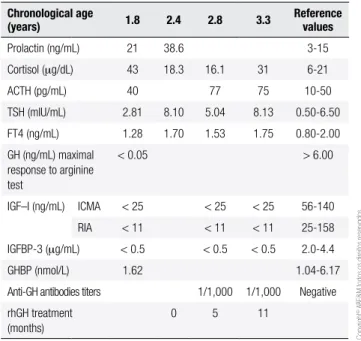
Table 2. Endocrinological evaluation
Chronological age
(years) 1.8 2.4 2.8 3.3 Reference values
Prolactin (ng/mL) 21 38.6 3-15
Cortisol (mg/dL) 43 18.3 16.1 31 6-21
ACTH (pg/mL) 40 77 75 10-50
TSH (mIU/mL) 2.81 8.10 5.04 8.13 0.50-6.50
FT4 (ng/mL) 1.28 1.70 1.53 1.75 0.80-2.00
GH (ng/mL) maximal response to arginine test
< 0.05 > 6.00
IGF–I (ng/mL) ICMA < 25 < 25 < 25 56-140
RIA < 11 < 11 < 11 25-158
IGFBP-3 (mg/mL) < 0.5 < 0.5 < 0.5 2.0-4.4
GHBP (nmol/L) 1.62 1.04-6.17
Anti-GH antibodies titers 1/1,000 1/1,000 Negative
rhGH treatment (months)
Cop
yright
© ABE&M t
odos os dir
eit
os r
eser
vados
.
Figure 1. A. Patient at 1.8 years of chronological age (written consent for publication was obtained from the parents). B. Brain MRI showing pituitary hypoplasia. C. Growth chart. Closed circles: height for chronological age. Open circles: bone age.
Hormone assays
Serum growth hormone (GH) secretion was evalu-ated by an arginine provocative test (0.5 g/kg body weight). GH, IGF-I, IGFBP-3, and ACTH serum lev-els were measured by chemiluminiscent immunometric assays (ICMA, IMMULITE® 2000 system, Siemens
Healthcare Diagnostics Products Ltd, Gwynedd, UK); cortisol, prolactin, TSH, and free T4 (FT4), by elec-trochemiluminescence assays (ECLIA, Roche Diagnos-tics GmbH, Mannheim, Germany) using a Cobas e411 analyzer. IGF-I levels were also measured by an in house RIA after serum extraction by the acid-ethanol method followed by cryoprecipitation (12), and GHBP serum concentration was determined by an in house
time-resolved luorometric immunofunctional assay modi-ied from Fisker and cols. (13,14). Anti-GH antibodies were determined by an in house ELISA as follows: A 96-well plate was coated with rhGH and non-speciic sites on the coated wells were blocked with phosphate buffer (PBS; pH = 7.4) containing BSA (2 g/L). Af-ter removal of the blocking solution, the serum sample (diluted 1/10 in PBS) was added and incubated over-night at 4°C. The plate was washed and incubated for two hours with rabbit polyclonal horseradish peroxi-dase-labeled total anti-human IgG (DakoCytomation, Glostrup, Denmark). The plate was washed and the substrate (tetramethyl benzidine) was added. After 5 minutes, the reaction was stopped by addition of sul-furic acid, and absorbance was determined at 450 nm.
A
B
C
Girls
Age (years)
120 CM
110
100
90
80
70
60
50
1 2 3 4 5 6
Cop
yright
© ABE&M t
odos os dir
eit
os r
eser
vados
.
Figure 2. A.GH1 gene ampliication (1.5% agarose gel electrophoresis, ethidium bromide staining). GH1 gene PCR ampliication yielded no product using the proband’s genomic DNA as template (P), while her parents (M, F) showed one band of the expected size (2,700 bp), similar to normal controls (C1, C2). Mk: 100 bp DNA ladder. B. Schematic representation of GH1 gene in the cluster context, and genomic organization resulting from 6.7 and 7.6 Kb deletions. 1F, 1R: oligonucleotide primers for GH1 gene PCR ampliication. 2F, 2R: oligonucleotide primers for simultaneous PCR ampliication of 5’ and 3’ homologous lanking sequences (white and shaded boxes, respectively) that give rise to the 6.7 Kb deletion. Grey boxes: homologous regions that give rise to 7.6 Kb deletion upon unequal recombination. Dotted arrows indicate SmaI restriction enzyme sites present in 3’ homologous lanking region.
C. SmaI digestion (5% polyacrylamide gel electrophoresis, ethidium bromide staining). Fragment pattern was consistent with the father (F) being heterozygous carrier for 6.7 Kb deletion, and the patient (P), compound heterozygous for 6.7 and 7.6 Kb deletions. Unfortunately, the band pattern for the mother (M), presumably carrier of a 7.6 Kb deletion, could not be distinguished from a normal control (C1). C3: Patient known to be homozygous for 6.7 Kb deletion. Mk: 100 bp DNA ladder. ND: non-digested PCR product. D. Diagram showing the expected size of the DNA fragments obtained after
SmaI digestion.
Magnetic resonance imaging (MRI): MRI exami-nation was carried out in sagittal and coronal T1 images of the brain, sellar and suprasellar structures, with and without gadolinium contrast.
Molecular characterization
Genomic DNA was isolated from peripheral venous blood by cetyltrimethylammonium bromide (CTAB) lysis buffer and chloroform-isoamyl alcohol extraction (15). Written informed consent for molecular studies was obtained from the parents.
PCR ampliication of the whole GH1 gene (Gene ID 2688, RefSeqGene: NG_011676.1) was performed us-ing GoTaq® DNA polymerase (Promega Corporation,
Madison, USA) and oligonucleotide primers GH1F (5’-ccagcaatgctcagggaaag-3’) and GH1R (5’-tgtcccac-cggttgggcatggcaggtagcc-3’) (16). PCR mixtures were denatured for 2 min at 94ºC and submitted to 30 cy-cles at 92ºC for 1 min; 61ºC for 45 sec; and 68ºC for 3 min, followed by inal extension at 68ºC for 10 minutes. The resulting PCR product (2700 bp) was visualized by agarose gel electrophoresis and ethidium bromide staining.
dele-Cop
yright
© ABE&M t
odos os dir
eit
os r
eser
vados
.
tions, were simultaneously ampliied by PCR with the fo-llowing primers: 5’-tccagcctcaaagagcttacagtc-3’ (GH2F) and 5’-cgttttctctagtctagatcttcccagag-3’ (GH2R). PCR mixtures were denatured at 94ºC for 3 min and submit-ted to 30 cycles at 94ºC for 1 min; 64ºC for 45 sec; and 72ºC for 3 min, followed by a 10-min inal extension at 72ºC. The resulting PCR fragments were digested overnight at 37°C with Smal restriction endonuclease (RO141S, New England Biolabs, MA, USA) according to the manufacturer’s protocol, and the digested prod-ucts were visualized by ethidium bromide staining after electrophoresis on a 5% polyacrylamide gel.
RESULTS
Biochemical evaluation:IGHD was conirmed by lack of response of GH to an arginine test, undetectable levels of IGF-I and IGFBP-3, normal thyroid function (normal to slightly elevated TSH with normal FT4 le-vels), normal ACTH, and elevated cortisol levels. Pro-lactin levels were slightly above the upper normal range (Table 2).
PCR ampliication of GH1 gene:GH1 gene PCR ampliication yielded no product using two different genomic DNA samples of the proband as template, while her parents showed one amplicon of the expected size, similar to DNA from normal controls (Figure 2A). This result was suggestive of GH1 gene deletion in the patient.
Characterization of GH1 gene deletion: follow-ing PCR ampliication of two homologous sequences lanking GH1 gene, the Smal restriction enzyme diges-tion band pattern obtained was consistent with the pa-tient being compound heterozygous for 6.7 and 7.6 Kb deletions, while her father displayed a pattern consist-ent with a heterozygous carrier of the 6.7 Kb deletion. The pattern obtained in the mother could not be dis-tinguished from that of a normal control, and was com-patible with both the mother having two normal alleles or being a heterozygous carrier of the 7.6 Kb deletion (Figure 2C). Unfortunately, only those heterozygous carriers for the 6.7 Kb deletion can be unambiguously detected by this assay, which does not enable the dif-ferentiation between normal homozygous individuals and 7.6 Kb deletion heterozygous carriers (19,20). As a consequence, we were not able to conirm whether the proband inherited the 7.6 Kb deletion from her mother, or if this deletion arose as a de novo event.
DISCUSSION
The characteristic phenotype of severe GH deiciency or resistance includes craniofacial disproportion, fron-tal bossing, truncal obesity, doll face, and acromicria. The absence of basal or stimulated GH together with normal secretion of other pituitary hormones, support the diagnosis of IGHD, suggesting a molecular defect in the GH1 gene. This gene is located in the long arm of chromosome 17 (17q24.2) as part of a cluster of 5 homologous genes, arranged from 5’ to 3’ as fol-lows: GH1, CSHL1 (chorionic somatommamotropin pseudogene), CSH1 (chorionic somatommamotropin gene 1, or placental lactogen), GH2 and CSH2. The cluster genes share a high degree of identity not only in coding, but also in intervening and lanking sequences. The three pairs of homologous sequences present up-stream and downup-stream of GH1 gene provide a basis for the high susceptibility of this gene to suffer unequal recombination events due to misalignment that give rise to the most common gene deletions (20,21).
To date, several different length deletions within the GH-gene cluster (6.7, 7.0, 7.6, 45, double dele-tions) have been characterized as molecular defects in IGHD (20-24), with the 6.7 Kb deletion as the most frequent one (80%). These patients show severe growth retardation early in infancy (irst 6 months of age), un-detectable GH levels and, in most of the cases, they develop anti-GH antibodies that impair growth re-sponse to exogenous GH treatment. However, in spite of having the same genetic defect and developing simi-lar anti-GH antibodies titers, growth response to GH treatment may be quite heterogeneous depending on the neutrali zing effects of these antibodies (25,26). In our patient, an initial 6-month good response to rhGH therapy slowed down when high titers of anti-GH an-tibodies developed. Therefore, it appears that rhIGF-I remains as the only alternative therapeutic approach.
Cop
yright
© ABE&M t
odos os dir
eit
os r
eser
vados
.
Acknowledgments: María Gabriela Ballerini, María Gabriela Ro-pelato and Ignacio Bergadá are research members of the “Carrera de Investigador en Salud, Gobierno de la Ciudad Autónoma de Buenos Aires”. The authors are grateful to Mrs. Perla Rossano for technical assistance and Mrs. Ana María Montese for her te-chnical assistance on hormonal measurements and optimization of the anti-GH antibodies methodology.
Disclosure: no potential conlict of interest relevant to this article was reported.
REFERENCES
1. Vimpani GV, Vimpani AF, Lidgard GP, Cameron EH, Farquhar JW. Prevalence of severe growth hormone deiciency. Br Med J. 1977;2(6084):427-30.
2. Mullis PE. Genetic control of growth. Eur J Endocrinol. 2005;152(1):11-31.
3. Alatzoglou KS, Dattani MT. Genetic causes and treatment of iso-lated growth hormone deiciency-an update. Nat Rev Endocrinol. 2010;6(10):562-76.
4. Mullis PE, Akinci A, Kanaka C, Eblé A, Brook CG. Prevalence of human Growth Hormone-1 gene deletions among patients with isolated growth hormone deiciency from different populations. Pediatr Res. 1992;31(5):532-4.
5. Kamijo T, Phillips JA 3rd, Ogawa M, Yuan L, Shi Y, Bao XL. Screen-ing for growth hormone gene deletions in patients with isolated growth hormone deiciency. J Pediatr. 1991;118(2):245-8. 6. Alatzoglou KS, Turton JP, Kelberman D, Clayton PE, Mehta A,
Buchanan C, et al. Expanding the spectrum of mutations in GH1 and GHRHR: genetic screening in a large cohort of patients with congenital isolated growth hormone deiciency. J Clin Endocrinol Metab. 2009;94(9):3191-9.
7. Phillips JA. Inherited defects in growth hormone synthesis and action. In: Scriver CR, Beaudet AL, Sly WS, Valle D (eds.). The met-abolic and molecular basis of inherited disease, 7th ed. McGraw-Hill, New York; 1995. p. 3023-44.
8. Procter AM, Phillips JA 3rd, Cooper DN. The molecular genetics of growth hormone deiciency. Hum Genet. 1998;103(3):255-72. 9. Illig R, Prader A, Ferrandez M, Zachmann M. Hereditary prenatal
growth hormone deiciency with increased tendency to growth hormone antibodies formation (“A-type” of isolated growth hor-mone deiciency). Acta Paediatr Scan (suppl). 1971;60:607. 10. Phillips JA III, Hjelle BL, Seeburg PH, Zachmann M. Molecular
basis for familial isolated growth hormone deiciency. Proc Natl Acad Sci USA. 1981;78:6372-5.
11. Lejarraga H, Orila G. Estándares de peso y estatura para niños y niñas argentinos desde el nacimiento hasta la madurez. Arch Argent Pediatr. 1987;85:209-22.
12. Martínez AS, Domené HM, Ropelato MG, Jasper HG, Pennisi PA, Escobar ME, et al. Estrogen priming effect on growth hormone (GH) provocative test: a useful tool for the diagnosis of GH dei-ciency. J Clin Endocrinol Metab. 2000;85(11):4168-72.
13. Fisker S, Frystyk J, Skriver L, Vestbo E, Ho KK, Orskov H. A simple, rapid immunometric assay for determination of functional and growth hormone-occupied growth hormone-binding protein in human serum. Eur J Clin Invest. 1996;26(9):779-85.
14. Ballerini MG, Ropelato MG, Domené HM, Pennisi P, Heinrich JJ, Jasper HG. Differential impact of simple childhood obesity on the components of the growth hormone-insulin-like growth fac-tor (IGF)-IGF binding proteins axis. J Pediatr Endocrinol Metab. 2004;17(5):749-57.
15. Del Sal G, Manioletti G, Schneider C. The CTAB-DNA precipita-tion method: a common mini-scale preparaprecipita-tion of template DNA from phagemids, phages or plasmids suitable for sequencing. Biotechniques. 1989;7(5):514-20.
16. Moseley CT, Mullis PE, Prince MA, Phillips JA 3rd. An exon splice enhancer mutation causes autosomal dominant GH deiciency. J Clin Endocrinol Metab. 2002;87(2):847-52.
17. Vnencak-Jones CL, Phillips JA 3rd, Wang DF. Use of polymerase chain reaction in detection of growth hormone gene deletions. J Clin Endocrinol Metab. 1990;70(6):1550-3.
18. Mone CM, Nigro V, Rotondi M, Del Buono A, Mazziotti G, Riondi-no M, et al. An improved polymerase chain reaction (PCR) proto-col for unambigous detection of growth hormone gene deletions. J Pediatr Endocrinol Metab. 1998;11(4):563-8.
19. Kamijo T, Phillips JA 3rd. Detection of molecular heterogeneity in GH-1 gene deletions by analysis of polymerase chain reaction ampliication products. J Clin Endocrinol Metab. 1992;74(4):786-9. 20. Hayashi Y, Kamijo T, Yamamoto M, Murata Y, Phillips JA 3rd,
Ogawa M, et al. A case with isolated growth hormone deiciency caused by compound heterozygous mutations in GH-1: a novel missense mutation in the initiation codon and a 7.6kb deletion. Growth Horm IGF Res. 2007;17(3):249-53.
21. Vnencak-Jones CL, Phillips JA 3rd, Chen EY, Seeburg PH. Molecu-lar basis of human growth hormone gene deletions. Proc Natl Acad Sci U S A. 1988;85(15):5615-9.
22. Goossens M, Brauner R, Czernichow P, Duquesnoy P, Rappaport R. Isolated growth hormone (GH) deiciency type 1A associated with a double deletion in the human GH gene cluster. J Clin En-docrinol Metab. 1986;62(4):712-6.
23. He YA, Chen SS, Wang YX, Lin XY, Wang DF. A Chinese familial growth hormone deiciency with a deletion of 7.1 kb of DNA. J Med Genet. 1990;27(3):151-4.
24. Arnhold IJ, Osorio MG, Oliveira SB, Estefan V, Kamijo T, Krishna-mani MR, et al. Clinical and molecular characterization of Brazil-ian patients with growth hormone gene deletions. Braz J Med Biol Res. 1998;31(4):491-7.
25. Rivarola MA, Phillips JA 3rd, Migeon CJ, Heinrich JJ, Hjelle BJ. Phenotypic heterogeneity in familial isolated growth hormone deiciency type I-A. J Clin Endocrinol Metab. 1984;59(1):34-40. 26. Laron Z, Kelijman M, Pertzelan A, Keret R, Shoffner JM, Parks JS.
Human growth hormone gene deletion without antibody forma-tion or growth arrest during treatment--a new disease entity? Isr J Med Sci. 1985;21(12):999-1006.
27. Igarashi Y, Ogawa M, Kamijo T, Iwatani N, Nishi Y, Kohno H, et al. A new mutation causing inherited growth hormone deiciency: a compound heterozygote of a 6.7 kb deletion and a two base deletion in the third exon of the GH-1 gene. Hum Mol Genet. 1993;2(7):1073-4.
28. Nishi Y, Ogawa M, Kamijo T, Igarashi Y, Iwatani N, Kohno H, et al. A case of isolated growth hormone (GH) deiciency with compound heterozygous abnormality at the GH-1 gene locus. J Pediatr En-docrinol Metab. 1997;10(1):73-6.