Cop
yright
© ABE&M t
odos os dir
eit
os r
eser
vados
.
editorial
1 PhD student, Department of
Internal Medicine, Erasmus University Medical Center, Rotterdam, The Netherlands
2 Professor, Department of
Internal Medicine, Erasmus University Medical Center, Rotterdam, The Netherlands
Correspondence to:
Theo J. Visser
Department of Internal Medicine, Erasmus MC, Room Ee 502 PO Box 2040, 3000
CA Rotterdam, The Netherlands t.j.visser@erasmusmc.nl
Tissue-speciic effects of mutations in
the thyroid hormone transporter MCT8
Simone Kersseboom1, Theo J. Visser2
T
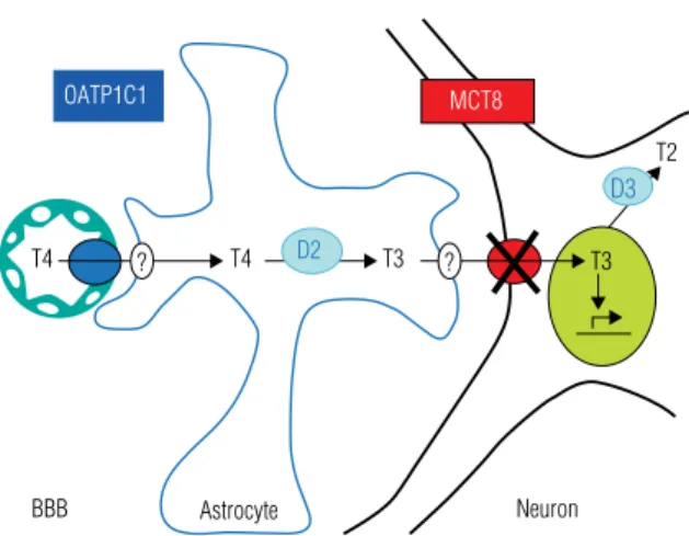
hyroid hormone (TH) is important for the development of different tissues, in particular the brain, as well as for the regulation of the metabolic activities of the tissues and thermogenesis throughout life. Most TH actions are initiated by bind-ing of the active hormone 3,3’,5-triiodothyronine (T3) to its nuclear receptor. This induces an alteration in proteins associated with the transcription initiation complex, resulting in the stimulation or suppression of the expression of TH responsive genes.The biological activity of TH is thus determined by the intracellular T3 concentra-tion, which is only indirectly dependent on the function of the thyroid gland which secretes predominantly the prohormone thyroxine (3,3’,5,5’-tetraiodothyronine, T4). In many target tissues, T3 availability is regulated in a paracrine manner, where T3 sup-ply to target cells is derived from T4 to T3 conversion in neighbouring cells. Brain and cochlea are examples of tissues with paracrine regulation of TH action. In other tissues, such as the pituitary and brown adipose tissue (BAT), T3 may be produced from T4 di-rectly in its target cells, representing an autocrine mechanism of TH action (Figure 1). In yet other tissues such as the liver and the kidneys, intracellular T3 is in rapid exchan-ge with circulating T3. This could be regarded as an endocrine action of T3, although it is still largely derived from peripheral conversion of T4 even in these same tissues.
In contrast to the activation of T4 by enzymatic outer ring deiodination (ORD) to T3, TH is inactivated by inner ring deiodination (IRD), which converts T4 to 3,3’,5’-triiodothyronine (reverse T3, rT3) and T3 to 3,3’-diiodothyronine (T2) (1). Three iodothyronine deiodinases (D1-3) are involved in these different deiodination reactions (1). D1 is expressed in liver, kidneys and thyroid and has both ORD and IRD activity. It is thought to contribute importantly to production of serum T3, in particular in eu- and hyperthyroid conditions. D2 has only ORD activity and is ex-pressed in brain, pituitary, BAT, thyroid and skeletal muscle. It is essential for local production of T3 in brain, pituitary and BAT, but the enzyme in thyroid and muscle may also be an important site for production of serum T3 in eu- and hypothyroid subjects. D3 is highly expressed in different fetal tissues, placenta and pregnant ute-rus, and also in adult brain and skin. It has only IRD activity and thus catalyzes the inactivation of TH. Together with D2, D3 has a crucial role in the region-speciic and time-dependent regulation of T3 in the developing brain. The deiodinases are homo-logous selenoprotein embedded in the membrane of the endoplasmic reticulum or the plasma membrane, such that the active sites are located in the cytoplasm (1).
Cop
yright
© ABE&M t
odos os dir
eit
os r
eser
vados
.
In addition, three transporters have been characterized showing considerable speciicity for iodothyronines, namely OATP1C1, MCT8, and MCT10 (2). The pa-thophysiological relevance of TH transporters has be-come clear in particular for MCT8, mutations in which are the cause of a syndrome of X-linked psychomotor retardation combined with abnormal TH levels (3,4). The clinical characteristics of a patient with a novel mu-tation in MCT8 are described by de Menezes Filho and cols. (5).
MOLECULAR CHARACTERISTICS OF MCT8
MCT8 and MCT10 are members of what is called the monocarboxylate transporter (MCT) family, although only 4 members of this family (MCT1-4) are known to transport monocarboxylates such as lactate and pyruva-te (6). The function of most of the 14 members of this family is still enigmatic. In 2001, MCT10 was identi-ied as an aromatic amino acid transporter and we have subsequently demonstrated that MCT8 and MCT10 are effective TH transporters (2).
The MCT8 gene is located on human chr Xq13.2 and has 6 exons and, thus, 5 introns of which intron 1 is ~100 kb in size. MCT8 has two possible translation start sites (TLSs), yielding proteins of 613 or 539 amino acids. In many animals MCT8 lacks the irst TLS giving rise to only the short MCT8 protein. If the N-terminal extension of the long human MCT8 protein has speciic functions remains to be determined. The MCT10 gene has a very similar structure, is located on human chr 6q21-q22, and codes for a protein of 515 amino acids. In all species, MCT10 has only one TLS, corresponding to the second TLS of human MCT8.
MCT8 and MCT10 have 12 putative transmembra-ne domains (TMDs), with both N-and C-terminal do-mains located intracellularly. MCT8 and MCT10 show a high degree of homology, especially in their TMDs, which its with their similar functions as TH transpor-ters. Both MCT8 and MCT10 transport different io-dothyronines; T3 is transported somewhat better by MCT10 than by MCT8, whereas the opposite is true for T4 (2). Both MCT8 and MCT10 facilitate cellular uptake as well as eflux of iodothyronines. Expression of MCT8 or MCT10 may thus induce only a modest in-crease in steady-state intracellular TH levels. However, transfection of cells with MCT8 or MCT10 strongly increases iodothyronine metabolism by D1, D2 or D3--expressing cells, indicating that they indeed effectively increase intracellular TH availability (2). Prevention of T4 and T3 eflux by co-transfection with the high-afi-nity cytoplasmic TH-binding protein CRYM, allows the proper study of TH uptake facilitated by MCT8 and MCT10 (2). TH eflux by MCT8 and MCT10 has been less well studied.
MCT8 and MCT10 are expressed in many human tissues. MCT8 is highly expressed in liver, kidney, adre-nal, ovary and thyroid. Studies in mice have shown that MCT8 is also expressed importantly in brain, in parti-cular in neurons in different brain regions, including cerebral cortex and cerebellum, but also in the choroid plexus, in capillaries and in tanycytes lining the 3rd ven-tricle (7). MCT8 has been localized in different nuclei in the human hypothalamus and in the human pituita-ry (8), where it appears to be expressed predominantly apparently in folliculostellate cells.
PATIENTS
Mutations in MCT8 are associated with X-linked psychomotor retardation and abnormal serum TH le-vels (2-4). The neurological syndrome is also known as the Allan-Herndon-Dudley syndrome (AHDS) af-ter the authors of the irst study of a large family with affected males published in 1944. AHDS comprises central hypotonia associated with poor head control and initially also peripheral hypotonia that progresses to hypertonia and spasticity. Most AHDS patients are unable to sit, stand or walk independently and have not developed speech. However, in some families patients have a milder phenotype and are able to walk and/or talk, albeit with great dificulty. All patients have severe mental retardation. AHDS patients are born without
OATP1C1 MCT8
T4 T4
Astrocyte
BBB Neuron
T3 T3
T2
?
D3
?
D2
Figure 1. Simplified schema of the regulation of T3 supply to neuronal
Cop
yright
© ABE&M t
odos os dir
eit
os r
eser
vados
.
apparent abnormality and the disease appears to deve-lop progressively, often associated with microcephaly. MRI of the brain usually shows delayed myelination before the age of 2 years which apparently normalizes with increasing age. For a more detailed description of the clinical characteristics, readers are referred to Holden and cols. (9) and the paper by de Menezes Fi-lho and cols. (5).
In addition to the psychomotor retardation, AHDS patients have abnormal serum TH levels. Serum T4 and FT4 levels vary from low-normal to truly reduced, whereas serum T3 levels are invariably increased. Like T4, serum rT3 is often decreased, and the serum T3/ rT3 and T3/T4 ratios are markedly elevated. Serum TSH varies between normal and elevated; mean TSH levels are about twice the normal mean. In view of the low (F)T4 and somewhat higher TSH levels, many AHDS patients have been treated with T4 substitution without any obvious beneit. This is also the case with the patient reported by de Menezes Filho and cols. (5).
TH is crucial for brain development. In AHDS patients, brain development is impaired despite the presence of elevated serum T3 levels, indicating some form of TH resistance. This is explained by the lack of T3 uptake in neuronal target cells where T3 action is required for optimal brain development (Figure 1). AHDS only occurs in males since the MCT8 gene is located on the X chromosome. However, one female has been described with AHDS due to chromosomal translocation with a breakpoint in MCT8 on one X chromosome and unfavourable inactivation of the in-tact X chromosome (10).
In over 50 families with AHDS studied so far various mutations in MCT8 have been identiied. These mu-tations include large deletions, frame-shift mumu-tations and non-sense mutations, which obviously inactivate MCT8. However, for mutations resulting in the dele-tion, insertion or substitution of single amino acids this is less obvious. The consequences of these mutations have been tested in cells transfected with wild-type (wt) or mutant MCT8 alone or in combination with cDNA coding for D3. These cells have subsequently been tested for TH uptake or TH metabolism, respectively. All tested mutations result in a marked decrease in TH transport, although the magnitude of the defect depen-ds on the cell type used for transfection (2,11). The results suggest some correlation between the severity of the clinical phenotype, the changes in serum TH levels and the defect in TH transport.
TISSUE AND HORMONE-SPECIFIC EFFECTS OF
MCT8 INACTIVATION
Inactivation of MCT8 appears to have different effects on TH availability in different tissues. As mentioned above, the neurological phenotype of patients with MCT8 mutations is explained by the lack of T3 uptake in central neurons. This appears to be supported by ob-servations that brain T3 uptake is almost completely blocked in Mct8 knockout (ko) mice, whereas brain T4 uptake is not affected (12). However, brain T3 uptake is probably largely determined by transport across the blood-brain barrier (BBB) and/or the blood-CSF bar-rier (BCB) and indeed MCT8 is importantly expressed in brain capillaries and in the choroid plexus (7,13). The lack of effect of Mct8 inactivation on brain up-take of T4, which is also a substrate for MCT8, may be explained by assuming that T4 is largely transported across the BBB and BCF by OATP1C1 (7,13).
In contrast to the subnormal T3 levels in brain, tis-sue T3 levels may be normal or even increased in other tissues, depending on the repertoire of TH transporters expressed. Although MCT8 is highly expressed in liver, hepatic T4 and T3 uptake do not seem to be affected in Mct8 ko mice (12). As the liver is exposed to increa-sed serum T3 levels, there is increaincrea-sed expression of T3 responsive genes such as Dio1 in the liver of Mct8 ko mice (12). This is in agreement with the elevated serum SHBG levels in AHDS patients, knowing that hepatic expression of SHBG is stimulated by T3 and increa-sed in patients with hyperthyroidism (14). The body composition of AHDS patients also suggest increased metabolic rate and muscle wasting due to tissue hyper-thyroidism. This is also reported in the present study by de Menezes Filho and cols (5). Also tachycardia has been observed in AHDS patients.
Mct8 ko have been generated to study the
patho-genic mechanism of AHDS, but disappointingly these mice do not show any obvious neurological phenotype (12,15). However, they do show the same changes in serum TH levels as AHDS patients, including a ma-rkedly decreased serum T4 and a mama-rkedly increased serum T3. Studies into the mechanisms of these altera-tions have indicated a) increased D1 expression in liver and kidney, b) increased D2 activity in brain and pitui-tary, and c) decreased D3 activity in brain of Mct8 ko vs. wt animals (12,16).
Cop
yright
© ABE&M t
odos os dir
eit
os r
eser
vados
.
T3 accumulation is increased in the kidneys of Mct8 ko vs. wt mice (12,16). This paradoxical change may sug-gest that MCT8 is more important for TH eflux than for TH uptake in the kidney. The increased tissue T4 content together with the high renal D1 activity may contribute to the large increase in peripheral T4 to T3 conversion, and thus, to the low T4 and high serum T3 levels in Mct8 ko mice and AHDS patients (16).
Recent studies have unveiled an important function of MCT8 also in TH secretion (17,18). Thyroidal T4 and T3 content is increased in Mct8 ko vs. wt mice. Furthermore, the increase in serum T4 induced by TSH injection is lower, whereas the increase in serum T3 is higher in Mct8 ko vs. wt mice (18). This suggests that MCT8 plays an important role in T4 secretion by thyroid cells. If MCT8 is inactivated, T4 accumulates in the thyrocyte leading to increased intra-thyroidal T4 to T3 conversion, and thus an increase in the T3/T4 secretion ratio.
Finally, MCT8 is expressed in both the hypothala-mus and the pituitary, and its inactivation may thus in-terfere with the negative feedback of TH at both sites (12,18). Indeed, serum TSH is usually modestly incre-ased in AHDS patients, which may be compatible with the modestly decreased serum T4 levels but not with the strongly elevated serum T3 levels. In Mct8 ko mice hypothalamic TRH expression is markedly increased which can only be suppressed by very high T3 doses, indicating T3 resistance particularly at the hypothala-mic level.
The study by de Menezes Filho and cols. (5) repor-ted in this issue presents important information regar-ding TH dysregulation in AHDS patients. In
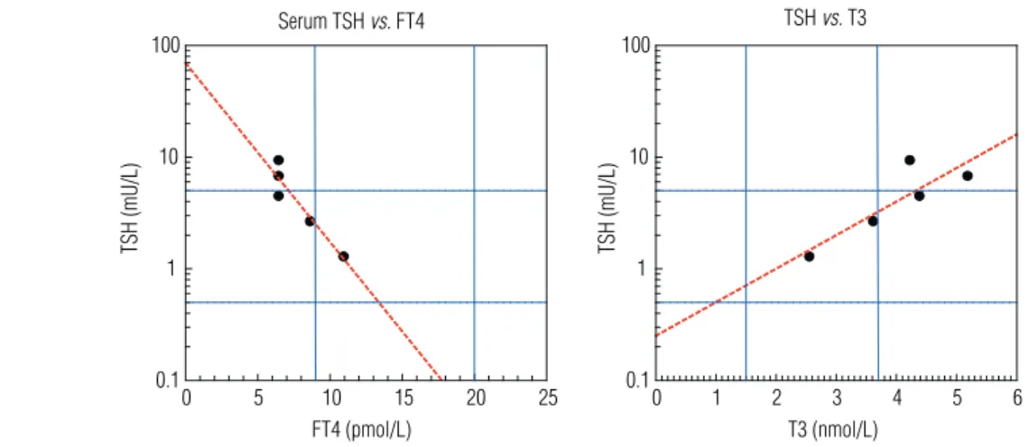
agree-ment with previous studies, their patient exhibits low serum FT4 and elevated T3 and TSH levels, in agree-ment with an impaired negative feedback of T3. Becau-se of the decreaBecau-sed T4 and increaBecau-sed TSH, the patient was treated with LT4. This resulted in a decrease in se-rum TSH and, remarkably, also a decrease in sese-rum T3. Plotting their data indicates that serum TSH shows a remarkably strong negative correlation with serum FT4 and an equally strong positive correlation with serum T3 (Figure 2). These results are compatible with the view that thyroidal T3 secretion may be an important source of serum T3 in AHDS patients.
TREATMENT
The lack of a neurological phenotype in Mct8 ko mice precludes their use as a model for development of an effective treatment of AHDS patients. The possible success of such a treatment will depend on a) the ex-tent to which irreversible damage in brain development has occurred at the time the disease is diagnosed, and b) the possibility to supply active hormone to impor-tant brain targets in the absence of functional MCT8. TH is essential for prenatal brain development, but TH-dependent brain maturation continues after birth (19). The lack of obvious abnormalities at birth and the progressive nature of AHDS may suggest that ear-ly postnatal initiation of an effective therapy may have substantial beneicial effect.
Because of the low serum T4 in combination with a modestly increased serum TSH, many AHDS patients have been treated with LT4 without obvious beneit. In fact, such treatment may have detrimental effects as
0 5 10 15 20 25
FT4 (pmol/L) 0.1
1 10 100
TSH (mU/L)
Serum TSH vs. FT4
0 1 2 3 4 5 6
T3 (nmol/L) 0.1
1 10 100
TSH (mU/L)
TSH vs. T3
Cop
yright
© ABE&M t
odos os dir
eit
os r
eser
vados
.
it may further increase T3 exposure of tissues which do not depend on MCT8 for TH uptake. For instance, it may induce or aggravate tachycardia. It could also be hypothesized that the abnormal serum T4 and T3 levels contribute importantly to the clinical phenotype and that their normalization may have beneicial thera-peutic effects. Such a study has been carried out in an adolescent AHDS patient in whom serum TH levels were normalized by block-and-replace therapy with PTU plus LT4. This produced favourable effects on body weight and heart rate but – as expected – did not result in an obvious mental improvement (14).
Obvious possibilities for effective therapy include early treatment of AHDS patients with a thyromimetic which is effectively transported into the brain even if MCT8 is mutated. The T3 analogue DITPA is curren-tly tested clinically in MCT8 patients (20). Preclinical studies are also in progress to test if the bioactive TH metabolite Triac or its precursor Tetrac may be used for effective treatment of AHDS patients.
Acknowledgment: Simone Kersseboom is supported by the Smile Foundation.
REFERENCES
1. Gereben B, Zavacki AM, Ribich S, Kim BW, Huang SA, Simonides WS, et al. Cellular and molecular basis of deiodinase-regulated thyroid hormone signaling. Endocr Rev. 2008;29:898-938. 2. Visser WE, Friesema EC, Visser TJ. Minireview: thyroid hormone
transporters: the knowns and the unknowns. Mol Endocrinol. 2011;25:1-14
3. Dumitrescu AM, Liao XH, Best TB, Brockmann K, Refetoff S. A no-vel syndrome combining thyroid and neurological abnormalities is associated with mutations in a monocarboxylate transporter gene. Am J Hum Genet. 2004;74:168-75.
4. Friesema EC, Grueters A, Biebermann H, Krude H, von Moers A, Reeser M, et al. Association between mutations in a thyroid hor-mone transporter and severe X-linked psychomotor retardation. Lancet. 2004;364:1435-7.
5. Menezes Filho HC, Marui S, Manna TD, Brust ES, Radonsky V, Kuperman H, et al. Novel mutation in MCT8 gene in a Brazilian boy with thyroid hormone resistance and severe neurologic ab-normalities. Arq Bras Endocrinol Metab. 2011;55(1):60-6.
6. Halestrap AP, Meredith D. The SLC16 gene family-from monocar-boxylate transporters (MCTs) to aromatic amino acid transpor-ters and beyond. Pflugers Arch. 2004;447:619-28.
7. Heuer H, Maier MK, Iden S, Mittag J, Friesema EC, Visser TJ, et al. The monocarboxylate transporter 8 linked to human psychomo-tor retardation is highly expressed in thyroid hormone-sensitive neuron populations. Endocrinology. 2005;146:1701-6.
8. Fliers E, Unmehopa UA, Alkemade A. Functional neuroanatomy of thyroid hormone feedback in the human hypothalamus and pituitary gland. Mol Cell Endocrinol. 2006;251:1-8
9. Holden KR, Zuniga OF, May MM, Su H, Molinero MR, Rogers RC, et al. X-linked MCT8 gene mutations: characterization of the pe-diatric neurologic phenotype. J Child Neurol. 2005;20:852-7. 10. Frints SG, Lenzner S, Bauters M, Jensen LR, Van Esch H, des
Portes V, et al. MCT8 mutation analysis and identification of the first female with Allan-Herndon-Dudley syndrome due to loss of MCT8 expression. Eur J Hum Genet. 2008;16:1029-37.
11. Kinne A, Roth S, Biebermann H, Koehrle J, Gruters A, Schweizer U. Surface translocation and T3 uptake of mutant MCT8 proteins are cell type-dependent. J Mol Endocrinol. 2009;43:263-71. 12. Trajkovic M, Visser TJ, Mittag J, Horn S, Lukas J, Darras VM, et al.
Abnormal thyroid hormone metabolism in mice lacking the mo-nocarboxylate transporter 8. J Clin Invest. 2007;117:627-35. 13. Roberts LM, Woodford K, Zhou M, Black DS, Haggerty JE, Tate
EH, et al. Expression of the thyroid hormone transporters MCT8 (SLC16A2) and OATP14 (SLCO1C1) at the blood-brain barrier. En-docrinology. 2008;149:6251-61.
14. Wemeau JL, Pigeyre M, Proust-Lemoine E, d’Herbomez M, Got-trand F, Jansen J, et al. Beneficial effects of propylthiouracil plus L-thyroxine treatment in a patient with a mutation in MCT8. J Clin Endocrinol Metab. 2008;93:2084-88.
15. Dumitrescu AM, Liao XH, Weiss RE, Millen K, Refetoff S. Tissue--specific thyroid hormone deprivation and excess in mono-carboxylate transporter (mct) 8-deficient mice. Endocrinology. 2006;147:4036-43.
16. Trajkovic M, Visser TJ, Darras WM, Friesema ECH, Schlott B, Mit-tag J, et al. Consequences of MCT8 deficiency for renal transport and metabolism of thyroid hormones in mice. Endocrinology. 2009;151:802-9.
17. Di Cosmo C, Liao XH, Dumitrescu AM, Philp NJ, Weiss RE, Re-fetoff S. Mice deficient in MCT8 reveal a mechanism regulating thyroid hormone secretion. J Clin Invest. 2010;120:3377-88. 18. Trajkovic-Arsic M, Müller J, Darras VM, Groba C, Lee S, Weih D, et
al. Impact of monocarboxylate transporter (Mct)-8 deficiency on the hypothalamus-pituitary-thyroid axis in mice. Endocrinology. 2010;151(10)5053-62.
19. Bernal J. Thyroid hormone receptors in brain development and function. Nat Clin Pract. 2007;3:249-59.

