ORIGINAL ARTICLE
Carbon dioxide use as contrast for vena cava filter implantation:
case series
Implante de filtro de veia cava com uso de dióxido de carbono como meio de contraste:
série de casos
Matheus Pessanha de Rezende1, Bernardo Massière2, Arno von Ristow3, Alberto Vescovi4, Alexandre A. Duarte1, Daniel A.
Drummond1, Leonardo Stambovsky1, Antonio Luiz de Medina5
Introduction
Pulmonary embolism is an apparent paradox of mod-ern Medicine – the greater the progress of medical tech-nology, the higher the number of clinical situations that lead to thromboembolism. However, several technological
developments have favored its diagnosis and treatment. Vena cava ilter implantation is a common therapeutic op-tion cava that is usually performed using iodine compounds as contrast media. Yet, some patients may develop contrast-induced nephropathy, which is the main cause of renal failure in hospitalized patients1,2.
Abstract
Objective: To assess the use of digital subtraction with carbon dioxide (CO2) for vena cava ilter implant.
Methods: From April 2010 to February 2011, seven patients underwent inferior vena cava ilter placement with digital subtraction angiography with the use of CO2 as contrast media. All patients had iliac and femoral deep venous thrombosis and contraindications for anticoagulation.
Results: Technical success was achieved in all cases. Inferior vena cava e renal veins were identiied in all cases. here were no evidences of complications related to the use of CO2 during or after the procedure.
Conclusion: he placement of inferior vena cava ilter with CO2 and digital subtraction angiography is safe and efective with good results in patients with renal insuiciency and allergy to iodine.
Keywords: angiography; carbon dioxide; venous thrombosis.
Resumo
Objetivo: Avaliar o resultado do implante de iltro em veia cava inferior empregando angiograia digital por subtração com dioxide de carbono (CO2) como meio de contraste.
Métodos: No periodo de abril de 2010 a fevereiro de 2011, sete pacientes foram submetidos ao implante de iltro na veia cava inferior, utilizando-se CO2 como meio de contraste em subtração digital. Os pacientes apresentaram como critério de inclusão trombose venosa profunda no setor iliacofemoral e contraindicação a anticoagulação.
Resultados: Foi obtido sucesso tecnico em todos os casos, com adequada visualização da veia cava e veias renais, não havendo complicações relacionadas ao uso do CO2 ou ao procedimento.
Conclusão: O implante de iltro de veia cava utilizando o CO2 como meio de contraste é segura e efetiva em pacientes portadores de alergia ao contraste iodado ou com insuiciência renal não dialítica.
Palavras-chave: angiograia; dióxido de carbono; trombose venosa.
Study carried out at the Department of Vascular and Endovascular Surgery at Centervasc-Rio – Rio de Janeiro (RJ), Brazil.
1 Physician; In Postgraduate Program in Vascular and Endovascular Surgery, Pontifícia Universidade Católica do Rio de Janeiro (PUC-Rio) – Rio de Janeiro (RJ), Brazil; Intern at Centervasc-Rio –
Rio de Janeiro (RJ), Brazil.
2 Vascular surgeon; Director, Centervasc-Rio, Brazil; Instructor Professor, Postgraduate Program in Vascular Surgery, PUC-Rio – Rio de Janeiro (RJ), Brazil. 3 Vascular surgeon; Director, Centervasc-Rio, Brazil; Associate Professor, Postgraduate Program in Vascular Surgery, PUC-Rio – Rio de Janeiro (RJ), Brazil. 4 Vascular Surgeon; Associate physician, Centervasc-Rio, Brazil; Instructor Professor, Postgraduate Program in Vascular Surgery, PUC-Rio – Rio de Janeiro (RJ), Brazil. 5 Vascular Surgeon; Titular Professor at the Postgraduate Program in Vascular Surgery of PUC-Rio – Rio de Janeiro (RJ), Brazil.
Vena cava ilter implantation with CO2 - Rezende MP et al. J Vasc Bras 2011, Vol. 11, Nº 1 19
Carbon dioxide (CO2) was irst used as contrast medium in the 1950’s to diagnose pericardial efusion. With the ad-vent of digital subtraction angiography (DSA) in 1980, angi-ography with CO2 has become a useful tool for the diagnosis, especially in patients with allergy to iodinated contrast or with chronic renal failure under medical treatment.
Angiography with CO2 can be used for the precise mea-surement of the inferior vena cava diameter, evaluation of anatomical features and evidence of non-occlusive thrombi, venous stenosis and even occlusions. It can also guide per-cutaneous interventions, such as vena cava ilter implanta-tion or vena cava recanalizaimplanta-tion3-5.
he objective of this study was to report a series of sev-en patisev-ents submitted to inferior vsev-ena cava ilter placemsev-ent using CO2 as contrast medium.
Material and methods
A single-center study was conducted from April 2010 to February 2011 in patients with deep venous thrombosis involving the iliac and femoral veins and contraindications to anticoagulation and to the use of iodinated contrast.
he procedures were performed in the operating room. he patients were submitted to local anesthesia and common femoral vein puncture through the Seldinger technique. he injection system employed a 60 mL syringe with Luer Lock
at-tached to a 3-way tap, which had one of its ways connected to a latex tube attached to the insulator normally used in lapa-roscopic surgery (Electronicendolator264305 20, Karl Storz Endoskope, Tuttlingen, Germany). Before using the CO2 in-jection system, all gas should be aspirated and the contents of the syringe should be purged in the environment three times, to avoid air contamination. Ater that, the remaining way is connected to the side way of the sheath that is part of the il-ter delivery system (Figure 1). In all cases, the Vena Tech LP® (BBraun, Melsungen, Germany) ilter was implanted.
he procedure started with pre-procedure cavography with the injection of 60 mL of CO2 for vena cava analysis, to determine the inferior vena cava morphology and to ind the ostia of the renal veins (Figure 2). Ater that, the ilter was implanted following the manufacturer’s instructions. A control cavography was performed ater ilter placement, with the injection of 60 mL of CO2 (Figure 3). A three-min-ute period between CO2 injections was observed
3.
Results
From April 2010 to February 2011, 45 patients were submitted to inferior vena cava ilter placement. Seven
Figure 1. System used to inject carbon dioxide (hose, 3-way tap and 60 mL syringe).
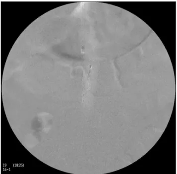
Figure 2. Inferior vena cavography with carbon dioxide.
patients presented contraindications to the use of iodinated
contrast and were submitted to the procedure using CO2
as contrast medium. We observed that four patients had non-dialysis renal failure and three patients had allergy to iodine. On average, 120 mL of CO2 were injected in each procedure.
Table 1 shows the characteristics of the studied popula-tion. All patients presented iliac and femoral deep venous thrombosis and contraindications to anticoagulation.
Vena cava ilter implantation with CO2 - Rezende MP et al. J Vasc Bras 2011, Vol. 11, Nº 1
20
Discussion
Evidence has been reported suggesting that the nephro-toxic efects of iodinated contrast on renal parenchyma are not temporary, but permanent and cumulative3,6,7. Patients with chronic renal failure under medical treatment, or with allergy to iodinated contrast beneit from vena cava ilter placement using alternative or no contrast media4-7.
Besides CO2, it is possible to implant vena cava ilters using gadolinium or the ultrasound-guided technique, which do not use contrast8. Some authors report the use of gadolinium as an alternative to iodinated compounds. However, when compared to CO2, it also presents lower ra-diographic density than iodinated contrast and it is associ-ated with the development of nephrogenic systemic ibrosis in patients with renal failure9,10.
CO2 is a low-cost, readily available medium in most operating rooms. It requires only a canister of pure CO2 , a laparoscopic insulator and a sterile hose to connect the insulator to the injection syringe.
Precautions should be adopted to avoid contamination with ambient air. CO2 is a colorless and odorless gas and it cannot be visibly distinguished from air. he incorrect appli-cation of this gas may result in air contamination, which may cause air embolism11. It is recommended to purge the injec-tion syringe three times, i.e., it should be illed with CO2 and emptied three times in order to keep only CO2 in the system.
he injected volume of CO2 and the time interval be-tween the injections should be observed, especially if the patient develops pain or hypotension. In our practice, we administered the injections with a minimum of three min-utes intervals. CO2 is about 20 times more soluble than oxygen. When injected into a blood vessel, bubbles of CO2 are fully dissolved within two to three minutes. For patients with chronic obstructive pulmonary disease, the amount per injection should be reduced and the time interval be-tween injections should be increased4,5,11.
CO2 is exhaled in a single passage through the lungs. However, the bubbles injected in the venous system may cross into the arterial system through the patent foramen ovale or other septal defects into the heart3,4. here are no absolute contraindications to CO2. However, it is prudent to avoid using it in the thoracic aorta due to the risk of gas embolism in the spinal, coronary and carotid arteries4,5.
Because of its lower radiographic density, the CO2 con-trast images present lower quality than iodinated concon-trast. he use of digital subtraction angiography helps improving image quality. In some cases, several injections of CO2 may be required, which increases the operator and the patient’s exposure to radiation.
Conclusion
he use of CO2 as a contrast medium for vena cava ilter placement is an option that presents satisfactory results in patients with allergy to iodinated contrast or non-dialysis renal failure.
References
1. Nash K, Hafeez A, Hou S. Hospital-aquired renal insuiciency. Am J Kidney Dis. 2002;39(5):930-6.
2. Solomon R. Contrast-medium-induced acute renal failure. Kidney Int. 1998;53(1):230-42.
3. Hawkins IF, Caridi JG. Carbon dioxide (CO2) digital subtraction angiography: 26-year experience at the University of Florida. Eur Radiol. 1998;8(3):391-402.
Table 1. Group of patients submitted to vena cava ilter implantation with carbon dioxide (CO2).
Patient Age Sex Contraindication to anticoagulation
Contraindication to CO2
DF 86 F digestive hemorrhage NDCRF
ET 71 F digestive hemorrhage allergy
RS 83 F digestive hemorrhage NDCRF
MB 77 F digestive hemorrhage NDCRF
HS 68 M gross hematuria allergy
SC 52 F polytrauma allergy
DP 78 F digestive hemorrhage NDCRF
M – male; F – female; NDCRF – non-dialysis chronic renal failure.
Vena cava ilter implantation with CO2 - Rezende MP et al. J Vasc Bras 2011, Vol. 11, Nº 1 21
4. Baiocchi MTP, Menezes FH, Luccas GC. Angiograia com gás dióx-ido de carbono. Rev Col Bras Cir. 1998;25(6):435-6.
5. Cho JK, Hawkins Jr IF. Carbon dioxide angiography. Medscape reference: drugs, diseases and procedures [internet]. [cited 2011 Oct 28]. Available from: http://emedicine.medscape.com/ article/423121-overview#showall.
6. Wong GTC, Irwin MG. Contrast-induced nephropathy. Br J Anaesth. 2007;99(4):474-83.
7. Dam MA, Wetzels JF. Toxicity of contrast media: an update. Neth J Med. 2008;66(10):416-22.
8. Neser RA, Capasso Filho M, Homa CMO. Implante de iltro de veia cava inferior guiado por ultra-som: relato de dois casos. J Vasc Bras. 2006;5(1):71-3.
9. Kaufman JA, Geller SC, Bazari H, et al. Gadolinium-based contrast agents as an alternative at vena cavography in patients with renal insuiciency – early experience. Radiology. 1999;212(1):280-4.
10. Girardi M, Kay J, Elston DM, et al. Nephrogenic systemic ibrosis: Clinicopathological deinition and workup recommendations. J Am Acad Dermatol. 2011.
11. Cho DR, Cho KJ, Hawkins IF. Potential air contamination during CO2 angiography using a hand-held syringe: theoretical consid-erations and gas chromatography. Cardiovasc Intervent Radiol. 2006;29(4):637-41.
Correspondence Bernardo Massière Departamento de Cirurgia Vascular e Endovascular – Centervasc-Rio Rua Sorocaba, 464 – 1º andar
CEP22271-110 – Rio de Janeiro (RJ), Brazil
E-mail: [email protected]