Med Radiol Diagn Imaging (2017)
DOI 10.1007/174_2017_51, © Springer International Publishing AG
João Lopes Dias and Teresa Margarida Cunha
J. Lopes Dias (*)
Centro Hospitalar de Lisboa Central, Lisbon, Portugal
Hospital Lusíadas de Lisboa, Lisbon, Portugal
New Medical School, Lisbon, Portugal e-mail: [email protected]
T.M. Cunha
Instituto Português de Oncologia de Lisboa Francisco Gentil, Lisbon, Portugal
e-mail: [email protected]
Abstract
Magnetic resonance imaging (MRI) and com- puted tomography (CT) are routinely used in female pelvis imaging. MRI is primarily use-ful for locoregional characterization of benign and malignant diseases. CT is less accurate in locoregional evaluation, but remains useful in the follow-up of treated gynecological malig-nancies, as well as in the setting of emergency and in the guidance of biopsies. Although transabdominal and transvaginal ultrasonog-raphy (US) is not under the scope of this chap-ter, it remains the first-line imaging method for most gynecological conditions.
Contents
1 Introduction ... 000
2 Magnetic Resonance Imaging ... 000
2.1 Introduction ... 000
2.2 Patient Preparation and Positioning ... 000
2.3 Coils, Scan Planes, and General Protocols ... 000
2.4 Gadolinium-Based Contrast Media ... 000
3 CT Technique ... 000
3.1 Introduction ... 000
3.2 Technical Disadvantages ... 000
3.3 Patient Preparation and Positioning ... 000
3.4 Oral and Rectal Contrast ... 000
3.5 Intravenous Iodine-Based Contrast Media ... 000
References ... 000
1
Introduction
pelvic infectious diseases) and in the guidance of biopsies. Although transabdominal and transvagi-nal ultrasonography (US) is not under the scope of this chapter, it remains the first-line imaging method for most gynecological conditions.
This chapter aims to focus on some important, hands-on topics regarding MRI and CT tech-niques, trying not to exhaustively develop issues with isolated historical interest.
2
Magnetic Resonance
Imaging
2.1 Introduction
The introduction of modern phased-array coils with eight or more elements led to high signal-to- noise ratios (SNR) and consequently increased image quality. Additionally, pelvic examinations have become faster as turbo (TSE) and fast spin- echo (FSE) sequences have replaced conven-tional spin-echo (SE) sequences. Most MRI examinations are now performed at 1.5 and 3 Tesla (T) magnets. Despite significant improve-ment on SNR with higher field strength, 3 T mag-nets are more prone to magnetic susceptibility artifacts, which may be particularly prominent on diffusion-weighted imaging (DWI).
2.2 Patient Preparation and Positioning
Before performing an MR exam, the patient should be informed about its approximate dura-tion as well as the necessity to use earplugs to protect against loud noises and to place a surface coil close to the skin. Moreover, all patients should be asked about contraindications and claustrophobia. Sedation may be required for those patients who would really benefit from the examination, but are unable to proceed due to claustrophobia. Any implanted device must be previously known and considered to be safe for the patient undergoing an MR procedure. Intrauterine devices (IUD) can be normally
scanned, and usually appear as a hypointense lin-ear structure within the endometrial cavity on both T2-weighted images (T2WI).
Finally, patients should also be informed about the utility of intravenous (IV) contrast and spasmolytic agent administration, as well as their side effects (see next sections).
Whenever a pregnant woman is undergoing an MR exam, issues about the fetus development are raised. As a general rule, the risk-benefit ratio should be evaluated for every patient. Fetal dele-terious effects have not been documented on 1.5 T magnets. However, some experts still rec-ommend avoiding the exam in the first trimester unless the potential benefits compensate the hypothetical risks. Since most of the studies were performed on 1.5 T scanners, far less is known about potential effects on 3 T (Masselli et al.
2013; Ray et al. 2016).
Some authors advocate that patients should void about 1 h before the examination to ensure that the bladder is only moderately filled. A full bladder may hamper T2WI and give rise to motion artifacts due to patient discomfort.
A 4-h fast helps to reduce bowel peristalsis and is recommended in some centers when intra-venous contrast administration is required. The administration of a fast-acting laxative enema to clean the bowel may also improve image quality.
It is uncommon to perform bowel preparation with diluted barium sulfate or other solutions as it increases preparation and imaging time and does not seem to bring significant advantages.
Vaginal tampon should be avoided. Vaginal opacification with ultrasound gel makes the eval-uation of vaginal walls easy and may be recom-mended when studying vagina tumors and deep endometriosis. Moreover, rectal and/or vaginal opacification with ultrasound gel may be useful in dynamic pelvic floor and deep endometriosis studies (Beddy et al. 2012; Sala et al. 2013; Bazot et al. 2016).
under the knees makes the examination more comfortable. Two important procedures are generally taken in order to decrease motion artifacts during abdominal and pelvic examina-tions: the use of a belt covering phased-array body coils, which restricts respiratory excur-sions, and the application of a spatial presatura-tion slab in a sagittal scout view, which is particularly useful in sequences without breath-hold, because of its ability to reduce artifacts from the movement of the anterior abdominal wall (Sala et al. 2013; Forstner et al. 2016; Froehlich et al. 2009).
2.2.1 Spasmolytic Medication
The administration of antispasmodic agents such as scopolamine N-butyl bromide (Buscopan®) or
glucagon is indicated to reduce artifacts from small bowel and bladder motion. It is particu-larly useful in the assessment of peritoneal implants on both morphological and functional sequences, especially on DWI. The administra-tion of intravenous Buscopan® (20–40 mg)
immediately before the examination is the most consensual option. Longer examinations may justify a second identical dose, because IV Buscopan® action only lasts about 15 min.
Intramuscular (IM) administration (20 mg) has an increased length of action (approximately 30–60 min). This anticholinergic drug should not be administered in patients who have demon-strated prior hypersensitivity to scopolamine N-butyl bromide, as well as in those with myas-thenia gravis, narrow- angle glaucoma, megaco-lon, tachycardia, prostatic enlargement with urinary retention, paralytic ileus, or mechanical stenosis in the gastrointestinal tract. Data regard-ing contraindications durregard-ing pregnancy is scarce; therefore Buscopan® is not recommended.
Common undesirable effects may be accommo-dation disorders, tachycardia, dizziness, or dry mouth. If accommodation changes occur, patients should be advised not to drive.
When Buscopan® is contraindicated,
gluca-gon (1 mg) can be administrated intravenously (Beddy et al. 2012; Sala et al. 2013; Bazot et al.
2016). In a study of Froehlich et al., glucagon
had more reliable onset of action and induced longer bowel paralysis when compared to sco-polamine N-butyl bromide. Glucagon is contra-indicated in patients with known hypersensitivity to the substance, as well as in those with known pheochromocytoma (due to the risk of stimulat-ing catecholamine release). In patients with a known insulinoma, glucagon should be adminis-tered cautiously since its initial hyperglycemic effect may stimulate the release of insulin and cause subsequent hypoglycemia (Froehlich et al.
2009).
2.3 Coils, Scan Planes, and General Protocols
Female pelvic MRI is generally performed with a phased-array body coil with at least four ele-ments. The introduction of modern coils improved SNR and allowed parallel imaging, thus reducing scan time on T1W and T2W con-ventional sequences. Intracavitary coils, either endovaginal or endorectal, have no current sci-entific support (Sala et al. 2013; Allen et al.
2014).
A general female pelvis protocol usually begins with a coronal localizer, which provides an anatomic overview of both lower abdomen and pelvis. It is helpful not only to guide pelvic sequences but also to exclude other conditions like hydronephrosis or renal malformations. Fast sequences like single-shot turbo or fast spin echo are generally used.
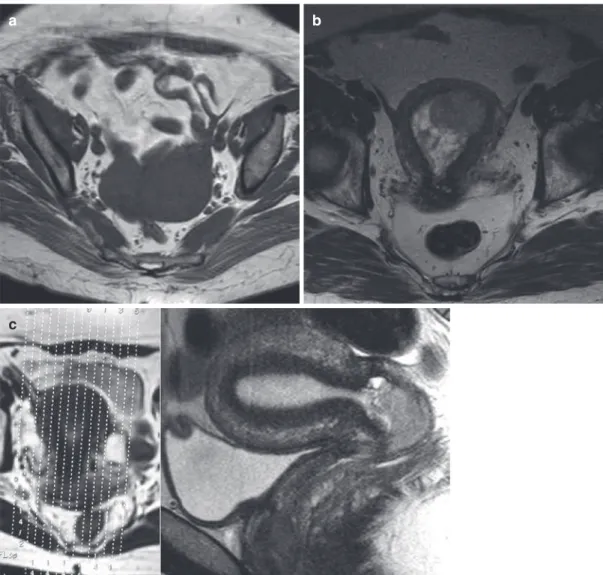
Specific protocols depend on the study target, but usually include an axial T1WI sequence and at least two T2WI sequences in different planes (Fig. 1). Dynamic contrast-enhanced magnetic resonance imaging (DCE-MRI) and DWI have become part of the standard imaging protocols for most of female pelvis MR examinations (Forstner et al. 2010, 2016; Allen et al. 2014; Sala et al. 2011).
Fig. 1 Examples of different orientations on MRI in dis-tinct patients. Axial T1W image of the pelvis (a). Axial T2W image through the pelvis showing the uterus with an endometrial carcinoma associated with a polyp (b). Oblique sagittal according to the uterine axis T2W image showing the normal uterine zonal anatomy and a cervical cancer (c). Oblique coronal according to the long axis of
the uterus, parallel to the endometrial cavity (d). Axial oblique T2W image, perpendicular to the long axis of the cervix, for evaluation of parametrial invasion while stag-ing a cervical cancer (e). Axial oblique TIW images after gadolinium with fat saturation, perpendicular to the long axis of the uterus, for local staging of an endometrial can-cer (f)
a b
f d
e
2.3.1 T1- and T2-Weighted Imaging
Typical SE T1WI is usually performed to assess spontaneous hyperintense content that may cor-respond to fat or blood. Axial fat-saturated T1WI in the same plane and with the same thickness should be performed for their distinction. Some centers perform T1W in- and opposed-phase images, which allow the detection of intravoxel lipid within masses (manifesting as a signal loss on the opposed-phase images). If a short scan
time is needed, gradient-echo (GRE) sequences may be applied; however anatomic detail will be decreased. Fat saturation may also be achieved by the Dixon method, a chemical shift-based technique that acquires both in- and opposed- phase images simultaneously, thus allowing mathematical combinations into fat-only and water-only sequences. It results in a more uni-form suppression of the fat signal, and allows the detection and quantification of microscopic lipid (Allen et al. 2014).
Multiplanar high-resolution nonfat-saturated T2W sequences are the most relevant for the majority of female pelvic diseases, because of their ability to depict uterine and ovarian zonal anatomy and to provide good contrast between normal and pathological tissues. TSE or FSE sequences are typically used. A single 3D T2W sequence with later multiplanar postprocessing can alternatively be performed. Single-shot TSE sequences during breath-hold (HASTE) may be useful in the coronal plane to exclude hydrone-phrosis or to assess renal malformations.
Fat-suppressed T2W sequences are not rou-tinely used for female pelvic imaging. However, they may be helpful to identify intraperitoneal, extraperitoneal, and endoluminal fluid; enlarged lymph nodes; and bone changes (for example, in the setting of bone metastases or of edematous changes due to postradiotherapy insufficiency fractures). When compared to nonfat-suppressed T2WI, it also helps to assess for macroscopic lipid content (Allen et al. 2014).
2.3.2 Diffusion-Weighted Imaging
DWI is a functional MR technique that assesses the random movement or the Brownian motion of water molecules in different physical media. The diffusion properties of a biological tissue are related to the amount of interstitial free water and permeability, therefore reflecting tissue cellularity and presence of intact cellular membranes. In general, high-cellularity tumors show restricted diffusion when compared to normal tissue, because of their higher cellular density. Coagulative necrosis, highly viscous fluid, and Table 1 Suitable general protocol for gynecological
MRI at a 1.5 T magnet
• 4–6 h of fasting
• Voiding and ingestion of two glasses of water 1 to 2 h before the exam
• Avoid the use of vaginal tampon • Vaginal opacification with ultrasound gel – May be useful for vaginal tumors and deep
endometriosis
• Rectal and/or vaginal opacification with ultrasound gel
– May be useful for dynamic pelvic floor studies and deep endometriosis • Administration of a fast-acting laxative enema to
clean the bowel, one on the day before the exam and another during the morning of the exam (optional) • Supine position
• Pelvic phased-array coils with at least four elements
• Belt covering phased-array body coils • Anterior and superior saturation band • Antispasmodic agents:
– Buscopan 20/40 mg IM/IV – Glucagon 1 mg IV • Matrix: 512 × 512
• Coronal localizer: single-shot turbo or fast spin-echo
• Morphological sequences: axial T1WI and at least two different planes on T2WI (or single 3D T2WI with posterior multiplanar post processing) • DWI: field-of-view and thickness equal to T2WI
(preferentially axial)
– Pelvis (b values: 0, 600 and 1000 s/mm2) – Abdomen (b values: 0, 500 and 1000 s/mm2) – High b values may reach 1200–1400 s/mm2 • DCE-MRI: 2D or 3D fat-suppressed T1W GRE
sequence
– Scan plane, region of interest, and acquisition timing depending on the specific target of the study
abscesses may behave similarly. DWI is per-formed using two or more b values (a measure of the gradient strength), including one or more low b values (0 or 50 s/mm2) and a high b value
(1000 s/mm2 or higher). Apparent diffusion
coef-ficients (ADC) are mathematical transformations of b value acquisitions that represent the slope of the line of the natural logarithm of signal inten-sity (y-axis) versus b values (x-axis). ADC maps are displayed parametrically as grayscale images. Suspicious areas with true water molecule move-ment restriction appear bright at high b values and dark on the ADC map. High b value sequences and the ADC map should always be interpreted together with morphological sequences to avoid potential pitfalls. In order to facilitate this evaluation, fusion images between T2WI and DWI may be generated. However, they can be altered by patient motion and bladder dis-tention during the examination, which may change the relative position of pelvic organs.
Whole-body diffusion-weighted MRI proto-cols have been developed over the last years, mainly for cancer staging and follow-up. Short- time examinations are now possible on both 1.5- and 3-T magnets due to the evolution of echo-planar and parallel imaging, generation of high-performance gradients, and introduction of phased-array multichannel surface coils. Advantages include absence of ionizing radiation and no injection of isotopes or intravenous con-trast media (Sala et al. 2013; Whittaker et al.
2009; Hameeduddin and Sahdev 2015; Qayyum
2009).
2.3.3 Dynamic Contrast Enhancement
Gadolinium-based contrast agents act by shorten-ing T1 relaxation time, which is better seen on T1WI. Thus, dynamic contrast-enhanced MRI (DCE-MRI) is usually performed using a 2D or 3D fat-suppressed T1W GRE sequence. 3D sequences like volumetric interpolated breath- hold examination (VIBE) allow the acquisition of thinner slices. The injection should be preferen-tially performed using an MR-compatible
auto-matic injector, but manual administration may also be performed. Serial image acquisitions are then performed every few seconds over a length of a few minutes. The protocol—including scan plane, region of interest, and acquisition time— will depend on the specific target of the study (see appropriate chapters). Qualitative, semi-quantitative, and quantitative analysis may be performed. The enhancement of a given structure may be directly and qualitatively accessed as an area of increased signal intensity (SI) on T1WI. Semiquantitative analysis implies record-ing the SI of a region of interest before and after contrast administration in order to get dynamic time-signal intensity (TSI) curves, which enable the extraction of some data like time to onset of enhancement, relative signal intensity, maximum postcontrast SI-to-precontrast SI ratio, rate of enhancement, and area under the curve (overall enhancement). Quantitative analysis—namely Ktrans (volume transfer constant between the plasma and the extracellular extravascular space)—remains under study and is not currently available in many centers (Beddy et al. 2012; Hameeduddin and Sahdev 2015; Bernardin et al.
2012).
Initial unenhanced imaging is helpful to detect hyperintense hemorrhagic or proteinaceous con-tent, and also allows performing subtraction imaging, which requires cautious breath-holding instructions to guarantee similar registration between unenhanced and contrast-enhanced images. Regardless of those instructions, some artifacts may be found due to bladder filling dur-ing the examination.
2.4 Gadolinium-Based Contrast Media
The risk of an acute reaction to a gadolinium- based contrast agent is low when compared to iodine-based contrast agents. However, similar cautions should be taken. Risk patients are those with a history of previ-ous acute reaction to gadolinium- based con-trast agent, asthma, and allergy requiring medical treatment. Unlike iodine-based con-trast agents, the risk of reaction to gadolinium-based contrast agents is not related to osmolality (Beckett et al. 2015).
Nephrogenic systemic fibrosis (NSF) is rec-ognized as a very late reaction to gadolinium-based contrast media since 2006. It usually starts with pain, pruritus, swelling, and erythema in the legs, and progresses to thickening of the skin and subcutaneous tissues, as well as to fibrosis of internal organs and respiratory muscles, which may lead to variable consequences ranging from contractures to cachexia and death. The severity of the disease implies the prompt recognition of high- risk patients, which are those with chronic kidney disease (CKD) 4 and 5 (GFR <30 mL/ min), including patients on dialysis, and those with acute kidney insufficiency. Gadolinium-based contrast agents with higher risk of NSF are gadodiamide (DTPA-BMA), gadopentetate dimeglumine (DTPA), and gadoversetamide (DTPA-BMEA). These high-risk agents should never be given in higher doses than 0.1 mmol/kg and are contraindicated in patients with stage 4 and 5 CKDM, including those on dialysis, in patients with acute renal insufficiency, in preg-nant women, and in neonates (Beckett et al.
2015; Thomsen et al. 2013, 2016; Mathur and Weinreb 2016).
Low-risk gadolinium contrast agents may be used in pregnancy when there is a strong need and no neonatal tests are necessary. Lactating women are frequently object of concern. If the above-mentioned high-risk agents are used, breastfeeding should be stopped for 24 h (Beckett et al. 2015; Thomsen et al. 2013, 2016; Mathur and Weinreb 2016).
3
CT Technique
3.1 Introduction
The advent of helical scanning movement and multislice data collection gave rise to a new era of fast and high-resolution acquisitions.
Modern CT scans show substantial improve-ment on volume coverage, scan speed, as well as a more efficient use of X-ray tubes. Helical CT scans allow near-isotropic acquisitions, thus enabling high-resolution multiplanar reconstruc-tions (MPR) and volume rendering. Moreover, the detector size decreased and multiple arrays were incorporated inside. Therefore, several slices can be recorded simultaneously, shortening the exposure time; thus thinner slices can be obtained and partial volume artifacts can be decreased.
The high speed of current CT scanners allows a complete thoracic, abdominal, and pelvic exam-ination in only one breath-hold. A CT pelvic acquisition is rarely performed alone. The scan is usually extended to the upper abdomen not only for staging and for the follow-up of malignant dis-eases but also when characterization of vascular, inflammatory, or infectious entities is needed.
a b
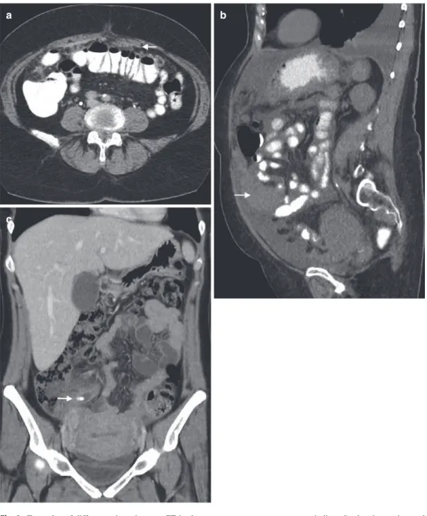
c
Fig. 2 Examples of different orientations on CT in three distinct patients. Axial image, after oral and intravenous contrast administration, depicting anterior peritoneal carci-nomatosis (arrow) in a patient with endometrial carcinoma (a). Sagittal image, after oral contrast administration
3.2 Technical Disadvantages
Among the main disadvantages of CT, there are two with great relevance in daily practice: ioniz-ing radiation exposure and metallic artifacts. Despite the kind of CT scan, radiation reduction and protection should always be considered. Both technicians and radiologists should be familiarized with some technical features includ-ing the tube current, X-ray beam collimation, and pitch, and cautiously select both the number and length of scan sequences. Dose quantification parameters like volume CT dose index (CTDlvol) and dose-length product (DLP) are usually dis-played in current CT scans and should be rou-tinely checked. Hip prosthesis causes substantial artifacts due to photon starvation and beam hard-ening, consequently hampering not only the joint and the surrounding muscles, but also pelvic organs. Methods may be used to reduce these
artifacts: filtered back projection (FBP), which uses information from areas adjacent to regions affected by metal artifacts and replaces the metal- corrupted raw data by the interpolated values; adaptive filtering, which corrects the excessive noise produced by the photon starvation effect; or iterative algorithms, which use a combination of different metal artifact reduction algorithms and algebraic reconstruction techniques (Morsbach et al. 2013). Iterative reconstruction techniques also enable performing reduced-dose CT exami-nations (due to either tube current or tube poten-tial lowering) without altering image quality (Padole et al. 2014).
3.3 Patient Preparation and Positioning
Before a CT scan, an inquiry about the patient medical history, routine medication, and potential contraindications is mandatory. Pregnancy is not a formal contraindication to CT; however, it should be avoided mainly during the first trimester. Patient preparation for CT scans is mainly related to contrast material administration (see next sec-tions). Due to the need of intravenous contrast, patients are optimally asked to fast at least for 4 h. Administrating a fast-acting laxative enema to clean the bowel may also be recommended.
Patients are usually scanned in supine and “foot-first” positions, with their arms raised above their heads. Claustrophobic patients tolerate lying with the head outside the gantry better. Moreover, these positions allow for a face-to-face communication with the patient and facil-itate the access to the patient whenever the examination has to be interrupted. “Foot-first” positioning also eases the connection of contrast medium tubes, which can be done immediately after inserting the venous access.
3.4 Oral and Rectal Contrast
Oral and rectal contrast media are frequently used in abdominal and pelvic CT imaging. Classically, it is given in cases of suspected bowel perforation or of anastomosis leakage. However, opacification Table 2 Suitable general protocol for gynecological CT
at a 64-detector scanner
• 4–6 h of fasting
• Administration of a fast-acting laxative enema to clean the bowel, one on the day before the exam and another during the morning of the exam (optional)
• Water or iodine-based oral contrast: 1000– 1500 mL of contrast medium administered in separate doses 45 to 60 min prior to the examination
• Water or iodine-based rectal contrast: catheter and enema bag; 200 mL to the rectosigmoid and 900–1200 mL to the entire colon
• Supine and “foot-first” positions • Range from diaphragm to pubic bone • Inspiration
• Scout image: 120 kV, 10–50 mA • Final acquisition: (20 kV, 100–200 mAs) • Sub-mm or mm volumetric acquisitions with
subsequent reconstruction into 2 to 5 mm thick axial, sagittal and coronal images
• Correction of hip prosthesis artifacts if need: filtered back projection, adaptive filtering or iterative algorithms
• Intravenous iodine-based contrast: 100–150 mL at 3–4 mL/s
• Scan delay
– 70–100 s (covering all range) – 3–5 min (if pelvic vein thrombosis is
suspected)
of the digestive tract is also helpful for distinguish-ing collapsed loops from lymph nodes, peritoneal implants, pelvic masses, and fluid collections.
Water as negative or positive iodinated solutions may be used for bowel opacification. In female pel-vis imaging, positive contrast is particularly useful for staging and follow-up of ovarian and endome-trial cancer. Iodinated solutions are preferable over barium suspensions due to its moderate effect on peristalsis and easier distribution along the diges-tive tract. Contrarily, barium suspensions are more prone to flocculate and may give rise to streak arti-facts that hamper wall evaluation. Both types of contrast are usually safe, with only rare cases of mild diarrhea being reported. However, if bowel perforation is suspected, only iodinated solution should be administered due to the higher peritoneal toxicity of barium-based contrast media. As 1–2% of oral contrast is absorbed through the gut patients who had previous moderate to severe IV contrast allergy should be managed carefully.
Protocols for oral contrast vary according to the center. Typically, 1000–1500 mL of contrast medium is ingested in separate doses 45–60 min prior to the examination. Some authors advocate the administration of 20 mg metoclopramide at the beginning of the ingestion, in order to shorten patient preparation.
Rectal contrast is administered when the patient lies on the scanning table by using a catheter and an enema bag. 200 mL of contrast usually opaci-fies the rectum and the sigmoid colon adequately, whereas the entire colon may require 900– 1200 mL of contrast. Sometimes, anal diseases like hemorrhoids and fissures hamper the intro-duction of the rectal tube due to pain. Special care should also be taken in patients with anal and lower third rectal carcinoma due to the risk of ulceration or perforation (Beckett et al. 2015).
3.5 Intravenous Iodine-Based Contrast Media
Intravenous iodine-based contrast media are usu-ally given as a rapid bolus via an IV cannula, by using a pump injector. A suitable protocol for abdominal and pelvic CT scan is to administer 100–150 mL of 350 mg iodine contrast media at
3–4 mL/s. As the time to contrast material arrival and peak enhancement are affected by the choice of intravenous access sites, particularly when forearm or hand veins are used, lower flow rates are desirable (Bae 2010).
A suitable routine gynecological CT scan includes the upper abdomen and is acquired 70–100 s after contrast injection. Liver metasta-ses from gynecological malignancies are typi-cally hypovascular, so an arterial phase is usually unnecessary. Delayed images are recommended whenever pelvic vein thrombosis is suspected (3–5 min) or bladder and ureteral opacification is needed (5–10 min).
In order to reduce the risk of an acute reaction to iodine-based contrast media, some procedures should be taken according to the European Society of Urogenital Radiology (ESUR) guide-lines: a nonionic contrast medium should be used; the patient should be kept in the Radiology Department for 30 min after the injection; and drugs and equipment for resuscitation have to be readily available. For patients at increased risk of reaction (history of previous moderate or severe acute reaction to an iodine-based contrast agent, asthma, and allergy requiring medical treatment), an alternative imaging tool not requiring an iodine-based contrast agent should be consid-ered. The use of premedication for allergy prevention is widely accepted, despite its underlying limited clinical evidence. The ESUR guidelines recommend oral administration of prednisolone 30 mg (or methylprednisolone 32 mg), 12 and 2 h before contrast medium administration (Thomsen et al. 2016).
There is usually some concern regarding thy-rotoxicosis, a potential very late reaction to iodine-based contrast media that usually occurs more than 1 week after injection. As a general rule, iodinated contrast media should not be given to patients with manifest hyperthyroidism. Patients at risk—those with untreated Graves’ disease, or multinodular goiter and thyroid auton-omy, especially if they are elderly and/or live in areas of dietary iodine deficiency—should be strictly monitored by endocrinologists after injection (Van der Molen et al. 2004).
Iodine-based contrast media may be given to pregnant patients in extraordinary conditions, but thyroid function should be checked in the neo-nate during the first week following the exam. When iodine-based agents are given to lactating mothers, breastfeeding may be continued nor-mally (Thomsen et al. 2016).
In patients with renal impairment (eGFR less 45 mL/min/1.73 m2 for intravenous
administra-tion), an alternative imaging method without iodine-based contrast media should be consid-ered. However, if it is really needed, volume expansion should be done. According to the ESUR guidelines, intravenous normal saline, 1.0–1.5 mL/kg/h, for at least 6 h before and after contrast medium administration is recommended. An alternative suitable protocol is intravenous sodium bicarbonate (154 mEq/L in dextrose 5% water), 3 mL/kg/h for 1 h before contrast medium administration and 1 mL/kg/h for 6 h after con-trast medium administration. Moreover, the low-est dose of a low or iso-osmolar contrast medium should be used (Thomsen et al. 2016).
Finally, some recommendations should be followed in patients taking metformin due to the risk of lactic acidosis after IV iodine-based con-trast exposure when significant renal impairment is present. Patients with an eGFR between 30 and 44 mL/min/1.73 m2 (CKD 3) should stop
metformin 48 h before contrast medium injec-tion and should only restart it 48 h after the examination if renal function has not deterio-rated. In patients with eGFR less than 30 mL/ min/1.73 m2 (CKD 4 and 5), iodine-based
con-trast media should be avoided (Beckett et al.
2015; Thomsen et al. 2016).
References
Allen BC, Hosseinzadeh K, Qasem SA, Varner A, Leyendecker JR (2014) Practical approach to MRI of female pelvic masses. Am J Roentgenol 202(6):1366– 1375. doi:10.2214/AJR.13.12023
Bae KT (2010) Intravenous contrast medium administra-tion and scan timing at CT: consideraadministra-tions and approaches. Radiology 256(1):32–61. doi:10.1148/ radiol.10090908
Bazot M, Bharwani N, Huchon C, Kinkel K, Cunha TM, Guerra A, Manganaro L, Buñesch L, Kido A, Togashi
K, Thomassin-Naggara I, Rockall AG (2016) European Society of Urogenital Radiology (ESUR) guidelines: MR imaging of pelvic endometriosis. Eur Radiol. doi:10.1007/s00330-016-4673-z2016
Beckett KR, Moriarity AK, Langer JM (2015) Safe use of contrast media: what the radiologist needs to know. Radiographics 35(6):1738–1750. doi:10.1148/ rg.2015150033
Beddy P, O’Neill AC, Yamamoto AK, Addley HC, Reinhold C, Sala E (2012) FIGO staging system for endometrial cancer: added benefits of MR imaging. Radiographics 32(1):241–254. doi:10.1148/rg.321115045
Bernardin L, Dilks P, Liyanage S, Miquel ME, Sahdev A, Rockall A (2012) Effectiveness of semi-quantitative multiphase dynamic contrast-enhanced MRI as a pre-dictor of malignancy in complex adnexal masses: radiological and pathological correlation. Eur Radiol 22(4):880–890. doi:10.1007/s00330-011-2331-z
Forstner R, Sala E, Kinkel K, Spencer JA (2010) ESUR guidelines: ovarian cancer staging and follow-up. Eur Radiol 20(12):2773–2780. doi:10.1007/ s00330-010-1886-4
Forstner R, Thomassin-Naggara I, Cunha TM, Kinkel K, Marselli G, Kubik-Huch R, Spencer JA, Rockall A (2016) ESUR recommendations for MR imaging of the sonographically indeterminate adnexal mass: an update. Eur Radiol. doi:10.1007/s00330-016-4600-3
Froehlich JM, Daenzer M, von Weymarn C, Erturk SM, Zollikofer CL, Patak MA (2009) Aperistaltic effect of hyoscine N-butylbromide versus glucagon on the small bowel assessed by magnetic resonance imag-ing. Eur Radiol 19:1387–1393. doi:10.1007/ s00330-008-1293-2
Goldman LW (2007) Principles of CT and CT technology. J Nucl Med Technol 35(3):115–130. doi:10.2967/ jnmt.107.042978
Hameeduddin A, Sahdev A (2015) Diffusion-weighted imaging and dynamic contrast-enhanced MRI in assessing response and recurrent disease in gynaeco-logical malignancies. Cancer Imaging 15:3. doi:10.1186/s40644-015-0037-1
Masselli G, Derchi L, McHugo J, Rockall A, Vock P, Weston M, Spencer J (2013) Acute abdominal and pelvic pain in pregnancy: ESUR recommendations. Eur Radiol 23:3485–3500. doi:10.1007/s00330-013-2987-7
Mathur M, Weinreb JC (2016) Imaging patients with renal impairment. Abdom Radiol 41(6):1108–1121. doi:10.1007/s00261-016-0709-8
Morsbach F, Bickelhaupt S, Wanner GA, Krauss A, Schmidt B, Alkadhi H (2013) Reduction of metal arti-facts from hip prostheses on CT images of the pelvis: value of iterative reconstructions. Radiology 268(1):237–244. doi:10.1148/radiol.13122089
Padole A, Khawaja R, Kalra M, Singh S (2014) CT radia-tion dose and iterative reconstrucradia-tion techniques. Am J Roentgenol 204:W384–W392. doi:10.2214/ AJR.14.13241
Ray J, Vermeulen M, Bharatha A, Montanera W, Park A (2016) Association between MRI exposure during pregnancy and fetal and childhood outcomes. JAMA 316(9):952–961. doi:10.1001/jama.2016.12126
Rydberg J, Buckwalter KA, Caldemeyer KS et al (2000) Multisection CT: scanning techniques and clinical applications. Radiographics 20(6):1787–1806. doi:10.1148/radiographics.20.6.g00nv071787
Sala E, Rockall A, Kubik-Huch R (2011) Advances in mag-netic resonance imaging of endometrial cancer. Eur Radiol 21(3):468–473. doi:10.1007/s00330-010-2010-5
Sala E, Rockall AG, Freeman SJ, Mitchell DG, Reinhold C (2013) The added role of MR imaging in treatment stratification of patients with gyneco-logic malignancies: what the radiologist needs to know. Radiology 266(3):717–740. doi:10.1148/ radiol.12120315
Thomsen HS, Morcos SK, Almen T et al (2013) Nephrogenic systemic fibrosis and gadolinium-based
contrast media: updated ESUR contrast medium safety committee guidelines. Eur Radiol 23(2):307– 318. doi:10.1007/s00330-012-2597-9
Thomsen HS et al (2016) ESUR Guidelines on Contrast Media Version:9.0. http://www.esur.org/esur-guidelines/
Van der Molen AJ, Thomsen HS, Morcos SK (2004) Effect of iodinated contrast media on thyroid function in adults. Eur Radiol 14(5):902–907. doi:10.1007/ s00330-004-2238-z
Whittaker CS, Coady A, Culver L, Rustin G, Padwick M, Padhani AR (2009) Diffusion-weighted MR imaging of female pelvic tumors: a pictorial review. Radiographics 29(3):759–774. doi:10.1148/ rg.293085130discussion 774–8