J Bras Pneumol. 2005;31(6):563-6
Primary effusion lymphoma in an immunocompetent patient 563
Primary effusion lymphoma in an immunocompetent
patient*
LEILA ANTONANGELO1, FRANCISCO S VARGAS2, LISETE RIBEIRO TEIXEIRA3, MARCELO A C VAZ4,
MARIA MIRTES SALES5, LUIS C MOREIRA6, ROBERTA KARLA BARBOSA DE SALES4
*Study carried out in the Pulmonology Department and Central Laboratory of the Hospital das Clínicas da Faculdade de Medicina da Universidade de São Paulo (HC-FMUSP, University of São Paulo School of Medicine Hospital das Clínicas), São Paulo, São Paulo, Brazil
1. Tenured Professor in the Pulmonology Department and Central Laboratory of the Hospital das Clínicas da Faculdade de Medicina da Universidade de São Paulo (HC-FMUSP, University of São Paulo School of Medicine Hospital das Clínicas), São Paulo, São Paulo, Brazil
2. PhD in Pulmonology in the Pulmonology Department and Central Laboratory of the Hospital das Clínicas da Faculdade de Medicina da Universidade de São Paulo (HC-FMUSP, University of São Paulo School of Medicine Hospital das Clínicas), São Paulo, São Paulo, Brazil
3. Head Professor in the Pulmonology Department and Central Laboratory of the Hospital das Clínicas da Faculdade de Medicina da Universidade de São Paulo (HC-FMUSP, University of São Paulo School of Medicine Hospital das Clínicas), São Paulo, São Paulo, Brazil
4. PhD in Pathology in the Pulmonology Department and Central Laboratory of the Hospital das Clínicas da Faculdade de Medicina da Universidade de São Paulo (HC-FMUSP, University of São Paulo School of Medicine Hospital das Clínicas), São Paulo, São Paulo, Brazil
5. Biologist in the Pulmonology Department and Central Laboratory of the Hospital das Clínicas da Faculdade de Medicina da Universidade de São Paulo (HC-FMUSP, University of São Paulo School of Medicine Hospital das Clínicas), São Paulo, São Paulo, Brazil
Correspondence to: Francisco S. Vargas. R. Itapeva, 500, Cj. 4C - Bela Vista, São Paulo, Brasil. CEP: 01332-000. Tel: 55 11 3069-5695. E-mail: [email protected] e [email protected]
Submitted: 18 January 2005. Accepted, after review: 5 April 2005. Primary effusion lymphoma (PEL) is classified
as a non-Hodgkin's lymphoma and is identified as a unique clinical and pathological entity according to the World Health Organization classification system.(1) It is characterized by the
presence of cavity effusion, the absence of nodal or extranodal involvement and the incorporation of genetic material (from human herpes virus type
ABSTRACT
Primary effusion lymphoma is an unusual non-Hodgkin's lymphoma rarely seen in immunocompetent patients. Herein, we present clinical and biochemical data obtained from an immunocompetent patient diagnosed with primary effusion lymphoma.
Keywords: Pleural effusion; Lymphoma, non-Hodgkin; HIV seronegativity; Case reports
8 and the Epstein-Barr virus) by the tumor cells, as well as by its association with Kaposi's sarcoma and Castleman's disease.(2-3)
Although PEL may occur in any of the serous cavities, it primarily affects the pleural cavity (in approximately 70% of cases), followed by the peritoneum and the pericardium. Involvement of more than one serous cavity is not uncommon.(3)
INTRODUCTION
564 Antonangelo L, Sales RKB, Vargas FS, Vaz MAC, Teixeira LR, Sales MM, Moreira LC
J Bras Pneumol. 2005; 31(6):563-6
Although PEL primarily affects immunocompromised patients, in recent years, cases of PEL have been described in individuals with no history of immunosuppression. Such individuals are typically over the age of sixty and present varied systemic diseases, such as congestive cardiac insufficiency, l i v e r c i r r h o s i s o r n e o p l a s i a .( 4 - 7 ) A m a r k e d
characteristic in this group of patients is that the great majority do not present the Epstein-Barr virus incorporated into the tumor genome.(4,7)
The present case is that of a pleural PEL in an elderly individual who was seronegative for human immunodeficiency virus. This is the first case of this type of lymphoma that has been described in Brazil.
CASE REPORT
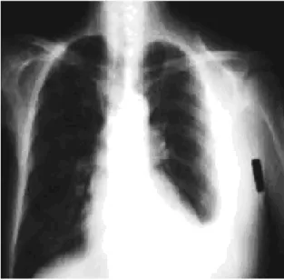
A 65-year-old male patient with a history of dyspnea and weight loss was referred to the Outpatient Clinic for Pleural Diseases of the University of São Paulo School of Medicine Hospital das Clínicas by his cardiologist. According to his medical records, the patient had been previously diagnosed with cardiac insufficiency and was under appropriate treatment. In the assessment, we found vocal fremitus, decreased breath sounds over the left lung and no signs of cardiac decompensation. The chest X-ray revealed moderate left pleural effusion, with no alterations in the mediastinum
(Figure 1). The patient was submitted to thoracentesis for symptom relief and for diagnostic purposes.
Serum and pleural fluid laboratory test results are shown in Table 1. Through the laboratory test results the pleural effusion was defined as an exudate with macroscopic evidence of hemorrhage, and the cytological examination revealed lymphocyte predominance with 88% blast cells. Morphologically, the neoplastic cells were described as polymorphic, with abundant and intensely basophilic cytoplasms, voluminous nuclei, prominent nucleoli and numerous mitotic figures (Figure 2). The results of a parietal pleura biopsy (Cope needle) indicated nonspecific chronic
Figure 1 - Posteroanterior chest X-ray
Figure 2 - Morphological characteristics of the blast cells. Leishamann, x1000
TABLE 1
Serum and pleural fluid test results
Hemoglobin - Hematocrit 10.3 g/dL - 30% Leukocytes (mm3)/ Lymphocytes (%) 6400/18.1%
Total serum protein 8.3 g/dL
Serum DHL 540 IU
Total protein - pleural fluid 5.8 g/dL (?)
DHL - pleural fluid 7685 IU
Total nucleated cells (mm3) 4600
Oncotic cytology 88% blast cells
Immunophenotypes
CD 45 Negative
CD3, CD10, CD19, CD20 and CD22 Negative
CD30, CD38 and CD71 Positive
Cytoplasmic IgM Positive
J Bras Pneumol. 2005;31(6):563-6
Primary effusion lymphoma in an immunocompetent patient 565
pleuritis (Figure 2). Since cytological evaluation revealed the presence of blast cells, the hypothesis of a lymphoma was taken into consideration. Therefore, we carried out a systemic radiological evaluation (in order to identify any primary nodal or extranodal sites), myelography and immunophenotypic study of the pleural fluid.
Tomography scans of the cervical, thoracic and abdominal regions revealed no evidence of nodal or extranodal involvement caused by the lymphoma. Blood count and myelography were also considered normal for the age, with no anomalous cellular elements.
The immunophenotypic study of the pleural fluid (Table 1) was essential for the diagnosis. For the immunophenotyping, we used an ample panel of monoclonal antibodies, similar to that used for the characterization of non-Hodgkin's lymphomas in general. These antibodies are generally expressed in cells that are hematological in origin and are not expressed in mesothelial cells. In the case in question, the neoplastic cells were CD30-, CD38- and CD71-positive, with monoclonality for IgM kappa demonstrated in the cytoplasms of the neoplastic cells after the use of permeabilization. The polymerase chain reaction for human herpes virus type 8 was positive in the cells of the pleural fluid.
The diagnosis of pleural PEL was made based on these findings, and chemotherapy was started. The patient died during the first chemotherapy cycle.
DISCUSSION
Classified as a non-Hodgkin's lymphoma, PEL affects the serous cavities. Since it is a rare pathology, there have been very few studies of its incidence. However, according to European data, PEL accounts for 3% of all non-Hodgkin's lymphomas affecting patients with acquired immunodeficiency syndrome and for 0.4% of those affecting the seronegative population in general.(4,8)
It is believed that the pathogenesis of PEL is multifactorial, and that the factors involved include breakdowns in the immune surveillance of i m m u n o c o m p r o m i s e d p a t i e n t s , a s w e l l a s qualitative alterations in immunoregulation due to immunosenescence.(3) Concomitance of chronic
diseases is another potential risk factor. Another aspect to be considered in PEL is co-infection with
human herpes virus type 8 or the Epstein-Barr virus, both frequently found in immunocompromised patients.(9) Although co-infection with the
Epstein-Barr virus is less frequent in cases of patients who tested negative in the serologic testing for human immunodeficiency virus, all cases presented co-infection with human herpes virus type 8, accompanied by Kaposi's sarcoma.(4-5)
A diagnosis of pleural PEL is initially based o n e x c l u d i n g t h e p o s s i b i l i t y o f o t h e r lymphoproliferative diseases that affect the pleural cavity and result in pleural effusion. In order to eliminate the possibility of secondary involvement of the pleural cavity caused by lymphoma, it is recommended that computed tomography scans of the neck, chest and abdomen be performed in o r d e r t o l o c a t e e n l a r g e d l y m p h n o d e s o r organomegalies, and that a blood count and fine-needle aspiration bone marrow biopsy be performed to investigate medullary infiltration. In this case, after these complementary tests, no enlarged lymph nodes, organomegalies or extranodal masses were found, nor was there any involvement of bone marrow or peripheral blood. Since there is no determination of primary nodal or extranodal site, a diagnosis of PEL is generally confirmed by evaluation of the cytological and immunophenotypic characteristics of fluid collected from the pleural cavity. The fluid is typically an exudate, with increased overall cellularity, a predominance of lymphocytes, a representative number of neoplastic cells, and blast cells similar to immunoblasts. From a cytological viewpoint, the morphology of the malignant cells imposes a differential diagnosis with diffuse large B-cell lymphoma (an immunoblastic variant) or with anaplastic lymphoma.(2-3,10-11) In the case described
herein, the pleural effusion was an exudate presenting more than 80% blast cells with immunoblastic aspects, numerous mitotic figures and considerable cellular degeneration.
566 Antonangelo L, Sales RKB, Vargas FS, Vaz MAC, Teixeira LR, Sales MM, Moreira LC
J Bras Pneumol. 2005; 31(6):563-6
invariably negative, as are the antigens HMB-45 and S-100.(7,10-11) In the immunophenotyping of the
pleural fluid of this case, the neoplastic cells were characterized by presenting the negative common leukocyte antigen (CD45), a finding that casts doubt on the lymphoid origin of these cells since the hematological lineage of the blast cells was not demonstrated. The antigens associated with the T and B lineages (CD2, CD3, CD5, CD19, CD20 and CD22) were also negative, as were the HMb45 antigens and the S100 protein. However, the cells expressed cell activation markers and mature B-lineage markers (CD30, CD38 and CD138). In the case described, we applied the technique of permeabilizing the cellular membrane of the neoplastic cells. It was thereby possible to demonstrate, in the cytoplasm, the expression of the Kappa light chain in CD38- and CD138-positive cells, confirming the clonal nature of these cells.
When immunophenotyping is insufficient to determine the lineage of the tumor cells, gene rearrangement through molecular techniques is generally used to confirm the presence of clonality.(12)
The cytological diagnosis of PEL in an elderly patient who was seronegative for human immunodeficiency virus, presented no clinical or radiological evidence of primary nodal or extranodal sites, and in whose tumor cells it was possible to demonstrate clonality and incorporation of the genetic material of human herpes virus type 8, was conclusive for the diagnosis of pleural PEL.
REFERENCES
1. Banks PM, Warnke RA. Primary effusion lymphoma. In: Jaffe ES, Harrris NL, Stein H, Vardiman JW, eds. Tumours of haematopoietic and lymphoid tissues. Lyon: IARC Press; 2001. p.179-80.
2. Cesarman E, Chang Y, Moore PS, Said JW, Knowles DM. Kaposi´s Sarcoma-associated Herpesvirus-like DNA s e q u e n c e s i n A I D S - r e l a t e d b o d y - c a v i t y - b a s e d lymphomas. N Engl J Med. 1995;332(18):1186-91. 3. H e n g g e U , R u z i c k a T, Ty r i n g S K , S t us c h k e M ,
Roggendorf M, Schwartz RA, et al. Update on Kaposi´s sarcoma and other HHV8 associated diseasea. Part 2: pathogenesis, Castleman´s disease, and pleural effusion lymphoma. Lancet Infect Dis.2002;2(6):344-52. 4. Carbone A, Goghini A, Vaccher E, Zagonel V, Pastore C,
Dalla Palma P, et al. Kaposi´s sarcoma associated herpesvirus DNA sequences in AIDS-related and AIDS-unrelated lymphomatous effusions. Br J Haematol. 1996;94(3):533-43.
5. Okada T, Katano H, Tsutsumi H, Kumakawa T, Sawabe M, Arai T, et al. Body-cavity-based lymphoma in an elderly AIDS-unrelated male. Int J Hematol. 1998;67(4):417-22. 6. Ascoli V, Scalzo C, Danese C, Vacca K, Pistilli A, Lo Coco F. Human herpes virus-8 associated primary effusion lymphoma of the pleural cavity in HIV-negative elderly men. Eur Respir J. 1999;14(5):1231-4. 7. Boulanger E, Hermine O, Fermand JP, Radford-Weiss I,
Brousse N, Meignin V, et al. Human herpesvirus 8 (HHV-8) - associated peritoneal primary effusion lymphoma (PEL) in two HIV-negative elderly patients. Am J Hematol. 2004;76(1):88-91.
8. Dal Maso L, Franceschi S. Epidemiology of non-Hodkin lymphomas and other haemolymphopoietic neoplasma in people with AIDS. Lancet Oncol. 2003;4(2):110-9. 9. Cesarman E. The role of Kaposi´s sarcoma-associated
herpesvirus (KSHV/HHV-8) in lymphoproliferative diseases. Recent Results Cancer Res. 2002;159:27-37.
1 0 . Rodríguez Salazar MJ, Raya Sánchez JM, Rodríguez Sánchez R, Alonso Socas MM, Brito Barroso ML, Hernandez Nieto L. [HIV-associated primary body-cavity-based lymphoma: clinico-biologic features in three patients diagnosed at the same institution]. An Med Interna. 2004; 21(4):175-8. Spanish.
11. Nador RG, Cesarman E, Chadburn A, Dawson DB, Ansari MQ, Sald J, et al. Primary effusion lymphoma: a distinct, clinopathologic entity associated with the Kaposi´s sarcoma-associated herpes virus. Blood. 1996;88(2):645-56.