347
1. MD. Substitute professor, Department of Pediatrics, Universidade Federalda Bahia (UFBA), Salvador, BA, Brazil
2. Pediatric hematologist. Associate professor, Universidade Federal da Bahia (UFBA), Salvador, BA, Brazil.
This paper is dedicated to Maria de Lourdes Santos Pardo - in memoriam.
Manuscript received Jan 16 2004, accepted for publication Mar 31 2004.
Suggested citation: Di Nuzzo DVP, Fonseca SF. Sickle cell disease and infection. J Pediatr (Rio J). 2004;80:347-54.
Abstract
Objective: To discuss the high prevalence of sickle cell disease in our environment and the increased morbidity and mortality as a result of infection associated with this condition.
Sources of data: Review of MEDLINE from 1986 to 2003. We found around 600 references about the subject. Thirty-five journal articles were reviewed, in addition to chapters in two text books.
Summary of the findings: We discuss general information concerning sickle cell disease as well as a few topics about the most frequently observed infections in these patients. Drug prophylaxis and immunizations are also covered.
Conclusions: This review hopes to provide the pediatric community with information concerning the association between sickle cell disease and infections, so as to minimize the occurrence of complications.
J Pediatr (Rio J). 2004;80(5):347-54: Sickle cell disease, infection, penicillin, immunization. Copyright © 2004 by Sociedade Brasileira de Pediatria
R
EVIEWA
RTICLESickle cell disease and infection
Dayana V. P. Di Nuzzo,1 Silvana F. Fonseca2
Sickle cell disease
A disease of genetic character, described for the first time by Herrick in 1910,1,2 frequently, but not exclusively, affecting individuals of African origin and originating from a mutation to chromosome 11,2 resulting in the substitution of a glutamic acid by valine at position six of the sixth position from the N-terminal of the ß-chain of the hemoglobin molecule, giving rise to hemoglobin S. Erythrocytes whose predominant content is hemoglobin S assume a shape reminiscent of a sickle under conditions of hypoxia from which the name sickle cell is derived resulting from the polymerization of hemoglobin S.3,4
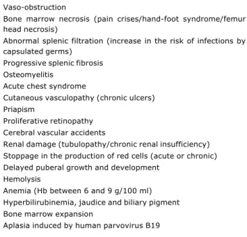
The red, sickle-shaped cells, do not circulate adequately within the microcirculation, resulting both in obstruction of capillary blood flow and their own premature destruction. This pathophysiologic mechanisms results in severe clinical manifestations, with greater frequency after 3 months of age (Table 1).3 During the first 6 months of life, these individuals are generally asymptomatic due to high hemoglobin F levels.1
Table 1 - Clinical manifestations of the sickle cell disease
Adapted1,2,4
Vaso-obstruction
Bone marrow necrosis (pain crises/hand-foot syndrome/femur head necrosis)
Abnormal splenic filtration (increase in the risk of infections by capsulated germs)
Progressive splenic fibrosis Osteomyelitis
Acute chest syndrome
Cutaneous vasculopathy (chronic ulcers) Priapism
Proliferative retinopathy Cerebral vascular accidents
Renal damage (tubulopathy/chronic renal insufficiency) Stoppage in the production of red cells (acute or chronic) Delayed puberal growth and development
Hemolysis
Anemia (Hb between 6 and 9 g/100 ml) Hyperbilirubinemia, jaudice and biliary pigment Bone marrow expansion
Aplasia induced by human parvovirus B19
The mortality rate among children under 5 with sickle-cell anemia is around 25 to 30%, and a majority of the deaths in this group are secondary to fatal infections, sequestration of the spleen or aplastic crises.2
While the greatest mortality rates occur during the first 2 years of life, obligatory inclusion of tests for hemoglobin disorders in the neonatal screening battery (Guthrie test) has been demonstrated to be an important step in the reduction of these rates, by means of the early identification of affected individuals and the introduction of suitable prophylaxis and regular outpatients follow-up.2,4,6
Current life expectancy for the American sickle-cell anemia population is 42 years for men and 48 years for women.7,8 While this is much better than the 14.3 years of 3 decades ago, it is still well below the life expectancy of the general population, which is evidence of the need for further investment and progress in the treatment of these patients.7
Laboratory diagnosis of sickle-cell anemia is made using electrophoresis of the hemoglobin, isoelectric focusing or high performance liquid chromatography (HPLC). Hemoglobin ß chains can be detected at an early stage of fetal development, from the 10th to 12th week of pregnancy, which makes prenatal diagnosis of sickle-cell anemia possible.3
Sickle cell disease manifests in individuals who are homozygous for hemoglobin S and in combination with other abnormal hemoglobin forms, which can result in sickle cell disease with a number of different degrees of severity: co-heredity with a gene for hemoglobin C (SC), a gene for ß+ thalassemia (SAF), or a gene for ß° thalassemia (SF), in decreasing order of frequency.3
Sickle cell disease and infection
Infections are the most frequent complications in individuals with sickle-cell anemia.1
In children from 0 to 6 years of age, splenomegaly is observed resulting from congestion in the red pulp due to sequestered sickle-shaped erythrocytes in the Billroth and sinusoidal cords, which progress with the formation of thromboses and infarction, culminating in atrophy and fibrosis of the organ. This phenomenon, known as auto-splenectomy, generally occurs by about 5 years of age.7,8 Nevertheless, even before auto-splenectomy, phagocytic capacity mediated by opsonin and antibody production are affected as a consequence of the persistent splenic aggression,9 leading to functional asplenia, which becomes permanent at around the sixth to eighth year of life.10
As a consequence of this asplenia, there will be increased susceptibility to infections by capsulated organisms, notably Haemophilus influenzae type b (Hib) and pneumococcus.10,11 The risk of infection by this last for children under five with sickle-cell anemia is approximately 30 to 100 times greater than for healthy children.10 These infections, accompanied by acidosis, hypoxia and dehydration, can trigger off and/or intensify the sickle cell crises, since they encourage the production of inflammatory cytokines, thus increasing the expression of endothelial adhesion cells and the adhesion of sickle-cells and polymorphonuclear sickle-cells in the vascular endothelium . Under these conditions, a vicious circle is formed that is dangerous for the patient, which can become lethal if not adequately treated. This fact justifies the search for effective prophylaxis and management.
Furthermore, patients with sickle-cell anemia have been observed to be at twenty-five fold risk of developing salmonella infections, especially older children and adults.12 Before 3 years of age, infections caused by pneumococcus and Hib still predominate.12,13
Viral infection and bone marrow aplasia
Certain types of virus are associated with transitory aplastic crises in sickle-cell anemia patients with special emphasis on parvovirus B19.14 Its primary target is immature erythroid cells.
Once patients suffering from chronic hemolytic anemia have accentuated compensatory hyperplasia of the erythroid series, infection by parvovirus B19, in addition to other virus, promotes destruction of immature erythroid cells, with a consequent stoppage in the production of red cells, leading to an exacerbation of the already existing anemia. Leukocytes and platelets are not generally a f f e c t e d , b u t , o c c a s i o n a l l y , l e u k o p e n i a a n d / o r thrombocytopenia may be observed, with a possibility of atypical lymphocytes and eosinophilia.15
Possible warning signs are: fever, general unwellness, mild gastrointestinal and respiratory pains and symptoms. Exanthema may occur in 23% of patients, which is difficult to see with melanodermous patients and, therefore, in the majority of individuals with sickle-cell anemia. The prodrome period is followed by a significant drop in hematocrit and severe reticulocitopenia.14
The aplastic crisis persists for around 10 to 12 days15 and occurs most often during winter and spring, with peaks in incidence every 2 to 3 years.14 Its prevalence increases with age, varying from 2 to 10% in children under 5 years and from 40 to 60% in adults over 20.16
Recently, nephrotic syndrome has been observed during infection by parvovirus B19 or within 6 weeks of one. There are also reports of cerebral-vascular complications, such as cerebral vascular accident concomitant with the acute anemia episode, meningitis, encephalitis and vasculitis.17
Specific etiologic diagnosis is performed by means of serum testing and/or by identification of the virus in tissues or blood. Tests employed are: IgG and IgM assay by immunoenzymatic methods, radioimmunoassay and immunofluorescence, detection by virus hybridization in situ, polymerase chain reaction (PCR) or electron microscope.14
Basic treatment consists of erythrocyte transfusion. The use of immunoglobulins, a good source of neutralizing antibodies, may be recommended for severely immunodepressed patients.16 Bone marrow recovers in between 7 and 10 days, and recurrent crises have not been described.3,14,17
The most common bacterial infections
In decreasing order of frequency, the main etiologic agents associated with episodes of invasive bacterial infection of individuals with sickle-cell anemia are: Streptococos pneumoniae, Salmonella spp, Hib, Escherichia coli and Klebsiella spp.18
Pneumococcus and Hib predominantly have incidence in children under 5 and are uncommon after this age. The latter agent affects more male children.18
Salmonella does not have any preference by age group, but there is a notable linear increase in incidence with increasing age.18
Klebsiella and Escherichia coli predominantly have incidence after 10 years of age and primarily after 20.18
It is extremely important to remember that any bacterial infection has a large potential for progressing to sepsis in individuals with sickle-cell anemia, very often with a lethal outcome if not identified and treated early.
Airways
Acute otitis media may often be observed, being primarily of pneumococcal etiology.19,20
Pneumonia by pneumococcus, Hib and salmonella is common. The first two of these agents are particularly more
prevalent and severe among children under 5 years old, primarily infants.20 Infections caused by atypical bacteria may occur at any age.21
Secondary infection may occur in areas of the pulmonary parenchyma that have suffered infarction in acute chest syndrome,22 which presents with clinical manifestations similar to pneumonia, such as fever, coughing, dyspnea and chest pain. Acute chest syndrome is the second most common cause of hospital admissions for all age groups of individuals with sickle-cell anemia.21 In acute chest syndrome, the use of antibiotics does not generally trigger a rapid improvement and progress is often unfavorable.3
Osteo-articular system
Bone marrow necrosis, secondary to bone infarction, predisposes a patient with sickle-cell anemia to complications such as osteomyelitis and septic arthritis.12,23 These complications are more common among the male sex (2:1), being rare under one year of age.24
The most common clinical findings are pain, edema, heat, rubor, increased local sensitivity and fever (temperatures above 38.2 °C). Laboratory tests may reveal leukocytosis (above 15,000/mm3) and increased erythrocyte sedimentation rate.24 Differential diagnosis between osteomyelitis and bone infarction is difficult,21 since their clinical signs are similar. High fever with shivering and an appearance of intoxication increase the suspicion of osteomyelitis, and vigilance must be maintained in such cases. Simple X-rays are of relatively limited value to differentiating between diagnoses of osteo-articular infection and bone infarction,24 except when a periosteal elevation in the painful area is observed, which favors a diagnosis of osteomyelitis. More sophisticated imaging studies, such as bone scintigraphy or magnetic resonance imaging, have greater sensitivity for the early diagnosis of osteomyelitis. The performance of blood cultures is recommended (blood, osteo-articular aspirate), and will possibly indicate bone biopsy of the affected area.
The most often identified etiologic agent in cases of osteomyelitis in the majority of samples is salmonella (57%), with cases also occurring because of S. aureus, S. pneumoniae, H. influenzae, ß-streptococcus/Klebsiella and Escherichia coli/Enterococcus.25
Greater predisposition to osteomyelitis by salmonella is observed among patients with sickle-cell anemia than in the general population. In osteomyelitis caused by salmonella, frequent multiple location bone involvement has been documented, in contrast with other etiologies.12
In cases of septic arthritis, there is no evidence of greater predisposition to any given pathogen, but streptococcus appears to be more prevalent.24
Nervous system
Meningitis presents a high mortality rate among individuals with sickle-cell anemia, in addition to acting as a precipitating factor for cerebral vascular accidents, primarily ischaemic.26
In a meta-analysis of studies in developing countries, it was observed that bacterial meningitis by pneumococcus caused more deaths and neurological sequelae than did either Hib or meningococcus.19
Meningitis by Hib has a universal distribution, generally being endemic, with predominance in temperate climates and during the winter. It is transmitted by means of Flügges droplets and nasopharyngeal secretions, ceasing 24 to 48 hours after antibiotic therapy is started. The incubation period is unknown, possibly varying from 2 to 4 days. Its clinical course is similar to that of other forms of meningitis, making it difficult to distinguish between pneumococcal and meningococcal meningitis. Mortality is greatest in the age group from 0 to 4 years (in particular among those less than 1 year old), decreasing from there on.27 It is important to emphasize that transmission may be possible throughout the entire period during which the microorganism is present and may be sustained even in the absence of nasal secretions, and Hib carriers also predominate in the same age group as peak incidence, which makes congregations (day-care centers, schools, institutions, etc.) the focus of vigilance and health care.27
Gastrointestinal system
In acute gastroenteritis caused by salmonella, symptoms have onset a few hours after the ingestion of contaminated food and manifest in nausea, and diarrhea, progressing with abdominal pain, fever and shivering.12
The invasive disease depends on the immune status of the host.12 Sepsis or osteomyelitis by salmonella may overcome gastrointestinal transport with a certain frequency by vaso-obstructive ischaemic episodes which break the mucosal barrier.23
Gastroenteritis of pneumococcal etiology is also encountered not infrequently.13
Abdominal pain in sufferer from sickle-cell disease is normally attributed to episodes of vaso-obstruction, with appendicitis being a rare event in such individuals, with incidence below that for the general population.28 The biological basis for this finding remains unknown.
Articles have been being published since 1950 demonstrating that hepatitis is one of the causes of liver disease among individuals with sickle-cell anemia, and the C virus has been identified as the primary etiologic agent of post-transfusion hepatitis. These patients present the risk of acquiring the infection by hepatitis C virus from the blood transfusions to which they are submitted,1,29 especially those who received blood transfusions before serum screening for anti-HCV antibodies in blood banks was started. A study performed in Pernambuco also showed an increased prevalence of infection by HCV among those individuals who had received more than 10
units of blood-based products.29 The prevalence of hepatitis C virus infection varies from 2 to 30% in individuals with sickle-cell anemia, when in the overall population it is estimated at 3%.29
Genito-urinary system
Individuals with sickle-cell anemia are susceptible to urinary tract infection. There is predominance among women, and the primary etiologic agents are gram-negative germs, in particular Escherichia coli, which predominantly has incidence after 20 years de age, possibly reflecting sexual activity, with an increased probability of urinary tract infections with ischaemic renal damage.18
There is also evidence of an increased incidence of urinary tract infections among expectant mothers: approximately twice that of the normal population,30 with the chance of developing septicemia.
Septicemia
This a permanent risk due to the lack or reduction of spleen function,31 primarily during the first 6 years of life,32 and it is the main cause of death among infants with sickle-cell anemia.18
We should remember that sepsis may be the initial event in the presentation of as-yet-undiagnosed sickle-cell anemia,13,33 since a majority of such events occur before 3 years of age.
The main etiologic agents, in decreasing order of frequency, are: pneumococcus, Hib, Salmonella sp, Escherichia coli, Enterobacter sp and Acinetobacter sp. In a Jamaican study, gram-negative microorganisms were identified in 50% of positive blood cultures.34
The incidence of sepsis by Hib has been falling, in contrast with what is observed of the behavior of pneumococcus.35
It is pneumococcal etiology that represents the greatest risk during the first 3 years of life, with peak incidence between 1 and 2 years, being rare before six moths and falling off after 5 years. It should be suspected in any severe case of sepsis involving children with sickle-cell anemia presenting high fevers. Initial clinical presentation is by fever, vomiting and toxemia, also occurring, albeit with less frequency, abnormal thoracic findings, meningism, petechiae, shock and disseminated intravascular coagulation.33 Confirmation is by blood culture.
The incidence of sepsis by salmonella is very high in Jamaica, being the most often identified organism after 6 years of age. In one published study, septicemia by salmonella was not observed in children under 3 years, but made up 30% of cases in children between 6 and 9 years of age, in the vast majority (77%) associated with osteomielite.12
In sepsis caused by Hib or salmonella, the patient presents seriously ill, with high fever and accentuated jaundice. Diagnostic confirmation is by blood culture. Early treatment should be instituted with ceftriaxone (100 mg/k/d, 12/12h, intravenous) or, in the absence of this, crystalline penicillin (100,000 to 200,000U/k/d in a single daily dose) associated with chloramphenicol (50 to 100 mg/k/d, 6/6h, intravenous).3
Patients with previous history of bacteremia have a greater susceptibility to recurrence,37 and should be submitted for diagnostic investigation in the swiftest and most aggressive manner possible. This susceptibility is directly interconnected with early loss of spleen function. The development of clinical splenomegaly during the first 6 months of life has been shown to be significantly associated with an increased risk of subsequent sepsis by pneumococcus. The possible importance of this early loss of spleen function is based on data from Saudi Arabia, in which fetal hemoglobin concentration is associated with preserved spleen function and a lower incidence of sepsis.18
Prophylaxis
Prophylaxis against the complications of the disease itself is indispensable for the least unfavorable prognosis possible for these individuals. The basic steps are four:
neonatal diagnosis followed by guidance and a family education program with regular outpatients care; pharmaceutical prophylaxis with penicillin;
vaccination against pneumococcus and Hib at the appropriate ages;
Early identification and appropriate management of febrile episodes, treating them as potential septic events.
Early sickle-cell anemia diagnosis makes it possible to keep the child under observation before the appearance of symptomology and its complications and allows antibiotic prophylaxis to be started from 3 months onwards, together with vaccination against capsulated germs.1,2,5 This significantly reduces the number of deaths associated with this infirmity, particularly due to infectious problems (from 30 to 1%),1 in addition to making better quality of life possible.5 In countries in which neonatal screening for hemoglobinopathies has been instituted, it has been demonstrated that when these children receive follow-up at specialized centers, mortality by pneumococcal infections can be reduced from 40 to 10% and overall mortality from 8 to 1.8%.6
The proposal of neonatal diagnosis of sickle-cell syndromes is already very well known and was given its impulse in the 70s in the USA and Jamaica, among other places.5 Neonatal diagnosis, associated with an aggressive approach to febrile episodes in infants, was effective at preventing deaths by septicemia before the era of prophylaxis with penicillin.32
One highly important point, essential to the adequate management of patients with sickle-cell anemia is rigorous outpatients follow-up, which should always be prioritized within health services.
Pharmaceutical prophylaxis
Antipneumococcal
This has been routine since 1986.37 When started early it has significantly reduced the incidence of bacteremia by pneumococcus.31,21 Some studies report 84% reduction is sepsis by pneumococcus in children given the correct prophylaxis with penicillin.39
Recommendations are to start at 4 months or as soon as a diagnosis of sickle-cell anemia is made, with maintenance until 4 years3,13,34 (some authors suggest continuing until 5 years37,40). After this age, a cost-benefit relationship has not been demonstrated that would justify continuing prophylaxis.32,41 In some health care centers, the last dose of penicillin is given together with the last dose of the antipneumococcal vaccine, in contrast with other centers that firmly suggest the maintenance of this type of prophylaxis, particularly in those children with recurring episodes of sepsis by pneumococcus. The Buffalo Hospital in New York, for example, recommends such a continuity, maintaining a posological penicillin regime administered orally and rigorous outpatients follow-up: every two months until 6 months, every three months from 6 months until 2 years and, thereafter, twice-yearly.40
It is important to emphasize that this type of prophylaxis, while necessary, may not be sufficient to avoid the occurrence of severe infections resulting from non-compliance with treatment, the appearance of pneumococcal resistance to penicillin, or from infections by other capsulated organisms.
The resistance of pneumococcus to penicillin is a growing problem all over the world. Rates of total or intermediate penicillin resistance are increasing, as studies performed in European countries show.13,19
The first reports of penicillin-resistant strains of pneumococcus were published in 1967. In 1974, the first infection in the USA by one of these strains occurred in a patient with sickle-cell anemia. Since then there has been an increase in cases of invasive infections by these strains, although their incidence varies widely depending upon geographical location.18
In a study using placebo versus penicillin, the incidence of sepsis by pneumococcus was 84% lower, and there were no deaths from this cause in the group given penicillin.41 This study was continued with older children, showing a low incidence of sepsis, but without demonstrating statistically significant benefits from continuing this type of prophylaxis after 5 years of age. They found that 4% of the patients were colonized by penicillin-resistant pneumococcus, but that prophylaxis with penicillin was not associated with increased rates of this colonization.41 A total of 33% of the strains isolated were resistant to penicillin, with 64% of these presenting intermediate resistance.35 In Jamaica, penicillin-resistant organisms are rare.13
Prophylaxis with penicillin does not appear to increase the rate of colonization by resistant pneumococcus strains.35
asymptomatic and, for many of them, the fact that the practice prevents a restricted risk of infection is unacceptable.42
In a study performed in order to assess patient compliance with this type of prophylaxis, it was found that this was greater among patients with private health insurance than among those dependent on the public health system. The reasons for this finding were not explained.40 In many studies, poor compliance is related with any type of pharmaceutical intervention on the part of patients suffering from chronic diseases. In the case of sickle-cell anemia, the lack of immediate results is added to this motive.40
The recommended regime is:
oral penicillin in a 125 mg dose for children under 2 years and 250 mg for those over 2, daily, every 12 hours; or
benzathine penicillin every 21 days, in a 300,000 UI dose for children weighing less than 10 kg, 600,000 UI for those weighing between 10 and 27 kg, and 1,200,000 UI for those over 27 kg.
For individuals who are allergic to penicillin the use of erythromycin is recommended in 125 mg doses, twice a day, every day, from 4 months to 3 years, and 250 mg, twice a day, every day, from 3 to 4 years of age.13
At a recent pediatric infectology convention, it was suggested that parents be instructed to administer 20 mg/kg/day of amoxycillin to children with sickle-cell anemia who present febrile episodes at home, followed by immediate medical attention (XIII Congresso Brasileiro de Infectologia Pediátrica, November 2002, Salvador, Bahia).
The sustained use of penicillin does not interfere in immunoresponse to re-vaccination.38
Immunization
Antipneumococcal vaccine
The polysaccharide capsule is the primary factor in the virulence of pneumococcus, of which 90 different serotypes have already been described. Their prevalence frequently varies with age group and geographical area.43
Preparation of the antipneumococcal vaccine is more complex than for the anti-Hib vaccine. There are six Hib serotypes that cause invasive infections, in contrast with pneumococcus, of which more than 90 serotypes have been identified. Furthermore, conjugated vaccines can only contain a certain number of serotypes because of problems with volume.43
Antipneumococcal vaccines were first licensed in the USA in 1946, but disappeared from the market with the advent of antibiotic usage, especially penicillin. However, despite the use of antimicrobials, diseases caused by pneumococcus maintained their virulence and high mortality rates all over the world, meaning that interest in antipneumococcal vaccines resurfaced in 1977. In 1983, the 23-valent antipneumococcal vaccine was licensed, indicated for children over two years old, in particular those with asplenia.40
Despite the immunological abnormalities observed in individuals with sickle-cell anemia, American studies have demonstrated that they do respond to the antipneumococcal vaccine,10 in contrast with a study performed by Bjornson, which reported a muted response to vaccination among patients with sickle-cell anemia.20
A recent study performed in the city of São Paulo assessed the response to immunization with the 23-valent antipneumococcal vaccine and reported an adequate level of antibody production, although the intensity of the response was greater in children over 6 years old. Efficacy was very low in those under 2 years old.10
Before 24 months, the 23-valent antipneumococcal vaccine did not exhibit good response, with low antibody production, rapid drops in serum levels and absence of immunoresponse.20
Currently on the market there are four types of conjugated antipneumococcal vaccine, depending of the serotypes contained:
7-valent: serotypes 4, 6B, 9V, 14, 18C, 19F, 23F, responsible for 69 to 79% of invasive pneumococcal disease cases in children under 5 years old (European epidemiological data);
9-valent: adds the serotypes 1 and 5 to the previous vaccine, covering 77 to 87% of cases;
11-valent: further adds serotypes 3 and 7F, covering 82 to 91%;
23-valent: serotypes 1, 2, 3, 4, 5, 6B, 7F, 8, 9N, 9V, 10A, 11A, 12F, 14, 15B, 17F, 18C, 19F, 19A, 20, 22F, 23F and 33F.
Response to the vaccine exhibits individual variations and is not homogeneous for all individuals. Vaccination affords a good level of protection, but not total. This being the case, it is recommended that antibiotic prophylaxis with penicillin be continued in association with immunization. Antipneumococcal vaccination appears to moderate the clinical course of the invasive disease, but it does not alter its mortality rate.13
Children vaccinated after 2 years of age exhibit a good response to immunization, although with a rapid fall in antibodies, requiring re-vaccination 5 years after the first dose.20 The World Health Organization recommends revaccination 10 years after the first dose, but immunodepressed children under 10 years old exhibit earlier falls in antibodies and should be re-vaccinated 3 to 5 years after the first dose.20
The 7-valent vaccine has been liberated for use from 2 months to 2 years (four doses).
The 23-valent vaccine is recommended at 2 years, with a booster dose at 5 years.
Currently, the development of vaccines with a good level of protection for infants is considered a priority.
vaccine, which, according to some studies, allows significantly higher antibody titers. In non-vaccinated children older than 7 months, the following regime is suggested: between 7 and 11 months give three doses (0-1 month to 1 year). Between 12 and 23 months, two doses (0-2 months). Between 2 and 9 years, a single dose.
Anti-Hib vaccine
The capsulated strains of Hib are split into six serotypes based on the antigenic structure of the polysaccharide capsule.43 The conjugated vaccine was developed in 1980 and has proven safe, immunogenic and highly effective when used on infants, which fact led to an extensive campaign to add this vaccine to the calendar of obligatory vaccinations, particularly in developing countries, where high rates of morbidity and mortality by Hib are estimated in children under 5. This led to the virtual disappearance of the invasive disease in many industrialized nations.44
Vaccination against Hib was begun around 11 years ago almost everywhere and has become the primary preventative measure against the most invasive clinical forms of this microorganism, especially meningitis.27 Routine vaccination is capable of preventing Hib carriers from being colonized, thus reducing transmission to the susceptible population. The vaccines efficacy is elevated, varying from 95 to 100%, but this effectiveness does not yet reach 100% of the target-population, remaining fixed at 87 and 88%. According to recommendations from the Centers for Disease Control and Prevention (CDC), failure of this vaccine is an uncommon event.27
The incidence of invasive disease by Hib has been drastically reduced by means of vaccination.34
The following vaccination regime is suggested: at 2, 4, 6 and 15 months. For non-vaccinated children between 12 and 18 months, give two doses with a two-month interval.
References
1. Gómez-Chiari M, Puigbert JT, Aramburu JO. Drepanocitosis: experiência de um centro. An Pediatr. 2003;58:95-9. 2. Iníguez ED, López MAC, Julian MEC, García PG. Detección precoz
neonatal de anemia falciforme y otras hemoglobinopatías en la comunidad autónoma de Madrid. Estudio piloto. An Pediatr. 2003;58:146-55.
3. Serjeant GR. A doença da célula falciforme. Anais Nestlè. 1999;58:11-22.
4. Costa FF. Anemia Falciforme. In: Zago MA, Falcão RP, Pasquini R. Hematologia Fundamentos e Prática. 1ª ed. Rio de Janeiro: Atheneu; 2001. p. 289-307.
5. Bandeira FM, Leal MC, Souza RR, Furtado VC, Gomes YM, Marques NM. Características de recém-nascidos portadores de hemoglobina S detectados através de triagem em sangue de cordão umbilical. J Pediatr. (Rio J). 1999;75:167-71. 6. Silla LMR. Doença falciforme: um grave e desconhecido problema
de saúde pública no Brasil. J Pediatr (Rio J). 1999;75:145-6. 7. Hokama NK, Hokama POM, Machado PEA, Matsubara LS.
Interferência da malária na fisiologia e na fisiopatologia do eritrócito (Parte 2 - Fisiopatologia da malária, da anemia falciforme e suas inter-relações). J Bras Med. 2002;83:40-8. 8. Lane PA. Sickle cell disease. Pediatr Clin North Am. 1996;73:
639-64.
9. Falcão RP, Donadi EA. Infecções e imunidade na doença falciforme. AMB Rev Assoc Med Bras. 1989;35:70-4.
10. Loggetto SR, Pellegrini-Braga JA, Costa-Carvalho BT, Solé D. Alterações imunológicas em pacientes com anemia falciforme. Rev Bras Alerg Imunopatol. 1999;22:77-82.
11. Wilkins BS. The spleen. Br J Haemat. 2002;117:265-74. 12. Ware RE. Salmonella infection in sickle cell disease: a clear and
present danger. J Pediatr. 1997;130:350-1.
13. Knight-Madden J, Serjeant GR. Invasive pneumococcal disease in homozygous sickle cell disease: Jamaican experience 1973-1997. J Pediatr. 2001;138:65-70.
14. Borsato ML, Bruniera P, Cusato MP, Spewien KE, Durigon EL, Toporovski J. Crise aplástica da anemia falciforme condicionada pelo parvovírus B19. J Pediatr (Rio J). 2000;76:458-60. 15. Saarinem UM, Chorba TL, Tattersall P, Young NS, Anderson LJ,
Palmer E, et al. Human parvovirus B19-induced epidemic acute red cell aplasia in patients with hereditary hemolytic anemia. Blood. 1986;67:1411-17.
16. Pinto MIM, Machado DM. Parvoviroses. In: Farhat CK, Carvalho ES, Carvalho LHF, Succi RCM. Infectologia Pediátrica. 2ª ed. Rio de Janeiro: Atheneu; 1998. p. 478-80.
17. Wierenga KJJ, Serjeant BE, Serjeant GR. Cerebrovascular complications and parvovirus infection in homozygous sickle cell disease. J Pediatr. 2001;139:438-42.
18. Magnus SA, Hambleton IR, Moosdeen F, Serjeant GR. Recurrent infections in homozygous sickle cell disease. Arch Dis Child. 1999;80:537-41.
19. Choo S, Finn A. New pneumococcal vaccines for children. Arch Dis Child. 2001;84:289-94.
20. Bricks LF. Vacina anti-pneumocócica: eficácia em diferentes grupos de risco e recentes avanços no desenvolvimento de uma vacina mais imunogênica atualização. J Pediatr (Rio J). 1994;70:75-81.
21. Claster S, Vichinsky EP. Managing sickle cell disease. BMJ. 2003;327:1151-55.
22. Rosse WF, Narla M, Petz LD, Steinberg NH. New views of sickle cell disease pathophysiology and treatment. Hematology. 2001;42:2-17.
23. Narchi HFRCP. Primary sternal osteomyelitis in children with sickle cell disease. Pediatr Infect Dis J. 1999;18:940-2. 24. Chambers JB, Forsythe DANW, Bertrand SL, Iwinski HJ, Steflik
DE. Retrospective review of osteoarticular infections in a pediatric sickle cell age group. J Pediat Orthop. 2000;20:682-5. 25. Robins E, Haile-Selassie T. Corynebacterium xerosis sepsis in a
pediatric patient with sickle cell disease (a case report). Clin Pediatr. 2001;40:181-2.
26. Mateos ME, López-Laso E, Simón R, Mateos F. Accidente cerebrovascular agudo asociado a drepanocitosis complicada con meningitis neumocócica en dos niños. Rev Neurol. 2000;30:1151-54.
27. Kmetzsch CI, Schermann MT, Santana JC, Estima CL, Faraco FJ, Silva CM, et al. Meningites por Haemophilus influenzae b após a implantação da vacina específica. J Pediatr (Rio J). 2003;79: 530-6.
28. Antal P, Gauderer M, Koshy M, Berman B. Is the incidence of appendicitis reduced in patients with sickle cell disease? Pediatrics. 1998;101:e7.
29. Torres MCMR, Pereira LMMB, Ximenes RAA, Araújo AS, Secaf M, Rodrigues SS, et al. Hepatitis C vírus infection in a Brazilian population with sickle-cell anemia. Braz J Med Biol Res. 2003;36:323-9.
30. Bruno D, Wigfall DR, Zimmerman SA, Rosoff PM, Wiener JS. Genitourinary complications of sickle cell disease. J Urol. 2001;166:803-11.
31. Costa FF, Zago MA, Covas DT, Bottura C. Asplenia e infecção. Rev Paul Med. 1986;104:323-6.
32. Castro O. Management of sickle cell disease (colon) recent advances and controversies. Brit J Haemat. 1999;107:2-11. 33. Schutze GE, Mason EOJ, Barson WJ, Kim KS, Wald ER, Givner LB,
et al. Invasive pneumococcal infections in children with asplenia. Pediatr Infect Dis J. 2002;21:278-82.
34. Wierenga KJJ, Hambleton IR, Wilson RM, Alexander H, Serjeant BE, Serjeant GR. Significance of fever in Jamaican patients with homozygous sickle cell disease. Arch Dis Child. 2001;84:156-9. 35. Norris CF, Mahannah SR, Smith-Whitley K, Ohene-Frempong K, Mcgowan KL. Pneumococcal colonization in children with sickle cell disease. J Pediatr. 1996;129:821-7.
Corresponding author: Dayana V. P. Di Nuzzo
Rua da Amoreira, 11, lote 32, Itapuã CEP 41620-250 Salvador, BA Brazil
Phone: +55 (71) 8103.6376 E-mail: bluemare@ig.com.br 37. Hogeng S, Wilimas JA, Harris S, Day SW, Wang WC. Recurrent
Streptococcus pneumoniae sepsis in children with sickle cell disease. J Pediatr. 1997;130:814-16.
38. Bjornson AB, Falletta JM, Verter JI, Buchanan GR, Miller ST, Pegelow CH, et al. Serotype-specific immunoglobulin G antibody responses to pneumococcal polysaccharide vaccine in children with sickle cell anemia: effects of continued penicillin prophylaxis. J Pediatr. 1996;129:828-35.
39. Steinberg MH. Drug therapy: management of sickle cell disease. N Engl J Med. 1999;340:1021-30.
40. Stephen J, Lillis KA, Grossi M. Compliance with penicillin prophylaxis in patients with sickle cell disease. Arch Pediatr Adolesc Med. 1998;152:274-8.
41. Pearson HA. Prevention of pneumococcal disease in sickle cell anemia. J Pediatr. 1996;129:788-9.
42. Keenan RD, Boswell T, Milligan DW. Do post-splenectomy patients take prophylactic penicillin? Br J Haemat. 1999;105: 509-10.
43. Wald ER. Pneumovax. Clin Pediatr. 2001;40:601-2.