Comparison of two different suture techniques in
laparoscopic partial nephrectomy
_______________________________________________
Onur Kaygisiz
1, Sinan Çelen
2, Berna Aytac Vuru
ş
kan
3, Hakan Vuru
ş
kan
11 Department of Urology, Uludag University, Faculty of Medicine, Bursa, Turkey; 2 Afyon Sandikli
Government Hospital, Afyon, Turkey; 3 Department of Surgical Pathology, Faculty of Medicine, Uludag University, Bursa, Turkey
ABSTRACT
ARTICLE
INFO
______________________________________________________________ ______________________
Objective: To comparatively evaluate the traditional interrupted knot-tying and run-ning suture renorrhaphy with Monocryl® in laparoscopic partial nephrectomy (LPN). Materials and Methods: A retrospective analysis of 62 consecutive patients undergo-ing LPN usundergo-ing traditional interrupted knot-tyundergo-ing suture renorrhaphy (Group 1; n=31) or running suture technique renorrhaphy with 2-0 monofilament polyglecaprone (Monocryl®, Ethicon) (Group 2; n=31) from December 2011 to October 2015 at the University. All patients underwent LPN performed by an experienced laparoscopic sur-geon. The demographic, perioperative and postoperative parameters were compared between the groups, and the effect of both suture techniques on the warm ischemic time (WIT) and trifecta were evaluated.
Results: The running suture renorrhaphy with Monocryl® reduced WIT, estimated blood lost and length of hospitalization stay significantly without increasing postoperative complication rate during LPN in comparison with interrupted knot-tying suture. Conclusion: The renorrhaphy using the running suture with Monocryl® is an effective and safe technique with the advantage of shortening WIT even in more challenging and larger tumors during LPN.
Keywords:
Nephrectomy; Laparoscopy; Suture Techniques
Int Braz J Urol. 2017; 43: 863-70
_____________________
Submitted for publication: October 04, 2016
_____________________
Accepted after revision: January 24, 2017
_____________________
Published as Ahead of Print: May 15, 2017
INTRODUCTION
Partial nephrectomy (PN) is recommended for localized renal cancers as an alternative to radi-cal nephrectomy (1). Previous studies reported less renal function loss and decreased risk of chronic kidney disease after PN than radical nephrectomy (2, 3). The laparoscopic approach in PN has become more common in recent years due to the advanta-ges of lower morbidity, shorter hospitalization stay, reduced postoperative pain, and increased patient satisfaction compared to the open approach (4).
The success of the PN technique is based on the negative surgical margin for oncological
outcome and the minimum warm ischemia time (WIT) for renal function preservation (5). Lapa-roscopic PN (LPN) and open PN have similar ou-tcomes in terms of positive surgical margin, but LPN has longer WIT compared to open PN (6, 7) due to technical and ergonomic challenges of la-paroscopic suturing. A recent study reported that increased WIT plays an important role in renal functional loss in the early postoperative period in elective LPN (8).
un-der warm ischemia (9-12). A limited number of studies compared the suture techniques using PDS II, self-retaining barbed sutures, and polyglactin sutures (8, 13).
However, to our knowledge, no study has compared suture techniques using poliglecaprone (Monocryl®; Ethicon) for running suture renorrha-phy. Thus, the aim of this study was to comparati-vely evaluate two alternative renorrhaphy techni-ques in laparoscopic PN using Monocryl®.
MATERIALS AND METHODS
Medical data from a series of 143 conse-cutive patients treated with LPN were evaluated between December 2011 and October 2015. The selected patients were operated on by a single experienced laparoscopic surgeon (HV). Sixty--six patients who were operated on by different surgeons were excluded. Ten patients who were followed up for less than 6 months were also ex-cluded, as well as five patients with a solitary kidney, other cancers, or history of kidney opera-tion. The remaining 62 consecutive patients were included in the study. LPN was performed by traditional interrupted knot-tying suture renor-rhaphy (Group 1, n=31), and the last 31 patients were treated with the running suture technique using Monocryl® (Group 2).
All patients were assessed with whole blood count, blood chemistry, and triphasic com-puted tomography. Preoperative variables included age, gender, body mass index (BMI), Charlson Co-morbidity Index, size and location of renal mass, hematocrit (HCT) value, and creatinine value. Pe-rioperative and postoperative parameters included the WIT, operative time, estimated blood loss (EBL), transfusion rate, complications, and length of hos-pital stay (LOS). Intraoperative and postoperative complications were classified according to SATA-VA and Clavien-Dindo classifications (14, 15).
Both groups were followed up for a me-dian of 7 months for the evaluation of renal func-tion and cancer status after LPN. Renal funcfunc-tion was assessed by eGFR using the Modification of Diet in Renal Disease (MDRD) study equation (16). The R.E.N.A.L. Nephrometry Score was analyzed by preoperative abdominal triphasic computed
to-mography by one specialist (17). Trifecta was defi-ned as a combination of negative surgical margin, WIT less than 25 minutes, and zero perioperative complications (18). To test the reproducibility of the assessments of scores, the same examiner reas-sessed all computed tomography scans two weeks after the first evaluation. The differences between double interpretations were statistically tested.
Surgical technique
Transperitoneoscopic PN was performed in all patients. All surgeries were performed by the same experienced laparoscopic surgeon. All patients were placed in a lateral decubitus po-sition. The pneumoperitoneum was established using the Veress technique. After insufflation of the abdomen, three or four ports were placed. Af-ter the renal mass was localized, Gerota’s fascia was dissected, leaving the tumor with overlying perinephric fat. The renal artery was dissected and mobilized gently. The renal capsule around the mass was incised to mark the position. Af-ter mannitol administration, the renal arAf-tery was cross-clamped using a Satinsky clamp or bulldog clamp. The renal vein was not clamped. The tumor was excised with cold scissors from the kidney. The renal mass was placed in an endobag at the end of the surgery.
In Group 1, the interrupted suture tech-nique was performed with 2-0 polypropylene su-ture (Prolene®; Ethicon, Somerville, NJ) and 3-0 polyglactin 910 suture (Vicryl®; Ethicon, Somer-ville, NJ). The blood vessels were repaired with 2-0 Prolene®, and nodes were done separately for each vessel. If there was an opened pelvicalyceal system, it was sutured with using 2-0 Vicryl® by interrupted suture closure with knot tying. Then, Surgicel® (Ethicon Endo-Surgery, Somerville, NJ) was placed on the parenchyma defect site. The renal parenchymal defect suture was performed using 2-0 Vicryl® over the Surgicel® by interrup-ted suture closure with knot tying. After the sutu-re closusutu-re was completed, the bulldog or Satinsky clamp was removed.
cutting to a length of 20cm. If there was an ope-ned pelvicalyceal system, it was firstly closed with 3-0 polyglactin 910 (Vicryl; Ethicon). After the initial suture using prepared 2-0 Monocryl® su-ture (poliglecaprone 25; Ethicon), the needle was passed from outside to inside through the renal parenchyma. The tumor bed was then sutured two or three times using running sutures.
At the end, the suture was removed from the renal capsule, tension was applied, and the suture was locked with Hem-o-lok® clips (Te-leflex® Medical, Research Triangle Park, NC). The clamps were removed, and the tumor bed was inspected to ensure hemostasis. If necessa-ry, additional sutures were supplied or only one Surgicel® (Ethicon Endo-Surgery, Somerville, NJ) was placed in the tumor bed. The outer pa-renchymal layer was repaired as described abo-ve after the clamps were remoabo-ved.
Statistical analysis
The data were statistically analyzed by using SPSS 23.0 (SPSS Inc, Chicago, Ill, USA). The variables were compared according to groups. The Shapiro-Wilk test was applied to test the normality of continuous variables. The normally distributed variables are presented as the mean±standard de-viation and compared using a student’s t-test. The non-normally distributed variables are presented as the median (minimum-maximum) and compa-red using the Mann-Whitney U test. The Wilcoxon signed rank test was used for non-normally distri-buted related samples. Nominal data are presented as a number or percentage and compared using the chi-square test. A p-value less than 0.05 was considered as statistically significant.
RESULTS
The mean age was 57.8±19.5 years. There were no significant differences between the groups with respect to age, gender, BMI, Charl-son Comorbidity Index, preoperative HCT level, preoperative creatinine, preoperative eGFR, or chronic kidney failure rate (Table-1). The intra-examiner correlation coefficient for repeated scores was 0.93, indicating high reliability. The
locations and pathology of the renal mass and R.E.N.A.L. Nephrometry Score were found to be similar in both groups, although the renal mass size was significantly higher in Group 2 (Ta-ble-1). Renal mass sizes were 3.0±1.03 cm and 3.97±1.47cm for Groups 1 and 2, respectively (Table-1). The median R.E.N.A.L. Nephrometry Scores were 4 (4-6) and 5 (4-10) for Groups 1 and 2, respectively (Table-1). The positive surgical margin rate did not differ between the groups. Positive surgical margins were 2 and 1 for groups 1 and 2, respectively (p=0.612). None of the pa-tients showed progression during a median of 36 months of follow-up.
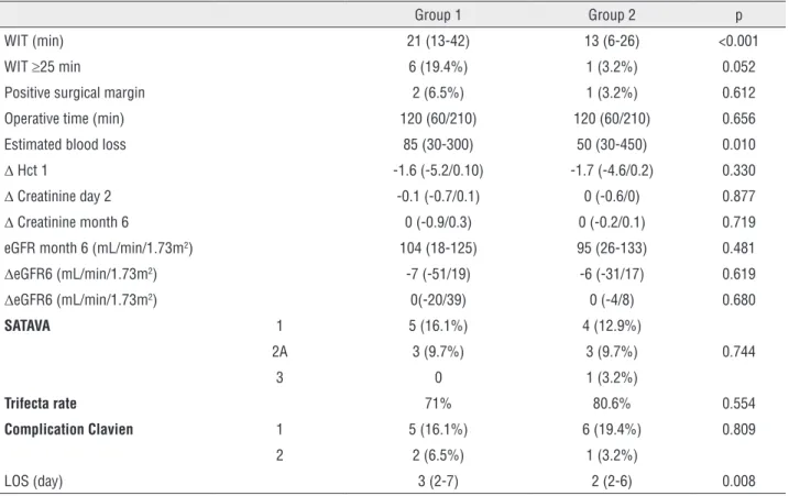
The median WITs were 21 (13-42) and 13 (6-26) minutes for Groups 1 and 2, respectively, and it was significantly lower in Group 2 than in Group 1 (p<0.001). WIT was over 25 minutes in six cases in Group 2 and in only one case in Group 1. The HCT differences and red blood cell transfusion rates were similar in both groups on the first day, but EBL was higher in Group 1 (Table-2). The perioperative opened pelvicalyce-al system rate and operative time were similar between groups (Table-2). There were no signifi-cant differences between the groups with regard to perioperative and postoperative complications (Table-2). In Group 1, diaphragmatic rupture was treated with laparoscopy in the same season by the same surgeon. In Group 2, one patient un-derwent angiography one week after the opera-tion for late hemorrhage. None of the cases were converted to open surgery. However, LOS was lo-wer in Group 2 (2.55±0.96 days) than in Group 1 (3.23±1.26 days; p=0.02, Table-2). Trifecta rates were similar in both groups.
The median serum creatinine levels were significantly increased and eGFR was significan-tly decreased after LPN in both Groups. However, there were no significant differences between the preoperative values and 6-month follow-up in both Groups (Table-3). The creatinine and eGFR differences were similar in both Groups (Table-2).
DISCUSSION
comparable oncologic outcomes to open PN while providing shorter hospital stay, decreased convalescence, and reduced pain (4). Despite the benefits of LPN, it is a technically demanding procedure with a higher rate of intraoperative complications and longer WIT compared to open surgery (19). Becker et al. (20) emphasized that WIT is the strongest modifiable surgical risk factor for postoperative chronic kidney disease. Lane et al. (19) also reported that the major surgical factor in renal function was WIT among the patient-specific, tumor-specific, and surgical factors after PN. Funahashi et al. (21) found that a WIT of 25 minutes or more caused irreversible
damage distributed diffusely throughout the operated kidney. Thompson et al. (22) reported that with prolonged WIT, ischemia reperfusion was positively associated with short- and long-term renal consequences, and they suggested that every minute counts for the severity of damage when the renal hilum is clamped. Therefore, WIT should be shortened as much as possible to help preserve renal function (20, 22).
Link et al. (23) stated that longer ischemia time in LPN is likely to result in difficulty in renorrhaphy with laparoscopy. Intracorporeal suturing was the most time-consuming stage during LPN. Thus, simplifying the suture Table 1 - Comparison of demographic, perioperative, and pathological outcomes between interrupted knot-tying suture renorrhaphy and running suture renorrhaphy with polyglecaprone
Group 1 Group 2 p
Age 53 (32-78) 59 (38-80) 0.123
Gender (Female/Male) 9/22 13/18 0.426
Charlson Comorbidity Index 3 (2-5) 3 (2-5) 0.052
BMI 27.21 (17-47) 27.12 (22-47) 0.833
Preoperative Hct 14.2 (10.3-18) 13.0 (10.3-16.8) 0.058
Preoperative creatinine 0.8 (0.6-2.8) 0.7 (0.6-1.9) 0.250
Preoperative eGFR (mL/min/1.73m2) 93.77±24.74 92.81±27.60 0.885
CKD 3A or more 3A 1 0
0.597
3B 1 2
4 1 1
Side (right) 54.5% 45.5% 0.611
Localization Upper 9 8
0.950
Middle 11 11
Lower 11 12
Pathology of renal mass
Clear Cell RCCa 10 14
0.804 Chromophobe RCCa 6 1
Papillary RCCa 7 8
Multicystic RCCa 2 2
Benign 6 6
Mass size (cm) 3 (2-5) 4 (2-8) 0.006
RENAL score 4 (4-6) 5 (4-10) 0.068
Table 2 - Comparison of perioperative and postoperative outcomes between groups.
Group 1 Group 2 p
WIT (min) 21 (13-42) 13 (6-26) <0.001
WIT ≥25 min 6 (19.4%) 1 (3.2%) 0.052
Positive surgical margin 2 (6.5%) 1 (3.2%) 0.612
Operative time (min) 120 (60/210) 120 (60/210) 0.656
Estimated blood loss 85 (30-300) 50 (30-450) 0.010
Δ Hct 1 -1.6 (-5.2/0.10) -1.7 (-4.6/0.2) 0.330
Δ Creatinine day 2 -0.1 (-0.7/0.1) 0 (-0.6/0) 0.877
Δ Creatinine month 6 0 (-0.9/0.3) 0 (-0.2/0.1) 0.719 eGFR month 6 (mL/min/1.73m2) 104 (18-125) 95 (26-133) 0.481 ΔeGFR6 (mL/min/1.73m2) -7 (-51/19) -6 (-31/17) 0.619 ΔeGFR6 (mL/min/1.73m2) 0(-20/39) 0 (-4/8) 0.680
SATAVA 1 5 (16.1%) 4 (12.9%)
0.744 2A 3 (9.7%) 3 (9.7%)
3 0 1 (3.2%)
Trifecta rate 71% 80.6% 0.554
Complication Clavien 1 5 (16.1%) 6 (19.4%) 0.809
2 2 (6.5%) 1 (3.2%)
LOS (day) 3 (2-7) 2 (2-6) 0.008 ΔCreatinine 6 - changes in creatinine in postoperative 6 month from baseline
Δ Hct 1 - changes in hematocrit creatinine in postoperative first day
Table 3 - Renal function changes overtime between groups.
Preoperative Postoperative day 2 Postoperative
month 6 p1 p2
Median Creatinine level G1 0.8 (0.6-2.8) 0.8 (0.6-3.5) 0.8 (0.6-3.7) 0.02 0.929
G2 0.7 (0.6-1.9) 0.8 (0.6-2.4) 0.7 (0.6-2.0) 0.01 0.414
eGFR (mL/min/1.73m2) G1 100 (25-138) 93 (18-117) 104 (18-125) 0.014 0.221
G2 95 (29-133) 90 (22-115) 95 (26-133) 0.03 1
p1 = statistical significance between preoperative and postoperative 2 day value; p2 = statistical significance between preoperative and postoperative 6 month value.
G1 = Group 1; G2 = Group 2
technique could reduce WIT and better preserve renal function. There are limited numbers of studies considering the effects of simplifying the suture technique on WIT, and none of them used polyglecaprone sutures (9-13). Therefore, the present study aimed to investigate the efficacy of renorrhaphy using running suture technique with monofilament polyglecaprone (Monocryl®,
Ethicon) on reducing renorrhaphy time and WIT during LPN in comparison with interrupted knot-tying suture renorrhaphy.
similar between groups, but WIT was significantly reduced in the group undergoing running suture technique renorrhaphy with Monocryl®. Erdem et al. (12) reported a significantly reduced WIT of 9 minutes in a group treated by self-retaining bar-bed suture rather than polyglactin suture. Jeon et al. also reported a shorter WIT of 7.4 minutes in a barbed suture group than a polyglactin suture group (24). The advantage of self-retaining bar-bed suture (V-Loc) in decreasing WIT might result from passing through the tissue in only one direc-tion, preventing the suture from slipping and eli-minating the need to maintain continuous tension while suturing and tying knots. In another study, the mean WIT was 21.5 minutes in running suture renorrhaphy using PDS suture, while it was 32.3 minutes in the interrupted suture group (13).
PDS II® suture was reported to be 1.4 times as stiff as Monocryl® suture (25). Bezwada et al. (25) showed that the lower stiffness and pliability of Monocryl® suture resulted in excellent handling and tensile properties with minimal resistance du-ring passage through tissue, and Monocryl® had very good tactile feedback. Vicryl® is a braided multifilament suture that causes resistance when passing through the kidney during LPN. Mono-cryl® suture stretches more than ViMono-cryl® and PDS at higher loads (26). In this study, Monocryl® su-ture was preferred for providing faster parenchy-mal suturing with miniparenchy-mal tissue damage, and it is also more cost-effective than V-Loc.
The median WIT was reduced 8 minutes in patients treated with running suture using Mo-nocryl® than traditional renorrhaphy, despite lar-ger tumors in running suture renorrhaphy Group. Likewise, WIT was higher than 25 minutes for only one patient in Group 2 but for 6 patients in Group 1. During running suture renorrhaphy, the surgeon does not need to see the bleeding vessels clearly to control the hemorrhage after unclamping. Thus, this technique facilitates re-nal parenchymal suturing and gives confidence to the surgeon for unclamping before repairing the renal parenchymal defect. These features also contribute to decreasing WIT.
The postoperative decline of renal func-tion after LPN was recovered to preoperative base-line values after 6 months post-operation in both
groups. There were no significant differences be-tween the groups with regard to renal function. WIT was significantly lower in Group 2. The me-dian WIT was 21 minutes in Group 1, and only 6 patients had WIT ≥25 minutes, which could be a reason for the similarity of renal function.
HCT difference from the first day, transfu-sion rate, operative time, perioperative and posto-perative complications, and trifecta rate were si-milar in both Groups. However, EBL was found to be higher in Group 1 than in Group 2. Olweny et al. (27) reported that barbed sutures reduce the in-cidence of serious intraoperative bleeding. Similar results were observed with other V-Loc series (24, 28). Our results are in accordance with these stu-dies. In Group 2, one patient underwent angiogra-phy one week after operation for late hemorrhage. After minimally invasive PN, Omea et al. (29) found an unexpectedly high rate of 21.7% for asymptomatic unruptured renal artery pseu-doaneurysm detected by computed tomography arteriography in the early period. However, in a systematic review and comparative analysis, Jain et al. (30) reported that the rate of symptomatic pseudoaneurysm was 1.96%. In our study, only one patient in Group 2 underwent angiography one week after the operation for symptomatic pseudoaneurysm. This ratio represents 1.6% of the total patients, which is in accordance with the literature. It is hard to make a conclusive de-cision about the comparison of the groups with regard to pseudoaneurysm due to the limited number of the patients.
LOS was shorter in the running suture re-norrhaphy Group than the traditional renorrha-phy Group. Running suture renorrharenorrha-phy provides a decline of 0.7 days in mean LOS. However, this study cannot provide a definite conclusion about this issue.
tradi-tional renorrhaphy group, although renal mass was smaller. Thirdly, the sample size was small, and the results should be confirmed with more cases.
CONCLUSIONS
Renorrhaphy using running sutures with Monocryl® is an effective and safe technique that
shortens WIT. EBL and LOS are also decreased with this technique.
CONFLICT OF INTEREST
None declared.
REFERENCES
1. Ljungberg B, Bensalah K, Canfield S, Dabestani S, Hofmann F, Hora M, et al. EAU guidelines on renal cell carcinoma: 2014 update. Eur Urol. 2015;67:913-24.
2. Weight CJ, Larson BT, Gao T, Campbell SC, Lane BR, Kaouk JH, et al. Elective partial nephrectomy in patients with clinical T1b renal tumors is associated with improved overall survival. Urology. 2010;76:631-7.
3. Weight CJ, Larson BT, Fergany AF, Gao T, Lane BR, Campbell SC, et al. Nephrectomy induced chronic renal insufficiency is associated with increased risk of cardiovascular death and death from any cause in patients with localized cT1b renal masses. J Urol. 2010;183:1317-23.
4. Springer C, Hoda MR, Fajkovic H, Pini G, Mohammed N, Fornara P, et al. Laparoscopic vs open partial nephrectomy for T1 renal tumours: evaluation of long-term oncological and functional outcomes in 340 patients. BJU Int. 2013;111:281-8.
5. Lane BR, Babineau DC, Poggio ED, Weight CJ, Larson BT, Gill IS, et al. Factors predicting renal functional outcome after partial nephrectomy. J Urol. 2008;180:2363-8; discussion 2368-9.
6. Gill IS, Kavoussi LR, Lane BR, Blute ML, Babineau D, Colombo JR Jr, et al. Comparison of 1,800 laparoscopic and open partial nephrectomies for single renal tumors. J Urol. 2007;178:41-6.
7. Gong EM, Orvieto MA, Zorn KC, Lucioni A, Steinberg GD, Shalhav AL. Comparison of laparoscopic and open partial nephrectomy in clinical T1a renal tumors. J Endourol. 2008;22:953-7.
8. Erdem S, Boyuk A, Tefik T, Yucel B, Naghiyev R, Ozsoy M, et al. Warm Ischemia-Related Postoperative Renal Dysfunction in Elective Laparoscopic Partial Nephrectomy Recovers During Intermediate-Term Follow-Up. J Endourol.
2015;29:1083-90.
9. Orvieto MA, Chien GW, Laven B, Rapp DE, Sokoloff MH, Shalhav AL. Eliminating knot tying during warm ischemia time for laparoscopic partial nephrectomy. J Urol. 2004;172:2292-5.
10. Agarwal D, O’Malley P, Clarke D, Rao R. Modified technique of renal defect. closure following laparoscopic partial nephrectomy. BJU Int. 2007;100:967-70.
11. Benway BM, Wang AJ, Cabello JM, Bhayani SB. Robotic partial nephrectomy with sliding-clip renorrhaphy: technique and outcomes. Eur Urol. 2009;55:592-9.
12. Erdem S, Tefik T, Mammadov A, Ural F, Oktar T, Issever H, et al. The use of self-retaining barbed suture for inner layer renorrhaphy significantly reduces warm ischemia time in laparoscopic partial nephrectomy: outcomes of a matched-pair analysis. J Endourol. 2013;27:452-8.
13. Kim KS, Choi SW, Kim JH, Bae WJ, Cho HJ, Lee JY, et al. Running-clip renorrhaphy reducing warm ischemic time during laparoscopic partial nephrectomy. J Laparoendosc Adv Surg Tech A. 2015;25:50-4.
14. Satava RM. Identification and reduction of surgical error using simulation. Minim Invasive Ther Allied Technol. 2005;14:257-61. 15. Dindo D, Demartines N, Clavien PA. Classification of surgical
complications: a new proposal with evaluation in a cohort of 6336 patients and results of a survey. Ann Surg. 2004;240:205-13. 16. Levey AS, Bosch JP, Lewis JB, Greene T, Rogers N, Roth
D. A more accurate method to estimate glomerular filtration rate from serum creatinine: a new prediction equation. Modification of Diet in Renal Disease Study Group. Ann Intern Med. 1999;130:461-70.
17. Kutikov A, Uzzo RG. The R.E.N.A.L. nephrometry score: a comprehensive standardized system for quantitating renal tumor size, location and depth. J Urol. 2009;182:844-53. 18. Khalifeh A, Autorino R, Hillyer SP, Laydner H, Eyraud R,
Panumatrassamee K, et al. Comparative outcomes and assessment of trifecta in 500 robotic and laparoscopic partial nephrectomy cases: a single surgeon experience. J Urol. 2013;189:1236-42.
19. Lane BR, Novick AC, Babineau D, Fergany AF, Kaouk JH, Gill IS. Comparison of laparoscopic and open partial nephrectomy for tumor in a solitary kidney. J Urol. 2008;179:847-51; discussion 852.
20. Becker F, Van Poppel H, Hakenberg OW, Stief C, Gill I, Guazzoni G, et al. Assessing the impact of ischaemia time during partial nephrectomy. Eur Urol. 2009;56:625-34. 21. Funahashi Y, Hattori R, Yamamoto T, Kamihira O, Kato K,
Gotoh M. Ischemic renal damage after nephron-sparing surgery in patients with normal contralateral kidney. Eur Urol. 2009;55:209-15.
clamped during partial nephrectomy. Eur Urol. 2010;58:340-5. 23. Link RE, Bhayani SB, Allaf ME, Varkarakis I, Inagaki T,
Rogers C, et al. Exploring the learning curve, pathological outcomes and perioperative morbidity of laparoscopic partial nephrectomy performed for renal mass. J Urol. 2005;173:1690-4.
24. Jeon SH, Jung S, Son HS, Kimm SY, Chung BI. The unidirectional barbed suture for renorrhaphy during laparoscopic partial nephrectomy: Stanford experience. J Laparoendosc Adv Surg Tech A. 2013;23:521-5.
25. Bezwada RS, Jamiolkowski DD, Lee IY, Agarwal V, Persivale J, Trenka-Benthin S, et al. Monocryl suture, a new ultra-pliable absorbable monofilament suture. Biomaterials. 1995;16:1141-8.
26. Weld KJ, Arzola J, Montiglio C, Bush AC, Cespedes RD. Lapra-Ty holding strength and slippage with various suture types and sizes. Urology. 2008;71:32-5.
27. Olweny EO, Park SK, Seideman CA, Best SL, Cadeddu JA. Self-retaining barbed suture for parenchymal repair during laparoscopic partial nephrectomy; initial clinical experience. BJU Int. 2012;109:906-9.
28. Sammon J, Petros F, Sukumar S, Bhandari A, Kaul S, Menon M, et al. Barbed suture for renorrhaphy during robot-assisted partial nephrectomy. J Endourol. 2011;25:529-33.
29. Omae K, Kondo T, Takagi T, Morita S, Hashimoto Y, Kobayashi H, et al. Renal sinus exposure as an independent fator predicting asymptomatic unruptured pseudoaneurysm formation detected in the early postoperative period after minimally invasive partial nephrectomy. Int J Urol. 2015;22:356-61.
30. Jain S, Nyirenda T, Yates J, Munver R. Incidence of renal artery pseudoaneurysm following open and minimally invasive partial nephrectomy: a systematic review and comparative analysis. J Urol. 2013;189:1643-8.
_______________________ Correspondence address: